Báo cáo khoa học: Alizarine derivatives as new dual inhibitors of the HIV-1 reverse transcriptase-associated DNA polymerase and RNase H activities effective also on the RNase H activity of non-nucleoside resistant reverse transcriptases pot
Bạn đang xem bản rút gọn của tài liệu. Xem và tải ngay bản đầy đủ của tài liệu tại đây (1.95 MB, 14 trang )
Alizarine derivatives as new dual inhibitors of the HIV-1
reverse transcriptase-associated DNA polymerase and
RNase H activities effective also on the RNase H activity of
non-nucleoside resistant reverse transcriptases
Francesca Esposito
1
, Tatyana Kharlamova
1
, Simona Distinto
2
, Luca Zinzula
1
, Yung-Chi Cheng
3
,
Ginger Dutschman
3
, Giovanni Floris
1
, Patrick Markt
4
, Angela Corona
1
and Enzo Tramontano
1
1 Department of Applied Sciences in Biosystems, University of Cagliari, Italy
2 Department of Pharmacobiological Sciences, University of Catanzaro, Italy
3 Deprtment of Pharmacology, Yale University Medical School, New Haven, CT, USA
4 Department of Pharmaceutical Chemistry, University of Innsbruck, Austria
Introduction
Acquired immunodeficiency syndrome is a pandemic
infection whose biological agent is HIV-1. In the third
decade of this pandemic, despite the availability of
20 antiretroviral drugs already approved for the
treatment of HIV-1 infection, current combination reg-
imens still face several challenges [1]. In particular,
newer broad-spectrum anti-HIV drugs are urgently
needed to improve convenience, reduce toxicity and
Keywords
anthraquinones; HIV-1 ribonuclease H;
NNRTI-resistant; RNase H; RT inhibitors
Correspondence
E. Tramontano, Department of Applied
Sciences in Biosystems, University of
Cagliari, Cittadella di Monserrato SS554,
09042, Monserrato, (Cagliari) Italy
Fax: +39 070 675 4536
Tel: +39 070 675 4538
E-mail:
(Received 30 December 2010, revised 7
February 2011, accepted 21 February 2011)
doi:10.1111/j.1742-4658.2011.08057.x
HIV-1 reverse transcriptase (RT) has two associated activities, DNA poly-
merase and RNase H, both essential for viral replication and validated drug
targets. Although all RT inhibitors approved for therapy target DNA poly-
merase activity, the search for new RT inhibitors that target the RNase H
function and are possibly active on RTs resistant to the known non-
nucleoside inhibitors (NNRTI) is a viable approach for anti-HIV drug
development. In this study, several alizarine derivatives were synthesized
and tested for both HIV-1 RT-associated activities. Alizarine analogues
K-49 and KNA-53 showed IC
50
values for both RT-associated functions of
10 l
M. When tested on the K103N RT, both derivatives inhibited the
RT-associated functions equally, whereas when tested on the Y181C RT,
KNA-53 inhibited the RNase H function and was inactive on the polymer-
ase function. Mechanism of action studies showed that these derivatives do
not intercalate into DNA and do not chelate the divalent cofactor Mg
2+
.
Kinetic studies demonstrated that they are noncompetitive inhibitors, they
do not bind to the RNase H active site or to the classical NNRTI binding
pocket, even though efavirenz binding negatively influenced K-49 ⁄ KNA-53
binding and vice versa. This behavior suggested that the alizarine deriva-
tives binding site might be close to the NNRTI binding pocket. Docking
experiments and molecular dynamic simulation confirmed the experimental
data and the ability of these compounds to occupy a binding pocket close
to the NNRTI site.
Abbreviations
AQ, anthraquinone; DKA, diketo acid; MD, molecular dynamic; NNRTI, non-nucleoside reverse transcriptase inhibitors; RDDP,
RNA-dependent DNA polymerase; RT, reverse transcriptase.
1444 FEBS Journal 278 (2011) 1444–1457 ª 2011 The Authors Journal compilation ª 2011 FEBS
provide antiretroviral activity against viral strains resis-
tant to the currently used antiretroviral agents [1].
The HIV-1 reverse transcriptase (RT) is responsible
for conversion of the genomic plus (+) single-stranded
viral RNA genome into the proviral double-stranded
DNA that is subsequently integrated into the cell host
chromosome by the viral-coded integrase [2,3]. HIV-1
RT is a multifunctional enzyme which has two different,
functionally related, catalytic activities: (a) DNA poly-
merase activity, which can be both RNA and DNA
dependent; and (b) RNase H activity that selectively
hydrolyzes the RNA strand of the RNA:DNA hybrid
formed during synthesis of the minus (–) strand DNA
that uses (+)-strand RNA as template [2,3]. HIV-1
RNase H, similar to all the RNase Hs and together with
transposases, retroviral integrases and RuvC resolvase,
belongs to the polynucleotidyl transferase family and
catalyzes the phosphoryl transfer through nucleophilic
substitution reactions on phosphate esters [4].
Even though both RT-associated activities are essen-
tial for virus replication and, therefore, both enzyme
functions are attractive targets for drug development,
all compounds targeting the HIV-1 RT, either
approved for treatment or under clinical evaluation,
inhibit the RT RNA-dependent DNA polymerase
(RDDP) associate activity, whereas none inhibit its
RNase H-associated activity [1,5,6]. Hence, the HIV-1
RNase H function is a valid and attractive viral target
whose inhibition is worth pursuing.
Anthraquinones (AQs) are common secondary
metabolites occurring in bacteria, fungi, lichens and
higher plants where they are found in a large number of
families [7]. AQ derivatives have been reported to have
diverse biological properties comprising DNA intercala-
tion ability, antitopoisomerase activity and telomerase
expression induction [8–10], and are active ingredients
of various Chinese traditional medicines [11]. Further-
more, AQs have been reported to have an effect against
the encephalomyocarditis virus in mice [12], inactivate
enveloped viruses [13], to inhibit human cytomegalovirus
[14,15], poliovirus [16] and hepatitis B viruses in cell-
based assays [17], and inhibit HIV-1 RT [18] and integr-
ase activities in biochemical assays [19]. In particular,
the AQ derivative alizarine has been reported to inhibit
human cytomegalovirus replication [15] and HIV-1
RT-associated RDDP and integrase activities [18,19].
Recently, we reported that some derivatives of the
AQ emodine inhibit HIV-1 RT-associated RNase H
activity without chelating the Mg
2+
ion at the catalytic
site [20], which is the proposed mode of action of other
RNase H inhibitors such as the diketo acid (DKA)
derivatives [6,21,22]. Continuing the search for agents
that might inhibit the HIV-RT activities with new
modes of action, we tested a novel series of AQ deriva-
tives based on the alizarine structure and found that
some are inhibitors of both RT-associated functions.
Mode-of-action studies demonstrate that these new
AQ derivatives are noncompetitive inhibitors that do
not bind to either the RNase H catalytic site or the
RT hybrid substrate. Interestingly, most of them were
similarly active on the mutant K103N RT-associated
functions, whereas only two analogues were able to
inhibit the RNase H activity of the Y181C RT. It was
hypothesized that they might bind to a site adjacent to
the non-nucleoside reverse transcriptase inhibitor
(NNRTI) pocket, which was originally reported by
Himmel et al. [23] as the binding site for some hydraz-
one derivatives. Hence, this binding site was investi-
gated using docking studies and molecular dynamic
(MD) simulation, leading to the hypothesis that AQ
inhibition of RNase H function may be because of a
change in the RNA:DNA hybrid RT accommodation,
induced by the AQs binding to this pocket, which
results in a possible variation in the nucleic acid trajec-
tory toward the RNase H catalytic site.
Results and Discussion
Inhibition of HIV-1 RT-associated RNase H
activity by alizarine derivatives
We have previously reported that analogues of the AQ
emodine inhibited the HIV-1 RT-associated RDDP
and RNase H activities [20]. In an effort to better
characterize the AQ derivative potentialities and mode
of action and, eventually, increase their potencies, we
tested the AQ derivative alizarine, which has previ-
ously been reported to inhibit the HIV-1 RT-associ-
ated RDDP function [18] but, to our best knowledge,
was never tested for its RNase H function. Results
showed that alizarine inhibited the HIV-1 RT-associ-
ated RDDP function with an IC
50
of 79 lm, as previ-
ously reported [18], but it was inactive on the HIV-1
RT-associated RNase H function (Table 1). With the
aim of increasing the alizarine potency of RT inhibi-
tion, we synthesized and assayed a series of derivatives
with different substituents at positions 1 and 2 of the
AQ ring. As shown in Table 1, when an acetophenon
group was inserted at position 2 of the AQ ring, com-
pound K-54 inhibited both HIV-1 RT-associated activ-
ities slightly. This was in agreement with what we
observed for the emodine derivative K-67, which was
able to inhibit both enzyme activities [20]. The further
introduction of a Br atom in the phenyl ring increased
the inhibition potency of the analogue KNA-53,
which showed IC
50
values of 21 and 5 lm for the
F. Esposito et al. Alizarines as new dual HIV-1 RT inhibitors
FEBS Journal 278 (2011) 1444–1457 ª 2011 The Authors Journal compilation ª 2011 FEBS 1445
polymerase-independent RNase H and RDDP func-
tions, respectively (Table 1). Interestingly, when the Br
atom was substituted with a second phenyl ring, com-
pound K-126 completely lost its inhibitory effect on
the RNase H function, although it retained the effect
on the RDDP function. Finally, when a phenylketo
group was inserted into both positions 1 and 2 of the
AQ ring, the analogue K-49 inhibited both enzyme
functions with IC
50
values of 12–13 lm.
Characterization of the mechanism of HIV-1 RT
inhibition by alizarine derivatives
Because it has been reported that the HIV-1 RNase H
activity might be influenced by the sequence of the
Table 1. Inhibition of the wild-type HIV-1 RT-associated activities by AQ derivatives.
Compound R
1
R
2
a
IC
50
(lM)
RNase H RDDP
Alizarine H H > 100 (92%)
b
79 ± 8
K-56
> 100 (100%) 82 ± 9
K-57
> 100 (100%) > 100 (80%)
K-45
> 100 (88%) > 100 (95%)
KNA-26
> 100 (90%) > 100 (59%)
K-52
> 100 (100%) 61 ± 8
K-53
> 100 (100%) > 100
K-54
39 ± 6 60 ± 5
KNA-53
21 ± 5 5 ± 2
K-126
100 ± 8 6 ± 1
K-61
> 100 (91%) 14 ± 3
K-111
> 100 (80%) > 100 (100%)
K-49
13 ± 3 12 ± 3
EFV > 10 0.003 ± 0.001
a
Compound concentration required to reduce by 50% enzyme activity ± SD.
b
Percentage enzyme activity at 100 l M compound concentration.
Alizarines as new dual HIV-1 RT inhibitors F. Esposito et al.
1446 FEBS Journal 278 (2011) 1444–1457 ª 2011 The Authors Journal compilation ª 2011 FEBS
RNA:DNA template utilized in the biochemical assay
[24], we also used a different, previously described [22]
RNA:DNA hybrid substrate to assess the effect of
alizarine analogues on the HIV-1 RNase H function.
Results showed that, also with this substrate, the newly
synthesized derivatives inhibited HIV-1 wild-type RT-
associated polymerase-independent RNase H function
with IC
50
values comparable to the one shown in
Table 1. In particular, compound K-49 inhibited wild-
type RNase H activity with an IC
50
value of 7 lm
without affecting the RNase H cleavage pattern
(Fig. 1). The DKA derivative RDS1643 was used as a
positive control and showed an IC
50
value of 13 lm
[22]. Subsequently, considering that hydrolysis of the
poly(dC)–poly(rG) hybrid substrate catalyzed by the
HIV-1 RNase H is a processive reaction which can be
monitored according to Michaelis–Menten kinetic
assumptions, we determined the inhibition kinetics of
the wild-type RT-associated polymerase-independent
RNase H function by K-49. In this system, the K
m
and k
cat
values were 1.5 nm and 0.82 s
)1
, respectively,
and K-49 resulted in noncompetitive inhibition of the
polymerase-independent RNase H activity with a K
i
value of 7 lm (Fig. 1). Similar results were also
obtained for the KNA-53 analogue (data not shown).
In addition, because it has been shown that some AQ
derivatives bind noncovalently to double-stranded
(ds)DNA [9] and given that the reaction substrates
used in our biochemical assays were RNA:DNA
hybrids, we asked whether the observed enzyme inhibi-
tion by the newly synthesized AQ analogues could be
due to intercalation into the hybrid substrate. There-
fore, as described previously [20], we evaluated the
ability of K-49 and KNA-53 analogues to bind to calf
thymus DNA in solution and found that they are not
able to intercalate into nucleic acids (data not shown).
Furthermore, it has been reported that the DKA deriv-
atives inhibit the HIV-1 RNase H function by chelat-
ing the RT metal cofactor [5,6], and in order to verify
whether the alizarine analogues also might interact
with the metal ions, we measured their visible spectrum
in the absence or presence of 6 mm MgCl
2
, observing
that addition of the cation did not significantly alter
the alizarine derivatives maximum absorbance (data
not shown).
Inhibition of HIV-1 K103N and Y181C RT-associ-
ated RNase H activity by alizarine derivatives
To date, four NNRTIs (nevirapine, delavirdine, efavi-
renz and etravirine) have been approved for clinical
use in combination with other antiviral agents [1]. It is
well known that treatment with NNRTI selects for
A
B
C
120
100
80
60
40
20
0
RNase H activity (% of control)
1 10 100 200
Compound concentraion (µ
M)
–RT +RT
K049 RDS1643
32-mer
28-mer
24-mer
15-mer
123456 7891011121314
0.060
0.045
0.030
0.015
0
–0.50 –0.25 0.00
1/[S] (1/µ
M)
0.25 0.50
1/V (1/fmoles)
0.20
0.15
0.10
Slope
0.05
0.00
–0.10 0 10 20 30 40 50
K49 (µ
M)
A
B
C
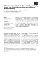
Fig. 1. Inhibition of wild-type HIV-1 RT-associated polymerase-
independent RNase H activity by K-49. (A) Inhibition curve of the
RNase H function by K-49 using poly(dC)–[
3
H]poly(rG) as the reaction
substrate. Reactions were carried out as described in Materials and
methods. Data represent mean values from three independent
determinations. (B) PAGE analysis of the RNase H function inhibition
by K-49 using the tC5U ⁄ p12 hybrid as the substrate. Reactions and
PAGE analysis were carried out as described in Materials and meth-
ods. Four major bands were resolved as reaction products, each
from a single cleavage event of the 32mer substrate. Lane 1, with-
out RT; lane 2, plus RT, lanes 3–8, plus RT and K-49 (100, 33, 11,
3.3, 1.1 and 0.33 l
M); lanes 9–14, plus RT and RDS 1634 (100, 33,
11, 3.3, 1.1 and 0.33 l
M). (C) Lineweaver–Burk plot of the inhibition
of the HIV-1 RNase H activity by K-49. Reactions were performed as
described in Materials and methods. HIV-1 RT was incubated in
the absence (s) or presence of 35 l
M (+), 10 lM (Ñ), 20 lM (e)
and 40 l
M (4) K-49. (Inset) Replot of the slopes obtained in the
Lineweaver–Burk plot against the K-49 concentration to calculate K
i
.
F. Esposito et al. Alizarines as new dual HIV-1 RT inhibitors
FEBS Journal 278 (2011) 1444–1457 ª 2011 The Authors Journal compilation ª 2011 FEBS 1447
HIV drug-resistant strains mutated in RT. In particu-
lar, mutations K103N and Y181C in the RT are the
most worrying, because they lead to resistance to many
different NNRTIs as a result of overlapping resistance
profiles [1]. In fact, new antiviral agents that may inhi-
bit HIV-1 strains mutated in these residues are actively
pursued [1]. Therefore, in order to assess the effect of
the AQ analogues on the mutant enzymes, compounds
even weakly active in at least one HIV-1 wild-type
RT-associated function were tested for both enzyme
activities of the K103N and Y181C RTs (Table 2).
Interestingly, when tested on the K103N RT, alizarine
derivatives mainly showed inhibition potencies similar
to those shown on the wild-type RT with three excep-
tions: (a) the K-54 analogue completely lost its ability
to inhibit the polymerase-independent RNase H activ-
ity, but retained its effect on the RDDP activity; (b)
the K-126 analogue, which was slightly active on the
wild-type RT RNase H function, showed a sixfold
increase in the inhibition potency of the RNase H
function, although it retained its inhibition potency on
the polymerase function; and (c) the K-61 analogue
showed a fourfold reduction in its RDDP activity inhi-
bition potency. By contrast, when the AQ derivatives
were tested on the Y181C RT, the results showed that
only KNA-53 and K-126 analogues retained their abil-
ity to inhibit the RT-associated RNase H function
with the same IC
50
values observed for the K103N
RT; all the other compounds were inactive (Table 2).
Interaction of alizarine derivatives and the DKA
RDS1643 on the HIV-1 RNase H activity
It has been proposed that DKA derivatives chelate the
RT metal cofactor in the active site [5,6]. Hence, in
order to ascertain whether the alizarine derivatives
could interact with the HIV-1 RT RNase H active site,
even without chelating the cofactor metal ion, we
determined the effect of the interaction between the
AQ analogue K-49 and the DKA analogue RDS1643
on the HIV-1 RT-associated polymerase-independent
RNase H activity by using the Yonetani–Theorell
model [25]. This graphical method allows us to deter-
mine whether two inhibitors of a certain enzyme com-
pete for the same binding site or act on two
nonoverlapping binding sites. The method has already
been used to dissect the effect of the interaction
between RNase H and RDDP inhibitors [20,26]. In
this revised model, the plot of the reaction velocity
reverse (1 ⁄ v) observed in the presence of different con-
centrations of the first inhibitor, in the absence or con-
temporaneous presence of the second inhibitor, leads
to a series of lines that are parallel if the two inhibitors
compete for the same binding site, whereas they inter-
sect if the inhibitors bind to different enzyme sites [25].
Therefore, the HIV-1 RT RNase H activity was mea-
sured in the presence of increasing concentrations of
both K-49 and RDS1643, and was analyzed using the
Yonetani–Theorell plot (Fig. 2). The results show that
both slope and intercepts of the plots of 1 ⁄ v versus
K-49 concentration increased as a linear function of
RDS1643, indicating that the two compounds do not
bind to overlapping sites.
In order to further investigate the possibility that
the AQ derivatives might bind to the RNase H active
site, the ability of K-49 and KNA-53 to inhibit the
enzyme activity of the isolated RNase H domain (p15)
was assessed [27]. In this system, the AQ derivatives
were not able to inhibit the RNA degradation (data
not shown).
Table 2. Inhibition of the mutant HIV-1 RT-associated activities and wild-type HIV-1 replication by AQ derivatives.
Compound
IC
50
(lM)
a
EC
50
(lM)
b
CC
50
(lM)
c
wt RT K103N RT Y181C RT
HIV-1 MT2
RNase H RDDP RNase H RDDP RNase H RDDP
Alizarine > 100 (92%)
d
79 ± 8 > 100 (58%) 68 ± 5 > 100 (100%) > 100 (100%) ND
e
ND
K-56 > 100 (100%) 82 ± 9 > 100 (100%) 100 ± 8 > 100 (100%) > 100 (100%) ND ND
K-52 > 100 (100%) 61 ± 8 > 100 (100%) 100 ± 9 > 100 (100%) > 100 (100%) ND ND
K-54 39 ± 6 60 ± 5 > 100 (98%) 35 ± 4 > 100 (86%) > 100 (74%) > 100 40
KNA-53 21 ± 5 5 ± 2 21 ± 4 9 ± 3 22 ± 4 > 100 (92%) > 100 > 100
K-126 100 ± 8 6 ± 1 16 ± 3 9 ± 2 16 ± 2 > 100 (100%) ND ND
K-61 > 100 (91%) 14 ± 3 > 100 (100%) 64 ± 8 > 100 (100%) > 100 (72%) ND ND
K-49 13 ± 3 12 ± 3 16 ± 4 28 ± 4 > 100 (79%) > 100 (87%) > 100 100
EFV > 10 0.003 ± 0.001 ND 0.68 ± 0.2 ND 0.40 ± 0.1
a
Compound concentration required to reduce enzyme activity by 50% ± SD.
b
Compound concentration required to reduce the HIV-1-
induced cytopathic effect in MT-2 cells by 50%.
c
Compound concentration required to reduce MT-2 cell multiplication by 50%.
d
Percentage
of enzyme activity at 100 l
M compound concentration.
e
ND, not done.
Alizarines as new dual HIV-1 RT inhibitors F. Esposito et al.
1448 FEBS Journal 278 (2011) 1444–1457 ª 2011 The Authors Journal compilation ª 2011 FEBS
Furthermore, because the RNase H domain contains
one tryptophan and six tyrosine residues as intrinsic
fluorophores, it has been reported that when the p15
domain is excitated at a wavelength of 290 nm the
contribution of tryptophan to the fluorescence signal is
maximized, whereas the fluorescence energy transfer
from the tyrosine residues to the tryptophan residue is
minimized [27]. Therefore, compounds interacting with
the HIV-1 RT-associated RNase H active site are able
to quench the intrinsic protein fluorescence of the iso-
lated HIV RNase H domain [27]. In fact, as described
previously [27], 2-hydroxy-1,2,3,4-tetrahydroisoquino-
line-1,3-dione, used as a positive control, was able to
reduce the p15 intrinsic fluorescence with an IC
50
of
52 lm, whereas only a small reduction (< 30%) in the
p15 intrinsic fluorescence was observed in the presence
of the highest (100 lm) K-49 or KNA-53 concentra-
tion (data not shown). Overall, these data support the
hypothesis that AQ derivatives do not inhibit the RT
catalysis by primarily binding to the RNase H active
site.
Interaction of alizarine derivatives with the
NNRTI efavirenz on the HIV-1 RDDP activity
Because the NNRTI-binding site is at a close spatial
distance from the substrate (dNTP)-binding site, the
NNRTIs have been shown to interfere with the poly-
merase catalytic site, impeding the normal RDDP per-
formance. Within the NNRTI-binding site, the amino
acid residues lysine (Lys103) and tyrosine (Tyr181)
interact with many NNRTIs. The observation that the
AQ analogues K-49 and KNA-53 inhibited both wild-
type and K103N RT-associated RDDP function while
they were inactive on the Y181C RT-associated RDDP
function, raised the question of whether they could
actually bind to the NNRTI-binding site, possibly with
low affinity. To answer this question, we measured the
effect of the interaction between K-49, or KNA-53,
and the NNRTI efavirenz on the wild-type RT RDDP
activity using the Yonetani–Theorell plot [25]. The
results showed that when the HIV-1 RT RDDP activ-
ity was measured in the presence of increasing concen-
trations of one of the two AQ derivatives and
efavirenz, and analyzed using the Yonetani–Theorell
plot, the slope and intercepts of the two plots of 1 ⁄ v
versus efavirenz concentration increased as a linear
A
B
C
0.10
0.08
0.06
0.04
0.02
0.00
–15
–10
–5 0
K49 (µ
M)
51015
0.30
0.25
0.20
0.15
0.10
0.05
0.00
0.5
0.4
0.3
0.2
0.1
0.0
–20 –10
–20
0204060
0102030
Efavirenz (n
M)
Efavirenz
(
nM
)
1/n (fmoles of product)1/n (1/fmoles of product)1/n (1/fmoles of product)
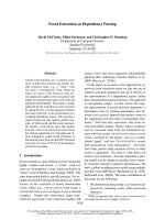
Fig. 2. Yonetani–Theorell plot of the interaction between AQ deriv-
atives and other RT inhibitors. (A) Yonetani–Theorell plot of the
combination of K-49 and RDS 1643 on the HIV-1 RT polymerase-
independent RNase H activity. HIV-1 RT was incubated in the pres-
ence of different concentrations of K-49 and in the absence (
•)or
presence of 3 l
M (.)or10lM ( ) RDS1643. (B) Yonetani–Theorell
plot of the interaction of K-49 and efavirenz on the HIV-1 RT RDDP
activity. HIV-1 RT was incubated in the presence of different con-
centrations of efavirenz and in the absence (
•) or presence of
1.9 l
M (s), 3.7 lM (.) and 7.5 lM (4) K-49. (C) Yonetani–Theorell
plot of the interaction of KNA-53 and efavirenz on the HIV-1 RT
RDDP activity. HIV-1 RT was incubated in the presence of different
concentrations of efavirenz and in the absence (
•) or presence of
1 l
M (.), 2 lM (4) and 4 lM ( ) KNA-53. Reactions were per-
formed as described in Materials and methods.
F. Esposito et al. Alizarines as new dual HIV-1 RT inhibitors
FEBS Journal 278 (2011) 1444–1457 ª 2011 The Authors Journal compilation ª 2011 FEBS 1449
function of the AQ derivatives concentrations, indicat-
ing that the AQ analogues and the NNRTI efavirenz
do not bind to overlapping sites (Fig. 2). However, it
is worth noting that the Yonetani–Theorell plot allows
us to calculate an interaction constant between the two
tested inhibitors, termed a [25]. When the two com-
pounds bind to the same site, a = ¥; when the two
compounds are strictly independent, a = 1; whereas
when the two compounds interact repulsively in the
enzyme–two inhibitors complex, the a > 1. Hence,
when we calculated the a value for the K-49 ⁄ efavirenz
and KNA-53 ⁄ efavirenz interactions we found that a
was 3.5 and 3.0, respectively, indicating that the bind-
ing of an AQ derivative results in a reduction of efavi-
renz binding and, vice versa, the binding of efavirenz
leads to a reduction of the AQ binding.
Docking studies
These results demonstrated that the AQs and NNRTI
binding sites are strictly functionally related. It has
been reported that some hydrazone derivatives that can
inhibit both RT-associated functions bind to a site near
the NNRTI binding pocket [23], therefore we wished to
verify whether the AQ derivatives could also bind to
this site. For this purpose, and to obtain a deeper
understanding of the RT–ligand interactions, QM
polarized docking (Schro
¨
dinger Inc, Portland, OR,
USA) was carried out. QM polarized docking workflow
combines docking with ab initio for ligand charges cal-
culation within the protein environment. This method-
ology has been showed to perform significantly better
than docking alone, enabling the modeling of biomolec-
ular systems at a reasonable computational effort while
providing the necessary accuracy [28]. A blind docking
experiment gave evidence of the existence of five possi-
ble binding areas that are shown in Fig. 3. However,
from energetic analysis, it appears that only two of the
five binding areas are favorable: one located close to
the RNase H catalytic cavity and the other close to the
NNRTI binding pocket. Because experimental data
derived from testing the compounds on the isolated
RNase H portion seem to suggest that the first binding
pocket is not primarily responsible for AQ activity,
more detailed analysis of K-49, KNA-53, K-54 and
K-126 putative binding mechanisms was carried out, in
both wild-type and mutants RTs, exploring the binding
site for RNase H inhibitors described by Himmel as a
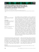
secondary binding site [23] (Fig. 4). The analysis of the
best poses of K-49 ⁄ wild-type RT highlighted that the
planar rings system guarantees a significant influence of
the p–p stacking interaction with Trp229 on the orien-
tation of the ligand in the binding site (Fig. 5A). Inter-
actions between the benzoic moiety and the
hydrophobic residues in the NNRTI pocket (Leu100,
Val106, Tyr188, Phe227, Leu234 and Tyr318) allowed a
sterically favorable allocation of this bulky portion
inside the pocket. These contacts appear to be essential
Fingers
Thumb
RNAse H
Polymerase
Palm
Fig. 4. Schematic representation of the overall structure of the
HIV-1 RT heterodimer. The p66 subunit (upper) is displayed in a col-
ored scheme for the individual subdomains, whereas the p51 sub-
unit is shown using the same color. The surface represents the
position of the new binding pocket for RNase H inhibitors. Close-up
of the binding cavity colored according to lipophilicity: light blue for
hydrophilic residues and pale yellow for hydrophobic residues.
Fig. 3. Representation of the overall structure of the HIV-1 RT
heterodimer with binding areas of alizarine derivatives found after
the blind docking experiment. The most favorable binding sites are
highlighted in blue.
Alizarines as new dual HIV-1 RT inhibitors F. Esposito et al.
1450 FEBS Journal 278 (2011) 1444–1457 ª 2011 The Authors Journal compilation ª 2011 FEBS
Asp110
Asp185
Asp186
Trp229
Tyr181
NNRTIbp
E
Tyr188
Asp110
Asp185
Asp186
Trp229
Cys181
NNRTIbp
B
Asp110
Asp185
Asp186
Trp229
Tyr181
NNRTIbp
Tyr188
F
Asp110
Asp185
Asp186
Trp229
Cys181
NNRTIbp
C
Asp110
Asp185
Asp186
Trp229
Tyr181
Tyr188
NNRTIbp
G
Tyr188
Tyr181
Trp229
Asn103
D
Asp110
Asp185
Asp186
Trp229
Tyr181
Tyr188
NNRTIbp
H
Asp110
Asp185
Asp186
Trp229
Cys181
NNRTIbp
A
K126
K126
I
Asp110
Asp185
Asp186
Tyr181
Trp229
Asn103
K126
K54
K54
KNA53
KNA53
K49
K49
VAL108A
VAL106A
PHE227A
LEU234A
TRP229A
TYR188A
TYR318A
LEU100A
TYR181A
VAL108A
TRP229A
LEU234A
PHE227A
PRO236A
TYR188A
VAL106A
LEU234A
PHE227A
VAL108A
TYR188A
TYR183A
TRP229A
LEU100A
LEU100A
TYR318A
TYR188A
TRP229A
LEU234A
ASN103A
VAL106A
VAL108A
TYR188A
VAL108A
TYR188A
MET230A
LEU234A
TRP229A
VAL108A
LEU228A
TRP229A
LEU100A
TYR188A
TYR188A
VAL106A
VAL108A
PHE227A
TYR318A
LEU234A
LEU100A
VAL108A
PHE227A
TYR188A
LEU234A
LEU100A
TYR318A
VAL106A
O
O
O
O
O
O
O
O
O
O
O
O
Br
O
O
O
O
O
O
O
O
O
O
O
O
O
O
O
O
O
O
O
O
O
O
O
O
O
O
O
O
O
O
O
O
O
O
O
O
O
O
O
O
O
O
Pr
O
Fig. 5. K-49 and KNA-53 (in sticks) best docked pose. Compound interactions with the RT were analyzed using Ligandscout: the yellow
spheres show hydrophobic contacts. Binding pocket surfaces are drawn as solid and colored according to lipophilicity: pale yellow indicates
lipophilic residues and light blue hydrophilic residues. (A) K-49, (B) KNA-53, (C) K-54, (D) K-126 binding mode into wild-type RT and a 2D
depiction of their respective interactions. (E) K-49, (F) KNA-53 and (H) K-126 binding mode into Y181C RT and a 2D depiction of their respec-
tive interactions. (G) K-54 and (I) K-126 binding mode into K103N RT and a 2D depiction of their respective interactions.
F. Esposito et al. Alizarines as new dual HIV-1 RT inhibitors
FEBS Journal 278 (2011) 1444–1457 ª 2011 The Authors Journal compilation ª 2011 FEBS 1451
for the RDDP inhibition activity. In the case of KNA-
53, the bulkier moiety 1-4-bromophenyl)-2-oxyetha-
none does not allow the compound to enter further into
the cavity (Fig. 5B). Several residues in the NNRTI
and the second binding pocket are involved in hydro-
phobic contacts that stabilize the complex.
Although the Lys103 mutation in Asn did not have
any effect on the RNase H inhibition by K-49 and
KNA-53, confirming that their binding does not involve
this portion of the NNRTI pocket, the Tyr181 mutation
in Cys led to a total loss of activity for K-49. In order to
better explain this observation, we studied the behavior
of the complex K-49 ⁄ wild-type RT in an aqueous
environment running 5 ns of MD simulation using Des-
mond Molecular Dynamics System 2.0 (Shaw Research,
New York, NY, USA) keeping the whole enzyme free to
move into explicit solvent. During QM polarized dock-
ing the receptor is treated as rigid, and the phenomena
of induced fit cannot be observed. Analysis of the trajec-
tory highlighted that Tyr181 rotated to better accommo-
date the ligand and interacted with the lower phenyl
ring adding an important p–p stacking (Fig. 6). Plots
for potential energy and RMSD fluctuations involving
the complex are depicted in Figs 6C,D, the analysis
shows that the structure reached equilibrium and the
low fluctuations support the stability of the intermolecu-
lar interactions. When Tyr181 is mutated in Cys, elec-
tronic and steric modifications occur. The binding mode
of K-49 in the Y181C RT is different, and the contribu-
tion of the p–p stacking interaction is lost. Furthermore,
the low number of good contacts with RT leads to insta-
bility of the complex and the compound can be easily
washed off the substrate cavity or displaced (Fig. 5E).
By contrast, from a deep insight in to the best KNA-53
docked pose in the Y181C RT (Fig. 5F), we were able
to observe that the enlarged cavity allowed KNA-53 to
go deeper and, at the same time, the bulky substituent in
position 2 did not interact with many NNRTI pocket
residues, leading to the loss of RDDP activity although
the RNase H activity was retained. Two other com-
pounds showed a significant difference in their RNase H
inhibition effects on mutant RTs: K-54 and K-126. In
particular, K-54 was characterized by loss of activity
versus the RNase H function and increased inhibition of
the RDDP function in K103N RT. This suggests that
K-54 may bind to the NNRTI pocket with higher affin-
ity when this mutation occurs. It is known that the
NNRTI pocket is highly flexible and shows induced fit
during NNRTIi binding [24], therefore, we usede a
docking simulation (data not shown) to exclude the
AB
C
D
ps
RMSD (Å)
Energy (kcal·mol
–1
)
0
0
–4.17E + 05
–4.18E + 05
–4.19E + 05
–4.20E + 05
2
4
Asp110
Asp186
Asp185
Trp229
Tyr181
1000 2000 3000 4000 5000
RMSD
E_p
Fig. 6. Superimposed structures of 5 ns MD simulations frames of K49-RT complex colored by timestep: initial (red), final (blue) along with
intermediate structures snapshots. (A) Overall structure of the HIV-1 RT heterodimer; (B) close-up of the binding cavity; (C) RMSD fluctua-
tions of the complex during the 5 ns trajectory; (D) potential energy of the complex during the MD simulation.
Alizarines as new dual HIV-1 RT inhibitors F. Esposito et al.
1452 FEBS Journal 278 (2011) 1444–1457 ª 2011 The Authors Journal compilation ª 2011 FEBS
possibility that our compounds can adopt a ‘butterfly-
like’ geometry like many NNRTI (e.g. nevirapine and
efavirenz) [29]. On the one hand, as previously shown by
Himmel et al. [23], RT ⁄ DHBNH and RT ⁄ CP-94,707
complexes have a similar conformation (RMS 0.57), on
the other hand, in the crystal reported by Pata et al. [30]
the NNRTI binding pocket most closely resembles the
RT unliganded conformation. Therefore, the AQ deriv-
atives were docked into the K103N RT in this confor-
mation. Under these conditions, we observed that K-54
not only entered into the NNRTI pocket but was also
stabilized by hydrogen bond interaction with Asn103.
Furthermore, the hydrogen bond between Tyr188 and
Asn103 maintains the position of the Tyr residue in a
conformation that allows a better fit (Fig. 5G). As high-
lighted for CP-94,707, even if this bond needs to be bro-
ken before ligand entrance, it could be reformed
immediately after because the compound does not inter-
fere with it [30]. Thus, this might explain the preference
for this binding mode when in K103N RT. In wild-type
RT, the steric and electrostatic effects of Lys hamper the
formation of the same interactions with this compound
and the binding shown in Fig. 5C is favored.
Finally, when analyzing the K-126 docking results
we observed that, because of the bulkiness of its diphe-
nyl substituent in the Y181C RT, the larger binding
pocket can better host the compound and higher RNa-
se H inhibition is observed (Fig. 5H). Furthermore,
also in the case of the K103N RT, and for the reasons
given above for K-54, K-126 is able to enter the bind-
ing pocket and act allosterically (Fig. 5I).
Conclusions
Targeting new RT drug binding pockets that may inhi-
bit one or both RT-associated enzymatic functions is an
attractive approach to allosterically inhibiting the HIV-
1 reverse transcription. We have identified a new series
of AQ derivatives that inhibit both HIV-1 RT-associ-
ated functions in the micromolar range in biochemical
assays, even though they are not able to inhibit viral rep-
lication in cell culture, possibly because of to a lack of
cell membrane penetration (Table 2 and data not
shown). However, some AQ derivatives showed a
unique profile of RT inhibition, resulting in their being
able to inhibit the RT-associated RNase H function of
mutant K103N and Y181C RTs. Experimental results
and modeling simulations led us to suppose that the
binding pocket lying between the polymerase catalytic
triad and NNRTI pocket [23] may be involved in AQs
binding to RT and in their ability to inhibit both
RT-associated RDDP and RNase H activities. This
conclusion is also in agreement with the very recent
demonstration, obtained using X-ray crystallography,
that a naphthyridinone derivative that is able to inhibit
the HIV-1 RT-associated RNase H function binds to
the same pocket adjacent to the NNRTI site [31]. How-
ever, given that blind docking experiments indicated the
existence of five possible binding pockets, we can not
completely exclude the possibility that the AQ deriva-
tives may additionally bind to other RT pockets and
that the binding stoichiometry of the compounds to RT
could also be different according to the RT mutations
and the relative compound-RT affinities.
The position of the proposed major AQs binding site
raises the question of the distance between this RNa-
se H inhibitor binding site and the RNase H catalytic
site. In this respect, it is worth noting that it has been
previously reported that NNRTIs binding to RT lead
to an increase in the RNase H activity and, in some
cases, to an alteration in the nucleic acid cleavage pat-
tern [26,32]. Furthermore, mutations in the primer grip,
which are essential for nucleic acid binding in either the
polymerase domain [33] or the RNase H domain [32–
36], alter the RNase H cleavage position of the
RNA:DNA hybrid. In addition, a subset of polymerase
domain primer grip residues (Phe227, Trp229, Leu234
and His235) also line the NNRTI-binding pocket, while
the mutant Y181C RT mutations showed an altered
RNase H cleavage kinetics [37,38].
All these observations, together with our results, led
us to speculate that the AQ binding to RT may induce
a variation in the RNA:DNA hybrid trajectory toward
the RNase H catalytic site. According to this mecha-
nism, the AQs inhibit the RT-associated RNase H by
avoiding the correct anchorage of the primer grip to
the nucleic acid, whereas they inhibit the RT-associ-
ated RDDP function due to a deep occupancy of the
NNRTI binding pocket and hydrophobic contacts with
the residues in this cavity. Further studies, providing
deeper understanding of the AQ–RT interactions, will
allow us to confirm this hypothesis and develop more
potent RT inhibitors with new modes of action.
Materials and methods
Materials
His-binding resin was obtained from GE Healthcare (Chal-
font St Giles, UK); [
32
P]ATP[cP] and [
3
H]-dGTP were
purchased from Perkin–Elmer (Boston, MA, USA); G-25
Sephadex quick spin column and T4 polynucleotide
kinase were from Roche (Switzerland). The p12 DNA oli-
gonucleotide (5¢-GTCTTTCTGCTC-3¢), the tC5U RNA
oligonucleotide (5¢-CCCCCUCUCAAAAACAGGAGCA
GAAAGACAAG-3¢) and the 12mer DNA oligonucleotide
F. Esposito et al. Alizarines as new dual HIV-1 RT inhibitors
FEBS Journal 278 (2011) 1444–1457 ª 2011 The Authors Journal compilation ª 2011 FEBS 1453
oligo(G)
12
were purchased from Operon (Ebersburg, Ger-
many). All buffer components and the other materials were
obtained from Sigma-Aldrich (St Louis, MO, USA).
HIV-1 RTs mutagenesis and purifications
HIV-1 wild-type RT was mutated into K103N RT and
Y181C RT using the Stratagene mutagenesis kit. Heterodi-
meric RT was expressed essentially as described previously
[20].
RT assays
For the RNase H polymerase-independent cleavage assay,
when the poly(dC)–[
3
H]poly(rG) hybrid was used as reac-
tion substrate the RNase H activity was measured as
described previously [22]. When the tC5U ⁄ p12 hybrid was
used as reaction substrate the RNase H activity was mea-
sured as described [20,39]. The RDDP activity of HIV-1
RT was measured as described previously [40].
Kinetic studies
Analysis of the kinetics of inhibition was performed accord-
ing to Lineaweaver–Burke plots; v was expressed as
fmolÆmin
)1
, K
i
was calculated by replotting the intercept
values versus the inhibitor concentration using sigma-
plot 9.0 software. The Yonetani–Theorell graphical
method was performed as described elsewhere [25].
HIV-1 replication assay
Drug-mediated inhibition of virus-induced cytotoxicity was
assayed in MT-2 cells as described previously [41] with
minor modifications [20].
Docking
The ligand structures and corresponding receptor protein
structures were prepared using utilities provided in the
Schro
¨
dinger Suite (Schro
¨
dinger Inc, New York, NY, USA).
Ligand preparation
Ligands were built within the Maestro platform, the geome-
try was optimized with MacroModel using the MMFFs
force field, the GB ⁄ SA solvation model, and the Polak-Ri-
bier Coniugate Gradient (PRCG) method converging on
gradient with a threshold of 0.05 kJÆ(molA
˚
)
)1
.
Protein preparation
Starting crystal coordinates of the complex RT-RNase
inhibitor were downloaded from the Protein Data Bank
( pdb accession code 2i5j [23]. The
protein was then prepared using the Schro
¨
dinger protein
preparation wizard. Hydrogen atoms were added to the sys-
tem. Partial atomic charges were assigned according to the
OPLS-2005 force field. A minimization was performed to
optimize hydrogen atoms and remove any high-energy con-
tacts or distorted bonds, angles and dihedrals. The com-
pounds were docked with the QM-polarized ligand docking
protocol utilizing Glide version 4.5, qsite version 4.5,
jaguar version 7.0 and maestro version 8.5 (Schro
¨
dinger
Inc, Portland, OR, USA) The QPLD workflow consists of
three steps: first, the protein–ligand complex is generated
with Glide (Grid Based Ligand Docking with Energetics).
The receptor van der Waals radii was scaled to 0.9 in order
to avoid overemphasizing steric repulsive interactions
that might otherwise be overemphasized, leading to rejec-
tion of overall correct binding modes of compounds. The
enzyme was divided into five boxes of the same size
(50 · 50 · 50 A
˚
) covering overall the whole enzyme and the
compounds were docked into each of them. Glide uses hier-
archical filters to explore plausible docking poses for a
given ligand within the receptor site. It examines the com-
plementarities of ligand–receptor interactions using a grid-
based method. Conformational flexibility is handled by an
extensive conformational search, improved by a heuristic
screen that eliminates unsuitable conformations. Poses
passed through these initial screens enter the final stage,
which involves the evaluation and minimization of a grid
approximation to the OPLS-AA non-bonded ligand–recep-
tor interaction energy. Final scoring is then carried out on
the energy-minimized poses. Finally, the minimized poses
are rescored using Schro
¨
dinger’s proprietary GlideScore
scoring function. In the second step, a mixed quantum
mechanical ⁄ molecular mechanics method is used to com-
pute the ligand charge distribution. For quantum mechani-
cal ⁄ molecular mechanics calculations, the qsite program is
used. The protein is defined as the MM region, and the
ligand is defined as the QM region. Polarizable ligand
charges were determined at 6-31G* ⁄ LACVP* basis sets
with the B3LYP density functional and Ultrafine SCF accu-
racy level. In the third step, the ligands are submitted to
another Glide docking run where the ligand charges are
substituted with the new charge sets calculated in the sec-
ond step. The extra-precision mode of Glide, which has a
higher penalty for nonphysical interactions, was used for
both the first and third steps [42]. For each ligand, Glide
was allowed to return up to 10 of the most energetically
favorable poses.
MD simulation
The best scored pose of complex K-49 ⁄ RT was used as the
starting point for a 5 ns MD simulation in Desmond [43],
keeping everything free of move into aqueous solvent. The
complex was solvated with an orthorhombic box with a
Alizarines as new dual HIV-1 RT inhibitors F. Esposito et al.
1454 FEBS Journal 278 (2011) 1444–1457 ª 2011 The Authors Journal compilation ª 2011 FEBS
buffer of 10 A
˚
transferable intermolecular potential 3-point
(TIP3P) water [44] and counterions were added to neutral-
ize the net charge of the system. Solvated models were
relaxed and minimized, and the Martyna–Tobias–Klein iso-
baric–isothermal ensemble (MTK_NPT) was subsequently
used. The default stages in the relaxation process for the
NPT ensemble comprise two minimizations and four simu-
lation steps. During the minimizations, two runs of 2000
steps were processed using the steepest descent method:
during the first run, the protein structure was fixed by a
force restraint constant of 50 kcalÆ(molA
˚
)
)1
and in the sec-
ond all restraints were released. With the first simulation, in
the volume and temperature constant (NVT) ensemble, the
system reached a temperature of 10K, whereas in the fol-
lowing three simulations in the NPT ensemble, the system
was heated to 300K and the pressure was kept constant at
1 bar, using the Berendsen thermostat–barostat. During the
production phase, temperature and pressure were kept con-
stant using the Nose
`
–Hoover thermostat–barostat. The
energy and trajectory were recorded every 4.8 ps and every
10.2 ps, respectively. For multiple time-step integration,
RESPA [45] was applied to integrate the equation of
motion with Fourier-space electrostatics computed every
6 fs, and all remaining interactions were computed every
2 fs. All chemical bond lengths involving hydrogen atoms
were fixed with SHAKE [46]. A short-range cut-off was set
to 9 A
˚
and the smooth particle mesh Ewald method (PME)
[47] was used for long-range electrostatic interactions. Anal-
ysis of the trajectories was performed with Desmond simu-
lation analysis event and VMD [48].
Y181C and K103N mutant enzymes
The wild-type RT residue 181 was mutated to Cys and the
103 residue in Asn. The enzymes were then minimized using
OPLS 2005 force field, the GB ⁄ SA solvation model and the
PRCG method converging on gradient with a threshold of
0.05 kJÆ(molA
˚
)
)1
allowing maximum 10 000 iterations.
Visualization of molecular modeling results
3D models of docking and MD simulation results for visu-
alization were created using the VMD and ligandscout
software [49].
Acknowledgements
This work was supported by Fondazione Banco di
Sardegna and by NIAID, NIH, grant n. AI-38204.
Yung-Chi Cheng is a fellow of the National Founda-
tion for Cancer Research while Tatyana Kharlamova
was visiting professor at the University of Cagliari.
The wild-type P6HRT-prot and p15 plasmids were
kindly provided by Dr S. Le Grice (NCI at Frederick).
We thank Elias Maccioni and Stefano Alcaro for help-
ful discussions, Vito Lipppolis and Claudia Caltagi-
rone for assisting in the fluorescent studies. Francesca
Esposito and Luca Zinzula were supported by RAS
fellowships, co-financed with funds of PO Sardinia
FSE 2007-2013 and of LR 7 ⁄ 2007, projects CRP2_683
and CRP2_682, respectively.
References
1 Mehellou Y & De Clercq E (2010) Twenty-six years of
anti-HIV drug discovery: where do we stand and where
do we go? J Med Chem 53, 521–538.
2 Hughes SH, Arnold E & Hostomsky Z (1998) RNase H
of retroviral reverse transcriptases. In Ribonucleases H
(Crouch RJ & Toulme JJ eds), pp 195–224. Les Edi-
tions INSERM, Paris.
3 Klarmann GJ, Hawkins ME & Le Grice SF (2002)
Uncovering the complexities of retroviral ribonucle-
ase H reveals its potential as a therapeutic target. AIDS
Rev 4, 183–194.
4 Rice PA & Baker TA (2001) Comparative architecture
of transposase and integrase complexes. Nat Struct Biol
8, 302–307.
5 Tramontano E (2006) HIV-1 RNase H: recent progress
in an exciting, yet little explored, drug target. Mini Rev
Med Chem 6, 727–737.
6 Tramontano E & Di Santo R (2010) HIV-1 RT-associ-
ated RNase H function inhibitors: recent advances in
drug development. Curr Med Chem 17, 2837–2853.
7 Singh R, Geetanjali R & Chauhan SMS (2004)
9,10-Anthraquinones and other biologically active
compounds from the genus Rubia. Chem Biodivers
1, 1241–1264.
8 Huang HS, Chiou JF, Fong Y, Hou CC, Lu YC,
Wang JY, Shih JW, Pan YR & Lin JJ (2003) Activation
of human telomerase reverse transcriptase expression by
some new symmetrical bis-substituted derivatives of the
anthraquinone. J Med Chem 46, 3300–3307.
9 Tan JH, Zhang QX, Huang ZS, Chen Y, Wang XD,
Gu LQ & Wu JY (2006) Synthesis, DNA binding and
cytotoxicity of new pyrazole emodin derivatives. Eur J
Med Chem 41, 1041–1047.
10 Wheate NJ, Brodie CR, Collins JG, Kemp S &
Aldrivch-Wright JR (2007) DNA intercalators in cancer
therapy: organic and inorganic drugs and their
spectroscopic tools of analysis. Mini Rev Med Chem 6,
627–648.
11 Huang Q, Lu G, Shen HM, Chung MC & Onq CN
(2007) Anticancer properties of anthraquinones from
rhubarb. Med Res Rev 27, 609–630.
12 Sill AD, Andrews ER, Sweet FW, Hoffman JW, Tier-
nan PL, Grisar JM, Fleming RW & Mayer GD (1974)
Bis-basic-substituted polycyclic aromatic compounds.
F. Esposito et al. Alizarines as new dual HIV-1 RT inhibitors
FEBS Journal 278 (2011) 1444–1457 ª 2011 The Authors Journal compilation ª 2011 FEBS 1455
A new class of antiviral agents. Bis-basic ethers of
anthraquinone and bisalkamine esters of anthraqui-
nonedicarboxylic acids. J Med Chem 17, 965–968.
13 Sydiskis RJ, Owen DG, Lohr JL, Rosler KHA &
Blomster RN (1991) Inactivation of enveloped viruses
by anthraquinones extracted from plants. Antimicrob
Agents Chemother 35, 2463–2466.
14 Barnard DL, Huffman JH, Morris JLB, Wood SG,
Hughes BG & Sidwell RW (1992) Evaluation of antivi-
ral activity of anthraquinones, anthrones and anthraqui-
none derivatives against human cyromegalovirus.
Antiviral Res 17, 63–77.
15 Bamard DL, Fairbaim DW, O’Neill KL, Gage TL &
Sidwell RW (1995) Anti-human cytomegalovirus activ-
ity and toxicity of sulfonated anthraquinones and
anthraquinone derivatives. Antiviral Res 28 , 317–329.
16 Semple SJ, Pyke SM, Reynolds GD & Flower FLP
(2001) In vitro antiviral activity of the anthraquinone
chrysophanic acid against poliovirus. Antiviral Res 49,
169–178.
17 Dang S, Zhang Z, Chen Y, Zhang X, Wang B, Yuan L
& Cheng Y (2006) Inhibition of the replication of hepa-
titis B virus in vitro by emodin. Med Sci Monit 12, 302–
306.
18 Higuchi H, Mori K, Kato A, Ohkuma T, Endo T,
Kaji H & Kaji A (1991) Antiretroviral activities of
anthraquinones and their inhibitory effects on reverse
transcriptase. Antiviral Res 15, 205–216.
19 Farnet CM, Wang B, Lipford JR & Bushman FD
(1996) Differential inhibition of HIV-1 preintegration
complexes and purified integrase protein by small mole-
cules. Proc Natl Acad Sci USA 93, 9742–9747.
20 Kharlamova T, Esposito F, Zinzula L, Floris G, Cheng
Y-C, Dutschman GE & Tramontano E (2009) Inhibi-
tion of HIV-1 ribonuclease H activity by novel frangu-
la–emodine derivatives. Med Chem 5, 398–410.
21 Shaw-Reid CA, Munshi V, Graham P, Wolfe A,
Witmer M, Danzeisen S, Olsen D, Carroll S, Embrey E,
Wai J et al. (2003) Inhibition of HIV-1 ribonuclease H
by a novel diketo acid, 4-[5-(benzoylamino)thien-2-yl]-
2,4-dioxobutanoic acid. J Biol Chem 278, 2777–
2780.
22 Tramontano E, Esposito F, Badas R, Di Santo R,
Costi R & La Colla P (2005) 6-[1-(4-Fluorophe-
nyl)methyl-1H-pyrrol-2-yl)]-2,4-dioxo-5-hexenoic acid
ethyl ester a novel diketo acid derivative which
selectively inhibits the HIV-1 viral replication in cell
culture and the ribonuclease H activity in vitro. Antiviral
Res 65, 117–124.
23 Himmel DM, Sarafianos SG, Dharmasena S,
Hossain MM, McCoy-Simandle K, Ilina T, Clark AD,
Knight JL, Julias JG, Clark PK et al. (2006) HIV-1
reverse transcriptase structure with RNase H inhibitor
dihydroxy benzoyl naphthyl hydrazone bound at a
novel site. ACS Chem Biol 1, 702–712.
24 Hang QL, Li Y, Yang Y, Cammack N, Mirzadegan T
& Klumpp K (2007) Substrate-dependent inhibition or
stimulation of HIV RNase H activity by non-nucleoside
reverse transcriptase inhibitors (NNRTIs). Biochem
Biophys Res Commun 352, 341–350.
25 Yonetani T (1982) The Yonetani–Theorell graphical
method for examining overlapping subsite of enzyme
centers. Methods Enzymol 87, 500–509.
26 Shaw-Reid CA, Feuston B, Munshi V, Getty K,
Jrueger J, Hazuda DJ, Parniak MA, Miller MD &
Lewis D (2005) Dissecting the effects of DNA
polymerase and ribonuclease H inhibitor combinations
on HIV-1 reverse-transcriptase activities. Biochemistry
44, 1595–1606.
27 Hang JQ, Rajendran S, Yang Y, Li Y, In PWK,
Overton H, Parkes KEB, Cammack N, Martin JA
& Klumpp K (2004) Activity of the isolated HIV RNa-
se H domain and specific inhibition by N-hydrosymides.
Biochem Biophys Res Commun 317, 321–329.
28 Chung JY, Hah J-M & Cho AE (2009) Correlation
between performance of QM ⁄ MM docking and simple
classification of binding sites. J Chem Inf Model 49,
2382–2387.
29 Sluis-Cremer N, Temiz NA & Bahar I (2004) Confor-
mational changes in HIV-1 reverse transcriptase
induced by nonnucleoside reverse transcriptase inhibitor
binding. Curr HIV Res 2, 323–332.
30 Pata JD, Stirtan WG, Goldstein SW & Steitz TA (2004)
Structure of HIV-1 reverse transcriptase bound to an
inhibitor active against mutant reverse transcriptases
resistant to other nonnucleoside inhibitors. Proc Natl
Acad Sci USA 101, 10548–10553.
31 So HP, Yan Y, Prasad G, Smith RF, Daniels CL,
Abeywickrema PD, Reid JC, Loughran HM, Kor-
nienko M, Sharma S et al. (2010) Structural basis for
the inhibition of RNase H activity of HIV-1 reverse
transcriptase by RNase H active site-directed inhibitors.
J Virol 84, 7625–7633.
32 Palaniappan C, Fay PJ & Bambara RA (1995) Nevira-
pine alters the cleavage specificity of ribonuclease H of
human immunodeficiency virus 1 reverse transcriptase.
J Biol Chem 270, 4861–4869.
33 Palaniappan C, Wisniewski M, Jacques PS, Le Grice
SF, Fay PJ & Bambara RA (1997) Mutations within
the primer grip region of HIV-1 reverse transcriptase
result in loss of RNase H function. J Biol Chem 272,
11157–11164.
34 Rausch JW, Lener D, Miller JT, Julias JG, Hughes SH
& Le Grice SF (2002) Altering the RNase H primer
grip of human immunodeficiency virus reverse transcrip-
tase modifies cleavage specificity. Biochemistry 41,
4856–4865.
35 Julias JG, McWilliams MJ, Sarafianos SG, Arnold E &
Hughes SH (2002) Mutations in the RNase H domain
of HIV-1 reverse transcriptase affect the initiation of
Alizarines as new dual HIV-1 RT inhibitors F. Esposito et al.
1456 FEBS Journal 278 (2011) 1444–1457 ª 2011 The Authors Journal compilation ª 2011 FEBS
DNA synthesis and the specificity of RNase H cleavage
in vivo. Proc Natl Acad Sci USA 99, 9515–9520.
36 Julias JG, McWilliams MJ, Sarafianos SG, Alvord WG,
Arnold E & Hughes SH (2003) Mutation of amino
acids in the connection domain of human immunodefi-
ciency virus type 1 reverse transcriptase that contact the
template-primer affects RNase H activity. J Virol 77 ,
8548–8554.
37 Archer RH, Dykes C, Gerondelis P, Lloyd A, Fay P,
Reichman RC, Bambara RA & Demeter LM (2000)
Mutants of human immunodeficiency virus type 1
(HIV-1) reverse transcriptase resistant to nonnucleoside
reverse transcriptase inhibitors demonstrate altered rates
of RNase H cleavage that correlate with HIV-1 replica-
tion fitness in cell culture. J Virol 74, 8390–8401.
38 Archer RH, Wisniewski M, Bambara RA & Demeter
LM (2001) The Y181C mutant of HIV-1 reverse trans-
criptase resistant to nonnucleoside reverse transcriptase
inhibitors alters the size distribution of RNase H cleav-
ages. Biochemistry 40, 4087–4095.
39 Esposito F, Fanti V, Marzeddu R, Randaccio P,
Tramontano E & Zinzula L (2009) Validation of a
computed radiography device to monitor the HIV-1
RNase H activity. Nucl Instrum Methods Phys Res 607,
226–228.
40 Tramontano E & Cheng Y-C (1992) HIV-1 reverse
transcriptase inhibition by dipyridodiazepinone deriva-
tive: BI-RG-587. Biochem Pharmacol 43, 1371.
41 Mellors JW, Dutschman GE, Im GJ, Tramontano E,
Winkler SR & Cheng Y-C (1992) In vitro selection and
molecular characterization of human immunodeficiency
virus-1 resistant to non-nucleoside inhibitors of reverse
transcriptase. Mol Pharmacol 41, 446–451.
42 Friesner RA, Murphy RB, Repasky MP, Frye LL,
Greenwood JR, Halgren TA, Sanschagrin PC & Mainz
DT (2006) Extra precision glide: docking and scoring
incorporating a model of hydrophobic enclosure for
protein–ligand complexes. J Med Chem 49, 6177–6196.
43 Bowers KJ, Chow E, Xu H, Dror RO, Eastwood MP,
Gregersen BA, Klepeis JL, Kolossvary I, Moraes MA,
Sacerdoti FD et al. (2006) Scalable algorithms for
molecular dynamics simulations on commodity clusters.
Proceedings of the ACM ⁄ IEEE Conference on Super-
computing (SC06), Tampa, FL, USA, doi:org/10.1145/
1188455.1188544.
44 Jorgensen WL, Chandrasekhar J, Madura JD, Impey
RW & Klein ML (1983) Comparison of simple poten-
tial functions for simulating liquid water. J Chem Phys
79, 926–935.
45 Gibson DA & Carter EA (1993) Time-reversible multi-
ple time scale ab initio molecular dynamics. J Phys
Chem 97, 13429.
46 Ryckaert JP, Ciccotti G & Berendsen HJC (1977)
Numerical integration of the Cartesian equations of
motion of a system with constraints: molecular dynam-
ics of n-alkanes. J Comput Phys 23, 327–341.
47 Darden T, York D & Pedersen L (1993) Particle mesh
Ewald: an N.log(N) method for Ewald sums in large
systems. J Chem Phys 98, 10089.
48 Humphrey W, Dalke A & Schulten K (1996) VMD:
visual molecular dynamics. J Mol Graph 14, 27–38.
49 Wolber G & Langer T (2005) LigandScout: 3-D phar-
macophores derived from protein-bound ligands and
their use as virtual screening filters. J Chem Inf Model
45, 160.
F. Esposito et al. Alizarines as new dual HIV-1 RT inhibitors
FEBS Journal 278 (2011) 1444–1457 ª 2011 The Authors Journal compilation ª 2011 FEBS 1457