Báo cáo khoa học: Wild-type p53 enhances annexin IV gene expression in ovarian clear cell adenocarcinoma docx
Bạn đang xem bản rút gọn của tài liệu. Xem và tải ngay bản đầy đủ của tài liệu tại đây (758.8 KB, 14 trang )
Wild-type p53 enhances annexin IV gene expression in
ovarian clear cell adenocarcinoma
Yusuke Masuishi
1,
*, Noriaki Arakawa
1,
*, Hiroshi Kawasaki
1
, Etsuko Miyagi
2
, Fumiki Hirahara
2
and
Hisashi Hirano
1
1 Department of Supramolecular Biology, Graduate School of Nanobioscience, Yokohama City University, Japan
2 Department of Obstetrics and Gynecology, Yokohama City University School of Medicine, Japan
Introduction
Epithelial ovarian carcinoma (EOC), which comprises
the majority of ovarian cancers, is a leading cause of
death among gynecological malignancies [1]. This dis-
ease is both morphologically and biologically heteroge-
neous, and can be divided into four major histological
subtypes based on morphological criteria: serous,
endometrioid, mucinous and clear cell carcinoma.
Clear cell adenocarcinoma (CCA) is distinct histopath-
ologically and clinically from the other EOC subtypes.
Although the incidence of CCA is not high, patients
with CCA have a markedly worse clinical prognosis
than patients with other EOC subtypes. The recurrence
of CCA is higher, even in the early stages, and the
3- and 5-year survival rates for CCA patients are sig-
nificantly lower than for patients with other subtypes
[2]. In addition, CCA shows a lower response to stan-
dard platinum-based chemotherapy. For these reasons,
CCA is considered a highly malignant type of EOC.
CCA has several features that distinguish it from the
other subtypes. The proliferative activity of CCA cells
Keywords
annexin IV; clear cell adenocarcinoma;
ovarian cancer; p53; promoter
Correspondence
N. Arakawa or H. Hirano, Department of
Supramolecular Biology Graduate School of
Nanobioscience Yokohama City University,
1-7-29 Suehiro-cho, Tsurumi-ku,
Yokohama 230-0045, Japan
Fax: +81 45 508 7667
Tel: +81 45 508 7247
E-mail:
*These authors contributed equally to this
work
(Received 13 December 2010, revised 25
January 2011, accepted 21 February 2011)
doi:10.1111/j.1742-4658.2011.08059.x
The protein annexin IV (ANX4) is elevated specifically and characteristi-
cally in ovarian clear cell adenocarcinoma (CCA), a highly malignant histo-
logical subtype of epithelial ovarian cancer. On the basis of the hypothesis
that the expression of ANX4 in CCA is regulated by a unique transcription
mechanism, we explored the cis-elements involved in CCA-specific ANX4
expression using a luciferase reporter. We compared the transcriptional
activities of the region from )1534 to +1010 relative to the ANX4 tran-
scription start site in CCA and non-CCA-type cell lines, and found that
two repeated binding motifs for the tumor suppressor protein, p53, in the
first intron of ANX4 were involved in CCA-specific transcriptional activity.
Furthermore, chromatin immunoprecipitation showed that endogenous p53
bound to this site in CCA cell lines. Moreover, the use of short interference
RNA to silence the p53 gene decreased the transcriptional activity and
mRNA expression of ANX4 in CCA cell lines. Thus, the ANX4 gene is, at
least in part, regulated by p53 in CCA cells. Mutations in the p53 gene
were absent and levels of p53 target genes were higher in several CCA-
derived cell lines. Although the expression of ANX4 is typically low in
these non-CCA cell lines, ANX4 levels were elevated more than three-fold
by the overexpression of wild-type but not mutant p53. Therefore, we con-
clude that the ANX4 gene is a direct transcriptional target of p53, and its
expression is enhanced by wild-type p53 in CCA cells.
Abbreviations
ANX4, annexin IV; CCA, clear cell adenocarcinoma; ChIP, chromatin immunoprecipitation; EOC, epithelial ovarian carcinoma; Mdm2, murine
double minute 2; MMC, mitomycin C; NF-jB, nuclear factor-jB RNAi, RNA interference; siRNA, small interfering RNA.
1470 FEBS Journal 278 (2011) 1470–1483 ª 2011 The Authors Journal compilation ª 2011 FEBS
is significantly lower than that of serous adenocarci-
noma cells [3,4], which may help explain why CCA
responds poorly to chemotherapy. Indeed, more
patients are diagnosed during stage I of disease for
CCA than for serous adenocarcinoma [5]. The tumor
repressor gene p53 is altered in 50–70% of advanced-
stage EOC cells of all subtypes except CCA cells [6,7],
in which it is only infrequently altered [8,9]. Further-
more, an immunohistochemical study of CCA tissue
revealed a significant increase in the expression of the
cyclin-dependent kinase inhibitor p21, a target of p53
[10]. Comprehensive gene expression profiling has
revealed that the pattern of gene expression in CCA
cells is clearly distinct from that of other EOC cells
[11,12]. In particular, the annexin IV (annexin A4,
ANX4) transcript is among a cluster of genes that are
up-regulated in CCA cells. In addition, based on fluo-
rescence 2D difference gel electrophoresis assays, it
was previously shown [13] that ANX4 protein expres-
sion is markedly elevated in CCA-type cell lines and
tissue compared to a mucinous adenocarcinoma-type
cell line and tissue. Subsequently, Zhu et al. [14] com-
pared proteomic patterns in 16 CCA and eight serous
tissue samples, and also reported the up-regulation of
ANX4 in all CCA tissues. More recently, in an immu-
nohistological chemical study of more than 100 tissue
samples of ovarian cancer patients, Kim et al. [15]
found that more than 30 of the 43 CCA-type tissue
samples were strongly positive for ANX4 compared to
only five of the 62 serous-type samples. These findings
suggest that the up-regulation of ANX4 is a unique
characteristic of ovarian CCA.
ANX4 belongs to a ubiquitous family of calcium-
dependent phospholipid-binding proteins. The function
of the protein is assumed to differ between ANX iso-
forms [16]. Although little is known about the detailed
physiological roles of ANX4, previous studies have
reported the involvement of this protein in membrane
permeability [17], exocytosis [18] and the regulation of
ion channels [19]. Han et al. [20] and Kim et al. [15]
reported that the level of ANX4 expression was associ-
ated with chemoresistance in human cancer cell lines.
Therefore, it was suggested that ANX4 might consti-
tute a novel therapeutic target for overcoming resis-
tance to cancer chemotherapy in patients with ovarian
CCA.
The elucidation of the molecular mechanisms regu-
lating CCA-specific ANX4 expression may lead to a
better understanding of the molecular biology unique
to CCA cells, which is important for overcoming the
malignancy of this disease. However, the mechanisms
regulating the transcription of the ANX4 gene have
not been elucidated. In the present study, we charac-
terized the flanking region of the transcription start
site for ANX4 and identified an intronic enhancer
essential to the up-regulation of ANX4 expression in
CCA cells. We also found that the wild-type p53 pro-
tein binds to this region and acts as a positive regula-
tor of ANX4 gene expression in ovarian CCA.
Results
CCA-specific expression of ANX4
We previously found (using 2D difference gel electro-
phoresis analysis) that the amount of ANX4 was sig-
nificantly higher in CCA than non-CCA cell lines and
tissues [13]. We confirmed this finding by western blot-
ting and real-time RT-PCR analyses using cell lines
originating from CCA, OVTOKO and OVISE cultured
cell lines, as well as the mucinous type of EOC,
MCAS. ANX4 was detected strongly in OVTOKO
and OVISE cells but not in MCAS cells (Fig. 1A). In
the real-time RT-PCR experiment, the expression level
of ANX4 mRNA was nine- and four-fold higher in
OVTOKO and OVISE cells, respectively, than in
MCAS
OVTOKO
OVISE
A
NX4
Actin
0
2
4
6
8
10
MCAS
OVTOKO
OVISE
ANX4 mRNA level (fold)
A
B
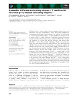
Fig. 1. ANX4 is up-regulated in CCA cell lines. Protein and RNA
were extracted from two CCA (OVTOKO and OVISE) cells lines
and one non-CCA (MCAS) cell line, and then ANX4 protein (A) and
mRNA (B) levels were compared by western blotting and real-time
RT-PCR analyses, respectively. Actin was included as a loading con-
trol. The values were normalized to the level of 18S ribosomal RNA
expression in each sample. Bars represent the mean ± SE of three
experiments.
Y. Masuishi et al. p53 is a positive regulator of annexin IV
FEBS Journal 278 (2011) 1470–1483 ª 2011 The Authors Journal compilation ª 2011 FEBS 1471
MCAS cells (Fig. 1B). These results indicate that the
expression level of ANX4 is increased in CCA cell
lines compared to non-CCA cell lines, as demonstrated
previously [13,15], and that ANX4 expression is con-
trolled at the level of transcription. To determine the
transcriptional factor responsible for these different
expression levels of ANX4, we performed pro-
moter ⁄ enhancer analysis of the ANX4 gene using these
three cell lines.
Determination of the 5¢-end of the ANX4 mRNA
To determine the 5¢-end of ANX4 mRNA, 5¢-RACE
analysis was performed using RNA isolated from
MCAS, OVTOKO and OVISE cultured cell lines. Sin-
gle DNA bands of the same size (170 bp) were
detected for each cell line by agarose gel electrophore-
sis of the 5¢-RACE products (Fig. 2A). Sequence anal-
yses verified that each band had the same sequence,
corresponding to the first through third exons of the
ANX4 cDNA reported in the GenBank database
(NM_001153.2), although the 5¢-end identified in the
present study was located upstream of the 5¢-end
reported in the database (Fig. 2B). We regarded the 5¢-
end determined by our 5¢-RACE analysis as a putative
transcription start site (+1) of ANX4.
The +180 region is essential for CCA-specific
transcriptional activity of ANX4
To identify the cis-elements essential for CCA-specific
expression of ANX4, we first isolated the region from
)1534 to +1010 relative to the transcriptional start
site and inserted it into a luciferase reporter vector
()1534 ⁄ +1010 luc). Consensus TATA-box sequences
were not found in the predicted positions of this
region, although the region from )586 to +402 was
identified as a CpG island (GC contents, 68%) using the
software cpg island researcher (http://cpgislands.
usc.edu/). The modified )1534 ⁄ +1010 luc vector was
MCAS
OVTOKO
OVISE
(kb)
300
200
100
500
1000
Size marker
A
B
Fig. 2. The 5¢-end of the ANX4 gene. To determine the transcriptional start site of ANX4 in EOC cells, the 5¢-end of ANX4 mRNA was inves-
tigated by 5¢-RACE analysis. (A) Agarose gel electrophoresis of PCR products from the 5¢-RACE procedure. The arrowhead indicates the
bands detected in three cell lines by 5¢-RACE. (B) The nucleotide sequence of the flanking region of the ANX4 transcription start site and
putative transcription factor-binding sites within this region. Uppercase letters indicate the first exon of ANX4. The asterisk and +1 show the
5¢-ends reported in the GenBank database (NM_001153.2) and identified newly in the present study, respectively. The putative binding
sequences for the representative transcription factors are underlined. The nucleotide positions at +180 and +270 are denoted by filled and
unfilled triangles, respectively.
p53 is a positive regulator of annexin IV Y. Masuishi et al.
1472 FEBS Journal 278 (2011) 1470–1483 ª 2011 The Authors Journal compilation ª 2011 FEBS
transfected into MCAS, OVTOKO and OVISE cells,
and the transcriptional activity was determined by
luciferase assay (Fig. 3A). The )1534 ⁄ +1010 region
demonstrated approximately nine- and four-fold higher
levels of transcriptional activity in OVTOKO and
OVISE cells, respectively, compared to MCAS cells.
This result is very similar to the real-time RT-PCR
data (Fig. 1B), suggesting that the )1534 ⁄ +1010
region contains an element essential for CCA-specific
expression of ANX4. Therefore, we constructed the
various 5¢-or3¢-deletion mutants of the modified
)1534 ⁄ +1010 luc vector, and measured the transcrip-
tional activity of each mutant (Fig. 3A). Deletion of
the 3¢-downstream region ()42 to +1010) resulted in a
marked decrease in luciferase activity in OVTOKO
and OVISE cells, although no change occurred in
MCAS cells. Further deletion of the 5¢-upstream
region from )181 decreased luciferase activity in all
three cell lines. By contrast, the deletion of the
5¢-upstream region from )43 alone also reduced lucif-
erase activity in all three cell lines, although it did not
completely diminish the higher activity seen in
OVTOKO and OVISE cells. This CCA-preferential
activity of the region between )43 and +1010 was
removed by deleting the 3¢-downstream region from
+28. These results suggest that an element essential
for CCA-specific expression of ANX4 is present
between +27 and +1010 in the downstream region of
the transcription start site. To further focus on the
region essential for CCA-specific gene expression,
serial 3¢-deletions were constructed and subjected to
luciferase reporter analysis (Fig. 3B). Deletion from
Luciferase activity (fold)
0 20406080100120
–1534
–43
+27
+1010
–181
Luciferase activity (fold)
0102050
–43
+1010
+397
+541
+282
+150
+27
OVTOKO
MCAS
OVISE
OVTOKO
MCAS
OVISE
Luciferase activity (fold)
del
del
01020304050607080
+160
+541
+180 +270
–
43
OVTOKO
MCAS
OVISE
A
B
C
Fig. 3. CCA-specific transcriptional activity
of ANX4 depends on the +180 region in the
first intron. The luciferase vector containing
the flanking region of the ANX4 transcrip-
tional start site )1534 ⁄ +1010 luc and its
deletion mutants were introduced into
OVTOKO, OVISE and MCAS cells, and the
transcriptional activities were measured.
Schematic diagrams of the ANX4 promoter–
luciferase plasmids are shown on the left,
where the 5¢- and 3¢-ends are indicated rela-
tive to the transcription start site. (A) The
3¢-downstream region of ANX4 is essential
for CCA-specific transcriptional activity. The
luciferase activities of the full-length
)1534 ⁄ +1010 luc vector and the mutants
with 5¢-upstream or 3¢-downstream dele-
tions were compared. (B) The transcriptional
activities of mutants with 3¢-deletions in the
region from )43 to +1010. (C) The effect of
deleting the +180 or +270 regions on the
transcriptional activities. Luciferase activity
is expressed as the fold change relative to
pGL3-basic vector activity in each cell. The
b-galactosidase control vector was co-trans-
fected as an internal control. Schematic
diagrams of the ANX4 promoter–luciferase
plasmids are shown on the left, where the
location of the 5¢- and 3¢-ends are indicated
relative to the transcription start site. Bars
represent the mean ± SE of at least three
experiments.
Y. Masuishi et al. p53 is a positive regulator of annexin IV
FEBS Journal 278 (2011) 1470–1483 ª 2011 The Authors Journal compilation ª 2011 FEBS 1473
+1010 up to +541, or from +541 to +397, resulted
in marked changes in luciferase activity in all three cell
lines. This suggests that the binding sites of both nega-
tive and positive regulatory transcription factors are
contained in these two regions, although their role in
ANX4 transcription is not specific to CCA cells. By
contrast, the deletion of +282 to +150 decreased
luciferase activity in OVTOKO and OVISE cells with-
out altering activity in MCAS cells, suggesting that
this region contains an element involved in CCA-spe-
cific expression of ANX4. In this region, the presence
of putative transcription factor-binding sites was
revealed by sequence analysis with the software
tfsearch ( />html) and motif ( searching
protein and nucleic acid sequence motifs. The nuclear
factor (NF)-jB and p53-binding sites were found at
position +180, and the GATA-binding site was found
at position +270 (Fig. 2B). To determine which site
was involved in CCA-specific ANX4 expression, repor-
ter analyses were performed using a luciferase construct
containing the region )43 to +541 ()43 ⁄ +541 luc)
and mutants of this construct with regions at either
+180 or +270 deleted. As shown in Fig. 3C, deleting
the +270 region did not change the transcriptional
activity of the )43 ⁄ +541 luc of any cell line, whereas
deleting the +180 region markedly decreased transcrip-
tional activity in OVTOKO and OVISE cells but not in
MCAS cells. Furthermore, CCA-specific transcriptional
activity conferred by the +180 region was diminished
by deleting the region upstream of +160. Accordingly,
the +180 region acts as a transcription enhancer essen-
tial for the up-regulation of ANX4 in CCA cells.
ANX4 expression is regulated by p53 in CCA
Potential binding sites for p53 and NF-jB were found
in the +180 region (Fig. 2B). To determine whether
these proteins conferred CCA-specific transcriptional
activation of ANX4, two kinds of mutation pat-
terns at the +180 region were designed. Both
mutations, +180 mutA (5¢-GG
CCAAGCGTA-3¢) and
+180 mutB (5¢-GGG
AAAGCCCC-3¢), abolished the
putative p53-binding site. In addition, +180 mutA
also destroyed the putative binding sequence for
NF-jB, whereas +180 mutB maintained the NF-jB-
binding sequence (5¢-GGRNNYCC-3¢). As shown in
Fig. 4a, both +180 mutA and +180 mutB markedly
decreased the transcriptional activity of the
)43 ⁄ +541 luc vector in OVTOKO and OVISE cells.
Mutations at the +180 region reduced the transcrip-
tional activity of the )1534 ⁄ +1010 luc vector by half
in CCA cells. Similar results were observed in the
other EOC cell lines. Mutations at the +180 region
significantly reduced transcriptional activity of the
)43 ⁄ +541 luc vector in the CCA cell lines RMG-I
and RMG-II compared to the non-CCA cell lines OV-
CAR-3 and RMUG-S (Fig. S1). These results suggest
that the +180 region acts as a p53-binding site in
CCA cells.
The p53 protein binds to two copies of the motif
5¢-RRRCWWGYYY-3¢, separated by a variable spacer
of length 0–13 bp [21]. The p53-binding motif in the
+180 region matched this sequence exactly. Three sites
at +161, +172 and +196 contained sequences similar
to the p53-binding motif, although each was an incom-
plete motif. To determine whether these act as other
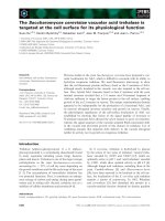
Fig. 4. p53 is a direct regulator of the ANX4 gene in CCA. (A) The effect of mutating the +180 region on transcriptional activity. Two muta-
tion patterns were made in the putative binding sequences for NF-jB and p53. In +180 mutA, both binding sequences were disrupted. In
+180 mutB, the p53-binding sequence was disrupted, whereas the NF-jB-binding sequence had 100% consensus. These mutations were
introduced into the indicated luciferase vectors. The b-galactosidase control vector was co-transfected with the luciferase vectors to normal-
ize transfection efficiency. *P < 0.05 and **P < 0.01 versus )1534 ⁄ +1010 luc. (B) The effect of mutating p53-binding motifs around the
+180 region. The p53-binding motif-like sequences around the +180 region in the )43 ⁄ +541 luc reporter were mutated (+180 mutA,
+161 mut, +172 mut and +196 mut). The mutants were transfected into OVISE cells, and transcriptional activities were measured via lucif-
erase assays. (C) p53 bound to the ANX4 gene. ChIP assay was performed with OVISE, OVTOKO and MCAS cells and antibodies against
p53. Immunoprecipitation of p53 protein–DNA complexes was conducted with control IgG or anti-p53 antibody (DO-1) or without antibody
(noAb). Total lysate was used as a control for PCR amplification (input). PCR was performed with gene-specific primers for p21 and ANX4.
As a positive control, p53 binding was tested with p21 specific primers targeting the genomic region harboring the p53-responsive element.
The results displayed are representative of the findings from three independent experiments. (D) The expression levels of p53 in cells trans-
fected with StealthÔ siRNA. Cell lines were transfected with siRNA and grown for 72 h, and then p53 protein levels were determined by
western blotting. Representative western blots of three experiments are shown. Actin was included as a loading control. (E) The CCA-spe-
cific transcriptional activities of )43 ⁄ +541 luc were suppressed by introducing p53 siRNA. siRNA-transfected cells were incubated for 24 h
in one well of a 24-well plate, and then transfected with the )43 ⁄ +541 luc vector and grown in culture for 24 h. The pRL-TK vector was
co-transfected with the luc vector used as an internal control. (F) siRNA-transfected cells were grown for 72 h, and then the mRNA levels of
ANX4 were quantified by real-time RT-PCR. All luciferase activity is expressed as the fold change relative to pGL3-basic vector activity.
Schematic diagrams of the ANX4 promoter–luciferase plasmids are shown on the left, where the location of the 5¢- and 3¢-ends are indicated
relative to the transcription start site (A, B and E). All bars represent the mean ± SE of at least three experiments (A, B, E and F).
p53 is a positive regulator of annexin IV Y. Masuishi et al.
1474 FEBS Journal 278 (2011) 1470–1483 ª 2011 The Authors Journal compilation ª 2011 FEBS
binding sites for p53, we introduced a mutation at
each of these predicted sites in the )43 ⁄ +541 luc vec-
tor, and compared the levels of transcriptional activity.
As shown in Fig. 4B, similar to the mutation at +180,
mutating the +196 region also significantly reduced
transcriptional activity. Although incomplete on its
own, the p53-binding motif in the +196 region was
6 bp distal to the motif in the +180 region. The two
motifs separated by a 6 bp spacer length is consistent
with the criteria for a p53-binding domain described
by Vogelstein et al. [21]. These findings suggest that
the motifs in the +180 and +196 regions might be
targets for p53 binding.
To examine whether endogenous p53 actually binds
to these regions in CCA cells, we performed chromatin
immunoprecipitation (ChIP) assays using PCR analysis
of the p53 binding domains regulating ANX4 and p21
after immunoprecipitation with the p53-specific anti-
–+–+–+
A
B
D
F
E
C
Y. Masuishi et al. p53 is a positive regulator of annexin IV
FEBS Journal 278 (2011) 1470–1483 ª 2011 The Authors Journal compilation ª 2011 FEBS 1475
body DO-1 or normal IgG as a negative control. As
shown in Fig. 4C, immunoprecipitation by the DO-1
antibody detected not only the p21 promoter, but also
the first intron of the ANX4 gene in OVTOKO and
OVISE cells but not in MCAS cells, indicating that
endogenous p53 protein directly binds to the ANX4
gene in CCA cells.
To verify the involvement of p53 in the CCA-specific
expression of ANX4, we performed a gene-silencing
experiment to suppress p53 protein expression. In cells
transfected with a chemically modified small interfering
RNA (siRNA) (StealthÔ siRNA) targeting p53
mRNA, the protein level of endogenous p53 markedly
decreased (Fig. 4D). As shown in Fig. 4E, knockdown
of p53 significantly reduced the transcriptional activity
of the )43 ⁄ +541 luc reporter in CCA cell lines but
not in MCAS cells. By contrast, knockdown of p53
did not affect the activity of the reporter in any cell
lines when the +180 region was mutated. These results
indicate that p53 enhanced the transcriptional activity
of ANX4 via the +180 region. Similar data were
obtained by knocking down p53 with another
StealthÔ siRNA that targets a different site on the p53
gene (data not shown). To confirm that the p53 pro-
tein actually regulates the expression of ANX4
mRNA, real-time RT-PCR analysis was conducted
using the siRNA-transfected cells. As shown in
Fig. 4F, introducing p53 siRNA reduced ANX4
mRNA in the CCA cell lines but did not affect ANX4
mRNA levels in the MCAS cells. These results indicate
that ANX4 is regulated by p53 in CCA cells.
Although the p53-directed siRNA completely dimin-
ished the CCA-specific transcriptional activity of the
)43 ⁄ +541 luc, it only reduced the ANX4 mRNA in
CCA cells by approximately half. This discrepancy was
also observed after mutations of the +180 region in
the luciferase reporter vectors. In CCA cell lines, muta-
tion of the +180 region completely diminished the
transcriptional activity of )43 ⁄ +541 luc, although the
same mutation in )1534 ⁄ +1010 luc, the reporter with
the longest region, decreased transcriptional activity
only by approximately half (Fig. 4A). Therefore, the
transcriptional activation of ANX4 in CCA is, at least
in part, caused by p53, and other transcription factors
with binding sites upstream of )43 or downstream of
+541 might provide moderate additional transcrip-
tional regulation.
ANX4 transcriptional activity correlates with the
functional status of p53 in EOC cells
In almost all human cancers, p53 activity is lost as a
result of mutation of the p53 gene [22]. However, the
above findings show that the ANX4 gene is regulated
by p53 in CCA cells, thereby suggesting that p53 is
functional in CCA cells. To examine whether there is
a correlation between the functional status of p53
and ANX4 transcriptional levels, we investigated p53
gene mutations, as well as the expression levels of
p53, ANX4 and typical p53 target genes. As shown
in Fig. 5A, the p53 antibody DO-1 detected major
bands near 53 kDa in EOC cell lines. Because the
DO-1 antibody would also recognize p53b and p53c,
C-terminal truncated forms of the typical full-length
p53 protein [23], the absence of bands at 46 kDa
indicate that these proteins were not expressed in any
of the EOC cell lines. Analysis of the p53 cDNA
sequences obtained from each cell line revealed no
mutations in the CCA cell lines, whereas all non-
CCA-type EOC cell lines had p53 mutations
(Table 1). Although the levels of p53 protein were
lower in CCA cell lines, those of p53 target genes,
p21 and murine double minute 2 (MDM2), as well as
ANX4, were significantly higher in CCA cell lines
than non-CCA-type EOC cell lines (Fig. 5A). In addi-
tion, in other cell lines carrying the wild-type p53
gene, HEK293 or LNCaP cell lines, protein levels of
ANX4, p21 and MDM2 were undetectable by wes-
tern blotting (data not shown). Similar results were
obtained by real time RT-PCR analyses; the mRNA
levels of p21 and MDM2 were relatively lower in
either HEK293, LNCaP or non-CCA-type EOC cell
lines, which did not abundantly express ANX4
(Fig. 5B). These results suggest that there is a corre-
lates between the functional status of p53 and ANX4
expression.
Wild-type p53 enhances the expression of the
ANX4 gene
The results reported above suggest that the activation
of wild-type p53 is one factor leading to ANX4 up-reg-
ulation in CCA. To examine whether wild-type p53 is
actually involved in the transcriptional activation of
ANX4, we transfected the
)43 ⁄ +541 luc and an
expression plasmid containing wild-type p53 cDNA
into MCAS, HEK293 and LNCaP cells (in which
ANX4 levels are very low) and then conducted a
luciferase assay. As shown in Fig. 6A, the overexpres-
sion of wild-type p53 resulted in a marked increase in
ANX4 transcriptional activity in each cell line. By con-
trast, transfection with the p53 mutants found in the
non-CCA-type EOC cell lines, MCAS or OVCAR-3,
did not alter luciferase activities in MCAS, HEK293
or LNCaP cells. As shown in Fig. 6B, ANX4 mRNA
levels were substantially increased with the induction
p53 is a positive regulator of annexin IV Y. Masuishi et al.
1476 FEBS Journal 278 (2011) 1470–1483 ª 2011 The Authors Journal compilation ª 2011 FEBS
0
1
2
3
4
5
6
0
1
2
3
4
5
6
RMG-II
RMUG-S
OVKATE
OVSAHO
OVCAR-3
RMG
-
I
OVISE
OVTOKO
MCAS
OVMANA
OVSAYO
CCA
Non-CCA
HEK293
LNCaP
EOC
Non-EOC
p21 mRNA levevl (fold)
RMG-II
RMUG-
S
OVKATE
OVSAHO
OVCAR-3
RMG-I
OVISE
OVTOKO
MCAS
OVMANA
OVSAYO
CCA
Non-CCA
HEK293
LNCaP
EOC Non-EOC
0
5
10
15
25
20
MDM2 mRNA levevl (fold)
Actin
OVCAR-
3
OVMANA
OVTOKO
OVSAHO
OVKATE
RMUG
-S
MCAS
OVISE
RMG-I
RMG-II
OVSAYO
CCANon-CCA
p53 (Do-1)
50 k
75 k
37 k
p21
ANX4
MDM2
RMG
-
II
RMUG-S
OVKATE
OVSAHO
OVCAR
-
3
RMG-I
OVISE
OVTOKO
MCAS
OVMANA
OVSAYO
CCANon-CCA
HEK293
LNCaP
EOC
Non-EOC
0
2
4
6
10
8
12
ANX4 mRNA level (fold)
A
B
Fig. 5. ANX4 expression level correlates with p53 functional status. Protein and total RNA were extracted from various EOC cell lines,
HEK293 and LNCaP cell lines. (A, B) Expression levels of protein and mRNA, and levels of p53, ANX4 and the known p53 targets, p21 and
MDM2, were analyzed by western blotting (A) and real-time RT-PCR analyses (B), respectively. Actin protein levels were included in the
western blotting analysis as a loading control. The relative mRNA levels were normalized to the level of 18S ribosomal RNA expression in
each sample.
Table 1. p53 mutation lines used in the present study.
Cell line Exon Codon Mutation Amino acid change EOC subtype
OVCAR-3 7 248 cgg fi cag R fi Q Serous
OVSAHO 10 342 cga fi tga R fi Stop Serous
OVKATE 8 282 cgg fi tgg R fi W Serous
RMUG-S 4, 10 72, 347 cgc fi ccc, gcc fi gtc R fi P, A fi V Mucinous
MCAS 4 114–125 Alternative sequence LHSGTAKSVTCT fi FTLWLP Mucinous
OVTOKO – – Not detected – Clear cell
OVISE – – Not detected – Clear cell
RMG-I – – Not detected – Clear cell
RMG-II – – Not detected – Clear cell
OVMANA – – Not detected – Clear cell
OVSAYO – – Not detected – Clear cell
HEK293 – – Not detected – –
LNCaP – – Not detected – –
Y. Masuishi et al. p53 is a positive regulator of annexin IV
FEBS Journal 278 (2011) 1470–1483 ª 2011 The Authors Journal compilation ª 2011 FEBS 1477
of p21 mRNA in HEK293 cells transfected with
the wild-type p53 expression vector. An increase in
ANX4 mRNA was not observed in response to the
overexpression of the p53 mutants. Moreover, when
LNCaP cells, which endogenously express wild-type
p53, were treated with the p53-activating reagent mito-
mycin C (MMC) or nutlin-3, p21 mRNA and protein
levels were elevated along with increase in endogenous
p53. Activation of endogenous p53 also increased
mRNA and protein levels for ANX4 in LNCaP cells
(Fig. 6C, D). These findings support the conclusion
that wild-type p53 plays a role in the up-regulation of
ANX4.
Discussion
The expression of ANX4 is specifically and characteris-
tically enhanced in ovarian CCA cells. This suggests
that the expression of ANX4 is regulated by a molecu-
lar mechanism that is unique to these cells. However,
the mechanisms for ANX4 up-regulation in CCA cells
have not been elucidated. In the present study, we
identified tandem repeats corresponding to the motif
for p53 binding in the first intron of the ANX4 gene,
and found (using reporter gene analysis) that this
region is a key site for CCA-specific expression. Gene
silencing of p53 by siRNA restricted ANX4 transcrip-
Nutlin-3 (μM)Nutlin-3 (μM)
Nutlin-3 (μ
M)
D
C
AB
MMC (μM)
MMC (μ
M)
MMC (μ
M)
Fig. 6. Wild-type p53 induces ANX4 gene expression. (A) The overexpression of wild-type p53 enhances the transcriptional activity of the
ANX4-luciferase reporter. The )43 ⁄ +541 luc was co-transfected into MCAS, HEK293 and LNCaP with pcDNA3 plasmids encoding the wild-
type or mutant forms of p53. Mutant forms 1 and 2 were p53 cDNA cloned from OVCAR-3 and MCAS, respectively. After 48 h, luciferase
activity was determined for each sample. The Renilla luciferase reporter vector was co-transfected as an internal control. (B) Overexpression
of wild-type p53 activates the expression of ANX4. Wild-type or mutated p53 expression vectors were transfected into HEK293. After 48 h,
total RNA was extracted and ANX4 mRNA levels were measured by real-time RT-PCR analyses. (C, D) ANX4 expression increased after p53
activation by MMC or nutlin-3 exposure. LNCaP cells were treated with MMC (C) or nutlin-3 (D). After treatment with MMC for 24 h or nut-
lin-3 for 12 h at the indicated concentrations, mRNA and protein levels of ANX4 and p21 were measured by western blotting and real-time
RT-PCR analyses. The p53 protein levels were also assessed by western blotting to verify that MMC and nutlin-3 activated p53 effectively.
The relative mRNA levels were normalized to the level of 18S ribosomal RNA expression in each sample. Actin protein levels were included
in the western blotting analysis as a loading control. Bars represent the mean ± SE of three experiments.
p53 is a positive regulator of annexin IV Y. Masuishi et al.
1478 FEBS Journal 278 (2011) 1470–1483 ª 2011 The Authors Journal compilation ª 2011 FEBS
tion in CCA cells but not in non-CCA-type EOC cells.
No mutations of the p53 gene were observed in any of
the CCA-derived cell lines used in the present study,
and p21 and MDM2 transcript levels were relatively
higher compared to those in other cell lines in which
ANX4 is not abundantly expressed. Moreover, the
mRNA levels of ANX4 in other types of cell lines were
significantly increased by the overexpression or activa-
tion of wild-type p53. Therefore, we conclude that
wild-type p53 acts as a positive regulator of ANX4
expression in CCA cells.
The characteristic up-regulation of ANX4 in CCA
led us to consider that the protein might be involved
in the malignance of CCA by conferring drug resis-
tance or accelerating cancer development. Unexpect-
edly, we found that the expression of the ANX4 gene
is directly regulated by the tumor suppressor protein
p53 in CCA cells. In general, p53 is known to serve as
a key player in responding to cellular stresses such as
DNA damage, oncogenic activation and microtubule
disruption [24,25]. When p53 is activated by such cellu-
lar stress, the protein exerts its effect mainly through
the transcriptional activation of target genes, including
p21, which arrests the cell cycle, and BAX, which
induces apoptosis. Thus, p53 typically suppresses can-
cer development, preventing the division of damaged
cells likely to contain mutations and exhibit abnormal
cellular growth [26]. Indeed, the p53 gene is mutated
frequently in almost all human cancers [22]. However,
among the EOC cell lines used in the present study,
p53 mutations were not observed in any of the CCA-
type cell lines, although they were detected in all non-
CCA cell lines, which express very low levels of ANX4
(Fig. 5B and Table 1). These findings are in good
agreement with studies reporting that p53 mutations
are infrequent in ovarian CCA but occur in at least
50% of the other subtypes of EOC [6–9]. Furthermore,
the overexpression of wild-type p53 resulted in an
increase in the number of p21 and ANX4 transcripts,
whereas overexpressing p53 mutants found in non-
CCA cell lines had no effect on the transcription of
either gene (Fig. 6). These results show that the p53
mutants in non-CCA cells were inert, compatible with
previous findings that p53 mutations generally result in
a loss of wild-type protein activity, dominant-negative
activity [27] or an increase in the half-life of the pro-
tein by preventing ubiquitination [28]. Therefore, the
absence of p53 mutation contributes to the up-regula-
tion of ANX4 in CCA cells. Furthermore, the func-
tional status of p53 was more important. Despite
having an intact p53 gene, HEK293 and LNCaP cell
lines expressed trace amounts of ANX4 (Fig. 5).
Expression levels of p21 or MDM2 are higher in CCA
cell lines than those of HEK293 and LNCaP cell lines,
showing a correlation with the expression level of
ANX4. Previous immunohistological studies also
showed that p21 and MDM2 protein is higher in many
ovarian CCA tissues compared to that found in the
other EOC subtypes [29,30]. The data obtained in the
present study together with those of these previous
reports suggest that p53 functional status is critical in
governing the ANX4 up-regulation in EOC cells.
Several previous studies have suggested a close rela-
tionship between wild-type p53 and ANX4 expression.
ANX4 expression is elevated in renal clear cell carci-
noma [31], where p53 gene mutations are rare [32], and
p21 expression has been confirmed by immunohisto-
chemical methods [33]. Moreover, comprehensive
expression analysis of p53-induced genes using the p53
temperature-sensitive cell model revealed that ANX4
mRNA was induced after the activation of p53 [34].
ChIP-on-chip analysis using lymphoblastoid cells
exposed to ionizing radiation identified 38 kinds of
p53-binding genes, and the ANX4 gene was among the
identified genes [35]. These studies strongly support
our finding that activated wild-type p53 directly regu-
lates the expression of ANX4 in CCA cells.
In general, p53 has been shown to induce not only
genes involved in tumor suppression, such as those
that arrest the cell cycle, induce apoptosis and show
anti-angiogenic activity, but also oncogenes such
as MDM2, p53-inducible protein with RING-H2
domain (PIRH2) and constitutively photomorphogenic
1(COP1) [36–38]. These oncogenes are cellular ubiqu-
itin-protein ligases that bind to the p53 protein directly
and regulate cellular p53 levels through ubiquitination.
The proteasomal degradation of the p53 protein, regu-
lated by a negative feedback mechanism, has been
shown to contribute to tumor development. Whether
ANX4 should be classified as an oncogene or as a
tumor suppressor remains unknown because little is
known about its functional role, although ANX4 is
reported to be involved in chemoresistance [15,20],
activation of chloride ion channels [19], exocytosis [18]
and membrane permeability [17]. To clarify the func-
tional and physiological role of the ANX4 protein in
ovarian CCA, we are currently conducting proteomic
analyses to identify its binding partners.
Because ovarian CCA shows a lower response to the
standard paclitaxel–carboplatin combination chemo-
therapy, a patient with this disease has a worse prog-
nosis than patients with other EOC subtypes,
especially serous adenocarcinoma [2]. In CCA, p53
mutation is infrequently observed [8,9]. Some studies
have investigated whether the presence of p53 muta-
tions correlates with the response to platinum-based
Y. Masuishi et al. p53 is a positive regulator of annexin IV
FEBS Journal 278 (2011) 1470–1483 ª 2011 The Authors Journal compilation ª 2011 FEBS 1479
chemotherapy in EOC patients. Lavarino et al. [39]
and Ueno et al. [40] found that, overall, EOCs with
wild-type p53 are less responsive to paclitaxel-carbopl-
atin chemotherapy than EOCs with mutated p53
[39,40]. Moreover, when Ueno et al. [40] investigated
individual EOC subtypes, they observed that this cor-
relation apparently existed in EOC subtypes other than
the serous type. Two other interesting studies have
reported the suggested involvement of ANX4 in the
chemoresistance of human cancer cell lines. Han et al.
[20] found that the level of ANX4 protein expression
was higher in a paclitaxel-resistant cell line derived
from a lung cancer cell line than in the parent cell line,
and that overexpression of ANX4 cDNA enhanced
resistance to paclitaxel in HEK293T cells. Moreover,
Kim et al. [15] also investigated whether ANX4 was
associated with chemoresistance in EOC cell lines, and
found that an ANX4-overexpressing cell line derived
from the serous-type EOC cell line OVSAHO exhibited
greater resistance to carboplatin compared to the
parental cell line [15]. Taken together, the findings of
these previous studies and our own reveal an associa-
tion between p53 and ANX4 expression that suggests
that tumor cells carrying wild-type p53, such as CCA,
may exhibit chemoresistance conferred by p53-depen-
dent ANX4 expression.
In conclusion, analysis of molecular mechanisms
underlying CCA-specific ANX4 expression has revealed
that the functional status of p53 is involved in the gene
regulation in EOC cells. This may lead to a better
understanding of the physiological significance of
ANX4 up-regulation and the mechanisms underlying
malignant progression and chemoresistance in CCA.
Experimental procedures
Cell cultures
Three ovarian cancer cell lines were used for most of the
experiments in this study: OVTOKO and OVISE established
from ovarian CCA [41], and MCAS, a cell line originating
from ovarian mucinous cystadenocarcinoma cloned, as
described previously [13]. In some experiments, eight more
ovarian cancer cell lines were also used to verify our results.
OVKATE, OVSAHO, OVMANA and OVSAYO were
established from metastasis ovarian tumors by Yanagibashi
et al. [42]. OVCAR-3 was obtained from the RIKEN (Tsu-
kuba, Japan) cell bank, and RMUG-S, RMG-I and RMG-
II were purchased from the Japanese Collection of Research
Bioresources (Tokyo, Japan). RMUG-S, RMG-I and
RMG-II were maintained in Ham’s F-12 medium, and the
other cell lines were cultured in RPMI medium. The human
embryonic kidney cell line HEK293 and the prostate adeno-
carcinoma cell line LNCaP were grown in Ham’s F-12 and
RPMI 1690 mediums, respectively. All media were supple-
mented with 10% fetal bovine serum (JRH Biosciences,
Inc., Lenexa, KS, USA). Cells were kept at 37 °Cina
humidified atmosphere supplemented with 5% CO
2
.
Western blotting
Protein was extracted from cells using 30 mm Tris-HCl
buffer (pH 7.5) containing 7 m urea, 2 m thiourea, 4%
Chaps and 1% dithithreitol. The protein extracts were sep-
arated by SDS ⁄ PAGE, transferred to poly(vinylidene diflu-
oride) membranes, and blocked by incubation in the
reagent Blocking One (Nacalai Tesque, Kyoto, Japan).
The blots were then reacted with one of the primary anti-
bodies: goat polyclonal anti-ANX4 (N-19), goat polyclonal
anti-actin (I-19), rabbit polyclonal anti-p21 (C-19) and
mouse monoclonal anti-MDM2 (SMP-14); all purchased
from Santa Cruz Biotechnology (Santa Cruz, CA, USA).
Mouse monoclonal anti-ANX4 (No. 50) and anti-p53
(DO-1) were purchased from Funakoshi (Tokyo, Japan)
and Calbiochem (San Diego, CA, USA), respectively. Pri-
mary antibodies were detected using the ECL Plus Wes-
tern Blotting Detection System (GE Healthcare,
Milwaukee, WI, USA).
Real-time RT-PCR
Total RNA was isolated from the various cell lines using
the RNeasy Plus Micro Kit (Qiagen, Hilden, Germany).
cDNA was synthesized from the isolated RNA by reverse
transcription with the oligo-dT primer and the 18S-rRNA
specific primer as described in Zhu and Altmann [43] with
one modification, namely, the use of the PrimeScript RT
reagent (Takara Bio Inc., Shiga, Japan). Real-time PCR
was performed using the Mx3000P Real-Time QPCR Sys-
tem (Agilent Technologies, Santa Clara, CA, USA) with
SYBR Premix Ex TaqÔ II Perfect Real Time (Takara Bio
Inc.). The primer pairs indicated in Table S1 were used for
the reactions at a concentration of 10 lm. The PCR prod-
ucts were detected by monitoring the increase in reporter
dye fluorescence. mRNA levels were normalized to 18S
ribosomal RNA levels.
5¢-RACE analysis
Total RNAs isolated from OVTOKO, OVISE and MCAS
were reverse-transcribed using the PowerScript reverse
transcriptase (Clontech Laboratories, Palo Alto, CA, USA)
with the ANX4-RT primer, which is complementary to the
nucleotide sequence of the human ANX4 mRNA (Gen-
Bank accession number: BC001153). dCTP tails were added
to the cDNAs using terminal deoxytransferase (Invitrogen,
Carlsbad, CA, USA), and then PCR amplification was per-
p53 is a positive regulator of annexin IV Y. Masuishi et al.
1480 FEBS Journal 278 (2011) 1470–1483 ª 2011 The Authors Journal compilation ª 2011 FEBS
formed with the oligo-dI-dG primer and the ANX4-R01
primer (Table S2). The RACE end determined by sequenc-
ing analysis was regarded as the transcription start site of
ANX4 and denoted as +1.
Plasmids
For production of luciferase reporter constructs, the flank-
ing region of the transcription start site of ANX4, from
)1534 to +1010, was amplified from human genomic
DNA (Novagen, Darmstadt, Germany) by PCR using
KOD-plus DNA polymerase (Toyobo Life Science, Osaka,
Japan), and then cloned into the SmaI ⁄ BglII site of pGL3-
basic vector (Promega, Madison, WI, USA). The 5¢-or
3¢-deletion constructs were produced by reacting the ampli-
fied PCR products using the primers shown in Table S2
with restriction enzymes. Deletions and mutations in the
+180 region were performed by ligating two PCR frag-
ments amplified with a mutation primer, as described previ-
ously [44]. To construct the wild-type and mutated p53
expression vectors, full-length p53 cDNAs were isolated
from OVISE, OVCAR-3 and MCAS by PCR amplification
and then cloned into the HindIII ⁄ EcoRV site of the
pcDNA3.1 plasmid (Invitrogen). All constructs were
sequenced to verify the orientation and fidelity of the insert.
Luciferase reporter assay
EOC cell lines were seeded on 24-well plates at a density of
2.0 · 10
5
, 3.0 · 10
5
and 2.5 · 10
5
cells per well for MCAS,
OVTOKO and the other cell lines, respectively. After 24 h,
cells were transfected with a pGL3 reporter vector and a
pSV-b-galactosidase control vector as an internal control
(Promega) using FuGENE HD (Roche, Indianapolis, IN,
USA) in accordance with the manufacturer’s instructions.
For experiments in which p53 was overexpressed, the pRL-
TK vector (Promega) was used as an internal control.
Then, 42 h after transfection, luciferase activity in cell
lysates was measured and normalized to either b-galactosi-
dase activity or Renilla luciferase activity.
ChIP
ChIP assays were performed using the ChIP-IT kit (Active
Motif, Carlasbad, CA, USA) in accordance with the manu-
facturer’s instructions. In brief, OVISE, OVTOKO and
MCAS cells at 70–80% confluence in 15 cm plates were
fixed for 15 min at room temperature with 1% formalde-
hyde. To shear genomic DNA, the nuclei were subjected to
enzymatic digestion with 5 units of enzymatic shearing mix-
ture solution (Active Motif) for 15 min at 37 °C. Sheared
chromatin was immunoprecipitated with 4 lg of anti-p53
(DO-1; Calbiochem) or control IgG (Active Motif). Cross-
linking was reversed and purified DNA was subjected to
PCR. The PCR products were analyzed by electrophoresis
on a 2% agarose gel stained with ethidium bromide. Prim-
ers employed were designed to detect the predicted p53
binding sites on ANX4 and p21 genes. The primer
sequences are indicated in Table S3.
Gene silencing of p53
StealthÔ siRNAs (Invitrogen) were used to silence the p53
gene. Two kinds of StealthÔ siRNAs were tested for their
RNA interference (RNAi) activity against the p53 gene,
and the one resulting in a higher level of knockdown was
selected for further use. The targeted sequence of the
selected siRNA was 5¢-UGGAAGACUCCAGUGGUA-
AUCUACU-3¢, corresponding to nucleotides 890–914 of
the p53 mRNA (GenBank accession number: BC003596).
Control experiments used the StealthÔ RNAi negative con-
trol MED (Invitrogen). EOC cells were transfected with the
StealthÔ siRNAs using Lipofectamine RNAi MAX (Invi-
trogen) in accordance with the manufacturer’s instructions.
For the luciferase assay, siRNA-transfected cells were incu-
bated for 24 h in one well of a 24-well plate, and then
transfected with reporter vectors. For western blotting or
real-time RT-PCR analyses, all cell lines were transfected
with siRNA and grown for 72 h.
p53 mutation analysis
The p53 cDNAs from various cell lines were amplified by
PCR using the KOD-plus DNA polymerase (Toyobo Life
Science) and p53-specific primers (sense 5¢-CACGACGGT
GACACGCTTCC-3¢ and antisense 5¢-CCTGGGTGCTT
CTGACGCAC-3¢) corresponding to nucleotides 64–83 and
1404–1423 of the p53 mRNA, respectively (GenBank acces-
sion number: BC003596). The PCR products were purified
using the Wizard SV Gel and the PCR Clean-Up System
(Promega) and then subjected to sequence analyses.
p53 activation by drug treatment
LNCaP cells were grown to 60–70% confluency in six-well
plates, and then treated with different concentrations of
MMC (Calbiochem) for 24 h, or nutlin-3 (Cayman Chemi-
cal, Ann Arbor, MI, USA) for 12 h. After treatment, cells
were subjected to real-time RT-PCR and western blotting.
Acknowledgements
This work was supported in part by a Grant-in-Aid
for young Scientists (B) 18790226 and 20790262 from
The Ministry of Education, Culture, Sports, Science
and Technology, Japan. We thank Dr Youhei Miyagi
(Kanagawa Cancer Center, Kanagawa, Japan) and Dr
Masato Katsuyama (Kyoto Prefectural University of
Medicine, Kyoto, Japan) for insightful discussions.
Y. Masuishi et al. p53 is a positive regulator of annexin IV
FEBS Journal 278 (2011) 1470–1483 ª 2011 The Authors Journal compilation ª 2011 FEBS 1481
References
1 Bray F, Loos AH, Tognazzo S & La Vecchia C (2005)
Ovarian cancer in Europe: cross-sectional trends in inci-
dence and mortality in 28 countries, 1953-2000. Int J
Cancer 113, 977–990.
2 Sugiyama T, Kamura T, Kigawa J, Terakawa N,
Kikuchi Y, Kita T, Suzuki M, Sato I & Taguchi K
(2000) Clinical characteristics of clear cell carcinoma of
the ovary: a distinct histologic type with poor prognosis
and resistance to platinum-based chemotherapy. Cancer
88, 2584–2589.
3 Itamochi H, Kigawa J, Akeshima R, Sato S, Kamazawa
S, Takahashi M, Kanamori Y, Suzuki M, Ohwada M
& Terakawa N (2002) Mechanisms of cisplatin resis-
tance in clear cell carcinoma of the ovary. Oncology 62,
349–353.
4 Itamochi H, Kigawa J, Sugiyama T, Kikuchi Y,
Suzuki M & Terakawa N (2002) Low proliferation
activity may be associated with chemoresistance in clear
cell carcinoma of the ovary. Obstet Gynecol 100, 281–287.
5 Itamochi H, Kigawa J & Terakawa N (2008) Mecha-
nisms of chemoresistance and poor prognosis in ovarian
clear cell carcinoma. Cancer Sci 99, 653–658.
6 Marks JR, Davidoff AM, Kerns BJ, Humphrey PA,
Pence JC, Dodge RK, Clarke-Pearson DL, Iglehart JD,
Bast RC Jr & Berchuck A (1991) Overexpression and
mutation of p53 in epithelial ovarian cancer. Cancer
Res 51, 2979–2984.
7 Kohler MF, Marks JR, Wiseman RW, Jacobs IJ,
Davidoff AM, Clarke-Pearson DL, Soper JT, Bast RC
Jr & Berchuck A (1993) Spectrum of mutation and fre-
quency of allelic deletion of the p53 gene in ovarian
cancer. J Natl Cancer Inst 85, 1513–1519.
8 Ho ES, Lai CR, Hsieh YT, Chen JT, Lin AJ, Hung
MH & Liu FS (2001) p53 mutation is infrequent in
clear cell carcinoma of the ovary. Gynecol Oncol 80,
189–193.
9 Okuda T, Otsuka J, Sekizawa A, Saito H, Makino R,
Kushima M, Farina A, Kuwano Y & Okai T (2003)
p53 mutations and overexpression affect prognosis of
ovarian endometrioid cancer but not clear cell cancer.
Gynecol Oncol 88, 318–325.
10 Saegusa M, Machida BD & Okayasu I (2001) Possible
associations among expression of p14(ARF),
p16(INK4a), p21(WAF1 ⁄ CIP1), p27(KIP1), and p53
accumulation and the balance of apoptosis and cell
proliferation in ovarian carcinomas. Cancer 92, 1177–
1189.
11 Schaner ME, Ross DT, Ciaravino G, Sorlie T, Troyans-
kaya O, Diehn M, Wang YC, Duran GE, Sikic TL,
Caldeira S et al. (2003) Gene expression patterns in
ovarian carcinomas. Mol Biol Cell 14, 4376–4386.
12 Schwartz DR, Kardia SL, Shedden KA, Kuick R,
Michailidis G, Taylor JM, Misek DE, Wu R, Zhai Y,
Darrah DM et al. (2002) Gene expression in ovarian
cancer reflects both morphology and biological behav-
ior, distinguishing clear cell from other poor-prognosis
ovarian carcinomas. Cancer Res 62, 4722–4729.
13 Morita A, Miyagi E, Yasumitsu H, Kawasaki H,
Hirano H & Hirahara F (2006) Proteomic search for
potential diagnostic markers and therapeutic targets for
ovarian clear cell adenocarcinoma. Proteomics 6, 5880–
5890.
14 Zhu Y, Wu R, Sangha N, Yoo C, Cho KR, Shedden
KA, Katabuchi H & Lubman DM (2006) Classifica-
tions of ovarian cancer tissues by proteomic patterns.
Proteomics 6, 5846–5856.
15 Kim A, Enomoto T, Serada S, Ueda Y, Takahashi T,
Ripley B, Miyatake T, Fujita M, Lee CM, Morimoto K
et al. (2009) Enhanced expression of annexin A4 in
clear cell carcinoma of the ovary and its association
with chemoresistance to carboplatin. Int J Cancer 125,
2316–2322.
16 Moss SE & Morgan RO (2004) The annexins. Genome
Biol 5, 219.
17 Hill WG, Kaetzel MA, Kishore BK, Dedman JR &
Zeidel ML (2003) Annexin A4 reduces water and
proton permeability of model membranes but does not
alter aquaporin 2-mediated water transport in isolated
endosomes. J Gen Physiol 121, 413–425.
18 Sohma H, Creutz CE, Gasa S, Ohkawa H, Akino T &
Kuroki Y (2001) Differential lipid specificities of the
repeated domains of annexin IV. Biochim Biophys Acta
1546, 205–215.
19 Xie W, Kaetzel MA, Bruzik KS, Dedman JR,
Shears SB & Nelson DJ (1996) Inositol 3,4,5,6-tetrakis-
phosphate inhibits the calmodulin-dependent protein
kinase II-activated chloride conductance in T84 colonic
epithelial cells. J Biol Chem 271, 14092–14097.
20 Han EK, Tahir SK, Cherian SP, Collins N & Ng SC
(2000) Modulation of paclitaxel resistance by
annexin IV in human cancer cell lines. Br J Cancer 83,
83–88.
21 el-Deiry WS, Kern SE, Pietenpol JA, Kinzler KW &
Vogelstein B (1992) Definition of a consensus binding
site for p53. Nat Genet 1, 45–49.
22 Levine AJ, Momand J & Finlay CA (1991) The p53
tumour suppressor gene. Nature 351, 453–456.
23 Bourdon JC, Fernandes K, Murray-Zmijewski F,
Liu G, Diot A, Xirodimas DP, Saville MK & Lane DP
(2005) p53 isoforms can regulate p53 transcriptional
activity. Genes Dev 19, 2122–2137.
24 Meek DW (1998) Multisite phosphorylation and the inte-
gration of stress signals at p53. Cell Signal 10, 159–166.
25 Lane DP (1992) Cancer. p53, guardian of the genome.
Nature 358, 15–16.
26 Ryan KM, Phillips AC & Vousden KH (2001) Regula-
tion and function of the p53 tumor suppressor protein.
Curr Opin Cell Biol 13, 332–337.
p53 is a positive regulator of annexin IV Y. Masuishi et al.
1482 FEBS Journal 278 (2011) 1470–1483 ª 2011 The Authors Journal compilation ª 2011 FEBS
27 Levesque MA, Katsaros D, Yu H, Zola P, Sismondi P,
Giardina G & Diamandis EP (1995) Mutant p53 pro-
tein overexpression is associated with poor outcome in
patients with well or moderately differentiated ovarian
carcinoma. Cancer 75, 1327–1338.
28 Eltabbakh GH, Belinson JL, Kennedy AW, Biscotti
CV, Casey G, Tubbs RR & Blumenson LE (1997) p53
overexpression is not an independent prognostic factor
for patients with primary ovarian epithelial cancer.
Cancer 80, 892–898.
29 Shimizu M, Nikaido T, Toki T, Shiozawa T & Fujii S
(1999) Clear cell carcinoma has an expression pattern of
cell cycle regulatory molecules that is unique among
ovarian adenocarcinomas. Cancer 85, 669–677.
30 Skomedal H, Kristensen GB, Abeler VM, Borresen-
Dale AL, Trope C & Holm R (1997) TP53 protein
accumulation and gene mutation in relation to overex-
pression of MDM2 protein in ovarian borderline
tumours and stage I carcinomas. J Pathol 181, 158–165.
31 Zimmermann U, Balabanov S, Giebel J, Teller S,
Junker H, Schmoll D, Protzel C, Scharf C, Kleist B &
Walther R (2004) Increased expression and altered
location of annexin IV in renal clear cell carcinoma: a
possible role in tumour dissemination. Cancer Lett 209,
111–118.
32 Hsueh C, Wang H, Gonzalez-Crussi F, Lin JN, Hung
IJ, Yang CP & Jiang TH (2002) Infrequent p53 gene
mutations and lack of p53 protein expression in clear
cell sarcoma of the kidney: immunohistochemical study
and mutation analysis of p53 in renal tumors of unfa-
vorable prognosis. Mod Pathol 15, 606–610.
33 Weiss RH, Borowsky AD, Seligson D, Lin PY, Dillard-
Telm L, Belldegrun AS, Figlin RA & Pantuck AD
(2007) p21 is a prognostic marker for renal cell carci-
noma: implications for novel therapeutic approaches.
J Urol 177, 63–68.
34 Robinson M, Jiang P, Cui J, Li J, Wang Y, Swaroop
M, Madore S, Lawrence TS & Sun Y (2003) Global
genechip profiling to identify genes responsive to
p53-induced growth arrest and apoptosis in human lung
carcinoma cells. Cancer Biol Ther 2, 406–415.
35 Jen KY & Cheung VG (2005) Identification of novel
p53 target genes in ionizing radiation response. Cancer
Res 65, 7666–7673.
36 Li M, Brooks CL, Wu-Baer F, Chen D, Baer R &
Gu W (2003) Mono- versus polyubiquitination:
differential control of p53 fate by Mdm2. Science 302,
1972–1975.
37 Leng RP, Lin Y, Ma W, Wu H, Lemmers B, Chung S,
Parant JM, Lozano G, Hakem R & Benchimol S (2003)
Pirh2, a p53-induced ubiquitin-protein ligase, promotes
p53 degradation. Cell 112, 779–791.
38 Dornan D, Wertz I, Shimizu H, Arnott D, Frantz GD,
Dowd P, O’Rourke K, Koeppen H & Dixit VM (2004)
The ubiquitin ligase COP1 is a critical negative regula-
tor of p53. Nature 429, 86–92.
39 Lavarino C, Pilotti S, Oggionni M, Gatti L, Perego P,
Bresciani G, Pierotti MA, Scambia G, Ferrandina G,
Fagotti A et al. (2000) p53 gene status and response to
platinum ⁄ paclitaxel-based chemotherapy in advanced
ovarian carcinoma. J Clin Oncol 18, 3936–3945.
40 Ueno Y, Enomoto T, Otsuki Y, Sugita N, Nakashima
R, Yoshino K, Kuragaki C, Ueda Y, Aki T, Ikegami H
et al. (2006) Prognostic significance of p53 mutation in
suboptimally resected advanced ovarian carcinoma trea-
ted with the combination chemotherapy of paclitaxel
and carboplatin. Cancer Lett 241, 289–300.
41 Gorai I, Nakazawa T, Miyagi E, Hirahara F,
Nagashima Y & Minaguchi H (1995) Establishment
and characterization of two human ovarian clear cell
adenocarcinoma lines from metastatic lesions with
different properties. Gynecol Oncol 57, 33–46.
42 Yanagibashi T, Gorai I, Nakazawa T, Miyagi E, Hira-
hara F, Kitamura H & Minaguchi H (1997) Complexity
of expression of the intermediate filaments of six new
human ovarian carcinoma cell lines: new expression of
cytokeratin 20. Br J Cancer 76, 829–835.
43 Zhu LJ & Altmann SW (2005) mRNA and 18S-RNA
coapplication-reverse transcription for quantitative gene
expression analysis. Anal Biochem 345, 102–109.
44 Kato Y, Arakawa N, Masuishi Y, Kawasaki H &
Hirano H (2009) Mutagenesis of longer inserts by the
ligation of two PCR fragments amplified with a
mutation primer. J Biosci Bioeng 107, 95–97.
Supporting information
The following supplementary material is available:
Fig. S1. The +180 region is essential for CCA-specific
transcriptional activity of ANX4.
Table S1. Nucleotide sequences of the primers used in
real-time RT-PCR.
Table S2. Nucleotide sequences of the primers used for
5¢-RACE and plasmid construction.
Table S3. Nucleotide sequences of the primers used in
the ChIP assay.
This supplementary material can be found in the
online version of this article.
Please note: As a service to our authors and readers,
this journal provides supporting information supplied
by the authors. Such materials are peer-reviewed and
may be re-organized for online delivery, but are not
copy-edited or typeset. Technical support issues arising
from supporting information (other than missing files)
should be addressed to the authors.
Y. Masuishi et al. p53 is a positive regulator of annexin IV
FEBS Journal 278 (2011) 1470–1483 ª 2011 The Authors Journal compilation ª 2011 FEBS 1483