Early and locally advanced breast cancer: diagnosis and treatment pdf
Bạn đang xem bản rút gọn của tài liệu. Xem và tải ngay bản đầy đủ của tài liệu tại đây (2 MB, 193 trang )
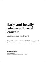
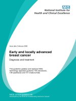
Early and locally
advanced breast
cancer:
diagnosis and treatment
This guideline updates and replaces NICE technology appraisal
guidance 109 (docetaxel), 108 (paclitaxel) and 107 (trastuzumab)
Full Guideline
February 2009
Developed for NICE by the National Collaborating Centre for Cancer
Published by the National Collaborating Centre for Cancer (2nd Floor, Front Suite, Park House, Greyfriars Road, Cardiff,
CF10 3AF) at Velindre NHS Trust, Cardiff, Wales
First published 2009
©2009 National Collaborating Centre for Cancer
No part of this publication may be reproduced, stored or transmitted in any form or by any means, without the prior
written permission of the publisher or, in the case of reprographic reproduction, in accordance with the terms of licenses
issued by the Copyright Licensing Agency in the UK. Enquiries concerning reproduction outside the terms stated here
should be sent to the publisher at the UK address printed on this page.
The use of registered names, trademarks etc. in this publication does not imply, even in the absence of a specific
statement, that such names are exempt from the relevant laws and regulations and therefore for general use.
While every effort has been made to ensure the accuracy of the information contained within this publication, the pub-
lisher can give no guarantee for information about drug dosage and application thereof contained in this book. In every
individual case the respective user must check current indications and accuracy by consulting other pharmaceutical
literature and following the guidelines laid down by the manufacturers of specific products and the relevant authorities
in the country in which they are practising.
The software and the textual and illustrative material contained on the CD-ROM accompanying this book are in copy-
right. The contents of the CD-ROM must not be copied or altered in any way, except for the purposes of installation. The
textual and illustrative material must not be printed out or cut-and-pasted or copied in any form except by an individual
for his or her own private research or study and without further distribution.
A library may make one copy of the contents of the disk for archiving purposes only, and not for circulation within or
beyond the library.
This CD-ROM carries no warranty, express or implied, as to fitness for a particular purpose. The National Collaborating
Centre for Cancer accepts no liability for loss or damage of any kind consequential upon use of this product.
By opening the wallet containing the CD-ROM you are indicating your acceptance of these terms and conditions.
ISBN 978-0-9558265-2-8
Cover and CD design by Newgen Imaging Systems
Typesetting by Newgen Imaging Systems
Printed in the UK by TJ International Ltd
Production management by Out of House Publishing Solutions
iii
Contents
Foreword v
Key priorities vi
Key research recommendations viii
Recommendations x
Methodology xvii
Algorithm xxvi
1 Epidemiology 1
1.1 Introduction 1
1.2 Incidence 1
1.3 Prognosis 4
1.4 Mortality 4
1.5 Survival 6
1.6 Prevalence 7
1.7 Treatment 7
1.8 Summary 13
1.9 Summary of findings from breast cancer teams peer review in England 2004-2007 14
2 Referral, diagnosis, preoperative assessment and psychological support 17
2.1 Introduction to Breast Cancer 17
2.2 Referral and Diagnosis 18
2.3 Preoperative Assessment of the Breast and Axilla 18
2.4 Preoperative Staging of the Axilla 21
2.5 Providing Information and Psychological Support 24
3
Surgery for early breast cancer 29
3.1
Surgery to the Breast 29
3.2
Surgery to the Axilla 32
3.3
Evaluation and Management of a Positive Sentinel Lymph Node 37
3.4
Breast Reconstruction 40
4
Postoperative assessment and adjuvant treatment planning 48
4.1
Introduction 48
4.2
Predictive Factors 48
4.3
Adjuvant Treatment Planning 50
4.4
Timing of Adjuvant Treatment 51
5
Adjuvant systemic therapy 54
5.1
Introduction 54
5.2
Endocrine Therapy for Invasive Disease 54
5.3
Endocrine Therapy for DCIS 60
5.4
Chemotherapy 61
5.5
Biological Therapy 63
5.6
Assessment and Treatment for Bone Loss 66
Early and locally advanced breast cancer: diagnosis and treatment
iv
6
Adjuvant radiotherapy 73
6.1
Introduction 73
6.2
Breast Conserving Surgery and Radiotherapy 73
6.3
Post-Mastectomy Radiotherapy 75
6.4
Dose Fractionation 77
6.5
Breast Boost 79
6.6
Radiotherapy to Nodal Areas 80
7 Primary systemic therapy 86
7.1 Early Breast Cancer 86
7.2 Locally Advanced or Inflammatory Breast Cancer 87
8 Complications of local treatment and menopausal symptoms 89
8.1
Introduction 89
8.2
Complications of Local Treatment 89
8.3
Menopausal Symptoms 92
9 Follow-up 97
9.1
Introduction 97
9.2
Follow-up Imaging 97
9.3
Clinical Follow-up 100
Appendices 104
1 Adjuvant! Online: review of evidence concerning its validity, and other considerations relating to its use in
the NHS
104
2 Algorithms taken from ‘Guidance for the management of breast cancer treatment-induced bone loss:
A consensus position statement from a UK expert group (2008)’
113
3 A cost effectiveness analysis of pretreatment ultrasound for the staging of the axilla in early breast cancer
patients
117
4 Abbreviations 138
5 Glossary 140
6 Guideline scope 150
7 List of topics covered by each chapter 154
8 People and organisations involved in production of the guideline 156
v
Foreword
Breast cancer is the most common cancer in women and its management often presents
patients and their healthcare professionals with difficult decisions about the most appropriate
treatment. For all those affected by breast cancer (including family and carers) it is important to
recognise the impact of this diagnosis, the complexity of treatment options and the wide rang-
ing needs and support required throughout this period of care and beyond. We hope that this
document will provide helpful and appropriate guidance to both healthcare professionals and
patients on the diagnosis and subsequent management of early and locally advanced breast
cancer.
The management of breast cancer is such a large topic that it has been necessary to divide it
into two separate guidelines: ‘Early and locally advanced breast cancer: diagnosis and treat-
ment’ and Advanced breast cancer: diagnosis and treatment’ (www.nice.org.uk/CG81) which
were developed at the same time. It should be appreciated that this guideline is not intended
to be an exhaustive textbook of early and locally advanced breast cancer. In addition it has
been impossible to cover every aspect of the patient pathway but instead we have concentrated
on those areas where it was felt uncertainty or variation in practice exists. We hope that those
who use the guideline will find it helpful and informative in decision making and management.
We are very grateful for all the hard work, commitment and common sense of the members of
the GDG, particularly the patient and carer members, whose views helped significantly in
shaping the document. We would also like to thank the staff at the NCC-C for their consider-
able support and hard work during the development of this guideline.
Mr James Smallwood Dr Adrian Harnett
Chair Clinical Lead
vi
Key priorities
1. Offer MRI of the breast to patients with invasive breast cancer:
− if there is discrepancy regarding the extent of disease from clinical examination,
mammography and ultrasound assessment for planning treatment
− if breast density precludes accurate mammographic assessment
− to assess the tumour size if breast conserving surgery is being considered for invasive
lobular cancer.
2. Pretreatment ultrasound evaluation of the axilla should be performed for all patients being
investigated for early invasive breast cancer and, if morphologically abnormal lymph
nodes are identified, ultrasound-guided needle sampling should be offered.
3. Minimal surgery, rather than lymph node clearance, should be performed to stage the axilla
for patients with early invasive breast cancer and no evidence of lymph node involvement on
ultrasound or a negative ultrasound-guided needle biopsy. SLNB is the preferred technique.
4. Discuss immediate breast reconstruction with all patients who are being advised to have a
mastectomy, and offer it except where significant comorbidity or (the need for) adjuvant
therapy may preclude this option. All appropriate breast reconstruction options should be
offered and discussed with patients, irrespective of whether they are all available locally.
5. Start adjuvant chemotherapy or radiotherapy as soon as clinically possible within 31 days
of completion of surgery
1
in patients with early breast cancer having these treatments.
6. Postmenopausal women with ER-positive early invasive breast cancer who are not consid-
ered to be at low risk
2
should be offered an aromatase inhibitor, either anastrozole or
letrozole, as their initial adjuvant therapy. Offer tamoxifen if an aromatase inhibitor is not
tolerated or contraindicated.
7. Patients with early invasive breast cancer should have a baseline dual energy X-ray absorp-
tiometry (DEXA) scan to assess bone mineral density if they:
− are starting adjuvant aromatase inhibitor treatment
− have treatment-induced menopause
− are starting ovarian ablation/suppression therapy.
8. Treat patients with early invasive breast cancer, irrespective of age, with surgery and
appropriate systemic therapy, rather than endocrine therapy alone, unless significant
comorbidity precludes surgery.
9. Offer annual mammography to all patients with early breast cancer, including DCIS, until
they enter the NHSBSP/BTWSP. Patients diagnosed with early breast cancer who are
already eligible for screening should have annual mammography for 5 years.
1
Department of Health (2007). Cancer reform strategy. London: Department of Health. (At present no equivalent target has been set by
the Welsh Assembly Government.)
2
Low-risk patients are those in the EPG or GPG groups in the Nottingham Prognostic Index (NPI) who have a 10 year predictive survival
of 96% and 93% respectively. They would have a similar prediction using Adjuvant! Online. High-risk patients are those in groups PPG
with 53% or VPG with 39%.
Key priorities
vii
10. Patients treated for breast cancer should have an agreed, written care plan, which should
be recorded by a named healthcare professional (or professionals), a copy sent to the GP
and a personal copy given to the patient. This plan should include:
− designated named healthcare professionals
− dates for review of any adjuvant therapy
− details of surveillance mammography
− signs and symptoms to look for and seek advice on
− contact details for immediate referral to specialist care, and
− contact details for support services, for example support for patients with
lymphoedema.
viii
Key research
recommendations
1. What is the effectiveness of cognitive behavioural therapy compared with other psychologi-
cal interventions for breast cancer patients?
There is currently a variation in the provision and quality of psychological approaches and
services offered to patients with breast cancer. As a consequence of the diagnosis of breast
cancer at least a quarter of patients report anxiety and depression and a third report sexual
problems.
Cognitive behavioural therapy (CBT) is one form of psychotherapy that has been proven to
treat and reduce depression in many patients including cancer patients. It is a time-limited,
structured and direct form of therapy that is well suited to patients with breast cancer.
Unfortunately there are no studies that compare CBT in breast cancer patients alone with
other forms of intervention. Other forms of psychotherapy include psychodynamic coun-
selling, Gestalt therapy or any other psychological intervention. The comparison group
could include support from the breast care nurse specialist, telephone support or pure
counselling.
2. In the absence of good data about differences in clinical outcome between axillary
radiotherapy and completion axillary lymph node dissection (ALND), entry into appropriate
clinical trials, e.g. AMAROS, is recommended for early breast cancer patients when the axilla
has been found by sentinel lymph node biopsy (SLNB) to contain metastasis.
Optimum treatment of the axilla, in patients with early breast cancer, when SLNB has shown
tumour positive lymph nodes remains unresolved: completion ALND or axillary radiotherapy
both have significant but differing morbidities. Studies, including AMAROS, are needed to
determine effectiveness of local control and overall survival, side effects and quality of life,
cost effectiveness, and whether the additional information of the total number of involved
lymph nodes obtained by ALND is relevant for optimum management. These alternative
management strategies would have significant impact on service delivery in the UK. The
piecemeal introduction of intraoperative sentinel lymph node assessment with immediate
ALND for a positive sentinel lymph node may make such research difficult in the near future.
3. How effective is trastuzumab in patients with invasive breast cancer: (a) as adjuvant therapy
without chemotherapy, (b) in terms of scheduling and duration of treatment in patients who
are also receiving or who have completed chemotherapy, and (c) as primary systemic treat-
ment in terms of quality of life, side effects, disease recurrence rates, disease-free survival and
overall survival?
In patients with human epidermal growth receptor 2 (HER2)-positive invasive breast cancer
trastuzumab is a routine adjuvant therapy, where appropriate, following surgery, chemo-
therapy and radiotherapy. The recommended scheduling at present is 3-weekly treatment
for 1 year but there may be more effective and cost effective regimens. Studies such as
PERSEPHONE and HERA 2 year treatment duration study arm have been designed to
address these issues. There are few studies assessing the role of trastuzumab as a primary
systemic treatment and even fewer using it in endocrine receptor-positive patients treated
with endocrine therapy alone and no chemotherapy.
Key research recommendations
ix
Studies are needed to resolve the questions of scheduling and duration, the place of
trastuzumab with endocrine therapy in the absence of adjuvant chemotherapy and its role
in primary systemic therapy.
4. What is the effectiveness in patients with early invasive breast cancer of: (a) different
hypofractionation radiotherapy regimens (b) partial breast radiotherapy and (c) newer radio-
therapy techniques (including intensity modulated radiotherapy), in terms of long term
outcomes such as, quality of life, side effects, disease recurrence rates, disease-free survival
and overall survival?
Following breast conservation surgery for invasive breast cancer the international standard
radiotherapy practice is to treat the whole breast, giving 50 Gy in 25 fractions of 2 Gy
fractions over 5 weeks. A 3-week schedule of 40 Gy in 15 fractions has been used in many
centres in the UK for years and this has been supported by the recent publication of the UK
Standardisation of Breast Radiotherapy (START) Trial. Further studies may show that it may
be possible to use even more hypofractionated regimens, which would be far more
convenient for patients and more cost effective if they are equally effective. In addition,
with technical advances in radiotherapy treatment planning and delivery, it is possible to
give partial breast radiotherapy or dose gradients across the breast in selected patients.
5. For patients who have been treated for early invasive breast cancer or ductal carcinoma in
situ (DCIS), what is the optimal frequency and length of surveillance of follow-up mammog-
raphy?
There is little evidence that routine follow-up of patients treated for early breast cancer to
detect recurrence early, or new primary disease, is either effective or offers any mortality
benefit. However, it remains routine practice in virtually all breast units in the UK to pro-
vide post-treatment follow-up with regular clinical examination and mammography for at
least 5 years. This routine follow-up is usually provided in the secondary care setting and
requires significant resources. The consensus of those providing breast cancer treatment is
that routine follow-up is beneficial for patient welfare and for monitoring effectiveness of
treatment. There are few data on which to base guidelines on the most effective methods of
providing follow-up, how frequently and for how long. Prospective randomised compara-
tive studies are required to ascertain the most effective methods for detecting recurrence
and new primary disease, and should include:
• how (by clinical examination and/or imaging and/or serum tumour markers)
• different patient populations, depending on their risks and toxicities from treatment
• where (in primary care and/or secondary care) and by whom (by patients, nurses or
doctors) these should be provided, and
• whether such care provides any benefits (such as reduced mortality, morbidity and treat-
ment costs).
x
Recommendations
Chapter 2: Referral, diagnosis, preoperative assessment
and psychological support
Preoperative assessment of the breast and axilla
The routine use of magnetic resonance imaging (MRI) of the breast is not recommended in
the preoperative assessment of patients with biopsy-proven invasive breast cancer or ductal
carcinoma in situ (DCIS).
Offer MRI of the breast to patients with invasive breast cancer:
• if there is discrepancy regarding the extent of disease from clinical examination, mammog-
raphy and ultrasound assessment for planning treatment
• if breast density precludes accurate mammographic assessment
• to assess the tumour size if breast conserving surgery is being considered for invasive lobu-
lar cancer.
Preoperative staging of the axilla
Pretreatment ultrasound evaluation of the axilla should be performed for all patients being
investigated for early invasive breast cancer and, if morphologically abnormal lymph nodes are
identified, ultrasound-guided needle sampling should be offered.
Providing information and psychological support
All members of the breast cancer clinical team should have completed an accredited
communication skills training programme.
All patients with breast cancer should be assigned to a named breast care nurse specialist who
will support them throughout diagnosis, treatment and follow-up.
All patients with breast cancer should be offered prompt access to specialist psychological
support, and where appropriate psychiatric services.
Chapter 3: Surgery for early breast cancer
Surgery to the breast
DCIS
For all patients treated with breast conserving surgery for DCIS a minimum of 2 mm radial
margin of excision is recommended with pathological examination to NHS Breast Screening
Programme reporting standards.
Re-excision should be considered if the margin is less than 2 mm after discussion of the risks
and benefits with the patient.
Enter patients with screen-detected DCIS into the Sloane Project
1
(UK DCIS audit).
All breast units should audit their recurrence rates after treatment for DCIS.
1
www.sloaneproject.co.uk
Recommendations
xi
Paget's disease
Offer breast conserving surgery with removal of the nipple-areolar complex as an alternative to
mastectomy for patients with Paget’s disease of the nipple, that has been assessed as localised.
Offer oncoplastic repair techniques to maximise cosmesis.
Surgery to the axilla
Invasive breast cancer
Minimal surgery, rather than lymph node clearance, should be performed to stage the axilla for
patients with early invasive breast cancer and no evidence of lymph node involvement on
ultrasound or a negative ultrasound-guided needle biopsy. Sentinel lymph node biopsy (SLNB)
is the preferred technique.
SLNB should only be performed by a team that is validated in the use of the technique, as iden-
tified in the New Start training programme
2
.
Perform SLNB using the dual technique with isotope and blue dye.
Breast units should audit their axillary recurrence rates.
DCIS
Do not perform SLNB routinely in patients with a preoperative diagnosis of DCIS who are
having breast conserving surgery, unless they are considered to be at a high risk of invasive
disease
3
.
Offer SLNB to all patients who are having a mastectomy for DCIS.
Evaluation and management of a positive sentinel lymph node
Offer further axillary treatment to patients with early invasive breast cancer who:
• have macrometastases or micrometastases shown in a sentinel lymph node
• have a preoperative ultrasound guided needle biopsy with histologically proven metastatic
cancer.
The preferred technique is axillary lymph node dissection (ALND) because it gives additional
staging information.
Do not offer further axillary treatment to patients found to have only isolated tumour cells in
their sentinel lymph nodes. These patients should be regarded as lymph node-negative.
Breast reconstruction
Discuss immediate breast reconstruction with all patients who are being advised to have a mas-
tectomy, and offer it except where significant comorbidity or (the need for) adjuvant therapy
may preclude this option. All appropriate breast reconstruction options should be offered and
discussed with patients, irrespective of whether they are all available locally.
Chapter 4: Postoperative assessment and adjuvant treatment
planning
Predictive factors
Assess oestrogen receptor (ER) status of all invasive breast cancers, using immunohistochemistry
with a standardised and qualitatively assured methodology, and report the results quantitatively.
Do not routinely assess progesterone receptor status of tumours in patients with invasive breast
cancer.
2
NEW START Sentinel Lymph Node Biopsy Training Programme, Raven Department of Education, Royal College of Surgeons, England.
3
Patients considered at high risk of invasive disease include those with a palpable mass or extensive microcalcifications.
Early and locally advanced breast cancer: diagnosis and treatment
xii
Test human epidermal growth receptor 2 (HER2) status of all invasive breast cancers, using a
standardised and qualitatively assured methodology.
Ensure that the results of ER and HER2 assessments are available and recorded at the multidis-
ciplinary team meeting when guidance about systemic treatment is made.
Adjuvant treatment planning
Consider adjuvant therapy for all patients with early invasive breast cancer after surgery at the
multidisciplinary team meeting and ensure that decisions are recorded.
Decisions about adjuvant therapy should be made based on assessment of the prognostic and
predictive factors, the potential benefits and side effects of the treatment. Decisions should be
made following discussion of these factors with the patient.
Consider using Adjuvant! Online
4
to support estimations of individual prognosis and the
absolute benefit of adjuvant treatment for patients with early invasive breast cancer.
Timing of adjuvant treatment
Start adjuvant chemotherapy or radiotherapy as soon as clinically possible within 31 days of
completion of surgery
5
in patients with early breast cancer having these treatments.
Chapter 5: Adjuvant systemic therapy
Endocrine therapy for invasive disease
Ovarian suppression/ablation
Do not offer adjuvant ovarian ablation/suppression to premenopausal women with ER-positive
early invasive breast cancer who are being treated with tamoxifen and, if indicated, chemo-
therapy.
Offer adjuvant ovarian ablation/suppression in addition to tamoxifen to premenopausal women
with ER-positive early invasive breast cancer who have been offered chemotherapy but have
chosen not to have it.
Aromatase inhibitors
Postmenopausal women with ER-positive early invasive breast cancer who are not considered
to be at low-risk
6
should be offered an aromatase inhibitor, either anastrozole or letrozole, as
their initial adjuvant therapy. Offer tamoxifen if an aromatase inhibitor is not tolerated or
contraindicated.
Offer an aromatase inhibitor, either exemestane or anastrozole instead of tamoxifen to post-
menopausal women with ER-positive early invasive breast cancer who are not low-risk
7
and
who have been treated with tamoxifen for 2-3 years.
Offer additional treatment with the aromatase inhibitor letrozole for 2-3 years to postmeno-
pausal women with lymph node-positive ER-positive early invasive breast cancer who have
been treated with tamoxifen for 5 years.
4
www.adjuvantonline.com
5
Department of Health (2007). Cancer reform strategy. London: Department of Health. (At present no equivalent target has been set by
the Welsh Assembly Government.)
6
Low-risk patients are those in the EPG or GPG groups in the Nottingham Prognostic Index (NPI) who have a 10 year predictive survival
of 96% and 93% respectively. They would have a similar prediction using Adjuvant! Online. High risk are patients in groups PPG with
53% or VPG with 39%.
7
Low-risk patients are those in the EPG or GPG groups in the Nottingham Prognostic Index (NPI) who have a 10 year predictive survival
of 96% and 93% respectively. They would have a similar prediction using Adjuvant! Online. High risk are patients in groups PPG with
53% or VPG with 39%.
Recommendations
xiii
The aromatase inhibitors anastrozole, exemestane and letrozole, within their licensed indica-
tions, are recommended as options for the adjuvant treatment of early ER-positive invasive
breast cancer in postmenopausal women.
8
The choice of treatment should be made after discussion between the responsible clinician and
the woman about the risks and benefits of each option. Factors to consider when making the
choice include whether the woman has received tamoxifen before, the licensed indications and
side-effect profiles of the individual drugs and, in particular, the assessed risk of recurrence.
9
Endocrine therapy for DCIS
Do not offer adjuvant tamoxifen after breast conserving surgery to patients with DCIS.
Chemotherapy
Offer docetaxel to patients with lymph node-positive breast cancer patients as part of an
adjuvant chemotherapy regimen.
Do not offer paclitaxel as an adjuvant treatment for lymph node-positive breast cancer.
Biological therapy
Offer trastuzumab, given at 3-week intervals for 1 year or until disease recurrence (whichever
is the shorter period), as an adjuvant treatment to women with HER2-positive early invasive
breast cancer following surgery, chemotherapy, and radiotherapy when applicable.
Assess cardiac function before starting treatment with trastuzumab. Do not offer trastuzumab
treatment to women who have any of the following:
• a left ventricular ejection fraction (LVEF) of 55% or less
• a history of documented congestive heart failure
• high risk uncontrolled arrhythmias
• angina pectoris requiring medication
• clinically significant valvular disease
• evidence of transmural infarction on electrocardiograph (ECG)
• poorly controlled hypertension.
Repeat cardiac functional assessments every 3 months during trastuzumab treatment. If the
LVEF drops by 10 percentage (ejection) points or more from baseline and to below 50% then
trastuzumab treatment should be suspended. Restart trastuzumab therapy only after further
cardiac assessment and a fully informed discussion of the risks and benefits with the woman.
Assessment and treatment for bone loss
Bone mineral density
Patients with early invasive breast cancer should have a baseline dual energy X-ray absorpti-
ometry (DEXA) scan to assess bone mineral density if they:
• are starting adjuvant aromatase inhibitor treatment
• have treatment-induced menopause
• are starting ovarian ablation/suppression therapy.
Do not offer a DEXA scan to patients with early invasive breast cancer who are receiving
tamoxifen alone, regardless of pretreatment menopausal status.
Bisphosphonates
Offer bisphosphonates to patients identified by algorithms 1 and 2 in ’Guidance for the
management of breast cancer treatment-induced bone loss: A consensus position statement
from a UK expert group (2008) (see Appendix 2).
8
This recommendation is from 'Breast cancer (early) – hormonal treatments’, NICE technology appraisal guidance 112.
9
This recommendation is from 'Breast cancer (early) – hormonal treatments’, NICE technology appraisal guidance 112.
Early and locally advanced breast cancer: diagnosis and treatment
xiv
Chapter 6: Adjuvant radiotherapy
Breast conserving surgery and radiotherapy
Patients with early invasive breast cancer who have had breast conserving surgery with clear
margins should have breast radiotherapy.
Offer adjuvant radiotherapy to patients with DCIS following adequate breast conserving
surgery
10
and discuss with them the potential benefits and risks.
Post-mastectomy radiotherapy
Offer adjuvant chest wall radiotherapy to patients with early invasive breast cancer who have
had a mastectomy and are at a high risk of local recurrence. Patients at a high risk of local
recurrence include those with four or more positive axillary lymph nodes or involved resection
margins.
Consider entering patients who have had a mastectomy for early invasive breast cancer and
who are at an intermediate risk of local recurrence, into the current UK trial (SUPREMO)
assessing the value of postoperative radiotherapy. Patients at an intermediate risk of local
recurrence include those with one to three lymph nodes involved, lympho-vascular invasion,
histological grade 3 tumours, ER-negative tumours, and those aged under 40 years.
Do not offer radiotherapy following mastectomy to patients with early invasive breast cancer
who are at low risk of local recurrence (for example, most patients who are lymph node-
negative).
Dose fractionation
Use external beam radiotherapy giving 40 Gy in 15 fractions as standard practice for patients
with early invasive breast cancer after breast conserving surgery or mastectomy.
Breast boost
Offer an external beam boost to the site of local excision to patients with early invasive breast
cancer who have a high risk of local recurrence following breast conserving surgery, with clear
margins, and whole breast radiotherapy.
If an external beam boost to the site of local excision following breast conservation is being
considered in patients with early invasive breast cancer inform the patient of the side effects
associated with this intervention, including poor cosmesis particularly in women with larger
breasts.
Radiotherapy to nodal areas
Do not offer adjuvant radiotherapy to the axilla or supraclavicular fossa to patients with early
breast cancer who have been shown to be histologically lymph node-negative.
Do not offer adjuvant radiotherapy to the axilla after ALND for early breast cancer.
If ALND is not possible following a positive axillary SLNB or 4-node sample, offer adjuvant
radiotherapy to the axilla to patients with early breast cancer
11
.
Offer adjuvant radiotherapy to the supraclavicular fossa in patients with early breast cancer and
four or more involved axillary lymph nodes.
Offer adjuvant radiotherapy to the supraclavicular fossa to patients with early breast cancer and
one to three positive lymph nodes if they have other poor prognostic factors (for example, T3
and/or histological grade 3 tumours, with good performance status).
Do not offer adjuvant radiotherapy to the internal mammary chain to patients with early breast
cancer who have had breast surgery.
10
See recommendation on DCIS margins in Chapter 3.
11
See recommendation in Chapter 3.
Recommendations
xv
Chapter 7: Primary systemic therapy
Early breast cancer
Treat patients with early invasive breast cancer, irrespective of age, with surgery and appropriate
systemic therapy, rather than endocrine therapy alone, unless significant comorbidity precludes
surgery.
Preoperative systemic therapy can be offered to patients with early invasive breast cancer who
are considering breast conserving surgery that is not advisable at presentation. However, the
increased risk of local recurrence with breast conserving surgery and radiotherapy rather than
mastectomy after systemic therapy should be discussed with the patient.
Locally advanced or inflammatory breast cancer
Offer patients with locally advanced or inflammatory breast cancer, who have been treated
with chemotherapy, local treatment by mastectomy (or in exceptional cases, breast conserving
surgery) followed by radiotherapy.
Chapter 8: Complications of local treatment and menopausal
symptoms
Complications of local treatment
Lymphoedema
Inform all patients with early breast cancer about the risk of developing lymphoedema and give
them relevant written information before treatment with surgery and radiotherapy.
Give advice on how to prevent infection or trauma that may cause or exacerbate lymphoedema
to patients treated for early breast cancer.
Ensure that all patients with early breast cancer who develop lymphoedema have rapid access
to a specialist lymphoedema service.
Arm mobility
All breast units should have written local guidelines agreed with the physiotherapy department
for postoperative physiotherapy regimens.
Identify breast cancer patients with pre-existing shoulder conditions preoperatively as this may
inform further decisions on treatment.
Give instructions on functional exercises, which should start the day after surgery, to all breast
cancer patients undergoing axillary surgery. This should include relevant written information
from a member of the breast or physiotherapy team.
Refer patients to the physiotherapy department if they report a persistent reduction in arm and
shoulder mobility after breast cancer treatment.
Menopausal symptoms
Discontinue hormone replacement therapy (HRT) in women who are diagnosed with breast
cancer.
Do not offer HRT (including oestrogen/progestogen combination) routinely to women with
menopausal symptoms and a history of breast cancer. HRT
12
may, in exceptional cases, be
offered to women with severe menopausal symptoms and with whom the associated risks have
been discussed.
12
The summaries of product characteristics state that HRT is contraindicated in women with known, past or suspected breast cancer.
Informed consent should be obtained and documented.
Early and locally advanced breast cancer: diagnosis and treatment
xvi
Offer information and counselling for all women about the possibility of early menopause and
menopausal symptoms associated with breast cancer treatment.
Tibolone or progestogens are not recommended for women with menopausal symptoms who
have breast cancer.
The selective serotonin re-uptake inhibitor antidepressants paroxetine
13
and fluoxetine
14
may be
offered to women with breast cancer for relieving menopausal symptoms, particularly hot
flushes, but not to those taking tamoxifen.
Clonidine, venlafaxine
15
and gabapentin
16
should only be offered to treat hot flushes in women
with breast cancer after they have been fully informed of the significant side effects.
Soy (isoflavone), red clover, black cohosh, vitamin E and magnetic devices are not recom-
mended for the treatment of menopausal symptoms in women with breast cancer.
Chapter 9: Complications of local treatment and menopausal
symptoms
Follow-up
Follow-up imaging
Offer annual mammography to all patients with early breast cancer, including DCIS until they
enter the NHS Breast Screening Programme (NHSBSP)/Breast Test Wales Screening Programme
(BTWSP). Patients diagnosed with early breast cancer who are already eligible for screening
should have annual mammography for 5 years.
On reaching the NHSBSP/BTWSP screening age or after 5 years of annual mammography
follow-up we recommend the NHSBSP/BTWSP stratify screening frequency in line with patient
risk category.
Do not offer mammography of the ipsilateral soft tissues after mastectomy.
Do not offer ultrasound or MRI for routine post-treatment surveillance in patients who have
been treated for early invasive breast cancer or DCIS.
Clinical follow-up
After completion of adjuvant treatment (including chemotherapy, and/or radiotherapy where
indicated) for early breast cancer, discuss with patients where they would like follow-up to be
undertaken. They may choose to receive follow-up care in primary, secondary, or shared care.
Patients treated for breast cancer should have an agreed, written care plan, which should be
recorded by a named healthcare professional (or professionals), a copy sent to the GP and a
personal copy given to the patient. This plan should include:
• designated named healthcare professionals
• dates for review of any adjuvant therapy
• details of surveillance mammography
• signs and symptoms to look for and seek advice on
• contact details for immediate referral to specialist care, and
• contact details for support services, for example support for patients with lymphoedema.
13
These drugs are not licensed for the stated use. Informed consent should be obtained and documented.
14
These drugs are not licensed for the stated use. Informed consent should be obtained and documented.
15
These drugs are not licensed for the stated use. Informed consent should be obtained and documented.
16
These drugs are not licensed for the stated use. Informed consent should be obtained and documented.
xvii
Methodology
Introduction
What is a clinical guideline?
Guidelines are recommendations for the care of individuals in specific clinical conditions or
circumstances – from prevention and self-care through to primary and secondary care and onto
more specialised services. NICE clinical guidelines are based on the best available evidence of
clinical and cost effectiveness, and are produced to help healthcare professionals and patients
make informed choices about appropriate healthcare. While guidelines assist the practice of
healthcare professionals, they do not replace their knowledge and skills.
Clinical guidelines for the NHS in England, Wales and Northern Ireland are produced as a
response to a request from the Department of Health (DH). They approve topics for guideline
development and before deciding whether to refer a particular topic to the National Institute for
Health and Clinical Excellence (NICE) they consult with the relevant patient bodies, profes-
sional organisations and companies. Once a topic is referred, NICE then commissions one of
seven National Collaborating Centres (NCCs) to produce a guideline. The Collaborating Cen-
tres are independent of government and comprise partnerships between a variety of academic
institutions, health profession bodies and patient groups. The National Collaborating Centre for
Cancer (NCC-C) was referred the topic of breast cancer in October 2003 as part of NICE’s ninth
wave work programme. Because of the size of this topic, the NCC-C used 2 guideline slots
(early breast cancer and advanced breast cancer) to fulfil this remit. However the guideline
development process began officially on 10 April 2006 when sufficient capacity became avail-
able at the NCC-C.
Who is the guideline intended for?
This guideline does not include recommendations covering every detail of the diagnosis and
treatment of early breast cancer. Instead we have tried to focus on those areas of clinical practice
that are (i) known to be controversial or uncertain; (ii) where there is identifiable practice varia-
tion; (iii) where there is a lack of high-quality evidence; or (iv) where NICE guidelines are likely
to have most impact. More detail on how this was achieved is presented later in the section on
‘Developing Clinical Evidence Based Questions’.
This guideline is relevant to all healthcare professionals who come into contact with patients
with early breast cancer, as well as to the patients themselves and their carers. It is also
expected that the guideline will be of value to those involved in clinical governance in both
primary and secondary care to help ensure that arrangements are in place to deliver appropri-
ate care to this group of patients.
The remit of the guideline
Guideline topics selected by the DH identify the main areas to be covered by the guideline in a
specific remit. The following remit for this guideline was received as part of NICE’s ninth wave
programme of work:
Early and locally advanced breast cancer: diagnosis and treatment
xviii
‘To prepare a guideline for the NHS in England and Wales on the clinical management of breast
cancer, to supplement existing service guidance. The guideline should cover:
• the key diagnostic and staging procedures
• the main treatment modalities including hormonal treatments
• the role of tumour-specific bisphosphonates.’
What the guideline covers - the scope
The remit was then translated into a scope document by the Guideline Development Group
(GDG) Chair and Lead Clinician and staff at the NCC-C. The purpose of the scope was to:
• provide an overview of what the guideline would include and exclude
• identify the key aspects of care that must be included
• set the boundaries of the development work and provide a clear framework to enable work
to stay within the priorities agreed by NICE and the NCC-C and the remit
• inform the development of the clinical questions and search strategy
• inform professionals and the public about the expected content of the guideline.
Prior to the commencement of the guideline development process, the scope was subject to a
four week stakeholder consultation in accordance with processes established by NICE in the
‘NICE guidelines manual’ (NICE, 2005, NICE 2006, NICE 2007). The full scope is shown in
Appendix 6. During the consultation period, the scope was posted on the NICE website
(www.nice.org.uk). Comments were invited from registered stakeholder organisations and the
NICE Guideline Review Panel (GRP). Further information about the GRP can also be found on
the NICE website. The NCC-C and NICE reviewed the scope in light of comments received,
and the revised scope was reviewed by the GRP, signed off by NICE and posted on the NICE
website.
Involvement of stakeholders
Key to the development of all NICE guidelines are the relevant professional and patient/carer
organisations that register as stakeholders. Details of this process can be found on the NICE
website or in the ‘NICE guidelines manual’ (NICE 2007). In brief, their contribution involves
commenting on the draft scope, submitting relevant evidence and commenting on the draft
version of the guideline during the end consultation period. A full list of all stakeholder organi-
sations who registered for the early breast cancer guideline can be found in Appendix 8.2.
Needs assessment
As part of the guideline development process the NCC-C invited specialist registrars to under-
take a needs assessment. The needs assessment aims to describe the burden of disease and
current service provision for patients with breast cancer in England and Wales, which informed
the development of the guideline. This document forms a supplement to the full guideline and
also appears on the accompanying CD-ROM to this guideline.
Assessment of the effectiveness of interventions is not included in the needs assessment, and
was undertaken separately by researchers in the NCC-C as part of the guideline development
process.
The information included in the needs assessment document was presented to the GDG. Most
of the information was presented in the early stages of guideline development, and other
information was included to meet the evolving information needs of the GDG during the
course of guideline development.
The Process of Guideline Development – Who Develops the
Guideline?
Overview
The development of this guideline was based upon methods outlined by the ‘NICE guidelines
manual’. A team of health professionals, lay representatives and technical experts known as the
Methodology
xix
GDG (see Appendix 8.1), with support from the NCC-C staff, undertook the development of
this clinical guideline. The basic steps in the process of developing a guideline are listed and
discussed below:
• using the remit, define the scope which sets the parameters of the guideline
• forming the guideline development group
• developing clinical questions
• systematically searching for the evidence
• critically appraising the evidence
• incorporating health economic evidence
• distilling and synthesising the evidence and writing recommendations
• agreeing the recommendations
• structuring and writing the guideline
• updating the guideline.
The Guideline Development Group (GDG)
The Early Breast Cancer GDG was recruited in line with the existing NICE protocol as set out in
the ‘NICE guidelines manual’. The first step was to appoint a Chair and a Lead Clinician.
Advertisements were placed for both posts and candidates were informally interviewed prior to
being offered the role. The NCC-C Director, GDG Chair and Lead Clinician identified a list of
specialties that needed to be represented on the GDG. Requests for nominations were sent to
the main stakeholder organisations and patient organisations/charities (see Appendix 8.2). Indi-
vidual GDG members were selected by the NCC-C Director, GDG Chair and Lead Clinician,
based on their application forms, following nomination from their respective stakeholder
organisation. The guideline development process was supported by staff from the NCC-C, who
undertook the clinical and health economics literature searches, reviewed and presented the
evidence to the GDG, managed the process and contributed to drafting the guideline. At the
start of the guideline development process all GDG members’ interests were recorded on a
standard declaration form that covered consultancies, fee-paid work, share-holdings, fellow-
ships and support from the healthcare industry. At all subsequent GDG meetings, members
declared new, arising conflicts of interest which were always recorded (see Appendix 8.1).
Guideline Development Group meetings
Fifteen GDG meetings were held between 10-11 April 2006 and 19-20 June 2008. During
each GDG meeting (either held over one or two days) clinical questions and clinical and
economic evidence were reviewed, assessed and recommendations formulated. At each meeting
patient/carer and service-user concerns were routinely discussed as part of a standing agenda
item.
NCC-C project managers divided the GDG workload by allocating specific clinical questions,
relevant to their area of clinical practice, to small sub-groups of the GDG in order to simplify
and speed up the guideline development process. These groups considered the evidence, as
reviewed by the researcher, and synthesised it into draft recommendations prior to presenting it
to the GDG as a whole. Each clinical question was led by a GDG member with expert knowl-
edge of the clinical area (usually one of the healthcare professionals). The GDG subgroups
often helped refine the clinical questions and the clinical definitions of treatments. They also
assisted the NCC-C team in drafting the section of the guideline relevant to their specific topic.
Patient/carer members
Individuals with direct experience of early breast cancer services gave an integral user focus
to the GDG and the guideline development process. The GDG included three patient/carer
members. They contributed as full GDG members to writing the clinical questions, helping to
ensure that the evidence addressed their views and preferences, highlighting sensitive issues
and terminology relevant to the guideline and bringing service-user research to the attention of
the GDG.
Early and locally advanced breast cancer: diagnosis and treatment
xx
Developing Clinical Evidence-Based Questions
Background
The scope, as described in Appendix 6, needs to be very clear about which patient groups are
included and which areas of clinical care should be considered. But within these boundaries it
does not usually specify which topics are considered a priority.
It was recognised by the NCC-C at an early stage that in order to complete the guideline devel-
opment work to an appropriate standard the GDG needed to restrict its work to approximately
30 clinical questions. Previously this prioritisation would have been carried out by the GDG at
its first two meetings but it was clear from some guidelines already published that this approach
had resulted in a much larger number of questions than 30 being addressed.
Clinical guidelines should be aimed at changing clinical practice and should avoid ending up
as ‘evidence-based textbooks’ or making recommendations on topics where there is already
agreed clinical practice. It was therefore felt important that the 30 clinical questions should be
prioritised into areas that were known to be controversial or uncertain, where there was identi-
fiable practice variation, or where NICE guidelines were likely to have most impact.
Method
An extensive list of potential topics for the guideline to investigate was compiled by the NCC-C
Director and GDG Chair and Lead Clinician in consultation with a small number of breast
cancer multidisciplinary teams across England and Wales.
This list was incorporated into a questionnaire which asked respondents to rate each topic as
low, medium or high clinical priority as well as low or high economic priority. It was made
clear that respondents would be rating the priority for each topic to be included in a clinical
guideline to be published in two years’ time. The questionnaire also asked respondents to
suggest any additional topics they would like to see included with an equivalent assessment of
their priority.
Questionnaires were subsequently sent to the Breast Cancer Advisory Groups of all 37 cancer
networks in England and Wales with a request for a 4-week turnaround. (A list of all cancer
networks can be found on the Cancer Action Team website at the DH). Questionnaires were
also sent via the Patient and Public Involvement Programme (PPIP) at NICE to all relevant
patient/carer stakeholder organisations.
The scores from each completed questionnaire were aggregated by NCC-C staff and ranked.
These results together with information on identifiable practice variation (see needs assessment)
were presented to the GDG at its first meeting. The list of prioritised topics produced via the
questionnaire survey was in no way definitive and the GDG used these results to agree their
final priorities for the clinical questions.
For clinical questions about interventions, the PICO framework was used. This structured
approach divides each question into four components: the patients (the population under study – P),
the interventions (what is being done - I), the comparisons (other main treatment options – C)
and the outcomes (the measures of how effective the interventions have been – O). Where
appropriate, the clinical questions were refined once the evidence had been searched and,
where necessary, sub-questions were generated.
The final list of clinical questions can be found in Appendix 7.
Care Pathway
Early in the development process the GDG drafted an outline care pathway (or algorithm) in order
to explore how patients with early breast cancer might access and be dealt with by the NHS.
Review of Clinical Literature
At the beginning of the development phase, initial scoping searches were carried out to identify
any relevant guidelines (local, national or international) produced by other groups or institutions.
Methodology
xxi
Additionally, stakeholder organisations were invited to submit evidence for consideration by
the GDG, provided it was relevant to the agreed list of clinical questions.
In order to answer each question the NCC-C information specialist developed a search strategy
to identify relevant published evidence for both clinical and cost effectiveness. Key words and
terms for the search were agreed in collaboration with the GDG. When required, the health
economist searched for supplementary papers to inform detailed health economic work, for
example modelling (see section on ‘Incorporating Health Economic Evidence’).
Papers that were published or accepted for publication in peer-reviewed journals were consid-
ered as evidence. Search filters, such as those to identify systematic reviews (SRs) and randomised
controlled trials (RCTs) were applied to the search strategies when there was a wealth of these
types of studies. No language restrictions were applied to the search; however, foreign language
papers were not requested or reviewed (unless of particular importance to that question).
The following databases were included in the literature search:
• The Cochrane Library
• Medline and Premedline 1950 onwards
• Excerpta Medica (Embase) 1980 onwards
• Cumulative Index to Nursing and Allied Health Literature (Cinahl) 1982 onwards
• Allied & Complementary Medicine (AMED) 1985 onwards
• British Nursing Index (BNI) 1994 onwards
• Psychinfo 1806 onwards
• Web of Science 1970 onwards. [specifically Science Citation Index Expanded
• (SCI-EXPANDED) and Social Sciences Citation Index (SSCI)]
• System for Information on Grey Literature In Europe (SIGLE) 1980–2005
• Biomed Central 1997 onwards
• National Research Register (NRR)
• Current Controlled Trials.
From this list the information specialist sifted and removed any irrelevant material based on the
title or abstract before passing to the researcher. All the remaining articles were then stored in a
Reference Manager electronic library.
Searches were updated and re-run 6–8 weeks before the stakeholder consultation, thereby
ensuring that the latest relevant published evidence was included in the database. Any evidence
published after this date was not included. For the purposes of updating this guideline, July
2008 should be considered the starting point for searching for new evidence.
Further details of the search strategies, including the methodological filters used, are provided
in the evidence review (and appear on the accompanying CD-ROM to this guideline).
Critical Appraisal and Evidence Grading
Following the literature search one researcher independently scanned the titles and abstracts of
every article for each question, and full publications were obtained for any studies considered
relevant or where there was insufficient information from the title and abstract to make a deci-
sion. The researcher then individually applied the inclusion/exclusion criteria to determine
which studies would be relevant for inclusion and subsequent appraisal. Lists of excluded
papers were generated for each question and the rationale for the exclusion was presented to
the GDG when required.
The researcher then critically appraised the full papers. Critical appraisal checklists were
compiled for each paper and one researcher undertook the critical appraisal and data extraction.
The researcher assessed the quality of eligible studies by referring to the SIGN criteria for
systematic reviews/meta-analyses and randomised control trials (Table A). Evidence relating to
clinical effectiveness was classified using this established hierarchical system. However this
checklist is less appropriate for studies reporting diagnostic tests of accuracy. In the absence of
a validated hierarchy for this type of test, NICE suggests levels of evidence that take into
account the factors likely to affect the validity of these studies.
Early and locally advanced breast cancer: diagnosis and treatment
xxii
Level Source of evidence
1++
High-quality meta-analyses, systematic reviews of randomised controlled trials (RCTs) or
RCTs with a very low risk of bias
1+ Well-conducted meta-analyses, systematic reviews of RCTs or RCTs with a low risk of bias
1− Meta-analyses, systematic reviews of RCTs or RCTs with a high risk of bias
2++
High-quality systematic reviews of case–control or cohort studies; high-quality case–control
or cohort studies with a very low risk of confounding, bias or chance and a high probability
that the relationship is causal
2+
Well-conducted case–control or cohort studies with a low risk of confounding, bias or
chance and a moderate probability that the relationship is causal
2−
Case–control or cohort studies with a high risk of confounding, bias or chance and a signifi-
cant risk that the relationship is not causal
3 Non-analytical studies (for example case reports, case series)
4 Expert opinion, formal consensus
Table A. Levels of evidence for intervention studies. Data source: ‘NICE guidelines manual’
(NICE 2007).
For all the relevant appraised studies for a particular question, data on the type of population,
intervention, comparator and outcomes (PICO) was recorded in evidence tables and an
accompanying evidence summary prepared for the GDG (see evidence review). All the evidence
was considered carefully by the GDG for accuracy and completeness.
All procedures were fully compliant with NICE methodology as detailed in the ‘NICE guide-
lines manual’.
In general, no formal contact was made with authors; however, there were ad hoc occasions
when this was required in order to clarify specific details.
Incorporating Health Economics Evidence
The aim of the economic input into the guideline was to inform the GDG of potential
economic issues relating to early breast cancer. It is important to investigate whether health
services are both clinically effective and cost effective, i.e. are they ‘value for money’.
The health economist helped the GDG by identifying priority topics within the guideline that
might benefit from economic analysis, reviewing the available economic evidence and, where
necessary, conducting economic analysis. Where published economic evaluation studies were
identified that addressed the economic issues for a clinical question, these are presented along-
side the clinical evidence wherever possible.
In order to assess the cost effectiveness of each priority topic, a comprehensive systematic
review of the economic literature was conducted. For those clinical areas reviewed, the infor-
mation specialists used a similar search strategy as used for the review of clinical evidence but
with the inclusion of a health economics and quality of life filter.
Each search strategy was designed to find any applied study estimating the cost or cost effec-
tiveness of the topic under consideration. A health economist reviewed abstracts and relevant
papers were ordered for appraisal.
Published economic evidence was obtained from a variety of sources:
• Medline 1966 onwards
• Embase 1980 onwards
• NHS Economic Evaluations Database (NHS EED)
• EconLit 1969 onwards.
Methodology
xxiii
Economic Modelling
In addition to the review of the relevant clinical evidence, the GDG were required to deter-
mine whether or not the cost effectiveness of each of the individual clinical questions should
be investigated. After the clinical questions were decided, the GDG agreed which topics were
an ‘economic priority’ for modelling. These ‘economic priorities’ were chosen on the basis of
the following criteria, in broad accordance with the ‘NICE guidelines manual:
Overall Relevance of the Topic
• The number of patients affected: interventions affecting relatively large numbers of patients
were given a higher economic priority than those affecting fewer patients
• The health benefits to the patient: interventions that that were considered to have a poten-
tially significant impact on both survival and quality of life were given a higher economic
priority
• The per patient cost: interventions with potentially high financial (cost/savings) implications
were given high priority compared to interventions expected to have lower financial impli-
cations
• Likelihood of changing clinical practice: priority was given to topics that were considered
likely to represent a significant change to existing clinical practice.
Uncertainty
• High level of existing uncertainty: higher economic priority was given to clinical questions in
which further economic analysis was considered likely to reduce current uncertainty over
cost effectiveness. Low priority was given to clinical questions when the current literature
implied a clearly ‘attractive’ or ‘unattractive’ incremental cost effectiveness ratio, which was
regarded as generalisable to a UK healthcare setting
• Likelihood of reducing uncertainty with further analyses (feasibility issues): when there was
poor evidence for the clinical effectiveness of an intervention, then there was considered to
be less justification for an economic analysis to be undertaken.
Once the economic priority clinical questions had been chosen, the next task was to perform a
systematic review of the cost effectiveness literature. When relevant published evidence was
identified and considered to be of sufficient quality, this information was used to inform the
recommendation for that specific clinical question. When no relevant cost effectiveness evidence
was identified, or when it was not considered to be of reasonable quality, consideration was
given to building a de novo economic model. This decision was made by the GDG based on
an assessment of the available evidence required to populate a potential economic model.
For those clinical questions where an economic model was required, the information specialist
performed supplemental literature searches to obtain additional data for modelling. Assumptions
and designs of the models were explained to and agreed by the GDG members during meet-
ings, and they commented on subsequent revisions.
The clinical question in this guideline selected for modelling was chosen because at the time it
was considered likely that the recommendations under consideration could substantially
change clinical practice in the NHS and have important consequences for resource use. The
details of the model are presented in the evidence review and Appendix 3. During the modelling
process the following general principles were adhered to:
• the GDG Chair and Clinical Lead were consulted during the construction and interpretation
of the model
• the model was based on the best evidence from the systematic review
• model assumptions were reported fully and transparently
• the results were subject to thorough sensitivity analysis and limitations discussed
• costs were calculated from a health services perspective.
Early and locally advanced breast cancer: diagnosis and treatment
xxiv
Linking to NICE technology appraisals
When this guideline was commissioned there were several published technology appraisals
(TAs) and some TAs in development which were relevant to the guideline. Two methodologi-
cal approaches were taken to link to these pieces of guidance.
1. Technology appraisals in development
Once the TA had been published, its recommendations were reproduced unchanged in the
most appropriate section of the guideline. To ensure accurate exchange of information
between the GDG and the appraisals team, a representative from the GDG attended all
Appraisal Committee meetings.
2. Published technology appraisals
Published TAs are periodically reviewed to determine if they need to be updated. If the
decision was taken by NICE, after consultation with stakeholders, that a TA should be
updated within this guideline the GDG determined whether any new evidence had become
available since the publication of the appraisal which meant the original recommendations
needed to be changed. Changes to recommendations needed to be supported by cost-
effectiveness analysis. Those TAs which were updated into this guideline were subject to
the same methodology as all other clinical questions.
For published TAs which were not due for review during the development of this guideline,
their recommendations were reproduced unchanged in the most appropriate section.
Agreeing the Recommendations
For each clinical question the GDG were presented with a summary of the clinical evidence,
and where appropriate economic evidence, derived from the studies reviewed and appraised.
From this information the GDG were able to derive the guideline recommendations. The link
between the evidence and the view of the GDG in making each recommendation is made
explicit in the accompanying qualifying statement.
Qualifying Statements
As clinical guidelines are currently formatted, there is limited scope for expressing how and
why a GDG made a particular recommendation from the evidence of clinical and cost effec-
tiveness. To make this process more transparent to the reader, the NCC-C felt the need for an
explicit, easily understood and consistent way of expressing the reasons for making each
recommendation.
The way we have chosen to do this is by writing a ‘qualifying statement’ to accompany every
recommendation and will usually cover:
• the strength of evidence about benefits and harms for the intervention being considered
• the degree of consensus within the GDG
• the costs and cost effectiveness (if formally assessed by the health economics team).
Where evidence was weak or lacking the GDG agreed the final recommendations through
informal consensus. Shortly before the consultation period, ten key priorities and five key
research recommendations were selected by the GDG for implementation and the patient
algorithm were agreed (see page xxvi for algorithm). To avoid giving the impression that higher
grade recommendations are of higher priority for implementation, NICE no longer assigns
grades to recommendations.
Consultation and Validation of the Guideline
The draft of the guideline was prepared by NCC-C staff in partnership with the GDG Chair and
Lead Clinician. This was then discussed and agreed with the GDG and subsequently forwarded
to NICE for consultation with stakeholders.
Methodology
xxv
Registered stakeholders (see Appendix 8.2) had one opportunity to comment on the draft
guideline and this was posted on the NICE website between 13 August 2008 and 8 October
2008. The GRP also reviewed the guideline and checked that stakeholder comments had been
addressed.
Following the consultation period the GDG finalised the recommendations and the NCC-C
produced the final document. This was then submitted to NICE for approval and publication on
their website. The other versions of the guideline (see below) were also discussed and
approved by the GDG and published at the same time.
Other Versions of the Guideline
This full version of the guideline is available to download free of charge from the NICE website
(www.nice.org.uk) and the NCC-C website (www.wales.nhs.uk/nccc).
NICE also produces three versions of the early breast cancer guideline which are available from
the NICE website:
• the NICE guideline, which is a shorter version of this guideline, containing the key priorities,
key research recommendations and all other recommendations
• the Quick Reference Guide (QRG), which is a summary of the main recommendations in the
NICE guideline. This is available in hard copy via NICE publications (phone 0845 003 7783)
• Understanding NICE Guidance (UNG), which describes the guideline using non-technical
language. It is written chiefly for patients with early breast cancer but may also be useful for
family members, advocates or those who care for patients with early breast cancer. This is
available in hard copy via NICE publications (phone 0845 003 7783).
Updating the Guideline
Literature searches were repeated for all of the clinical questions at the end of the GDG devel-
opment process, allowing any relevant papers published before July 2008 to be considered.
Future guideline updates will consider evidence published from this cut-off date.
Two years after publication of the guideline, NICE will commission a National Collaborating
Centre to determine whether the evidence base has progressed significantly to alter the guide-
line recommendations and warrant an early update. If not, the guideline will be updated
approximately 4 years after publication.
Funding
The National Collaborating Centre for Cancer was commissioned by NICE to develop this
guideline. Health economic analysis for this guideline was provided by the London School of
Hygiene and Tropical Medicine and funded by the National Collaborating Centre for Cancer.
Disclaimer
The GDG assumes that healthcare professionals will use clinical judgment, knowledge and
expertise when deciding whether it is appropriate to apply these guidelines. The recommenda-
tions cited here are a guide and may not be appropriate for use in all situations. The decision
to adopt any of the recommendations cited here must be made by the practitioner in light of
individual patient circumstances, the wishes of the patient and clinical expertise.
The NCC-C disclaims any responsibility for damages arising out of the use or non-use of these
guidelines and the literature used in support of these guidelines.
References
National Institute for Health and Clinical Excellence (2005) The guidelines manual. London: National Institute for Health and Clinical
Excellence.
National Institute for Health and Clinical Excellence (2006) The guidelines manual. London: National Institute for Health and Clinical
Excellence.
National Institute for Health and Clinical Excellence (2007) The guidelines manual. London: National Institute for Health and Clinical
Excellence.


