Báo cáo khoa học: Fatty acid omega-oxidation as a rescue pathway for fatty acid oxidation disorders in humans pot
Bạn đang xem bản rút gọn của tài liệu. Xem và tải ngay bản đầy đủ của tài liệu tại đây (390.86 KB, 13 trang )
MINIREVIEW
Fatty acid omega-oxidation as a rescue pathway for fatty
acid oxidation disorders in humans
Ronald J. A. Wanders, Jasper Komen and Stephan Kemp
Academic Medical Center, University of Amsterdam, The Netherlands
Introduction
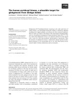
In general, fatty acids (FAs) can be degraded via dif-
ferent mechanisms, including a-, b- and x-oxidation
(Fig. 1). In humans a-oxidation takes place in peroxi-
somes only, whereas both peroxisomes and mitochon-
dria are able to b-oxidize FAs. Importantly, in recent
years a great number of genetically determined disor-
ders in humans has been described in which either FA
a-oxidation or FA b-oxidation in mitochondria or per-
oxisomes is deficient.
As discussed in more detail below, treatment options
for each of the different groups of FA oxidation disor-
ders is limited, which prompted us to investigate x-oxi-
dation as a rescue pathway for these disorders. This is
based on the notion that if it was possible to upregulate
the x-oxidation of specific FAs known to accumulate in
the different disorders, one could reduce the accumula-
tion of these FAs under in vivo conditions and thereby
counteract the detrimental effects associated with the
accumulation of these FAs and their derivatives, which
are the basis of the clinical signs and symptoms
observed in the different (groups of) disorders.
We first briefly describe FA a- and b-oxidation path-
ways and the disorders involved and then describe the
current state of knowledge regarding x-oxidation as
a rescue pathway for Refsum disease and X-linked
adrenoleukodystrophy (X-ALD).
Keywords
adrenoleukodystrophy; cytochrome P450;
fatty acids; mitochondria; peroxisomes;
Refsum disease; Zellweger syndrome;
a-oxidation; b-oxidation; x-oxidation
Correspondence
R. J. A. Wanders, Genetic Metabolic
Diseases, Room F0-226, Academic Medical
Center, University of Amsterdam,
Meibergdreef 9, 1105 AZ Amsterdam,
The Netherlands
Fax: +31 (0)20 6962596
Tel: +31 (0)20 5665958 ⁄ 5664197
E-mail:
(Received 22 June 2010, revised 28
September 2010, accepted 3 November
2010)
doi:10.1111/j.1742-4658.2010.07947.x
Fatty acids (FAs) can be degraded via different mechanisms including
a-, b- and x-oxidation. In humans, a range of different genetic diseases has
been identified in which either mitochondrial FA b-oxidation, peroxisomal
FA b-oxidation or FA a-oxidation is impaired. Treatment options for most
of these disorders are limited. This has prompted us to study FA x-oxida-
tion as a rescue pathway for these disorders, based on the notion that if
the x-oxidation of specific FAs could be upregulated one could reduce the
accumulation of these FAs and the subsequent detrimental effects in the
different groups of disorders. In this minireview, we describe our current
state of knowledge in this area with special emphasis on Refsum disease
and X-linked adrenoleukodystrophy.
Abbreviations
ATRA, all-trans-retinoic acid; CCALD, childhood cerebral adrenoleukodystrophy; FA, fatty acid; LTB4, leukotriene B4; PPAR, peroxisome
proliferator-activated receptor; VLCFA, very long-chain fatty acid; X-ALD, X-linked adrenoleukodystrophy.
182 FEBS Journal 278 (2011) 182–194 ª 2010 The Authors Journal compilation ª 2010 FEBS
General aspects of fatty acid oxidation
Beta-oxidation is the preferred way of oxidizing FAs.
In principle, each FA can be b-oxidized, including
straight- and branched-chain FAs, as well as mono-
and polyunsaturated FAs. There is one exception,
however, and that is if the carbon-3 has a methyl- or
any other functional group attached. In such cases,
degradation can only occur by a-orx-oxidation.
Until a few years ago, the enzymology of the a-oxi-
dation system remained unresolved and its subcellular
localization heavily disputed. This has now changed;
the basic chemistry of the pathway has been delineated
and the enzymes involved in a-oxidation and their sub-
cellular localization have been identified and character-
ized, although some questions remain [1,2].
Mitochondrial fatty acid b-oxidation
and its disorders
Mitochondria catalyze the b-oxidation of the majority
of FAs and contain the full enzymatic machinery to
oxidize straight-chain, 2-methyl-branched-chain, and
mono- and polyunsaturated FAs. After uptake of FAs
into the cells via a mechanism which remains incom-
pletely understood, but probably involves CD36 [3],
FAs are rapidly converted into coenzyme A (CoA)-
esters by one of the many acyl-CoA synthetases either
of the long-chain or very long-chain acyl-CoA synthe-
tase family [4]. Subsequently, the acyl-CoA esters are
transferred across the mitochondrial membrane by
means of the carnitine cycle, which involves carnitine
palmitoyltransferase I, mitochondrial carnitine acylcar-
nitine translocase and carnitine palmitoyltransferase II
[5–7]. In case of the straight-chain and 2-methyl-branched
chain FAs, b-oxidation can start right away via the
well-established cascade of four steps involving dehy-
drogenation, hydratation, dehydrogenation again and
thiolytic cleavage of the acyl-CoA esters. Each step of
the b-oxidation spiral is not catalyzed by one single
enzyme but by multiple chain-length-specific enzymes.
For example, at least three different acyl-CoA dehy-
drogenases are involved in the oxidation of saturated
long-chain FAs. These include very long-chain acyl-
CoA dehydrogenase, medium-chain acyl-CoA dehy-
drogenase and short-chain acyl-CoA dehydrogenase.
The same is true for the third step in mitochondrial
fatty acid b-oxidation, with at least two different
enzymes involved including short-chain 3-hydroxyacyl-
CoA dehydrogenase and long-chain 3-hydroxyacyl-
CoA dehydrogenase. The latter enzyme is part of a
larger enzyme complex called mitochondrial trifunc-
tional protein with additional enoyl-CoA hydratase
and 3-ketothiolase activities. Defects in each of these
enzymes have been identified (see Table 1). Although
the clinical signs and symptoms of patients vary
depending on the type of enzyme defect and the extent
of the deficiency, a general characteristic of all disor-
ders of mitochondrial FA oxidation is hypoketotic
hypoglycemia which may be life threatening, and car-
diomyopathy, especially in the case of the long-chain
fatty oxidation defects such as mitochondrial carnitine
acylcarnitine translocase deficiency, carnitine palmito-
yltransferase II deficiency, very long-chain acyl-CoA
dehydrogenase deficiency and long-chain 3-hydroxya-
cyl-CoA dehydrogenase ⁄ mitochondrial trifunctional
protein deficiency [8,9]. With the exception of dietary
measures consisting of a diet rich in carbohydrates and
low in fat taken at frequent intervals, there are virtu-
ally no realistic treatment options.
Fig. 1. Schematic diagram depicting the
different mechanisms by which fatty acids
can be oxidized (see text).
R. J. A. Wanders et al. Fatty acid oxidation disorders
FEBS Journal 278 (2011) 182–194 ª 2010 The Authors Journal compilation ª 2010 FEBS 183
Peroxisomal a-oxidation and its
disorders
FA a-oxidation allows the chain-shortening of FAs by
one carbon atom and takes place in peroxisomes only.
A typical 3-methyl-branched-chain FA like phytanic
acid (3,7,11,15-tetramethylhexadecanoic acid), is com-
pletely dependent on a normal functioning a-oxidation
system in order to be oxidized. A defect in the a-oxida-
tion system is reflected in the accumulation of phytanic
acid in the tissues and body fluids of patients [1,2,10].
Alpha-oxidation of phytanic acids starts with the
formation of the CoA-ester, i.e. phytanoyl-CoA, fol-
lowed by hydroxylation to generate 2-hydroxyphy-
tanoyl-CoA, a reaction catalyzed by the enzyme
phytanoyl-CoA 2-hydroxylase. Subsequently, 2-hydr-
oxyphytanoyl-CoA is cleaved by the enzyme 2-hydr-
oxyacyl-CoA lyase to pristanal and formyl-CoA, which
is then hydrolyzed to formic acid and coenzyme A
(CoASH). Pristanal is then oxidized to pristanic acid
(2,6,10,14-tetramethylpentadecanoic acid), as catalyzed
by a yet undefined peroxisomal aldehyde dehydroge-
nase. After activation to its CoA-ester, pristanoyl-CoA
undergoes three cycles of b-oxidation in peroxisomes,
after which the end-products are transported to mito-
chondria for full oxidation [11,12].
Alpha-oxidation is deficient in different peroxi-
somal disorders including the peroxisome biogenesis
disorders, in which the primary genetic defect is in one
of the many genes involved in peroxisome biogenesis
[13–15]. To date, however, only one single enzyme defi-
ciency in the a-oxidation pathway per se has been
described. This is phytanoyl-CoA hydroxylase defi-
ciency with Refsum disease as its disease counterpart
[10]. Patients suffering from Refsum disease show a
late-onset phenotype, dominated by retinitis pigmen-
tosa, culminating in blindness with anosmia, cerebellar
ataxia and a range of other more variable abnormali-
ties. The only therapy available to date is a life-long
diet low in phytanic acid, which may stop further pro-
gression of some, but not all, of the symptoms
provided the diet is meticulously maintained.
Peroxisomal fatty acid b-oxidation and
its disorders
Peroxisomes contain a FA b-oxidation system just like
mitochondria, but the individual reactions of the b-oxi-
dation spiral are catalyzed by different enzymes
encoded by distinct genes compared with the mito-
chondrial b-oxidation system [11]. Importantly, peroxi-
somes oxidize a unique set of FAs which cannot be
b-oxidized in mitochondria. Most important from a
clinical point of view are: (a) very long-chain fatty
acids (VLCFAs), notably C24:0 and C26:0; (b) pris-
tanic acid (2,6,10,14-tetramethylpentadecanoic acid), as
Table 1. The mitochondrial and peroxisomal beta-oxidation deficiencies
Mitochondrial fatty acid oxidation disorders Mutant gene Deficient enzyme Locus OMIM
Carnitine palmitoyl-CoA transferase-1 deficiency CPT1A CPT1A 11q13 600528 ⁄ 255120
Carnitine ⁄ acylcarnitine translocase deficiency SLC25A2 CACT 3p21 212138
Carnitine palmitoyl-CoA transferase-2 deficiency CPT2 CPT2 1p32 600649 ⁄ 255110
Very long-chain acyl-CoA dehydrogenase deficiency ACADVL VLCAD 17p11 201475
Medium-chain acyl-CoA dehydrogenase deficiency ACADM MCAD 1p31 201450
Short-chain acyl-CoA dehydrogenase deficiency ACADS SCAD 12q22 201470
Isolated long-chain 3-hydroxyacyl-CoA dehydrogenase deficiency HADHA LCHAD 2p23 600890
Isolated long-chain 3-ketothiolase deficiency HADHB LCKAT 2p23 143450
Complete mitochondrial trifunctional protein deficiency HADHA LCHAD 2p23 600890
HADHB LCKAT 2p23 143450
Short-chain 3-hydroxyacyl-CoA dehydrogenase deficiency HADHSC SCHAD 4q22 601609
Medium-chain 3-ketoacyl-CoA thiolase deficiency ACAA2 MCKAT 602199
ETF dehydrogenase deficiency ETFDH ETFDH 4q32 231675
ETF-alpha deficiency ETFA ETFa 15q23 608053
ETF-beta deficiency ETFB ETFb 19q13 130410
2,4-dienoyl-CoA reductase deficiency DECR1 DECR1 8q21 222745
Peroxiomal fatty acid oxidation disorders
X-linked adrenoleukodystrophy ABCD1 ALDP Xq28 300100
Acyl-CoA oxidase deficiency ACOX1 ACOX1 17q25.1 264470
D-Bifunctional protein deficiency HSD17B4 DBP ⁄ MFP2 5q2 261515
2-methylacyl-CoA racemase deficiency AMACR AMACR 5p13.3-p12 604489
Peroxisomal sterol carrier protein x (SCPx) deficiency SCP2 SCPx 1p32 –
Fatty acid oxidation disorders R. J. A. Wanders et al.
184 FEBS Journal 278 (2011) 182–194 ª 2010 The Authors Journal compilation ª 2010 FEBS
derived directly from dietary sources and indirectly
from phytanic acid upon a-oxidation; and (c) di- and
trihydroxycholestanoic acid (see [11] for review). Per-
oxisomes are unable to b-oxidize FAs to completion.
Instead FAs are only chain-shortened to shorter chain
FAs followed by the transfer of these chain-shortened
FAs to mitochondria for full oxidation. This has been
established most firmly for pristanic acid which under-
goes three cycles of b-oxidation in peroxisomes to
produce propionyl-CoA, acetyl-CoA and 4.8-dimethyl-
nonanoyl-CoA followed by the transfer of these
CoA-esters either as carnitine-ester or as free fatty acid
to mitochondria for full oxidation to CO
2
and H
2
O
[16,17].
At present, five different genetically determined sin-
gle-enzyme deficiencies have been described in humans.
These include: (a) X-ALD, (b) acyl-CoA oxidase defi-
ciency, (c) D-bifunctional protein deficiency, (d) sterol
carrier protein x deficiency and (e) 2-methylacyl-CoA
racemase deficiency [18]. All five disorders are rela-
tively rare with sterol carrier protein x deficiency
described in a single patient only to date [19], 2-meth-
ylacyl-CoA racemase-deficiency described in six
patients [20] and acyl-CoA oxidase deficiency described
in 30 patients [21]. Most frequent is X-ALD with an
incidence of 1 : 15 000, followed by D-bifunctional
protein deficiency [22]. X-ALD is a devastating neuro-
logical disease which comes in two main phenotypes
including childhood cerebral ALD (CCALD) and
adrenomyeloneuropathy, together constituting > 80%
of all X-ALD patients. The most devastating pheno-
type is CCALD which is characterized by a rapidly
progressive cerebral demyelination causing severe dis-
ability and death, usually within 2 years after the onset
of symptoms. Adrenomyeloneuropathy has a much
milder course characterized by a gradually progressive
myelopathy and peripheral neuropathy, causing severe
disability.
X-ALD is caused by mutations in the ABCD1 gene
which codes for a peroxisomal half-ABC transporter
adrenoleukodystrophy protein (ALDP), localized in
the peroxisomal membrane as a homodimer. ALDP
catalyzes the transport of very long-chain FAs across
the peroxisomal membrane in the CoA-ester form
[23,24]. If ALDP is absent or dysfunctional, oxidation
of VLCFA is impaired and this leads to the accumula-
tion of VLCFAs in plasma and tissues including the
brain of X-ALD patients. The VLCFAs that accumu-
late are not so much derived from the diet, but are
synthesized endogenously via chain elongation [24],
which explains why a diet low in VLCFAs is of no
benefit for X-ALD patients (Fig. 2). The only thera-
peutic options for X-ALD are bone-marrow transplan-
tation and gene therapy, as recently reported by
Cartier and Aubourg [25] in three X-ALD boys.
Fatty acid x -oxidation by CYP450
proteins in humans
Early work on FA x-oxidation dates back to the
1930s when Verkade et al. [26,27] performed a series of
systematic studies that revealed the formation of dicar-
boxylic acids after administration of medium-chain tri-
glycerides to healthy individuals. It was the 1960s
before enzymatic studies could be performed using
subcellular fractions prepared from guinea-pig, rat and
human livers. This allowed identification of the path-
way intermediates and the subsequent discovery that
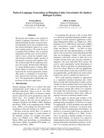
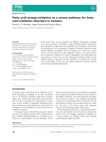
Fig. 2. Schematic diagram illustrating the
homeostatic mechanisms involved in C26:0
metabolism. Very long-chain fatty acids are
predominantly derived from long-chain fatty
acids via chain-elongation and degraded via
b-oxidation in the peroxisome. Several
diseases are known in which b-oxidation
is deficient including X-linked
adrenoleukodystrophy. Omega-oxidation of
C26:0 involves the participation of different
enzymes including CYP4F2 and CYP3FB
plus ALDH3A2. The latter converts the
x-keto form of C26:0 into the dicarboxylic
acid.
R. J. A. Wanders et al. Fatty acid oxidation disorders
FEBS Journal 278 (2011) 182–194 ª 2010 The Authors Journal compilation ª 2010 FEBS 185
the enzyme catalyzing the first step of the pathway was
in fact a hemoprotein belonging to the ubiquitous dis-
covered family of CYP450s, with members in eukary-
otic and prokaryotic species [28–30].
The CYP4A subfamily
After the successful cloning of CYP4A1 coding for
clofibrate-inducible arachidonic acid ⁄ lauric acid
x-hydroxylase from rat liver [31], the human homolog
of this enzyme was identified and named CYP4A11
[32,33]. CYP4A11 turned out to have a broad sub-
strate spectrum and is able to x-hydroxylate the satu-
rated FAs lauric acid, myristic acid (tetradecanoic
acid), palmitic acid (hexadecanoic acid) and the unsat-
urated FAs oleic acid [(Z)-octadec-9-enoic acid] and
arachidonic acid (all-cis-5,8,11,14-eicosatetraenoic acid)
[34]. Recently, another CYP4A subfamily member in
humans was identified and designated CYP4A22 [35].
This protein is highly homologous with CYP4A11
and not abundantly present in tissues. Kawashima
et al. have expressed the CYP4A22 protein in Escheri-
chia coli, and showed that this protein has lauric acid
x-hydroxylase activity [36], but erroneously reported it
to be CYP4A11.
The CYP4F subfamily
CYP4A is not the only subfamily of CYP4 proteins in
humans that are capable of x-hydroxylation of fatty
acids. During the early 1980s, Hansson et al. reported
on the x-oxidation of leukotriene B4 (LTB4) in human
leukocytes [37]. The x-oxidation pathway is necessary
for the degradation (and thereby inactivation) of this
compound, which plays an important role in the inflam-
mation process. The CYP450 involved in this pathway
belonged to a, at that moment, unidentified subfamily,
the CYP4F subfamily (reviewed in [38,39]), and was
designated as CYP4F3. Later it was found that the
CYP4F3 gene could give rise to two different transcripts
by alternative promoter usage and tissue-specific gene
splicing, which results in two different proteins [40,41].
The isoform originally detected in leukocytes was desig-
nated CYP4F3A and the other, which was detected in
liver, was designated CYP4F3B. These proteins differ
from each other due to the alternative use of only one
exon. However, this leads to a substantial difference in
substrate specificity, with CYP4F3A being specific for
LTB4, whereas CYP4F3B has a higher specificity
towards arachidonic acid [40,42].
Shortly after the cloning of CYP4F3A from human
leukocytes in 1993, Kikuta et al. identified a novel
LTB4-hydroxylating CYP450 in human liver [43]. This
isoform was named CYP4F2 and has a high homology
with the CYP4F3B protein. CYP4F2 was shown to be
the major arachidonic acid x-hydroxylase in human
liver and kidney with a higher substrate specificity for
arachidonic acid than the already established arachi-
donic acid x-hydroxylase CYP4A11 [44,45]. The
formation of x-hydroxylated arachidonic acid (20-hy-
droxyeicosatetraenoic acid) plays an important role in
the regulation of the cardiovascular system because it
is a known vasoconstrictor (reviewed in [46]). CYP4F2
was also shown to x-hydroxylate LTB4 in liver, which
suggests that this protein might play a role in the
inflammatory system [47]. Furthermore, CYP4F2 is
responsible for x-hydroxylation of the phytyl tail of
the tocopherols and tocotrienols that are collectively
called vitamin E [48]. Omega-hydroxylation is the
initial step for the degradation of vitamin E via x-oxi-
dation and subsequent b-oxidation [48,49].
Three additional members of the CYP4F subfamily
have been identified in humans, as reviewed by Kalso-
tra and Strobel [38]. These were recently discovered
and have been only partially characterized. CYP4F8
is present in epithelial linings and catalyzes the
(x-1)-hydroxylation of prostaglandin H2. CYP4F11 is
mainly expressed in liver, followed by kidney, heart,
brain and skeletal muscle. No endogenous substrates
have been found to date for CYP4F11, but it has been
shown that recombinant CYP4F11 is quite active in
hydroxylating some xenobiotics. Finally, the CYP4F12
protein detected in human liver, heart, gastrointestinal
and urogenital epithelia is active towards both eicosa-
noids and xenobiotics.
Other CYP4 homologs
The CYP4B1 protein, which is predominantly
expressed in lung, forms another subfamily of x-hy-
droxylases. However, this protein has no clear sub-
strate spectrum, but it is capable of x-hydroxylating
medium-chain FAs and xenobiotics (see [50] for
review). Other CYP450s belonging to family 4 have
been identified in humans. Their homology with the
known CYP4 subfamilies suggests that these orphans
(i.e. CYPs with unknown substrate specificity) might
be able to x-hydroxylate FAs and ⁄ or FA-like com-
pounds [51].
The most important and well-characterized x-hy-
droxylases, the CYP450s belonging to the CYP4A and
4F subfamilies, are present not only in humans;
CYP4A ⁄ F homologs are also well represented in other
animals, such as the mouse, rat and rabbit [38,52,53].
Moreover, these animals contain more CYP4A and
CYP4F subfamily members than humans, which
Fatty acid oxidation disorders R. J. A. Wanders et al.
186 FEBS Journal 278 (2011) 182–194 ª 2010 The Authors Journal compilation ª 2010 FEBS
makes interpretation of results found in studies using
these animals problematic.
Induction of fatty acid x-hydroxylases
CYP4A gene regulation and the role of
peroxisome proliferator-activated receptor a in
CYP4A induction
The induction of drug-metabolizing enzymes by for-
eign compounds has been the topic of many research
studies during the last several decades. Halfway the
20th century it was already known that the x-hydroxy-
lase activity in microsomal fractions prepared from rat
livers was much higher when laboratory animals were
fed with certain kinds of xenobiotics, including: (a)
polycyclic aromatic hydrocarbons and (b) barbiturates,
as described in Conney [54]. A third type of metabolic
enzyme inducers are the hypolipidemic drugs of the
fibrate class, which have been in use since the early
1960s, and were found to upregulate the x-hydroxyl-
ation of lauric acid [55,56]. All of these compounds are
able to induce one or more CYP450s. However, the
precise mechanism which is the basis of this induction
has remained unclear for several decades. The discov-
ery that a receptor (the aryl or aromatic hydrocarbon
receptor) was involved in the induction by polycyclic
aromatic hydrocarbons of the CYP450s responsible for
the hydroxylation of polycyclic aromatic hydrocarbons
(CYP1 family) was the first step in unraveling the com-
plex mechanism of CYP450 regulation (reviewed in
[57]). Another breakthrough in CYP450 regulation was
the finding that peroxisome proliferator-activated
receptor alpha (PPARa) was involved in the induction
of CYP4A enzymes (reviewed by Johnson et al. [58]).
PPARa is a member of the large receptor superfamily
of ligand-activated transcription factors (also referred
to as the nuclear receptor family) [59]. Moreover,
many members of this superfamily have been found to
be involved in the regulation of multiple CYP450s
[60,61].
PPARa is a member of the larger family of PPARs
which also consists of a b-(d) and c-isoform. All iso-
forms play important roles in physiological processes
as lipid sensors and regulators of lipid and glucose
homeostasis. However, the different PPARs have spe-
cific substrate specificities and tissue distributions, and
control specific subsets of transcriptional profiles (see
[62,63] for review). Activation of the PPARs by the
so-called peroxisome proliferators (a structurally unre-
lated class of compounds among which are FAs, plast-
icizers, herbicides and the fibrate class of
hypolipidemic drugs) enables the receptor to dimerize
with another nuclear receptor, the retinoid X receptor
[64]. The ligand-activated heterodimer can bind to spe-
cific sequences of DNA known as peroxisome prolifer-
ator responsive elements in the promoter regions of
target genes, thereby inducing gene expression of the
target gene. Most of these target genes are involved in
lipid metabolism. Particularly pronounced is the induc-
tion of proteins involved in peroxisomal fatty acid
metabolism, which leads to an increase in peroxisomal
number (i.e. peroxisome proliferation) and size [65].
Induction of hepatic peroxisome proliferation by
PPARa activation in rodents ultimately leads to hepa-
tomegaly and hepatocarcinogenesis (see Gonzalez [66]
for review). Fortunately, these toxic effects of PPARa
ligands are not observed in humans [67]. Therefore,
fibrates are still in use as important drugs for the treat-
ment of patients with dyslipidemia and ⁄ or metabolic
syndrome (reviewed in [68,69]).
Besides increasing peroxisomal FA oxidation,
PPARa is also involved in the upregulation of mito-
chondrial b-oxidation, FA transport and the already
mentioned FA x-hydroxylation via the CYP4A sub-
family. Initial studies, which focused on the induction
of the CYP4A subfamily in rats and mice, showed that
levels of certain subtypes did indeed increase in these
rodent animal models after PPARa activation [70,71].
In humans, uncertainties remain with respect to the
induction of the CYP4A subtype. Overexpression of
PPARa in the hepatoma cell line HepG2 led to an
increase in CYP4A11 ⁄ A22 under specific growth con-
ditions, suggesting the involvement of PPARa in the
regulation of human CYP4A expression [72]. Another
study showed that fibrates are able to induce
CYP4A11 mRNA expression in primary cultures of
human hepatocytes [73]. By contrast, the peroxisome
proliferators responsive elements present in the promo-
tor regions of the genes coding for members of the
CYP4A subfamily in rodents have not been identified
in human CYP4A genes [36].
Recently, another regulatory pathway for CYP4A11
gene expression was discovered. Activation of a differ-
ent member of the nuclear hormone receptor family,
retinoic acid receptor, by all-trans retinoic acid
(ATRA) in the hepatoma cell line HepaRG was shown
to decrease CYP4A11 gene and protein expression,
ultimately leading to a decrease in catalytic activity
(lauric acid hydroxylation) in this cell line [74].
In mice, three different CYP4A genes have been
identified. Cyp4a10 is highly expressed in both sexes,
whereas Cyp4a12 (consisting of two gene products,
Cyp4a12a and Cyp4a12b) is predominantly male spe-
cific and Cyp4a14 is a female-specific isoform. Further-
more, the protein levels of these Cyp4a isoforms vary
R. J. A. Wanders et al. Fatty acid oxidation disorders
FEBS Journal 278 (2011) 182–194 ª 2010 The Authors Journal compilation ª 2010 FEBS 187
in different mouse strains and tissues. PPARa also
plays an important role in the regulation of the expres-
sion of the different Cyp4a isoforms in mice. Fibrates
are able to induce gene expression of Cyp4a10 and
Cyp4a14 [71,75] in both liver and kidney. Cyp4a12 is
constitutively expressed in kidney and liver of male
mice, whereas in kidney and liver of female mice
Cyp4a12 is expressed at low levels. Moreover, Cyp4a12
gene expression cannot be induced by fibrates in kid-
ney and liver of male mice, whereas in female mice,
kidney and liver Cyp4a12 RNA levels were increased
to male levels after treatment with fibrates. In addi-
tion, Cyp4a12 gene expression is also upregulated in
female mice by treatment with androgens via an as yet
unknown mechanism [75,76].
CYP4F induction
By contrast to the CYP4A subfamily, relatively few
studies have appeared on the regulation of genes
comprising the CYP4F subfamily (reviewed in
[37,38,77]). Regulation of the CYP4F2 gene has been
studied most intensively of all human isoforms.
Zhang et al. [78] found that CYP4F2 gene expression
was regulated by retinoic acid and fibrates. Peroxi-
some proliferators suppressed CYP4F2 promotor
activity, whereas both 9-cis-retinoic acid and ATRA
induced promoter activity through activation of reti-
noic acid receptor and retinoid X receptor. However,
further research revealed that protein expression of
CYP4F2 was increased by 9-cis-retinoic acid in the
hepatoma cell line HepG2, in marked contrast to
ATRA, which only gave rise to an induction of
CYP4F2 promotor activity [79]. From these results,
Zhang and Hardwick concluded that CYP4F2 gene
expression is regulated by 9-cis-retinoic acid and
ATRA. Activation of retinoid X receptor induces
gene expression (and protein content) and retinoic
acid receptor activation results in repression of gene
expression. Recently, Hsu et al. showed that in
HepG2 cells and primary hepatocytes, CYP4F2 gene
expression and protein content could be induced by
statins, which are well-known drugs used for the
treatment of hypercholesterolemia [80]. Furthermore,
Hsu et al. showed that the CYP4F2 transcriptional
activation is mediated by sterol regulatory element
binding proteins (SREBP; reviewed in [81]) and that
activation of the sterol regulatory element binding
protein-2 isoform is involved in the induction
CYP4F2 by statins [80].
Parallel studies on the induction of CYP4F3 showed
that this enzyme was induced in HL60 cells and
human leukocytes after treatment of these cells with
ATRA [82,83]. However, the mechanism behind this
induction remains to be determined since the receptor
for ATRA, i.e. retinoic acid receptor, seems only indi-
rectly involved in this process.
Studies in rats and mice have shown that the expres-
sion of some isoforms of the CYP4 subfamily changes
during inflammation. During an inflammatory
response, induction of CYP4F isoforms occurs in
rodents needed for the breakdown of inflammatory
mediators such as the eicosanoid LTB4 (reviewed in
[38,77]). Recent studies by Kalsotra et al. [84] provided
evidence that specific cytokines are involved in regula-
tion of the CYP4F enzymes levels during inflamma-
tion. The pro-inflammatory cytokines interleukin-1b,
interleukin-6 and tumor necrosis factor-a are able to
induce CYP4Fs, whereas the anti-inflammatory cyto-
kine interleukin-10 suppresses CYP4F expression [84].
Peroxisomal fatty acid b-oxidation
disorders including X-ALD and
x-oxidation
Despite the profound increase in our knowledge about
X-ALD, treatment options are very limited and are
mostly symptomatic. Lorenzo’s oil reduces plasma
C26:0 but does not halt progression of the disease
[85,86]. Lovastatin also lowered plasma VLCFA [87],
but a recent placebo-controlled trial revealed that lova-
statin has no effect on C26:0 levels in peripheral blood
lymphocytes and erythrocytes, or on the VLCFA con-
tent of the low-density lipoprotein fraction [88].
Hematopoietic stem cell transplantation can halt or
reverse clinical deterioration [89]. However, it is only
effective in patients at the earliest stage of CCALD.
Recent breakthroughs in gene therapy have to date
been applied to CCALD only [25]. Therefore new
therapeutic options aimed at the reduction of VLCFA
are warranted.
We have previously demonstrated that VLCFA can
undergo x-oxidation [90]. This would provide an alter-
native mode of degradation. We demonstrated that
CYP4F2 and CYP4F3B are key enzymes in this
pathway [91]. In the first step of the metabolism of
VLCFA via x-oxidation, VLCFAs are converted into
x-hydroxy-VLCFA by CYP4F2 or CYP4F3B (Fig. 1).
Subsequently, this product is readily oxidized to a
dicarboxylic-VLCFA by an alcohol and aldehyde
dehydrogenase or via subsequent hydroxylation
reactions by CYP4F2 and CYP4F3B [92]. The
dicarboxylyl-VLCFAs that are generated can be
metabolized further in peroxisomes via b-oxidation.
Beta-oxidation of dicarboxylyl VLCFA takes place in
peroxisomes and this process is not deficient in
Fatty acid oxidation disorders R. J. A. Wanders et al.
188 FEBS Journal 278 (2011) 182–194 ª 2010 The Authors Journal compilation ª 2010 FEBS
X-ALD. This is concluded from the finding that
b-oxidation of long-chain dicarboxylic acid is not
affected in fibroblasts from X-ALD patients, whereas
oxidation was deficient in fibroblasts from patients
with a peroxisomal biogenesis disorder [93]. These
findings indicate that peroxisomes are essential for the
degradation of dicarboxylic acids, but that ALDP is
not required for the transport of dicarboxylic acids
across the peroxisomal membrane. Because the trans-
port of dicarboxylic acids may involve other ABC
half-transporters, i.e. ALDRP or PMP70, the x-oxida-
tion of VLCFA may function as an escape route.
Under normal physiological conditions, the x-oxida-
tion pathway accounts for 5–10% of total FA oxida-
tion. Because expression levels of cytochrome P450
enzymes can be induced by a variety of drugs and
chemicals [94], stimulation of VLCFA x-oxidation
may reduce or normalize VLCFA levels and might
therefore be beneficial for X-ALD patients (Fig. 1).
The possibility of upregulating VLCFA x-oxidation
and its consequences for VLCFA homeostasis are now
being studied in a mouse model of X-ALD.
Mitochondrial b-oxidation and
x-oxidation
In the case of mitochondrial FA b-oxidation disorders,
there is accumulation of certain FAs either as free
FAs, or in an esterified form as in CoA and carnitine
esters. The types of FAs and FA derivates that accu-
mulate are determined by the site of the enzyme defect.
The different acylcarnitine profiles observed in the var-
ious mitochondrial b-oxidation deficiencies emphasize
this notion [95]. Specific induction of the capacity to
x-oxidize these FAs would reduce the FA burden and
may ameliorate the signs and symptoms in these
patients. No studies on this point have been published
in the literature.
Refsum disease, phytanic acid and
x-oxidation
Brenton and Krywawych [96] reported on the excretion
of 3-methyladipic acid in the urine of Refsum patients,
which suggested that phytanic acid does undergo x-oxi-
dation under in vivo conditions. This was soon followed
by another report, which documented the identification
of 2,6-dimethyloctanedioic acid, a metabolite derived
from x-oxidation of phytanic acid in Refsum’s patients.
The finding by Wierzbicki et al. [97] that the amounts of
3-methyladipic acid in urine from Refsum’s patients
correlated with plasma levels of phytanic acid in these
patients, has lent further support to the notion that
3-methyladipic acid is indeed formed upon x-oxidation
of phytanic acid. Based on these results, we have begun
to characterize the enzymology of the x-oxidation
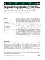
Fig. 3. Schematic diagram depicting phytanic acid homeostasis. Phytanic acid is derived solely from dietary sources and can be oxidized by
either a-oxidation or x-oxidation (see text for further details). The product of the peroxisomal a-oxidation of phytanic acid is pristanic acid
which first undergoes three cycles of b-oxidation in the peroxisome to produce propionyl-CoA (in the first and third cycle of b-oxidation) and
acetyl-CoA (in the second cycle of b-oxidation) plus 4,8-dimethylnonanoyl-CoA. These CoA-esters are all transferred to mitochondria for
further oxidation.
R. J. A. Wanders et al. Fatty acid oxidation disorders
FEBS Journal 278 (2011) 182–194 ª 2010 The Authors Journal compilation ª 2010 FEBS 189
pathway, first in rat liver microsomes [98] and then in
human liver microsomes [99,100]. In rat liver micro-
somes we found that phytanic acid undergoes both (x-)
and (x-1)-hydroxylation [98]. In human microsomes,
however, there was a virtually exclusive production of
x-hydroxy phytanic acid [99]. In order to identify the
CYP450 involved we first performed studies with selec-
tive inhibitors including 17-octadecynoic acid, diet-
hyldithiocarbamate, ketoconazole, troleandomycin,
omeprazole, trimethoprim, furafylline, quinidine and
sulfaphenozole [100]. These studies already pointed to
the CYP4 family of x-hydroxylases as likely candidates.
The availability of individually expressed CYP4s pro-
duced in baculovirus-infected insect cells (SupersomesÔ)
allowed this possibility to be tested directly. CYP4F3A
turned out to be most reactive towards phytanic acid,
followed by CYP4F3B, CYP4F2 and CYP4A11 with
catalytic efficiencies of 0.87, 0.22, 0.06 and 0.02, respec-
tively [100]. The question now is whether upregulation
of one or more of these CYP450s is feasible under in vivo
conditions and if this is associated with an increased rate
of phytanic acid x-oxidation or not [12] (see Fig. 3).
With respect to CYP4F3A and CYP4F3B, there is no
information about whether expression can be upregulat-
ed. CYP4A11 expression is controlled by PPARa in
conjunction with retinoid X receptor so that fibrates or
other PPAR ligands should be successful in upregulating
CYP4A11 activity. Finally, with respect to CYP4F2, it
has been established experimentally that the promoter of
the CYP4F2 gene contains a sterol-regulatory element,
as described above. Activation of the classical sterol
regulatory element binding protein (SREBP) route, for
example by means of statins, inhibitors of 3-hydroxy-
3-methylglutaryl-CoA (HMG-CoA) reductase, would
then lead to the increased expression of CYP4F2. The
availability of a mouse model for Refsum disease allows
for future studies aimed at resolving whether upregu-
lation of CYP4A11 by fibrates and ⁄ or CYP4F2 by
statins leads to the enhanced degradation of phytanic
acid and amelioration of the clinical signs and symptoms
[101].
Conclusions
Omega-oxidation as a rescue pathway for different
genetic diseases in humans in which either peroxisomal
or mitochondrial FA oxidation is impaired, is an
attractive possibility to allow breakdown of FAs which
accumulate as a consequence of an enzyme or trans-
porter defect. Identification of the specific cytochrome
P450s involved in the x-oxidation of phytanic acid and
VLCFAs, added to the fact that the different CYPs
involved can be induced pharmacologically, now
allows us to study whether our in vitro data can be
extrapolated successfully to intact organisms. We will
first perform such studies in mouse models for Refsum
disease and X-ALD.
Acknowledgements
This work was supported by grants from the European
Leukodystrophy Association [ELA 2008-05111A (RJW)],
the Prinses Beatrix Fonds [WAR08-20 (SK)] and the
Netherlands Organization for Scientific Research
[VIDI-grant No. 91786328 (SK)]. Mrs Maddy Festen is
gratefully acknowledged for preparation of the manu-
script and Mr Jos Ruiter for design of the figures.
References
1 Wanders RJA, Jansen GA & Lloyd MD (2003) Phytanic
acid alpha-oxidation, new insights into an old problem:
a review. Biochim Biophys Acta 1631, 119–135.
2 Casteels M, Foulon V, Mannaerts GP & Van Veldho-
ven PP (2003) Alpha-oxidation of 3-methyl-substituted
fatty acids and its thiamine dependence. Eur J Biochem
270, 1619–1627.
3 Glatz JF, Luiken JJ & Bonen A (2010) Membrane fatty
acid transporters as regulators of lipid metabolism: impli-
cations for metabolic disease. Physiol Rev 90, 367–417.
4 Watkins PA (2008) Very-long-chain acyl-CoA syntheta-
ses. J Biol Chem 283, 1773–1777.
5 Rinaldo P, Matern D & Bennett MJ (2002) Fatty acid
oxidation disorders. Annu Rev Physiol 64, 477–502.
6 Wanders RJA, Vreken P, Den Boer MEJ, Wijburg FA,
Van Gennip AH & IJlst L (1999) Disorders of mito-
chondrial fatty acyl-CoA beta-oxidation. J Inherit
Metab Dis 22, 442–487.
7 Houten SM & Wanders RJA (2010) A general intro-
duction to the biochemistry of mitochondrial fatty acid
beta-oxidation. J Inherit Metab Dis 33, 469–477.
8 Bonnet D, Martin D, Pascale DL, Villain E, Jouvet P,
Rabier D, Brivet M & Saudubray JM (1999) Arrhyth-
mias and conduction defects as presenting symptoms of
fatty acid oxidation disorders in children. Circulation
100, 2248–2253.
9 Saudubray JM, Martin D, de Lonlay P, Touati G,
Poggi-Travert F, Bonnet D, Jouvet P, Boutron M, Sla-
ma A, Vianey-Saban C et al. (1999) Recognition and
management of fatty acid oxidation defects: a series of
107 patients. J Inherit Metab Dis 22, 488–502.
10 Wierzbicki AS (2007) Peroxisomal disorders affecting
phytanic acid alpha-oxidation: a review. Biochem Soc
Trans 35, 881–886.
11 Wanders RJA & Waterham HR (2006) Biochemistry of
mamalian peroxisomes revisited. Annu Rev Biochem 75,
295–332.
Fatty acid oxidation disorders R. J. A. Wanders et al.
190 FEBS Journal 278 (2011) 182–194 ª 2010 The Authors Journal compilation ª 2010 FEBS
12 Wanders RJA & Komen JC (2007) Peroxisomes,
Refsum’s disease and the alpha- and omega-oxidation
of phytanic acid. Biochem Soc Trans 35, 865–869.
13 Weller S, Gould SJ & Valle D (2003) Peroxisome
biogenesis disorders. Annu Rev Genomics Hum Genet 4,
165–211.
14 Wanders RJA & Waterham HR (2005) Peroxisomal
disorders I: biochemistry and genetics of peroxisome
biogenesis disorders. Clin Genet 67, 107–133.
15 Steinberg SJ, Dodt G, Raymond GV, Braverman NE,
Moser AB & Moser HW (2006) Peroxisome biogenesis
disorders. Biochim Biophys Acta 1763, 1733–1748.
16 Verhoeven NM, Roe DS, Kok RM, Wanders RJA,
Jakobs C & Roe C (1998) Phytanic acid and pristanic
acid are oxidized by sequential peroxisomal and mito-
chondrial reactions in cultured fibroblasts. J Lipid Res
39, 66–74.
17 Verhoeven NM & Jakobs C (2001) Human metabolism
of phytanic acid and pristanic acid. Prog Lipid Res 40,
453–466.
18 Wanders RJA & Waterham HR (2006) Peroxisomal
disorders: the single peroxisomal enzyme deficiencies.
Biochim Biophys Acta 1763, 1707–1720.
19 Ferdinandusse S, Kostopoulos P, Denis S, Rusch H,
Overmars H, Dillmann U, Reith W, Haas D, Wanders
RJA, Duran M et al. (2006) Mutations in the gene
encoding peroxisomal sterol carrier protein X (SCPx)
cause leukencephalopathy with dystonia and motor
neuropathy. Am J Hum Genet 78, 1046–1052.
20 Ferdinandusse S, Denis S, Clayton PT, Graham A,
Rees JE, Allen JT, Mclean BN, Brown AY, Vreken P,
Waterham HR et al. (2000) Mutations in the gene
encoding peroxisomal alpha-methylacyl-CoA racemase
cause adult-onset sensory motor neuropathy. Nat Genet
24, 188–191.
21 Ferdinandusse S, Denis S, Hogenhout EM, Koster J,
van Roermund CWT, IJlst L, Moser AB, Wanders
RJA & Waterham HR (2007) Clinical, biochemical,
and mutational spectrum of peroxisomal acyl-coenzyme
A oxidase deficiency. Hum Mutat 28, 904–912.
22 Ferdinandusse S, Denis S, Mooyer PA, Dekker C,
Duran M, Soorani-Lunsing RJ, Boltshauser E, Macaya
A, Gartner J, Majoie CB et al. (2006) Clinical and bio-
chemical spectrum of D-bifunctional protein deficiency.
Ann Neurol 59, 92–104.
23 van Roermund CWT, Visser WF, IJlst L, van Cruchten
AG, Boek M, Kulik W, Waterham HR & Wanders
RJA (2008) The human peroxisomal ABC half
transporter ALDP functions as a homodimer and
accepts acyl-CoA esters. FASEB J 22, 4201–4208.
24 Ofman R, Dijkstra IM, van Roermund CW, Burger N,
Turkenburg M, van Cruchten A, van Engen CE,
Wanders RJA & Kemp S (2010) The role of ELOVL1
in very long-chain fatty acid homeostasis and X-linked
adrenoleukodystrophy. EMBO Mol Med 2, 90–97.
25 Cartier N, Hacein-Bey-Abina S, Bartholomae CC,
Veres G, Schmidt M, Kutschera I, Vidaud M, Abel U,
Dal-Cortivo L, Caccavelli L et al. (2009)
Hematopoietic stem cell gene therapy with a lentiviral
vector in X-linked adrenoleukodystrophy. Science 326,
818–823.
26 Verkade PE & Van der Lee J (1934) Researches on fat
metabolism II. Biochem J 28, 31–40.
27 Verkade PE, Elzas M, Van der Lee J, De Wolff
HHV-SA & Van der Sande A (1932) Research on fat
metabolism I. Proc K Ned Akad Wet 35, 251.
28 Van Bogaert INA, Groeneboer S, Saerens K & Soetaert W
(2010) The role of cytochrome P450 monooxygenases in
microbial fatty acid metabolism. FEBS J.
29 Pinot F & Beisson F (2010) Cytochrome P450
metabolizing fatty acids in plants: characterization and
physiological roles. FEBS J.
30 Omura T & Sato R (1962) A new cytochrome in liver
microsomes. J Biol Chem 237, 1375–1376.
31 Hardwick JP, Song BJ, Huberman E & Gonzalez FJ
(1987) Isolation, complementary DNA sequence, and
regulation of rat hepatic lauric acid omega-hydroxylase
(cytochrome P-450LA omega). Identification of a
new cytochrome P-450 gene family. J Biol Chem 262,
801–810.
32 Palmer CN, Richardson TH, Griffin KJ, Hsu MH,
Muerhoff AS, Clark JE & Johnson EF (1993)
Characterization of a cDNA encoding a human kidney,
cytochrome P-450 4A fatty acid omega-hydroxylase
and the cognate enzyme expressed in Escherichia coli.
Biochim Biophys Acta 1172, 161–166.
33 Imaoka S, Ogawa H, Kimura S & Gonzalez FJ (1993)
Complete cDNA sequence and cDNA-directed
expression of CYP4A11, a fatty acid omega-
hydroxylase expressed in human kidney. DNA Cell Biol
12, 893–899.
34 Hoch U, Zhang Z, Kroetz DL & Ortiz de Montellano
PR (2000) Structural determination of the substrate
specificities and regioselectivities of the rat and human
fatty acid omega-hydroxylases. Arch Biochem Biophys
373, 63–71.
35 Bellamine A, Wang Y, Waterman MR, Gainer JV III,
Dawson EP, Brown NJ & Capdevila JH (2003) Charac-
terization of the CYP4A11 gene, a second CYP4A gene
in humans. Arch Biochem Biophys 409, 221–227.
36 Kawashima H, Naganuma T, Kusunose E, Kono T,
Yasumoto R, Sugimura K & Kishimoto T (2000)
Human fatty acid omega-hydroxylase, CYP4A11:
determination of complete genomic sequence and
characterization of purified recombinant protein. Arch
Biochem Biophys 378, 333–339.
37 Hansson G, Lindgren JA, Dahlen SE, Hedqvist P &
Samuelsson B (1981) Identification and biological activ-
ity of novel omega-oxidized metabolites of leukotriene
B4 from human leukocytes. FEBS Lett 130, 107–112.
R. J. A. Wanders et al. Fatty acid oxidation disorders
FEBS Journal 278 (2011) 182–194 ª 2010 The Authors Journal compilation ª 2010 FEBS 191
38 Kalsotra A & Strobel HW (2006) Cytochrome P450 4F
subfamily: at the crossroads of eicosanoid and drug
metabolism. Pharmacol Ther 112, 589–611.
39 Kikuta Y, Kusunose E & Kusunose M (2002) Prosta-
glandin and leukotriene omega-hydroxylases. Prosta-
glandins Other Lipid Mediat 68–69, 345–362.
40 Christmas P, Ursino SR, Fox JW & Soberman RJ
(1999) Expression of the CYP4F3 gene. Tissue-specific
splicing and alternative promoters generate high and
low K(m) forms of leukotriene B(4) omega-hydroxy-
lase. J Biol Chem 274, 21191–21199.
41 Christmas P, Carlesso N, Shang H, Cheng SM,
Weber BM, Preffer FI, Scadden DT & Soberman RJ
(2003) Myeloid expression of cytochrome P450 4F3 is
determined by a lineage-specific alternative promoter.
J Biol Chem 278, 25133–25142.
42 Christmas P, Jones JP, Patten CJ, Rock DA, Zheng Y,
Cheng SM, Weber BM, Carlesso N, Scadden DT, Ret-
tie AE et al. (2001) Alternative splicing determines the
function of CYP4F3 by switching substrate specificity.
J Biol Chem 276, 38166–38172.
43 Kikuta Y, Kusunose E, Kondo T, Yamamoto S,
Kinoshita H & Kusunose M (1994) Cloning and
expression of a novel form of leukotriene B4
omega-hydroxylase from human liver. FEBS Lett 348,
70–74.
44 Powell PK, Wolf I, Jin R & Lasker JM (1998) Metabo-
lism of arachidonic acid to 20-hydroxy-5,8,11,14-eicosa-
tetraenoic acid by P450 enzymes in human liver:
involvement of CYP4F2 and CYP4A11. J Pharmacol
Exp Ther 285, 1327–1336.
45 Lasker JM, Chen WB, Wolf I, Bloswick BP, Wilson
PD & Powell PK (2000) Formation of 20-hydroxyeico-
satetraenoic acid, a vasoactive and natriuretic eicosa-
noid, in human kidney. Role of Cyp4F2 and Cyp4A11.
J Biol Chem 275, 4118–4126.
46 Roman RJ (2002) P-450 metabolites of arachidonic
acid in the control of cardiovascular function. Physiol
Rev 82, 131–185.
47 Jin R, Koop DR, Raucy JL & Lasker JM (1998) Role
of human CYP4F2 in hepatic catabolism of the proin-
flammatory agent leukotriene B4. Arch Biochem Bio-
phys 359, 89–98.
48 Sontag TJ & Parker RS (2002) Cytochrome P450
omega-hydroxylase pathway of tocopherol catabolism.
Novel mechanism of regulation of vitamin E status.
J Biol Chem 277, 25290–25296.
49 Birringer M, Drogan D & Brigelius-Flohe R (2001)
Tocopherols are metabolized in HepG2 cells by side
chain omega-oxidation and consecutive beta-oxidation.
Free Radic Biol Med 31, 226–232.
50 Baer BR & Rettie AE (2006) CYP4B1: an enigmatic
P450 at the interface between xenobiotic and endobiotic
metabolism. Drug Metab Rev 38, 451–476.
51 Guengerich FP, Wu ZL & Bartleson CJ (2005)
Function of human cytochrome P450s: characterization
of the orphans. Biochem Biophys Res Commun 338, 465–
469.
52 Okita RT & Okita JR (2001) Cytochrome P450 4A
fatty acid omega hydroxylases. Curr Drug Metab 2,
265–281.
53 Nelson DR, Zeldin DC, Hoffman SM, Maltais LJ,
Wain HM & Nebert DW (2004) Comparison of
cytochrome P450 (CYP) genes from the mouse
and human genomes, including nomenclature
recommendations for genes, pseudogenes and
alternative-splice variants. Pharmacogenetics 14, 1–18.
54 Conney AH (1967) Pharmacological implications
of microsomal enzyme induction. Pharmacol Rev 19,
317–366.
55 Gibson GG, Orton TC & Tamburini PP (1982) Cyto-
chrome P-450 induction by clofibrate. Purification and
properties of a hepatic cytochrome P-450 relatively
specific for the 12- and 11-hydroxylation of dodecanoic
acid (lauric acid). Biochem J 203
, 161–168.
56 Orton TC & Parker GL (1982) The effect of
hypolipidemic agents on the hepatic microsomal
drug-metabolizing enzyme system of the rat. Induction
of cytochrome(s) P-450 with specificity toward terminal
hydroxylation of lauric acid. Drug Metab Dispos 10,
110–115.
57 Nebert DW, Eisen HJ, Negishi M, Lang MA,
Hjelmeland LM & Okey AB (1981) Genetic
mechanisms controlling the induction of polysubstrate
monooxygenase (P-450) activities. Annu Rev Pharmacol
Toxicol 21, 431–462.
58 Johnson EF, Hsu MH, Savas U & Griffin KJ (2002)
Regulation of P450 4A expression by peroxisome
proliferator activated receptors. Toxicology 181–182,
203–206.
59 Issemann I & Green S (1990) Activation of a member
of the steroid hormone receptor superfamily by
peroxisome proliferators. Nature 347, 645–650.
60 Waxman DJ (1999) P450 gene induction by structurally
diverse xenochemicals: central role of nuclear receptors
CAR, PXR and PPAR. Arch Biochem Biophys 369,
11–23.
61 Honkakoski P & Negishi M (2000) Regulation of
cytochrome P450 (CYP) genes by nuclear receptors.
Biochem J 347, 321–337.
62 Lemberger T, Desvergne B & Wahli W (1996)
Peroxisome proliferator-activated receptors: a nuclear
receptor signaling pathway in lipid physiology. Annu
Rev Cell Dev Biol 12, 335–363.
63 Berger J & Moller DE (2002) The mechanisms of
action of PPARs. Annu Rev Med 53, 409–435.
64 Miyata KS, McCaw SE, Marcus SL, Rachubinski RA
& Capone JP (1994) The peroxisome proliferator-
Fatty acid oxidation disorders R. J. A. Wanders et al.
192 FEBS Journal 278 (2011) 182–194 ª 2010 The Authors Journal compilation ª 2010 FEBS
activated receptor interacts with the retinoid X receptor
in vivo. Gene 148, 327–330.
65 Green S (1995) PPAR: a mediator of peroxisome
proliferator action. Mutat Res 333, 101–109.
66 Gonzalez FJ (2002) The peroxisome
proliferator-activated receptor alpha (PPARalpha):
role in hepatocarcinogenesis. Mol Cell Endocrinol 193 ,
71–79.
67 Cattley RC, DeLuca J, Elcombe C, Fenner-Crisp P,
Lake BG, Marsman DS, Pastoor TA, Popp JA,
Robinson DE, Schwetz B et al. (1998) Do peroxisome
proliferating compounds pose a hepatocarcinogenic
hazard to humans? Regul Toxicol Pharmacol 27, 47–60.
68 Barbier O, Torra IP, Duguay Y, Blanquart C, Fruchart
JC, Glineur C & Staels B (2002) Pleiotropic actions of
peroxisome proliferator-activated receptors in lipid
metabolism and atherosclerosis. Arterioscler Thromb
Vasc Biol 22, 717–726.
69 Brown JD & Plutzky J (2007) Peroxisome proliferator-
activated receptors as transcriptional nodal points and
therapeutic targets. Circulation 115, 518–533.
70 Kimura S, Hardwick JP, Kozak CA & Gonzalez FJ
(1989) The rat clofibrate-inducible CYP4A subfamily
II. cDNA sequence of IVA3, mapping of the Cyp4a
locus to mouse chromosome 4, and coordinate and
tissue-specific regulation of the CYP4A genes. DNA 8,
517–525.
71 Bell DR, Plant NJ, Rider CG, Na L, Brown S,
Ateitalla I, Acharya SK, Davies MH, Elias E & Jenkins
NA (1993) Species-specific induction of cytochrome P-
450 4A RNAs: PCR cloning of partial guinea-pig,
human and mouse CYP4A cDNAs. Biochem J 294,
173–180.
72 Savas U, Hsu MH & Johnson EF (2003) Differential
regulation of human CYP4A genes by peroxisome
proliferators and dexamethasone. Arch Biochem
Biophys 409, 212–220.
73 Raucy JL, Lasker J, Ozaki K & Zoleta V (2004)
Regulation of CYP2E1 by ethanol and palmitic acid
and CYP4A11 by clofibrate in primary cultures of
human hepatocytes. Toxicol Sci 79, 233–241.
74 Antoun J, Amet Y, Simon B, Dreano Y, Corlu A,
Corcos L, Salaun JP & Plee-Gautier E (2006)
CYP4A11 is repressed by retinoic acid in human liver
cells. FEBS Lett 580, 3361–3367.
75 Heng YM, Kuo CS, Jones PS, Savory R, Schulz RM,
Tomlinson SR, Gray TJ & Bell DR (1997) A novel
murine P-450 gene, Cyp4a14, is part of a cluster of
Cyp4a and Cyp4b, but not of CYP4F, genes in mouse
and humans. Biochem J 325(Pt 3), 741–749.
76 Holla VR, Adas F, Imig JD, Zhao X, Price E
Jr, Olsen N, Kovacs WJ, Magnuson MA, Keeney DS,
Breyer MD et al. (2001) Alterations in the regulation of
androgen-sensitive Cyp 4a monooxygenases
cause hypertension. Proc Natl Acad Sci USA 98,
5211–5216.
77 Kroetz DL & Xu F (2005) Regulation and inhibition of
arachidonic acid omega-hydroxylases and 20-HETE
formation. Annu Rev Pharmacol Toxicol 45, 413–438.
78 Zhang X, Chen L & Hardwick JP (2000) Promoter
activity and regulation of the CYP4F2 leukotriene B(4)
omega-hydroxylase gene by peroxisomal proliferators
and retinoic acid in HepG2 cells. Arch Biochem Biophys
378, 364–376.
79 Zhang X & Hardwick JP (2000) Regulation of CYP4F2
leukotriene B4 omega-hydroxylase by retinoic acids in
HepG2 cells. Biochem Biophys Res Commun 279, 864–
871.
80 Hsu MH, Savas U, Griffin KJ & Johnson EF (2007)
Regulation of human cytochrome P450 4F2 expression
by sterol regulatory element-binding protein and lova-
statin. J Biol Chem 282, 5225–5236.
81 Brown MS & Goldstein JL (1997) The SREBP path-
way: regulation of cholesterol metabolism by proteoly-
sis of a membrane-bound transcription factor. Cell 89,
331–340.
82 Mizukami Y, Sumimoto H & Takeshige K (2004)
Induction of cytochrome CYP4F3A in all-trans-retinoic
acid-treated HL60 cells. Biochem Biophys Res Commun
314, 104–109.
83 Kikuta Y, Yamashita Y, Kashiwagi S, Tani K, Okada
K & Nakata K (2004) Expression and induction of
CYP4F subfamily in human leukocytes and HL60 cells.
Biochim Biophys Acta 1683, 7–15.
84 Kalsotra A, Anakk S, Brommer CL, Kikuta Y, Mor-
gan ET & Strobel HW (2007) Catalytic characterization
and cytokine mediated regulation of cytochrome
P450 4Fs in rat hepatocytes. Arch Biochem Biophys
461, 104–112.
85 Aubourg P, Adamsbaum C, Lavallard-Rousseau MC,
Rocchiccioli F, Cartier N, Jambaque I, Jakobezak C,
Lemaitre A, Boureau F & Wolf C (1993) A two-year
trial of oleic and erucic acids (‘Lorenzo’s oil’) as treat-
ment for adrenomyeloneuropathy. N Engl J Med 329,
745–752.
86 van Geel BM, Assies J, Haverkort EB, Koelman
JHTM, Verbeeten B Jr, Wanders RJA & Barth PG
(1999) Progression of abnormalities in adrenomyeloneu-
ropathy and neurologically asymptomatic X-linker
adrenoleukodystrophy despite treatment with ‘Lore-
nzo’s oil’. J Neurol Neurosurg Psychiatry 67, 290–299.
87 Singh I, Khan M, Key L & Pai S (1998) Lovastatin for
X-linked adrenoleukodystrophy. N Engl J Med 339,
702–703.
88 Engelen M, Ofman R, Dijkgraaf MG, Hijzen M, van der
Wardt LA, van Geel BM, de Visser M, Wanders RJA,
Poll-The BT & Kemp S (2010) Lovastatin in X-linked
adrenoleukodystrophy. N Engl J Med 362, 276–277.
R. J. A. Wanders et al. Fatty acid oxidation disorders
FEBS Journal 278 (2011) 182–194 ª 2010 The Authors Journal compilation ª 2010 FEBS 193
89 Peters C, Charnas LR, Tan Y, Ziegler RS, Shapiro
EG, DeFor T, Grewal SS, Orchard PJ, Abel SL,
Goldman AI et al. (2004) Cerebral X-linked adreno-
leukodystrophy: the international hematopoietic cell
transplantation experience from 1982 to 1999. Blood
104, 881–888.
90 Sanders RJ, Ofman R, Valianpour F, Kemp S &
Wanders RJA (2005) Evidence for two enzymatic path-
ways for {omega}-oxidation of docosanoic acid in rat
liver microsomes. J Lipid Res 46, 1001–1008.
91 Sanders RJ, Ofman R, Duran M, Kemp S & Wanders
RJA (2006) Omega-oxidation of very long-chain fatty
acids in human liver microsomes. Implications for
X-linked adrenoleukodystrophy. J Biol Chem 281,
13180–13187.
92 Sanders RJ, Ofman R, Dacremont G, Wanders RJA &
Kemp S (2008) Characterization of the human omega-
oxidation pathway for omega-hydroxy-very-long-chain
fatty acids. FASEB J 22, 2064–2071.
93 Ferdinandusse S, Denis S, van Roermund CWT, Wan-
ders RJA & Dacremont G (2004) Identification of the
peroxisomal {beta}-oxidation enzymes involved in the
degradation of long-chain dicarboxylic acids. J Lipid
Res 45, 1104–1111.
94 Dickins M (2004) Induction of cytochromes P450. Curr
Top Med Chem 4, 1745–1766.
95 Wanders RJA, Ruiter JPN, IJlst L, Waterham HR &
Houten SM (2010) The enzymology of mitochondrial
fatty acid beta-oxidation and its application to
follow-up analysis of positive neonatal screening
results. J Inherit Metab Dis 33, 479–494.
96 Brenton DP & Krywawych S (1982) 3-Methyladipate
excretion in Refsum’s disease. Lancet 319, 624.
97 Wierzbicki AS, Mayne PD, Lloyd MD, Burston D,
Mei G, Sidey MC, Feher MD & Gibberd FB (2003)
Metabolism of phytanic acid and 3-methyl-adipic acid
excretion in patients with adult Refsum disease. J Lipid
Res 44, 1481–1488.
98 Komen JC, Duran M & Wanders RJA (2004)
Omega-hydroxylation of phytanic acid in rat liver
microsomes: implications for Refsum disease. J Lipid
Res 45, 1341–1346.
99 Komen JC, Duran M & Wanders RJA (2005)
Characterization of phytanic acid omega-hydroxylation
in human liver microsomes. Mol Gen Metab 85,
190–195.
100 Komen JC & Wanders RJA (2006) Identification of
the cytochrome P450 enzymes responsible for the
omega-hydroxylation of phytanic acid. FEBS Lett 580,
3794–3798.
101 Ferdinandusse S, Zomer AW, Komen JC, Van den
brink CE, Thanos M, Hamers FP, Wanders RJA, Van
Der Saag PT, Poll-The
´
BT & Brites PM (2008) Ataxia
with loss of Purkinje cells in a mouse model for
Refsum disease. Proc Natl Acad Sci USA 105,
17712–17717.
Fatty acid oxidation disorders R. J. A. Wanders et al.
194 FEBS Journal 278 (2011) 182–194 ª 2010 The Authors Journal compilation ª 2010 FEBS