Role of C-Reactive Protein and Procalcitonin in Differentiation of Tuberculosis from Bacterial Community Acquired Pneumonia potx
Bạn đang xem bản rút gọn của tài liệu. Xem và tải ngay bản đầy đủ của tài liệu tại đây (486.48 KB, 6 trang )
Role of C-Reactive Protein and Procalcitonin in Differentiation
of Tuberculosis from Bacterial Community Acquired Pneumonia
Young Ae Kang
1
, Sung-Youn Kwon
2
, Ho IL Yoon
2
, Jae Ho Lee
2
, and Choon-Taek Lee
2
1
Department of Internal Medicine, Yonsei University College of Medicine, Seoul;
2
Department of Internal Medicine,
Respiratory Center, Seoul National University Bundang Hospital, Seongnam, Korea
DOI: 10.3904/kjim.2009.24.4.337
ORIGINAL ARTICLE
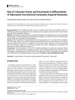
Background/Aims: We investigated the utility of serum C-reactive protein (CRP) and procalcitonin (PCT) for
differentiating pulmonary tuberculosis (TB) from bacterial community-acquired pneumonia (CAP) in South Korea,
a country with an intermediate TB burden.
Methods: We conducted a prospective study, enrolling 87 participants with suspected CAP in a community-based
referral hospital. A clinical assessment was performed before treatment, and serum CRP and PCT were measured.
The test results were compared to the final diagnoses.
Results: Of the 87 patients, 57 had bacterial CAP and 30 had pulmonary TB. The median CRP concentration
was 14.58 mg/dL (range, 0.30 to 36.61) in patients with bacterial CAP and 5.27 mg/dL (range, 0.24 to 13.22) in
those with pulmonary TB (p<0.001). The median PCT level was 0.514 ng/mL (range, 0.01 to 27.75) with bacterial
CAP and 0.029 ng/mL (range, 0.01 to 0.87) with pulmonary TB (p<0.001). No difference was detected in the
discriminative values of CRP and PCT (p=0.733).
Conclusions: The concentrations of CRP and PCT differed significantly in patients with pulmonary TB and
bacterial CAP. The high sensitivity and negative predictive value for differentiating pulmonary TB from bacterial
CAP suggest a supplementary role of CRP and PCT in the diagnostic exclusion of pulmonary TB from bacterial
CAP in areas with an intermediate prevalence of pulmonary TB. (Korean J Intern Med 2009;24:337-342)
Keywords: C-reactive protein; Pneumonia, community acquired; Procalcitonin; Tuberculosis
Received: December 13, 2008
Accepted: March 4, 2009
Correspondence to Choon-Taek Lee, M.D.
Department of Internal Medicine, Respiratory Center, Seoul National University Bundang Hospital, 300 Gumi-dong, Bundang-gu, Seongnam
463-707, Korea
Tel: 82-31-787-7002, Fax: 82-31-787-4052, E-mail:
INTRODUCTION
Community-acquired pneumonia (CAP) is a major
cause of hospital admission and the most important
infectious cause of death [1]. A rapid diagnosis and
appropriate antibiotic treatment are essential to reduce
the morbidity and mortality from CAP. In countries with a
high tuberculosis (TB) burden,
Mycobacterium tuberculosis
is a frequent cause of CAP [2-4], and the differential
diagnosis of TB from common bacterial pneumonia is
difficult. The varying clinical and radiographic presentation
of CAP and TB according to patient age and comorbidity
and the low sensitivity of acid-fast bacillus microscopy
make it even more difficult to distinguish TB from
common bacterial pneumonia [5-7]. Therefore, an adjunct
diagnostic method that can determine whether CAP is
caused by pulmonary TB or other bacterial pathogens
would have a clinical role in terms of isolating patients
with TB and administering appropriate anti-TB medication
or antibiotic treatment at an early stage.
C-reactive protein (CRP) is an acute-phase protein and
nonspecific marker of systemic inflammation [8]. The
ability of the serum CRP concentration to identify the
etiology of CAP and to predict the prognosis of CAP has
been investigated [9-14]. Procalcitonin (PCT), a 116-
amino-acid protein, is a useful marker of severe systemic
bacterial infection [15-18]. Recently, PCT has also been
introduced as a promising alternative to CRP in guiding
the antibiotic treatment of CAP and acute exacerbations
of chronic obstructive pulmonary disease [19,20] based
on the ability of PCT to discriminate between patients
with or without bacterial infection. In addition, PCT does
not appear to be significantly elevated in patients with
pulmonary TB [21-23], making it an attractive potentially
rapid diagnostic method for differentiating pulmonary TB
from bacterial CAP.
Therefore, we investigated the utility of serum CRP and
PCT for differentiating pulmonary TB from other bacterial
CAP in South Korea, a country with an intermediate TB
burden. We also investigated whether serum CRP and
PCT could help distinguish CAP according to pneumonia
severity.
METHODS
Participants
Participants were recruited between March 2007 and
November 2007 after the study protocol had been
approved by the Seoul National University Bundang
Hospital Ethics Review Committee. Adult patients who
visited the emergency department or outpatient clinic
with respiratory symptoms and chest radiograph
abnormalities were eligible for enrollment in this study.
After providing written informed consent, all participants
were enrolled in this study. Of the 115 eligible patients, 28
were excluded because the final diagnosis was
inconclusive or they had other diagnoses, such as
pulmonary embolism, acute exacerbation of interstitial
lung disease, or non-small cell lung cancer. Eighty-seven
patients were classified with pulmonary TB or bacterial
CAP. None of the patients in this study was HIV-positive.
Patients were considered to have pulmonary TB when
M. tuberculosis was cultured from their sputum or lavage
fluid, and the concentration of adenosine deaminase in
the effusion was >65 IU/dL in lymphocyte-predominant
exudative pleural effusions combined with a lung
parenchymal lesion. Bacterial CAP was diagnosed when
the subjects had clinical signs of pneumonia and a new
infiltrate on chest X-ray, and these resolved completely
with antibiotic treatment and cultures of sputum or lavage
fluid were negative for
M. tuberculosis during follow-up.
For the microbiologic evaluation of the patients with
CAP, we performed sputum Gram stains and cultures, two
blood cultures, and urinary antigen assays to detect
Legionella pneumophila and Streptococcus pneumoniae.
All participants had a complete physical examination,
and blood samples were obtained for measuring CRP and
PCT before starting treatment. Additionally, demographic
data, a white blood cell (WBC) count and differential, and
the Pneumonia Patient Outcomes Research Team (PORT)
[24] score were collected. The results of these tests were
compared to the final diagnostic group scores.
Methods
The serum CRP level was measured using an automated
latex-enhanced turbidimetric immunoassay in a clinical
laboratory within 1 hour of collecting the samples
(Dimension; Dade Behring, Newark, DE, USA; TBA-
200FR; Toshiba, Tokyo, Japan).
The PCT level was measured using a monoclonal
immunoluminometric assay (LIA PCT sensitive; BRAHMS
Diagnostica, Berlin, Germany). After separating the
serum, it was aliquoted and frozen at -70˚C until analyzed.
The functional assay sensitivity for PCT with a 20%
inter-assay variation coefficient was 0.05 ng/mL.
Statistics
Differences between the two groups were tested using
the nonparametric Mann-Whitney
U-test for continuous
variables. Pearson’s
χ
2
test or Fisher’s exact test was used
for categorical variables, and the Spearman rank correlation
coefficient was calculated. Optimal cutoffs for predicting
pulmonary TB or bacterial CAP were investigated using
receiver-operating characteristics (ROC) analysis, and the
diagnostic accuracy was assessed from the area under the
ROC curves (AUCs). A
p<0.05 was regarded as statistically
significant, and analyses were performed using SPSS
version 15.0 (SPSS Inc., Chicago, IL, USA).
RESULTS
Clinical and laboratory characteristics of the
patients
Of the 87 patients who met the inclusion criteria, 57 had
bacterial CAP and 30 had pulmonary TB. The median age
of the bacterial CAP and pulmonary TB groups was 71
years (range, 18 to 88) and 48 years (range, 18 to 82),
respectively. The responsible pathogen was determined in
22 patients (38.6%) with bacterial CAP; nine patients had
positive cultures for respiratory secretions and 13 patients
338
The Korean Journal of Internal Medicine Vol. 24, No. 4, December 2009
had positive urinary pneumococcal antigen tests. Twenty-
seven patients (90%) with pulmonary TB had positive
respiratory specimen cultures for
M. tuberculosis. The
patients’ demographic characteristics, symptoms, and
laboratory results are compared in Table 1.
The median CRP concentration was 14.58 mg/dL
(range, 0.30 to 36.61) in patients with bacterial CAP
and 5.27 mg/dL (range, 0.24 to 13.22) in those with
pulmonary TB (
p<0.001). The respective median PCT
level was 0.514 ng/mL (range, 0.013 to 27.754) and 0.029
ng/mL (range, 0.01 to 0.873) (
p<0.001). A significant
positive correlation was detected between the CRP and
PCT concentrations (
r=0.648, p=0.01).
Diagnostic accuracy for discriminating TB from
bacterial CAP
In the ROC curve analysis, the CRP concentration had a
Kang YA, et al. C-reactive protein and procalcitonin for the diagnosis of tuberculosis
339
Table 1. Clinical and laboratory characteristics of the participants
Bacterial pneumonia Tuberculosis p value
(n=57) (n=30)
Demographic characteristics
Age, yr 71 (18-88) 48 (18-82) <0.001
*
Sex, male/female 36 / 21 18 / 12 0.77
†
History of tuberculosis 14 (24.6) 6 (20.0) 0.63
†
Symptoms
Cough 43 (75.4) 27 (90.0) 0.10
†
Sputum 48 (84.2) 22 (73.3) 0.22
†
Fever 52 (91.2) 15 (50.0) <0.001
†
Dyspnea 34 (59.6) 12 (40.0) 0.08
†
Night sweats 0 (0) 7 (23.3) <0.001
‡
Weight loss 1 (1.8) 8 (26.7) 0.001
‡
Chest pain 11 (19.3) 9 (30.0) 0.30
†
Laboratory test
White blood cell,
×10
3
/µL 13.21 (2.29-39.92) 8.38 (5.07-22.99) <0.001
Neutrophils,
×10
3
/µL 11.06 (1.70-37.92) 5.85 (3.07-20.23) <0.001*
Monocyte, µL 503 (0-1210) 535 (253-5009) 0.053*
C-reactive protein, mg/dL 14.58 (0.30-36.61) 5.27 (0.24-13.22) <0.001*
Procalcitonin, ng/mL 0.514 (0.013-27.754) 0.029 (0.01-0.873) <0.001*
Radiographic findings
Upper lobe dominance 16 (28.1) 23 (76.7) <0.001
†
Cavitary lesion 0 (0) 11 (36.7) <0.001
‡
Effusion 11 (19.3) 9 (30.0) 0.26
†
PORT score 87 (18-187) 54.5 (10-126) <0.001*
Values are presented as number (%) or median (range).
PORT, Pneumonia Patient Outcomes Research Team.
* Mann-Whitney U-test.
†
Pearson χ
2
test.
‡
Fisher’s exact test.
Figure 1. Receiver-operating characteristics curve for
discriminating between pulmonary tuberculosis and bacterial
community-acquired pneumonia for C-reactive protein (CRP)
and procalcitonin (PCT). No difference was detected in the
discriminative value between CRP and PCT.
discriminative value of 0.857 (95% confidence interval
[CI], 0.778 to 0.936), and the PCT concentration had a
discriminative value of 0.872 (95% CI, 0.792 to 0.951). No
difference was found in the discriminative value between
CRP and PCT (
p=0.733). At a cutoff value of 12.5 mg/dL,
the CRP concentration had a sensitivity of 90.0% and a
specificity of 58.9%; at a cutoff value of 0.25 ng/mL, the
PCT concentration had a sensitivity of 93.1% and a
specificity of 59.6% (Fig. 1, Table 2).
CRP and PCT concentrations according to the
PORT Pneumonia Severity Index (PSI) risk classes
The median CRP and PCT concentrations were calculated
according to the PSI risk class in the bacterial CAP group.
The respective median CRP and PCT values were 3.8
mg/dL (range, 3.04 to 11.63) and 0.023 ng/mL (range,
0.013 to 1.974) in class I, 15.0 mg/dL (range, 10.48 to
35.63) and 0.164 ng/mL (range, 0.035 to 1.609) in class
II, 18.9 mg/dL (range, 0.30 to 32.85) and 0.723 ng/mL
(range, 0.013 to 3.279) in class III, 12.0 mg/dL (range,
3.55 to 34.42) and 0.707 ng/mL (range, 0.018 to 22.994)
in class IV, and 18.3 mg/dL (range, 8.52 to 36.61) and
0.525 ng/mL (range, 0.049 to 27.754) in class V. Patients
in risk classes III-V had a higher median PCT value of
0.659 ng/mL (range, 0.013 to 27.754) compared to 0.159
ng/mL (range, 0.013 to 1.974) for those in classes I and II
(
p=0.012), whereas no significant difference was observed
in the CRP concentrations between those groups classified
340
The Korean Journal of Internal Medicine Vol. 24, No. 4, December 2009
Table 2. Diagnostic validity of C-reactive protein (CRP) and procalcitonin (PCT) in differentiating pulmonary
tuberculosis from bacterial community-acquired pneumonia according to the different value
Sensitivity Specificity Positive predictive value Negative predictive value
CRP, mg/dL
<5.0 50.0 89.3 71.4 76.9
<10.0 83.3 75.0 64.1 89.4
<12.5 90.0 58.9 54.0 91.7
<15.0 100.0 50.0 51.7 100.0
PCT, ng/mL
<0.1 86.2 78.9 67.6 91.8
<0.25 93.1 59.6 54.0 94.4
<0.5 93.1 50.9 49.1 93.5
<1.0 100.0 31.6 42.6 100.0
Values are presented as percentages.
Figure 2. C-reactive protein (CRP) and procalcitonin (PCT) concentration according to the pneumonia severity index in bacterial
community-acquired pneumonia. Patients in risk classes III and V had a higher median PCT value compared to those in classes I and
II, whereas no significant difference was observed in the CRP concentrations between those groups classified as Pneumonia Severity
Index (PSI) I-II or PSI III-V.
with PSI I-II or PSI III-V (Fig. 2).
DISCUSSION
The results of this study suggest that CRP and PCT
can help to discriminate between pulmonary TB and other
common bacterial CAP in a setting of intermediate TB
prevalence. Significantly lower CRP and PCT serum
concentrations were found with pulmonary TB compared
to the other bacterial CAP in the initial diagnosis stage.
About 46,000 cases of TB are newly diagnosed annually
in South Korea [25], and the rapid, accurate differential
diagnosis of TB from common bacterial CAP has important
public health implications for the isolation care of patients
with TB and early appropriate anti-TB medication or
antibiotic treatment. Discriminating pulmonary TB from
bacterial CAP is frequently impossible based on patient
history, physical examination, and radiographic findings.
Therefore, CRP and PCT might have a role in the diagnostic
algorithm as rapid, noninvasive tests.
No difference was observed in the discriminating power
of CRP and PCT for differentiating pulmonary TB and
other bacterial infections in this study. CRP is an acute-
phase protein and nonspecific marker for systemic inflam-
mation, and the utility of CRP level as a marker for bacterial
infection of the lower respiratory tract has been studied in
several populations [26]. PCT has also been investigated
as a predictor of bacterial infection and is considered a
more accurate marker of various bacterial infections than
CRP [16,27]. Therefore, the absence of a difference between
CRP and PCT in our study should be considered in light of
several factors. First, the low yield of a causative pathogen
in bacterial CAP (38.6%) suggests the possibility of
including bacterial CAP with an atypical etiology, such as
Mycoplasma pneumoniae, Chlamydia pneumoniae, and
respiratory viruses. These atypical pathogens produce
lower PCT levels than classical bacterial pneumonia such
as pneumococcal pneumonia [11,28]. Second, because the
hospital in which this study was conducted is a secondary
referral hospital, although it is a community-based
hospital, more than 24 hours had passed from the onset of
symptoms to the time some patients visited the hospital.
The variable time interval from the onset of symptoms
before evaluating PCT and CRP might have affected the
results because of the kinetics of each inflammatory
marker [29,30]. Considering this point, a follow-up PCT
or CRP measurement after the initial evaluation will affect
the treatment strategy.
Although we found no difference in the discriminating
power of CRP and PCT for distinguishing pulmonary TB
from bacterial infection, our results showed the superiority
of PCT for predicting the severity of bacterial CAP
compared to CRP. This is consistent with the finding that
PCT is a good predictor of the severity of pneumonia and
sepsis, as described previously [31,32], and implies that
PCT can be used effectively for site-of-care decisions and
for predicting CAP prognosis based on clinical con-
siderations [33].
To appreciate our results fully, we must consider the
limitations of this study. First, the patients with pulmonary
TB were younger than those with bacterial CAP, and few
had far-advanced disease; this was reflected in the
difference in the PORT score between patients with
pulmonary TB and those with bacterial CAP. Further
study, including a study of advanced pulmonary TB,
might complement our study. Second, the low yield of
possible pathogens in bacterial CAP and the small number
of study subjects should be considered when generalizing
the results using CRP and PCT to determine pulmonary
TB and bacterial CAP.
In conclusion, serum CRP and PCT concentrations
differed significantly in patients with pulmonary TB
and those with bacterial CAP at the initial diagnosis stage.
The high sensitivity and negative predictive value for
differentiating the diagnosis of pulmonary TB from
bacterial CAP suggest a supplementary role for CRP and
PCT in the diagnostic exclusion of pulmonary TB from
bacterial CAP in areas with an intermediate prevalence of
active pulmonary TB.
REFERENCES
1.Marrie TJ. Community-acquired pneumonia. Clin Infect Dis
1994;18:501-513.
2. Ishida T. Etiology of community-acquired pneumonia among
adult patients in Japan. Jpn J Antibiot 2000;53(Suppl B):3-12.
3. Scott JA, Hall AJ, Muyodi C, et al. Aetiology, outcome, and risk
factors for mortality among adults with acute pneumonia in
Kenya. Lancet 2000;355:1225-1230.
4. Liam CK, Pang YK, Poosparajah S. Pulmonary tuberculosis
presenting as community-acquired pneumonia. Respirology
2006;11:786-792.
5. Kiyan E, Kilicaslan Z, Gurgan M, Tunaci A, Yildiz A. Clinical and
radiographic features of pulmonary tuberculosis in non-AIDS
immunocompromised patients. Int J Tuberc Lung Dis 2003;7:
Kang YA, et al. C-reactive protein and procalcitonin for the diagnosis of tuberculosis
341
764-770.
6. Perez-Guzman C, Torres-Cruz A, Villarreal-Velarde H, Salazar-
Lezama MA, Vargas MH. Atypical radiological images of
pulmonary tuberculosis in 192 diabetic patients: a comparative
study. Int J Tuberc Lung Dis 2001;5:455-461.
7. Lieberman D, Lieberman D, Schlaeffer F, Porath A. Community-
acquired pneumonia in old age: a prospective study of 91 patients
admitted from home. Age Ageing 1997;26:69-75.
8. Black S, Kushner I, Samols D. C-reactive protein. J Biol Chem
2004;279:48487-48490.
9. Almirall J, Bolibar I, Toran P, et al. Contribution of C-reactive
protein to the diagnosis and assessment of severity of community-
acquired pneumonia. Chest 2004;125:1335-1342.
10. Kerttula Y, Leinonen M, Koskela M, Makela PH. The aetiology of
pneumonia: application of bacterial serology and basic laboratory
methods. J Infect 1987;14:21-30.
11. Hedlund J, Hansson LO. Procalcitonin and C-reactive protein
levels in community-acquired pneumonia: correlation with
etiology and prognosis. Infection 2000;28:68-73.
12. Castro-Guardiola A, Armengou-Arxe A, Viejo-Rodriguez A,
Penarroja-Matutano G, Garcia-Bragado F. Differential diagnosis
between community-acquired pneumonia and non-pneumonia
diseases of the chest in the emergency ward. Eur J Intern Med
2000;11:334-339.
13. Smith RP, Lipworth BJ, Cree IA, Spiers EM, Winter JH. C-
reactive protein: a clinical marker in community-acquired
pneumonia. Chest 1995;108:1288-1291.
14. Ortqvist A, Hedlund J, Wretlind B, Carlstrom A, Kalin M.
Diagnostic and prognostic value of interleukin-6 and C-reactive
protein in community-acquired pneumonia. Scand J Infect Dis
1995;27:457-462.
15. Uzzan B, Cohen R, Nicolas P, Cucherat M, Perret GY. Procalcitonin
as a diagnostic test for sepsis in critically ill adults and after
surgery or trauma: a systematic review and meta-analysis. Crit
Care Med 2006;34:1996-2003.
16. Simon L, Gauvin F, Amre DK, Saint-Louis P, Lacroix J. Serum
procalcitonin and C-reactive protein levels as markers of bacterial
infection: a systematic review and meta-analysis. Clin Infect Dis
2004;39:206-217.
17. Muller B, Becker KL. Procalcitonin: how a hormone became a
marker and mediator of sepsis. Swiss Med Wkly 2001;131:595-
602.
18. de Werra I, Jaccard C, Corradin SB, et al. Cytokines, nitrite/
nitrate, soluble tumor necrosis factor receptors, and procalcitonin
concentrations: comparisons in patients with septic shock,
cardiogenic shock, and bacterial pneumonia. Crit Care Med
1997;25:607-613.
19. Stolz D, Christ-Crain M, Bingisser R, et al. Antibiotic treatment of
exacerbations of COPD: a randomized, controlled trial comparing
procalcitonin-guidance with standard therapy. Chest 2007;131:9-19.
20.Christ-Crain M, Stolz D, Bingisser R, et al. Procalcitonin guidance
of antibiotic therapy in community-acquired pneumonia: a
randomized trial. Am J Respir Crit Care Med 2006;174:84-93.
21. Polzin A, Pletz M, Erbes R, et al. Procalcitonin as a diagnostic tool
in lower respiratory tract infections and tuberculosis. Eur Respir J
2003;21:939-943.
22.Lawn SD, Obeng J, Acheampong JW, Griffin GE. Serum pro-
calcitonin concentrations in patients with pulmonary tuberculosis.
Trans R Soc Trop Med Hyg 1998;92:540-541.
23.Schleicher GK, Herbert V, Brink A, et al. Procalcitonin and C-
reactive protein levels in HIV-positive subjects with tuberculosis
and pneumonia. Eur Respir J 2005;25:688-692.
24.Fine MJ, Auble TE, Yealy DM, et al. A prediction rule to identify
low-risk patients with community-acquired pneumonia. N Engl J
Med 1997;336:243-250.
25.World Health Organization. Global Tuberculosis Control:
Surveillance, Planning, Financing (WHO/HTM/TB/2007.376).
WHO Report 2007. Geneva: World Health Organization, 2007.
26.van der Meer V, Neven AK, van den Broek PJ, Assendelft WJ.
Diagnostic value of C reactive protein in infections of the lower
respiratory tract: systematic review. BMJ 2005;331:26.
27. Muller B, Harbarth S, Stolz D, et al. Diagnostic and prognostic
accuracy of clinical and laboratory parameters in community-
acquired pneumonia. BMC Infect Dis 2007;7:10.
28.Moulin F, Raymond J, Lorrot M, et al. Procalcitonin in children
admitted to hospital with community acquired pneumonia. Arch
Dis Child 2001;84:332-336.
29.Pepys MB, Hirschfield GM. C-reactive protein: a critical update. J
Clin Invest 2003;111:1805-1812.
30.Dandona P, Nix D, Wilson MF, et al. Procalcitonin increase after
endotoxin injection in normal subjects. J Clin Endocrinol Metab
1994;79:1605-1608.
31. Hausfater P, Garric S, Ayed SB, Rosenheim M, Bernard M, Riou
B. Usefulness of procalcitonin as a marker of systemic infection in
emergency department patients: a prospective study. Clin Infect
Dis 2002;34:895-901.
32.Masia M, Gutierrez F, Shum C, et al. Usefulness of procalcitonin
levels in community-acquired pneumonia according to the
patients outcome research team pneumonia severity index. Chest
2005;128:2223-2229.
33. Mandell LA, Wunderink RG, Anzueto A, et al. Infectious Diseases
Society of America/American Thoracic Society consensus guidelines
on the management of community-acquired pneumonia in adults.
Clin Infect Dis 2007;44(Suppl 2):S27-S72.
342
The Korean Journal of Internal Medicine Vol. 24, No. 4, December 2009