Risk of Arrhythmia and Sudden Death pdf
Bạn đang xem bản rút gọn của tài liệu. Xem và tải ngay bản đầy đủ của tài liệu tại đây (7.12 MB, 429 trang )
Risk of Arrhythmia
and
Sudden Death
Edited by
Marek Malik
BMJ Books
Risk of Arrhythmia and
Sudden Death
To my parents
Risk of Arrhythmia and
Sudden Death
Edited by
Marek Malik
Professor of Cardiac Electrophysiology
St George’s Hospital Medical School, London, UK
©BMJ Books 2001
BMJ Books is an imprint of the BMJ Publishing Group
All rights reserved. No part of this publication may be reproduced, stored in a retrieval system, or transmitted, in any
form or by any means, electronic, mechanical, photocopying, recording and/or otherwise, without the prior written
permission of the publishers.
First published in 2001
by BMJ Books, BMA House, Tavistock Square,
London WC1H 9JR
www.bmjbooks.com
British Library Cataloguing in Publication Data
A catalogue record for this book is available from the British Library
ISBN 0-7279-1581-9
Typeset by Phoenix Photosetting, Chatham, Kent
Printed and bound by Creative Print and Design Ltd
Contents
Contributors ix
Preface xiii
Abbreviations xv
Part I: Problem and methodology
1 Clinical goals of risk stratification 3
Roberto Elosua, Josep Guindo, Xavier Viñolas, Antonio Martinez-Rubio,
Toni Bayés-Genis and Antoni Bayés de Luna
2 Definition of arrhythmic risk 10
Steen Z Abildstrom, Christian Torp-Pedersen and Lars Køber
3 Statistical methods for risk-stratification studies 19
Timothy R Church
4 Step-wise risk-stratification strategies 29
Stefan H Hohnloser
5 Risk-stratification studies for prospective trial design 37
Marek Malik
Part II: Techniques of risk assessment
6 Basic clinical assessment 49
Branco Mautner
7 Left ventricular ejection fraction and wall motion score 57
Steen Z Abildstrom, Christian Torp-Pedersen and Lars Køber
8 Risk assessment: the 12-lead electrocardiogram 65
Rory Childers
9 Electrophysiological study for risk stratification of cardiac patients 98
Velislav Batchvarov
10 QT dispersion 117
Velislav Batchvarov and Marek Malik
v
11 Electrocardiographic assessment of myocardial ischaemia
(with a note on the ischaemia–arrhythmia connection) 135
Shlomo Stern
12 Exercise electrocardiography for the assessment of arrhythmias 144
Michael Cusack and Simon Redwood
13 Analysis of monophasic action potentials 155
Peter Taggart and Peter Sutton
14 Ventricular signal averaged electrocardiography 167
Piotr Kulakowski
15 Signal averaged P wave 180
Antonio Michelucci, Luigi Padeletti, Andrea Colella, Maria Cristina Porciani,
Paolo Pieragnoli, Alessandro Costoli and Gian Franco Gensini
16 Non-invasive investigation of Wedensky modulation 187
Katerina Hnatkova and Marek Malik
17 Ambulatory electrocardiography: use in arrhythmia risk assessment 194
Morrison Hodges and James J Bailey
18 Event loop recorders and implantable monitors 202
Mark L Brown
19 Basic autonomic tests 209
Federico Lombardi
20 Heart rate assessment and monitoring 213
Xavier Copie, Olivier Piot, Thomas Lavergne, Louis Guize and
Jean-Yves Le Heuzey
21 Heart rate variability 221
Robert E Kleiger and Phyllis K Stein
22 Baroreflex sensitivity 234
Maria Teresa La Rovere, Andrea Mortara and Gian Domenico Pinna
23 Heart rate turbulence 242
Georg Schmidt
24 T wave dynamicity 249
Pierre Maison Blanche and Philippe Coumel
25 Repolarisation alternans 256
Daniel M Bloomfield and Richard J Cohen
26 Social and psychosocial influences on sudden cardiac death, ventricular
arrhythmia and cardiac autonomic function 266
Harry Hemingway
vi
Risk of Arrhythmia and Sudden Death
Part III: Clinical studies of risk assessment
27 Risk stratification after myocardial infarction 287
Yee Guan Yap
28 Ventricular arrhythmias in chronic heart failure 297
Steven Lindsay and Jim Nolan
29 Ventricular arrhythmias in hypertrophic cardiomyopathy 309
Gang Yi and William J McKenna
30 Ventricular arrhythmias in apparently healthy athletes 316
Francesco Furlanello, Fredrick Fernando, Amedeo Galassi and
Annalisa Bertoldi
31 Paroxysmal atrial fibrillation 325
Johan EP Waktare
32 Persistent atrial fibrillation 336
Joseph T Dell’Orfano and Gerald V Naccarelli
33 Permanent atrial fibrillation 348
Isabelle C Van Gelder and Harry JGM Crijns
34 Arrhythmias associated with the long QT syndrome 353
Peter J Schwartz
35 Arrhythmia associated with other cardiac diseases 360
Patrick Lam and Paul Schweitzer
36 Arrhythmias associated with non-cardiac disease 365
Josef Kautzner
Part IV: Antiarrhythmic trials
37 Antiarrhythmic device trials 379
Arthur J Moss
38 Recent antiarrhythmic drug trials 389
Michiel J Janse
Index 395
vii
Contents
Department of Cardiology, Gentofte University Hospital,
Copenhagen, Denmark
James J Bailey
Center for Information Technology, National Institutes of
Health, Bethesda, Maryland, USA
Velislav Batchvarov
Department of Cardiological Sciences, St George’s
Hospital Medical School, London, UK
Antoni Bayés de Luna
Departamento de Cardiología, Hospital de la Santa Creu i
Sant Pau, Barcelona, Spain
Toni Bayés-Genis
Fellow, Mayo Clinic, Rochester, Minnesota, USA
Annalisa Bertoldi
Department of Cardiology, S. Chiara Hospital, Trento, Italy
Daniel M Bloomfield
Assistant Professor of Medicine, Division of Cardiology,
College of Physicians and Surgeons, Columbia University,
USA
Mark L Brown
Staff Scientist, Tachyarrhythmia Research Department,
Atrial Fibrillation Research Group, Medtronic,
Minneapolis, Minnesota, USA
Rory Childers
Professor of Medicine, Section of Cardiology, University
of Chicago Medical Centre, Chicago, USA
Timothy R Church
Division of Environmental and Occupational Health,
School of Public Health, University of Minnesota,
Minneapolis, Minnesota, USA
Richard J Cohen
Whitaker Professor, Harvard University–Massachusetts
Institute of Technology, Division of Health Sciences and
Technology, Massachusetts, USA
Andrea Colella
Istituto di Clinica Medica e Cardiologia, Università di
Firenze, Florence, Italy
Xavier Copie
Department of Cardiology, Broussais Hospital, Paris,
France
Alessandro Costoli
Istituto di Clinica Medica e Cardiologia, Università di
Firenze, Florence, Italy
Philippe Coumel
Hôpital Lariboisière, Paris, France
Harry JGM Crijns
Department of Cardiology, Thoraxcenter, University
Hospital Groningen, Groningen, The Netherlands
Michael Cusack
Clinical Research Fellow, Department of Cardiology,
Rayne Institute, St Thomas’ Hospital, London, UK
Joseph T Dell’Orfano
Department of Medicine, Division of Cardiology, The
State University of New York at Stony Brook, Stony
Brook, New York, USA
Roberto Elosua
Unidad de Lípidos y Epidemiología Cardiovascular,
Instituto Municipal de Investigación Médica, Barcelona,
Spain
Fredrick Fernando
Sports Science Institute, Italian National Olympic
Committee, Rome, Italy
ix
Contributors
Francesco Furlanello
S. Raffaele Scientific Institute, Milan-Rome, Italy
Amedeo Galassi
Arrhythmia and Electrophysiological Center, S Donato
Institute, Milan, Italy
Gian Franco Gensini
Istituto di Clinica Medica e Cardiologia, Università di
Firenze, Florence, Italy
Louis Guize
Department of Cardiology, Broussais Hospital, Paris, France
Josep Guindo
Departmento di Cardiologia, Hospital de la Santa Creui i
Sant Pau, Barcelona, Spain
Harry Hemingway
Senior Lecturer in Epidemiology, International Centre for
Health and Society, Department of Epidemiology and
Public Health, University College London Medical School,
London; Director of Research and Development,
Department of Research and Development, Kensington &
Chelsea and Westminster Health Authority, London, UK
Morrison Hodges
Minneapolis Heart Institute Foundation, Minnesota, USA
Stefan H Hohnloser
Department of Medicine, Division of Cardiology, J.W.
Goethe University, Frankfurt, Germany
Katerina Hnatkova
Department of Cardiological Sciences, St George’s
Hospital Medical School, London, UK
Michiel J Janse
Cardiovascular Research, Academic Medical Center,
Amsterdam, The Netherlands
Josef Kautzner
Department of Cardiology, Institute for Clinical and
Experimental Medicine, Prague, Czech Republic
Robert E Kleiger
Professor of Medicine, Washington University School of
Medicine; Medical Director of Washington University
School of Medicine HRV Laboratory, St Louis, USA
Lars Køber
Department of Cardiology, Gentofte University Hospital,
Copenhagen, Denmark
Piotr Kulakowski
Department of Cardiology, Postgraduate Medical School,
Grochowski Hospital, Warsaw, Poland
Patrick Lam
Beth Israel Medical Center, New York, USA
Maria Teresa La Rovere
Fondazione “Salvatore Maugeri”, IRCCS, Divisione di
Cardiologia, Centro Medico Montescano, Pavia, Italy
Thomas Lavergne
Department of Cardiology, Broussais Hospital, Paris,
France
Jean-Yves Le Heuzey
Department of Cardiology, Broussais Hospital, Paris,
France
Steven Lindsay
Consultant Cardiologist, Bradford Royal Infirmary,
Bradford, UK
Federico Lombardi
Cardiologia, Dipartimento di Medicina, Chirurgia e
Odontoiatria Ospedale S Paolo, Università degli Studi di
Milano, Milan, Italy
Pierre Maison Blanche
Hôpital Lariboisière, Paris, France
Marek Malik
Professor of Cardiac Electrophysiology, Department of
Cardiological Sciences, St George’s Hospital Medical
School, London, UK
Antonio Martinez-Rubio
Departamento de Cardiología, Hospital de la Santa Creu i
Sant Pau, Barcelona, Spain
Branco Mautner
President, International Society for Holter and Non-
Invasive Electrocardiology; Academic Vice-Chancellor,
Favaloro University, Belgrano, Buenos Aires, Argentina
William J McKenna
Professor of Cardiac Medicine, Department of
Cardiological Sciences, St George’s Hospital Medical
School, London, UK
Antonio Michelucci
Istituto di Clinica Medica e Cardiologia, Università di
Firenze, Florence, Italy
Risk of Arrhythmia and Sudden Death
x
Andrea Mortara
Fondazione “Salvatore Maugeri”, IRCCS, Divisione di
Cardiologia, Centro Medico Montescano, Pavia, Italy
Arthur J Moss
Professor of Medicine (Cardiology), Director, Heart
Research Follow-up Program, University of Rochester
Medical Center, Rochester, New York, USA
Gerald V Naccarelli
Chief, Division of Cardiology, MS Hershey Medical Center
of the Pennsylvania State University, Hershey,
Pennsylvania, USA
Jim Nolan
Consultant Cardiologist, Cardiothoracic Centre, North
Staffordshire Hospital, Stoke on Trent, UK
Luigi Padeletti
Istituto di Clinica Medica e Cardiologia, Università di
Firenze, Florence, Italy
Paolo Pieragnoli
Istituto di Clinica Medica e Cardiologia, Università di
Firenze, Florence, Italy
Gian Domenico Pinna
Servizio di Bioingegneria, Fondazione “Salvatore
Maugeri”, IRCCS, Centro Medico Montescano, Pavia,
Italy
Olivier Piot
Department of Cardiology, Broussais Hospital, Paris,
France
Maria Cristina Porciani
Istituto di Clinica Medica e Cardiologia, Università di
Firenze, Florence, Italy
Simon Redwood
Senior Lecturer / Honorary Consultant Cardiologist,
Department of Cardiology, Rayne Institute, St Thomas’
Hospital, London, UK
Georg Schmidt
Deutsches Herzzentrum und Medizinische Klinik der
Technischen Universität München, Munich, Germany
Peter J Schwartz
Professor and Chairman, Department of Cardiology,
Policlinico S Matteo, IRCCS and University of Pavia, Italy
Paul Schweitzer
Beth Israel Medical Center, New York, USA
Phyllis K Stein
Research Assistant Professor of Medicine, Washington
University School of Medicine; Director of Washington
University School of Medicine HRV Laboratory, St Louis,
USA
Shlomo Stern
Emeritus Professor of Medicine, Hebrew University, and
the Bikur Cholim Hospital, Jerusalem, Israel
Peter Sutton
Departments of Cardiology, UCL Hospitals and Hatter
Institute, University College Hospital, London, UK
Peter Taggart
Departments of Cardiology, UCL Hospitals and Hatter
Institute, University College Hospital, London, UK
Christian Torp-Pedersen
Department of Cardiology, Gentofte University Hospital,
Copenhagen, Denmark
Isabelle C Van Gelder
Department of Cardiology, Thoraxcenter, University
Hospital Groningen, Groningen, The Netherlands
Xavier Viñolas
Departamento de Cardiología, Hospital de la Santa Creu i
Sant Pau, Barcelona, Spain
Johan EP Waktare
Cardiological Sciences, St George’s Hospital Medical
School, London UK
Yee Guan Yap
Specialist Registrar in Cardiology, Department of
Cardiological Sciences, St George’s Hospital Medical
School, London, UK
Gang Yi
Department of Cardiological Sciences, St George’s
Hospital Medical School, London, UK
Contributors
xi
rising and practically every clinical, pharmacological or
technological breakthrough brings not only an improved
quality of life and prolonged patient survival but also a sig-
nificant burden on healthcare providers irrespective of
whether they are privately organised or government con-
trolled. Despite the differences in the economies and
healthcare arrangements of the Western world countries,
the discussions about the ever rising cost of medical care
are everywhere similarly heated. Clearly, there are no easy
solutions since removing a potentially life-saving treatment
from any human being is not ethical. At the same time, no
country of the world is so wealthy that it could afford the
best available treatment and care for all its citizens.
Cardiology is no exception to this trend. For instance,
studies that have confirmed the efficacy and appropriate-
ness of the prophylactic use of implantable defibrillators
are examples of findings that have both clinical and finan-
cial implications. Still, compared to comprehensive safety
nets and resuscitation programs designed to save patients
only after they have suffered from malignant ventricular
arrhythmias, prophylactic use of implantable defibrillators
is most likely a cost-saving option.
In general, prophylactic medicine is one of the most
effective ways of reducing the overall costs of medical care
while, at the same time, maintaining adequate quality of
life and improving the survival of the overall population. It
is therefore not surprising that prophylactic methods are
presently receiving substantial attention from both clinical
community and healthcare providers. The advances in pro-
phylactic medicine are clearly dependent on improved risk
markers and advanced risk stratification approaches. Again,
cardiology is no exception. Cardiac risk stratification is
presently being investigated not only to identify patients
who might benefit from more cost-effective modes of pro-
phylactic treatment but also, even more importantly, to
save patients who would otherwise succumb to cardiac
death and sudden cardiac death in particular.
For all these reasons, I was very pleased when I was
offered the opportunity of editing this book on risk strati-
fication of arrhythmias and sudden cardiac death. Indeed,
some of the principles and methodologies of arrhythmia
risk stratification are rather complex and, in some cases,
perhaps not fully appreciated and understood by clinical
cardiologists and electrophysiologists. In order to offer a
comprehensive coverage of the field, I have divided the
contents of the book into four sections. The first part con-
tains chapters dealing with the general principles of risk
stratification, explaining the individual facets of methodol-
ogy and technology and summarising the goals of arrhyth-
mic risk stratification studies. The second part is devoted
to a detailed description of individual investigation tech-
niques aimed at identification of patients at high risk of
arrhythmia or sudden death. The third part describes the
present experience with applying the risk stratification
technologies and tests to patients of different clinically
defined groups. Finally, the two chapters of the final part
summarise the presently conducted clinical trials utilising
the risk stratification techniques.
As with every multi-authored book, I faced the usual edi-
toral dilemma of finding the proper balance between having
the book compact with cross-references within individual
chapters and having the chapters suited for separate read-
ing. Eventually, I felt that with a book of this size aimed at
providing a source of standard references, each chapter
should contain a standard coverage of its subject. Hence, I
am happy to recommend the reader to select chapters cor-
responding to his/her particular needs and interest.
Needless to say, reading the book in its entirety offers much
more comprehensive learning of the whole field.
Without the kind positive response of the authors of the
individual chapters, this book would have never been writ-
ten. I truly appreciate the efforts of the individual contribu-
tors and am very grateful for their kind help. My deep
thanks also go to my secretary, Mrs Melanie Monteiro, who
carefully organised the editorial office of this book and who
helped me in many other ways. I am grateful to the pub-
lisher for their useful suggestions, significant technical
help, and kind flexibility. My apologies go to my wife and
children since far too frequently I have devoted the time
that I should have spent with them to the editing of this
text.
Marek Malik
xiii
Preface
AF/AT atrial fibrillation/atrial tachycardia
APC atrial premature contraction
AT attributable risk
BRS baroreflex sensitivity
CHF congestive heart failure
DAD delayed after-depolarisations
EAD early after-depolarisations
ECG electrocardiograph
EGM electrogram
ERP effective refractory period
FRP functional refractory period
HCM hypertrophic cardiomyopathy
HF high frequency
HRV heart rate variability
ICD cardioverter/defibrillator
ILR implantable loop recorder
LF low frequency
LVEF left ventricular ejection fraction
LVH left ventricular hypertrophy
MAPs monophasic action potentials
MI myocardial infarction
NPV negative predictive value
NSVT non-sustained ventricular tachycardia
PCA principal component analysis
pNN50 percentage of normal to normal RR
intervals that differ ≥50 ms from the
preceding interval
PPV positive predictive value
PSVT paroxysmal supraventricular tachycardia
rMSSD root mean square of successive differences
of normal to normal RR intervals
ROC receiver-operating characteristic
SACT sinoatrial conduction time
SDNN standard deviation of normal to normal RR
intervals
SDNNIDX SDNN index
SNRT sinus node recovery time
TO turbulence onset
TP total power
ULF ultralow frequency
VA ventricular arrhythmias
VPC ventricular premature contraction
VT ventricular tachycardia
xv
Abbreviations
Problem and methodology
1
The clinical goals of risk stratification of sudden death
(SD) are to identify subjects who are at high risk and,
eventually, to reduce the incidence of SD. We will focus
in this chapter on aspects related to epidemiological and
clinical issues to stratify risk of SD. We consider SD as a
syndrome that may be associated with different diseases
and situations. The term “sudden death” has been used in
different ways by epidemiologists, clinicians, forensic
pathologists, etc. Clinically, the term is used for deaths
due to natural causes that occur within 1 hour of the
onset of symptoms in a person with or without pre-exist-
ing heart disease, but in whom the time and mode of
death are unexpected. In approximately one-third of cases
the death is instantaneous, without symptoms or coincid-
ing only during a few seconds with the presence of symp-
toms. If the patient is found dead, death is considered to
be “sudden” if the subject was seen alive and well in the
preceding 24 hours.
1
In this chapter we shall discuss the need for identifica-
tion of high-risk populations for malignant arrhythmias
and sudden death. It is especially important to emphasise
the paradox that the highest-risk subgroups (cardiac arrest
survivors or patients with an ejection fraction <30%)
account for a small proportion of SD events in a popula-
tion. We emphasise how different is the clinical approach
for risk stratification in the general population and in
patients with already clear evidence of heart diseases, for
example in post-myocardial infarction patients.
Clinical epidemiological aspects
Sudden death usually appears in the presence of clinical
or silent heart disease.
2,3
Ischaemic heart disease (IHD) is
by far the most common associated situation found in
patients presenting with sudden cardiac death (SCD) (Box
1.1). Nevertheless, other heart diseases may present with
SCD, especially in the presence of heart failure and/or left
ventricular hypertrophy.
4,5
In some cases, there are only
isolated electrophysiological abnormalities (WPW, long QT
syndrome, Brugada syndrome, etc.)
6–8
or congenital
defects of coronary arteries.
9
In a few cases (≅3%), there
is no evidence of associated heart disease, probably
because, with the current technology used, we are unable
to define or demonstrate some undetectable, minor abnor-
malities. Such cases, now named “idiopathic SCD”, will
be fewer in the future.
10
SCD is considered to be of ischaemic origin if it
appears during an acute ischaemic event or in the pres-
ence of severe IHD. From a pathological point of view,
SCD is considered ischaemic when stenosis of at least
75% of the area of the lumen of one coronary artery is
found. Intraluminal or intra-intimal thrombi may or may
not be present.
11
Severe coronary atherosclerosis may
exist whether or not there are clinical symptoms (acute
myocardial infarction or angina) before SCD.
12
We will comment on the following epidemiological
aspects related to SCD:
●
the size of denominator pools in different population
subgroups that determine the ability to identify
potential victims within population subgroups of vari-
ous sizes;
13
●
the time dependence of risk, which expresses risk as
non-linear function of time after a conditioning clini-
cal event;
13,14
Box 1.1 Principal causes of sudden cardiac death
●
Ischaemic heart disease
●
Cardiomyopathies
● idiopathic dilated cardiomyopathy – heart failure
● hypertrophic cardiomyopathy
● arrhythmogenic right ventricular dysplasia
●
Valvular heart disease
● aortic stenosis
● mitral valve prolapse
●
Electrophysiologic abnormalities
● pre-excitation syndromes
● long QT syndrome
● conduction system abnormalities
● Brugada syndrome
●
Congenital cardiac abnormalities
●
Sudden death without apparent structural heart dis-
ease
3
1
Clinical goals of risk stratification
Roberto Elosua, Josep Guindo, Xavier Viñolas, Antonio Martinez-Rubio,
Toni Bayés-Genis and Antoni Bayés de Luna
●
the importance of the different risk factors and envi-
ronment in the appearance of SCD.
1–3
Population subgroups and sudden cardiac death
According to Myerburg,
13
if we consider all cases of SCD
occurring in the USA, the overall incidence is in the range
of 1–2/1000 population per year (0.1–0.2%). This large
population base contains both those victims where SCD
occurs as a first cardiac event, in which the possibility to
predict sudden death is very limited (i.e. the general pop-
ulation), and those victims whose deaths may be predicted
with greater accuracy because they come from higher risk
subgroups (i.e. survivors of sudden cardiac death) (Fig.
1.1). Because of the size of the denominator, any inter-
vention designed for the general population must be
applied to more than 99% of population who will not have
an event during the course of a year. These numbers limit
the nature of a broad-based intervention and encourage
the identification of specific higher-risk clinical subgroups.
Nevertheless, with increasing specificity of subgroups, the
absolute number of potential victims who can be identi-
fied decreases. Figure 1.1 expresses the incidence (per-
cent per year) of SCD among various subgroups.
In clinical epidemiology it is very important to consider
two different levels of risk: attributable risk versus relative
risk.
15,16
Relative risk (RR) is the ratio of the incidence of
an event, SCD in this case, in persons with and without a
risk factor; whereas, attributable risk (AR) refers to the
proportion of the incidence of an event that can be
explained by the presence of the risk factor.
1
By moving from the total adult population to a sub-
group with higher risk there may be a more than 10-fold
increase in the incidence of events annually, an important
RR increase. As shown in Fig. 1.1, there is a progressive
increase in the incidence of SCD as one moves from the
general population to subgroups having had a prior coro-
nary event and to those with low ejection fractions and
heart failure, and to survivors of out-of-hospital cardiac
arrest, and to those experiencing ventricular tachycardia
or fibrillation (VT/VF) during the convalescent phase after
myocardial infarction. However, the corresponding
absolute number of deaths becomes progressively smaller
as the subgroups become more focused, an important AR
decrease. Thus, in these selected subgroups although the
RR is higher the AR is lower.
Clinical application of new technologies and procedures
appears to have had a favourable impact on the highest
risk subgroups. Progress measured in terms of prevention
of large numbers of sudden cardiac deaths in the general
population, however, will be limited until it is possible to
identify more easily higher risk individuals in the general
population. At the moment it is impossible to know which
patients in the general population present vulnerable
plaques. Thus, the only way to prevent SCD in this popu-
lation is to fight against classical cardiovascular risk factors
(smoking, hypertension, lipid disorders).
Time dependence of risk
Furukawa and Myeburg
13,14
have also emphasised that risk
of SCD does not appear to be linear as a function of time
after a change in cardiovascular status. Survival curves
after major cardiovascular events (cardiac arrest survivors,
post-myocardial infarction patients, patients with recent
onset of heart failure, etc.), which identify populations at
high risk for both sudden and total cardiac death, generally
demonstrate that the most dangerous period occurs during
the first 6–18 months after the index event. By 24
months, the slope of a survival curve is not very different
from one describing a similar population that has remained
free of an interposed major cardiovascular event. Thus,
there is a time dependence of risk after a major event,
which stresses the importance of the most intensive inter-
vention in the early period. The higher risk subgroup, as
happens in survivors of cardiac arrest, has not only a differ-
ent overall mortality rate but also a different pattern of
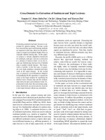
attrition as a function of time. In Fig. 1.2 the actuarial
analysis of recurrences among a population of 101 cardiac
arrest survivors with IHD is shown. The risk is high in the
first 6 months (11.2%) and then falls to 3.3% in the next
Risk of Arrhythmia and Sudden Death
4
Figure 1.1 Bar graphs showing the relation between inci-
dence and total numbers of sudden cardiac deaths in overall
and population subgroups. The numbers are calculated for
the US population. (Adapted form reference 13.)
(Abbreviations: EF = ejection fraction; MI = myocardial
infarction; VF = ventricular fibrillation; VT = ventricular tachy-
cardia.)
Overall incidence
in adult population
High coronary
risk subgroup
Any prior
coronary event
EF < 30%
heart failure
Out–of–hospital
cardiac arrest
survivors
Convalescent phase
VT/VF after MI
Percent/Year Events/Year
0 1 2 5 10 20 30 0 100 200 300
( ¥ 1000 )
three 6-month blocks thereafter. Low ejection fraction is
the most powerful predictor of death during the first 6
months, subsequently, persistent inducibility during pro-
grammed stimulation is the most powerful predictor of
death.
13,14
In a study of a post-myocardial infarction popula-
tion, Moss et al.
8
pointed out that 50% of the deaths that
occur during the 48 months after myocardial infarction
occur within the first 6 months. Therefore, the use of time
as a dimension for measuring risk is extremely important.
Risk factors and the environment
The incidence of SCD varies considerably from country to
country with relation to the prevalence of IHD. Since IHD
is characteristic of industrialised societies and its ageing
population, the prevalence of SCD is greater in the west-
ern countries than in the rest of the world. According to
the World Health Organization,
17
the incidence of SCD in
industrialised areas varies between 19 and 159 per
100 000 inhabitants per year among men between the
ages of 35 and 64.
In the USA and other industrialised countries, the inci-
dence of SCD is decreasing parallel to the overall decrease
in IHD mortality.
18
This decrease has been attributed to a
decrease in the incidence of IHD and to a lower fatality
related to an improved medical therapy and a more effec-
tive system for resuscitating victims of out-of-hospital car-
diac arrest. In spite of this, the incidence of SCD remains
high and is still a major challenge confronting contempo-
rary cardiology. On the other hand, the global burden of
cardiovascular diseases including IHD and consequently of
SCD is increasing in developing countries. Thus, we have
to extend our preventive campaigns world-wide.
A number of studies have shown that SCD displays a
prominent circadian pattern, with a primary peak occur-
ring between 7 and 11 am.
19
In the Framingham Study,
20
the risk of SCD is approximately 70% greater from 7 to 9
am than during the rest of the day. A circadian variation
of SCD, similar to that of the occurrence of non-fatal
myocardial infarction and episodes of myocardial
ischaemia, was observed.
Although the incidence of SCD parallels the incidence
of IHD, which increases with age, the proportion of those
who die suddenly is higher in younger patients. This dif-
ferential effect of age may be due to older patients having
more advanced coronary heart disease, and therefore the
incidence of death due to heart failure is greater.
SCD is more common in males than in females by a
ratio of 3 to 1. This is due to the lower prevalence of IHD
in premenopausal women. Females attain a comparable
incidence of SCD 20 years older than men. However,
more females than males die suddenly without clinical evi-
dence of IHD. Since the frequency of SCD is much higher
in postmenopausal women compared to premenopausal
women of the same age, it is likely that a hormonal factor
influences the result.
Multivariate logistic analysis in the Framingham
Study,
21
including all coronary risk factors, indicates that,
in males, age, systolic blood pressure, cigarette smoking,
and relative bodyweight are all independently related to
the incidence of sudden death. In females, aside from
age, only hypercholesterolaemia and vital capacity are
associated independently with an increased risk of sud-
den death. Using these parameters, there is a wide varia-
tion in the risk of sudden death. Forty-two percent of
sudden deaths in males and 53% in females occur in one-
tenth of the population in the top decile of multivariate
risk.
It is important to conceptualise sudden cardiac death
as a manifestation of IHD occurring as a consequence of a
variety of risk factors and living habits. Small changes in
blood pressure, cholesterol levels, and cigarette smoking
pattern may not be of great importance if they are iso-
lated, but they can interact to increase the risk of SCD.
Genetic factors are also important in less common
causes of SCD, such as in the congenital long QT syn-
drome,
22
hypertrophic cardiomyopathy,
23
arrhythmogenic
right ventricle dysplasia, or Brugada syndrome,
24
etc.
Parental sudden death has been also identified as a risk
factor for SCD.
25
Clinical goals of risk stratification
5
Percent
100
90
80
70
60
0
11.2%
3.3%
3.3%/
6 months
0.8%/
6 months
EF
(<35%)
Persistent inducibility
0 6 12 18 24 30 36 42
Months ( F/U )
n = 101
at t = 0
Figure 1.2 Time dependence of rate of recurrences
among survivors of cardiac arrest. Actuarial analysis or recur-
rences among a population of 101 cardiac arrest survivors
with coronary heart disease. Risk was higher in the first 6
months and then fell to 3.3% for the next three 6-month peri-
ods. After 24 months, the rate fell to 0.8% for each 6-month
period thereafter. The most powerful predictor of death was
a low ejection fraction (EF) during the 6 first months and
persistent inducibility during programmed stimulation after-
wards. (Adapted from reference 14.)
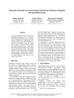
Pathophysiology of sudden cardiac death
Ischaemic heart disease
Most cases of SCD occur in patients with IHD. In these
patients the chain of events leading to SCD may occur in
two ways:
●
an acute ischaemic syndrome (AIS), especially acute
myocardial infarction (AMI), or
●
a primary arrhythmic event (PAE) (Fig. 1.3).
It is practically impossible to predict the appearance of
acute ischaemic syndrome (acute MI) when this is the
first manifestation of the disease without previous herald-
ing symptoms. This would only be possible if we could
detect, easily and non-invasively, not only the presence of
coronary atherosclerosis, but especially the existence of
vulnerable plaques. Such plaques are at high risk of rup-
ture or thrombosis.
26–28
From a pathological point of view,
plaques are considered vulnerable when they have large
lipid cores occupying more than 40% of the overall plaque
volume, thin and inflamed fibrous caps separating the
cores from the arterial lumen, a high density of
macrophages, and a low density of smooth-muscle cells in
the caps.
26
On the other hand, the danger of a primary arrhythmic
event often occurs in post-MI patients. In these cases, the
danger of an acute ischaemic syndrome (severe and persis-
tent ischaemia, usually appearing as acute myocardial
infarction) is not the only cause of the vulnerability of the
myocardium to SCD, although some degree of residual or
transient ischaemia may be present. The most important
markers that express this arrhythmic risk type of vulnera-
bility are:
●
clinical/ECG
●
morphological (anatomic substrate), and
●
related to autonomic nervous system imbalance (Fig.
1.4).
In all these circumstances these markers represent an
increase, often very important, in electrical instability,
which bears no relation to the presence of an acute
ischaemic syndrome. This instability is a substrate to
develop a malignant ventricular arrhythmia in the pres-
ence of different triggers.
The comparative role of acute ischaemic
syndrome versus primary arrhythmic event in
the presentation of SCD: a 50% responsibility
As we stated at the beginning of this article (Fig. 1.1), the
population pool of SD represents an overall incidence of
1–2/1000 per year. This large population base include vic-
tims of SCD as a first manifestation of disease; in the
majority of patients with first MI, in whom SCD is related
to acute ischaemic syndrome (severe and persistent
ischaemia), the danger of SCD is very difficult to predict.
In this group there are also included a small number of
cases of hypertrophic cardiomyopathy, primary ventricular
fibrillation, etc. This group represents 40–60% of SCD
cases. On the other hand, the remaining cases of SCD may
be predicted with greater accuracy because they are
included in higher-risk subgroups. The majority of these
patients present IHD, but often the cause of the presenta-
tion of the final event is not an acute ischaemic syndrome,
although some transient ischaemia may be present. There
is considerable evidence
29,30
that, in patients with
implanted defibrillator, thus in patients at a higher risk to
present SCD, the cause of death is, in the majority of
cases, a primary arrhythmic event and not an ischaemic-
Risk of Arrhythmia and Sudden Death
6
Ischaemic
heart
disease
Vulnerable
plaque
Anatomic
substrate
Vulnerable
myocardium
Ventricular
fibrillation
Ventricular
fibrillation
Final
arrhythmia
Acute
ischaemic
syndrome
Sustained
ventricular
tachycardia
Final
step
Sudden
cardiac
death
· clinical
· circadian
· autonomic
nervous
system
Triggers &
modulators
Figure 1.3 The chain of events that leads to final arrhyth-
mia and sudden cardiac death. Some triggers and/or modu-
lators acting in a vulnerable myocardium lead to the final
step and final arrhythmia. The triggers and modulators may
be similar in the two ways but the vulnerable myocardium is
different: vulnerable plaque in the case of acute ischaemic
syndrome leading to a ventricular fibrillation and anatomic
substrate leading to a sustained ventricular tachycardia and
ventricular fibrillation.
Anatomic Substrate
Electrical
instability
RISK
Trigger
Malignant Ventricular Arrhythmia
Clinical and ECG
markers
Autonomic
nervous system
Left ventricular
dysfunction
Residual
ischaemia
Figure 1.4 The triangle of risk in post-myocardial infarc-
tion patients with satellite triangle in the angle of electrical
instability with different markers of bad outcome for a pri-
mary arrhythmic event.
thrombotic acute syndrome. Henry et al.
29
demonstrated
that in patients with implantable cardioverter–defibrillator
(ICD) appropriate shocks for ECG-verified ventricular
tachyarrhythmias were only very rarely (<3% of cases) pre-
ceded or followed by signs of acute myocardial infarction.
Thereby supporting the idea that, in patients at higher risk
of SCD, this is usually due to a primary arrhythmic event.
On the other hand, Mont et al.
30
demonstrated that, in
patients who have survived VF as the sole documented
arrhythmia at the time of resuscitation and are currently
receiving ICD therapy, VT is by far the most common ven-
tricular arrhythmia recorded in device-incorporated elec-
trograms during follow-up. This adds further support to
the consideration that the primary cause of VF was a sus-
tained VT not triggered by an acute ischaemic syndrome.
On the whole we may consider that acute ischaemic
syndrome with thrombosis accounts for approximately
40–50% of cases of SCD. There are several arguments to
support this. Clinically, the incidence of symptoms sugges-
tive of ischaemia in cases of SCD is between 30 and
50%.
1,2
Furthermore, in our survey of patients who died
while wearing a Holter device,
31
evidence of ischaemia
detected by Holter monitoring was around 30%. From an
angiographic point of view, Spaulding demonstrated
32
that
the presence of acute coronary artery occlusion in a coro-
nariography performed immediately in survivors of out-of-
hospital cardiac arrest was 48%. Lastly, from a pathological
point of view the frequency of active lessions varies greatly
(from 13% to 95%)
11,12,33,34
depending on the type of
patients studied (ambulatory versus CCU patients,
methodological differences, etc.). Burke et al.
33
recently
demonstrated that, in patients with IHD and presenting
with a SCD, acute thrombosis was present in 52% of cases.
As a conclusion, it seems clear that in approximately
50% of cases SCD is explained by acute ischaemic syn-
drome and in the other 50% of cases by a primary arrhyth-
mic event.
In other associated diseases
The chain of events leading to SCD in cases of other asso-
ciated diseases flows a similar step-by-step approach. In
Fig. 1.5 we may see this chain of events in hypertrophic
cardiomyopathy, heart failure, Wolff–Parkinson–White
(WPW) and congenital long QT syndrome.
Clinical goals
The clinical goals are to prevent the appearance of the dis-
ease if possible, and when the disease exists to avoid pro-
gression from silent to clinical, and when it has already
been diagnosed to decrease the incidence of new events
and complications. We will comment on some aspects
focusing on IHD but also consider this problem in other
situations.
In case of ischaemic heart disease
To detect the presence of ischaemic heart
disease and especially of vulnerable plaque
before the clinical appearance of the disease
At present, as previously described, the presence of vul-
nerable plaque by non-invasive methods cannot be investi-
Clinical goals of risk stratification
7
Triggers &
modulators
Vulnerable
myocardium
Final
step
Final
arrhythmia
Sudden
cardiac
death
Hypertrophic
cardiomyopathy
Heart
failure
Wolff–Parkinson–
White
Congenital long
QT syndrome
· Physical
activity
· Supraventricular
arrhythmias
· Ionic/Metabolic
imbalance
· Drugs
· Supraventricular
arrhythmias
· Pulmonary
embolism
· Atrial fibrillation
with rapid
ventricular
response
· Physical-
psychologial
stress
Hypertrophy
(disarray)
Electrical
instability
Ventricular
fibrillation
Ventricular
fibrillation
Ventricular
fibrillation
Ventricular
fibrillation
Electrical
instability
Electrical
instability
Torsade de
pointes
Dilated
(fibrosis)
High risk
bypass tract
Repolarization
abnormalities
Figure 1.5 Sequence of events
leading to sudden cardiac death in
other associated diseases:
hypertrophic cardiomyopathy, heart
failure, Wolff–Parkinson–White, and
congenital long QT syndrome.
gated. There is some evidence that magnetic resonance
may be useful in the future.
35
What is possible is to sus-
pect the presence of coronary atherosclerosis by special
computed scanner and when some non-invasive ECG tests
are very pathological (e.g. exercise testing). The sensitivity
and specificity are also very high if this pathological non-
invasive test is positive in patients with many risk factors
or evidence of atherosclerotic disease in other locations. In
this high-risk subgroup of asymptomatic patients, it may be
necessary to perform other tests, even in some cases coro-
nariography to rule out IHD, and if possible, to identify
vulnerable plaques with more sophisticated techniques.
36
Preventive campaigns are very important because, if we
treat hypertension, decrease level of lipid disorders and
smoking, we will decrease the incidence of IHD. If IHD is
present, we can slow atherosclerosis progression or sta-
bilise vulnerable plaques and consequently decrease the
incidence of SCD. Nevertheless, there are still 20–30% of
cases in which IHD is present in the absence of classical
risk factors. Thus it is also very important to identify
genetic aspects or other non-conventional risk factors that
increase the risk of IHD.
To avoid recurrences of ischaemic syndrome
and the presentation of primary arrhythmic
event
When IHD is already established, our goal is to fight
against the factors that are related to a negative outcome:
residual ischaemia, left ventricular dysfunction, and elec-
trical instability (Fig. 1.4). Therefore to decrease SCD we
have to proceed with medical treatment or revascularisa-
tion procedures to decrease or, if possible, abolish resid-
ual ischaemia, and to give the best treatment
(beta-blockers, ACE inhibitors, etc.) to decrease left ven-
tricular dysfunction. If patients show markers of a high-
risk group for SCD from a primary arrhythmic event (low
ejection fraction, evidence of substrate, ventricular tachy-
cardia in Holter, or previous sustained ventricular tachy-
cardia, especially if it occurs in the first month after
myocardial infarction), we may advise an ICD as a primary
preventive measure.
In other associated diseases
In other cases, as in those of SCD associated with other
different diseases (hypertrophic cardiomyopathy, electrical
disorders, etc.) we have to proceed to stratify the risk of
SCD. In case of high risk, the solution as you will see in
different chapters of this book may be different, but usu-
ally we will have enough information to solve, sometimes
definitively, the problem as happens in WPW syndrome,
congenital abnormalities, etc. In other cases, as in hyper-
trophic cardiomyopathy, dilated cardyomyopathy, congeni-
tal long QT syndrome, or idiopathic ventricular fibrillation,
the only solution at the present time in high-risk groups
may be an ICD.
Conclusion
In the majority of cases the approach to reduce the num-
ber of SD victims has been based on secondary prevention
among patients who have survived potentially fatal
arrhythmias, or those who are identified as being at extra-
ordinarily high risk because of a recent major cardiovascu-
lar event or specific clinical risk factors. Nevertheless, this
approach does not emphasise that a large number of fatali-
ties occur as a primary event. In patients with high risk of
SCD, as in those who have already presented with out-of-
hospital cardiac arrest, the most optimistic figures avail-
able for 1-year survival are in the range of 25–30%. In
these cases an ICD implant is mandatory. However, pri-
mary prevention of cardiac arrest should have a much
greater impact on overall outcome, but at the moment it
is practically impossible to identify individual patients at
high risk from amongst large population pools who usually
do not present with markers of any real danger of SCD.
Therefore we have to increase our ability to detect candi-
dates of SCD in the global population if we want to fight
efficaciously for SCD prevention in the future.
References
1. De Vreede-Swagemakers JJ, Gorgels AP, Dubois-Arbouw WI
et al. Out-of-hospital cardiac arrest in the 1990s: a popula-
tion-based study in the Maastrich area on incidence, char-
acteristcs and survival. J Am Coll Cardiol 1997;30:1500–5.
2. Hinkle LE, Thaler HT. Clinical classification of cardiac
deaths. Circulation 1982;65:457–4.
3. Goldstein S, Bayés de Luna A, Guindo Soldevila J. Sudden
cardiac death. Mount Kisko, NY: Futura Publishing Co.,
1994.
4. Maron BJ, Fananapazir L. Sudden death in hypertrophic
cardiomyopathy. Circulation 1992;85:I57–63.
5. Stevenson WG, Stevenson LW, Middlekauff HR, Saxon LA.
Sudden death prevention in patients with advanced ven-
tricular dysfunction. Circulation 1993;88:2953–61.
6. Torner P, Brugada P, Smeets J et al. Ventricular fibrillation
in Wolff–Parkinson–White syndrome. Eur Heart J
1991;12:144–50.
7. Brugada J, Brugada R, Brugada P. Right bundle branch
block and ST-segment elevation in leads V1 through V3: a
marker for sudden death in patients without demonstrable
structural heart disease. Circulation 1998;97:457–60.
8. Moss AJ, Schwartz PJ, Crampton RS, Locati E, Carleen E.
The long QT syndrome: a prospective international study.
Circulation 1985;71:17–21.
9. Cheitlin MD, De Castro CM, McAllister HA. Sudden death
Risk of Arrhythmia and Sudden Death
8