Báo cáo khoa học: Neuroprotective effects of naturally occurring polyphenols on quinolinic acid-induced excitotoxicity in human neurons ppt
Bạn đang xem bản rút gọn của tài liệu. Xem và tải ngay bản đầy đủ của tài liệu tại đây (892.16 KB, 15 trang )
Neuroprotective effects of naturally occurring polyphenols
on quinolinic acid-induced excitotoxicity in human
neurons
Nady Braidy
1
, Ross Grant
1,2
, Seray Adams
1
and Gilles J. Guillemin
1,3
1 University of New South Wales, Faculty of Medicine, Sydney, Australia
2 Australasian Research Institute, Sydney Adventist Hospital, Sydney, Australia
3 St Vincent’s Centre for Applied Medical Research, Sydney, Australia
Introduction
Quinolinic acid (QUIN) cytotoxicity is known to be
involved in the pathogenesis of several central nervous
system disorders, including Alzheimer’s disease (AD)
[1–3], amyotrophic lateral sclerosis [4], Huntington’s
disease [5] and the AIDS dementia complex [6]. We
have previously shown that the N-methyl-d-aspartic
acid (NMDA) receptor can be activated by pathophys-
iological concentrations of QUIN in both human
astrocytes and neurons, rendering these cells suscepti-
ble to injury via an excitotoxic process [7]. Excitotoxic-
Keywords
Alzheimer’s disease; excitotoxicity; NAD
+
;
polyphenols; quinolinic acid
Correspondence
G. J. Guillemin, Department of
Pharmacology, Faculty of Medicine,
University of NSW, Sydney 2052, Australia
Fax: +61 02 9385 1059
Tel: +61 02 9385 2548
E-mail:
(Received 5 June 2009, revised 22 October
2009, accepted 9 November 2009)
doi:10.1111/j.1742-4658.2009.07487.x
Quinolinic acid (QUIN) excitotoxicity is mediated by elevated intracellular
Ca
2+
levels, and nitric oxide-mediated oxidative stress, resulting in DNA
damage, poly(ADP-ribose) polymerase (PARP) activation, NAD
+
deple-
tion and cell death. We evaluated the effect of a series of polyphenolic
compounds [i.e. epigallocatechin gallate (EPCG), catechin hydrate, curcu-
min, apigenin, naringenin and gallotannin] with antioxidant properties on
QUIN-induced excitotoxicity on primary cultures of human neurons. We
showed that the polyphenols, EPCG, catechin hydrate and curcumin can
attenuate QUIN-induced excitotoxicity to a greater extent than apigenin,
naringenin and gallotannin. Both EPCG and curcumin were able to atten-
uate QUIN-induced Ca
2+
influx and neuronal nitric oxide synthase
(nNOS) activity to a greater extent compared with apigenin, naringenin
and gallotannin. Although Ca
2+
influx was not attenuated by catechin
hydrate, nNOS activity was reduced, probably through direct inhibition of
the enzyme. All polyphenols reduced the oxidative effects of increased
nitric oxide production, thereby reducing the formation of 3-nitrotyrosine
and poly (ADP-ribose) polymerase activity and, hence, preventing NAD
+
depletion and cell death. In addition to the well-known antioxidant proper-
ties of these natural phytochemicals, the inhibitory effect of some of these
compounds on specific excitotoxic processes, such as Ca
2+
influx, provides
additional evidence for the beneficial health effects of polyphenols in excit-
able tissue, particularly within the central nervous system.
Abbreviations
3-NT, 3-nitrotyrosine; AD, Alzheimer’s disease; EPCG, epigallocatechin gallate; iNOS, inducible nitric oxide synthase; LDH, lactate
dehydrogenase; NMDA, N-methyl-
D-aspartic acid; nNOS, neuronal nitric oxide synthase; NO•, nitric oxide; PAR, poly(ADP-ribose); PARP,
poly(ADP-ribose) polymerase; QUIN, quinolinic acid; RNS, reactive nitrogen species; ROS, reactive oxygen species.
368 FEBS Journal 277 (2010) 368–382 ª 2009 The Authors Journal compilation ª 2009 FEBS
ity can occur through over-activation of the NMDA
receptor, with subsequent influx of Ca
2+
, activation of
both neuronal nitric oxide synthase (nNOS) and induc-
ible nitric oxide synthase (iNOS), and excess genera-
tion of nitric oxide (NO•) [8].
NO• is a potent vasodilator and an important neu-
rotransmitter that is not considered toxic at physiologi-
cal concentrations [9]. However, the NO• radical is
largely unstable in the cellular system, and can react
via complex pathways to yield tertiary reactive nitro-
gen species (RNS), such as NO
)
2
and the peroxynitrite
free radical [10]. These molecules can cause DNA dam-
age leading to activation of the nuclear DNA nick
sensing enzyme poly(ADP-ribose) polymerase-1
(PARP-1) [11]. Activated PARP-1 synthesizes ADP-
ribose polymers from NAD
+
[11]. Over-activation of
PARP-1 can lead to the depletion of intracellular
NAD
+
and ATP stores, leading to a number of delete-
rious processes, including mitochondrial permeability
[12], overproduction of superoxide [12] and the release
of cell death mediators [11]. We have previously shown
that QUIN can induce PARP activation and subse-
quent NAD
+
depletion and cell death in primary
human neurons at pathophysiological concentrations
[7]. Therefore, strategies directed at reducing QUIN-
induced NO• production and free radical damage may
prove beneficial in treatments of neurodegenerative dis-
ease.
Extensive investigations have been undertaken to
determine the neuroprotective effect of polyphenolic-
rich beverages, such as teas and red wine [13–16]. Sev-
eral neuroprotective mechanisms of action have been
proposed, including antioxidant and ⁄ or anti-inflamma-
tory properties [17]. Studies have shown that frequent
consumption of fruit and vegetable juices, which are
high in polyphenols, are associated with a substantially
decreased risk of AD [18]. The Kame Project found
that subjects who reported drinking juices three or
more times per week were 76% less likely to develop
signs of AD than those who drank less than one serv-
ing per week. Even drinking juices once or twice a
week was found to reduce the risk by 16% [18].
Numerous studies have shown that green tea polyphe-
nols can protect against excitotoxicity in neuronal
cells, although the exact mechanism remains unclear
[19]. Tea consumption ad libitum by rodents was
shown to afford neuroprotection against oxidative
damage in normal aging [20], and through combina-
tion with the NMDA channel blocker memantine
against brain excitotoxicity [21]. Some studies have
shown that tea- and wine-derived catechins, in parallel
with the individual flavonol quercetin, can reduce the
concentrations of increased reactive oxygen species
(ROS) and RNS [22–25] and intracellular Ca
2+
levels
in the synapse [26]. Other studies have indicated a
significant inhibitory effect of catechins and apigenin
upon iNOS activity [27,28]. However, to our knowl-
edge, no study has reported the potential inhibitory
effect of naturally occurring polyphenolic compounds
on nNOS activity and intracellular Ca
2+
influx in
human neurons following exposure to pathophysiologi-
cal concentrations of QUIN.
In the present study we evaluated the potential inhib-
itory effect of several polyphenolic compounds present
in green tea, namely epigallocatechin gallate (EPCG),
Table 1. Structure of the green tea polyphenols used in the pres-
ent study.
Polyphenol Chemical structure
EPCG
Catechin hydrate
Curcumin
Apigenin
Naringenin
Gallotannin
N. Braidy et al. Neuroprotective effects of polyphenols
FEBS Journal 277 (2010) 368–382 ª 2009 The Authors Journal compilation ª 2009 FEBS 369
catechin hydrate, curcumin, apigenin, naringenin and
gallotannin (Table 1) on QUIN-mediated elevations in
nNOS activity in cultured human neurons using the
citrulline assay. nNOS activity was verified by nitrite
determination in culture supernatant using the fluoro-
metric Griess diazotization assay. Intracellular Ca
2+
influx was measured using a fluorometric assay. The
potential neuroprotective effects of these polyphenols
on QUIN-mediated NAD
+
depletion and PARP-1
activation were also investigated using well-established
spectrophotometric assays. Immunohistochemistry was
used to detect the formation of poly(ADP-ribose)
(PAR) polymers. PAR formation is directly correlated
to DNA strand breaks [11].
Results
Effect of EPCG, catechin hydrate, curcumin,
apigenin, naringenin and gallotannin on
QUIN-induced nNOS activity and extracellular
nitrite production in human neurons
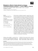
We investigated the effect of QUIN on nNOS activity
in cultured human neurons. Primary human neurons
were treated with QUIN for 30 min at increasing
concentrations. A dose-dependent increase in nNOS
activity was observed with increased concentrations of
QUIN (Fig. 1A). As expected, the increase in nNOS
activity correlated well with an increasing release of
nitrite into the extracellular medium (Fig. 1B).
To determine if polyphenols can influence QUIN-
induced nNOS activity due to QUIN in human
neurons, we tested the effect of selected polyphenolic
compounds on nNOS activity in cultures pretreated
with selected polyphenols for 15 min. All polyphenols
tested produced a dose-dependent decrease in nNOS
activity in human neurons, with EPCG, catechin
hydrate and curcumin showing higher potency than
apigenin, naringenin and gallotannin. These results
correlate well with the reduced extracellular nitrite
release from the same neuronal cell cultures (Fig. 1D).
Effect of EPCG, catechin hydrate, curcumin,
apigenin, naringenin and gallotannin on
intracellular NAD
+
levels, extracellular lactate
dehydrogenase (LDH) and PARP activation in
human neurons
To determine the effect of polyphenols on intracellular
NAD
+
levels, endogenous PARP activation and cell
viability, we measured intracellular NAD
+
levels,
PARP and extracellular LDH activities in human neu-
0
100
200
300
QUIN conc. (nM)
ng L-citrulline/mg
protein/30 minutes
0.0 150.0 350.0 550.0 750.01200.0 0.0 150.0 350.0 550.0 750.0 1200.0
0
100
200
300
400
500
QUIN conc. (n
M
)
µM NO
2
production/mg
protein
µM NO
2
production/mg
protein
0
10
20
30
40
*
*
*
*
*
*
*
*
*
*
*
*
0
10
20
*
*
*
*
EPCG Catechin Hydrate Curcumin Apigenin Naringenin Gallotannin
ng L-citrulline/mg
protein/30 minutes
QUIN
(550 n
M
)
–+ + + + +
Polyphenol
(1 µ
M
)
Polyphenol
(10 µ
M
)
Polyphenol
(50 µ
M
)
Polyphenol
(100 µ
M
)
QUIN
(550 n
M
)
Polyphenol
(1 µ
M
)
Polyphenol
(10 µ
M
)
Polyphenol
(50 µ
M
)
Polyphenol
(100 µ
M
)
–
–+–––
–
––+––
–
–––+–
–
–– – –+
–
+++++
––
+
–––
–– –
+
––
–– – –
+
–
–– –
––
+
AB
CD
Fig. 1. Effect of polyphenols on
QUIN-induced nNOS activity and nitrite
production in human neurons. Effect of: (A)
QUIN on nNOS activity for 30 min
(*P < 0.05 compared with previous dose);
(B) QUIN on extracellular nitrite production
(*P < 0.05 compared with previous dose);
(C) EPCG, catechin hydrate, curcumin, api-
genin, naringenin and gallotannin on nNOS
activity in the presence of QUIN (550 n
M)
for 30 min (*P < 0.05 compared with
550 n
M QUIN alone); (D) EPCG, catechin
hydrate, curcumin, apigenin, naringenin and
gallotannin on extracellular nitrite production
in the presence of QUIN (550 n
M)
(*P < 0.05 compared with 550 n
M QUIN
alone); n = 4 for each treatment group.
Neuroprotective effects of polyphenols N. Braidy et al.
370 FEBS Journal 277 (2010) 368–382 ª 2009 The Authors Journal compilation ª 2009 FEBS
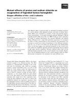
rons after 24 h of treatment. Treatment with EPCG
and curcumin significantly increased intracellular
NAD
+
levels in a dose-dependent manner (Fig. 2A),
but no significant difference was observed for PARP
(Fig. 2B) and LDH activities (Fig. 2C). On the con-
trary, gallotannin induced a dose-dependent decrease
in intracellular NAD
+
levels (Fig. 2A) and a dose-
dependent increase in extracellular LDH activity
(Fig. 2C). No significant difference was observed for
PARP activity (Fig. 2B). Similarly, no significant dif-
ferences were observed in intracellular NAD
+
levels
(Fig. 2A), PARP (Fig. 2B) and extracellular LDH
activities (Fig. 2C) for apigenin and naringenin.
Effect of EPCG, catechin hydrate, curcumin,
apigenin, naringenin and gallotannin on
QUIN-mediated NAD
+
depletion, extracellular
LDH and PARP activation in human neurons
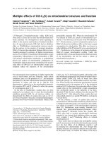
To assess the effects of polyphenols on QUIN-
mediated NAD
+
depletion, PARP activation and
extracellular LDH release (cell death), we measured
intracellular NAD
+
levels, PARP and extracellular
LDH activities in human neurons after 24 h of
treatment. The addition of EPCG, catechin hydrate
and curcumin (50 lm) significantly attenuated QUIN-
mediated NAD
+
depletion after 24 h (Fig. 3A). Apige-
nin, naringenin and gallotannin also prevented NAD
+
depletion at the same concentration (50 lm), but to a
lesser extent (Fig. 3A). As previously shown, neurons
treated with QUIN at 550 nm for 1 h had significantly
increased PARP activity compared with the control
(Fig. 3B). Concomitant treatment of these cells with
EPCG, catechin hydrate and curcumin (50 lm) signifi-
cantly reduced PARP activity compared with QUIN
treatment alone. Treatment with apigenin, naringenin
and gallotannin (50 lm) also reduced PARP activity,
but to a significantly lower degree than EPCG, cate-
chin hydrate or curcumin (Fig. 3B). These results clo-
sely correlate with results presented for NAD
+
(Fig. 3A). Neurons treated with QUIN (550 nm) in the
presence of selected polyphenols (50 lm) showed sig-
nificantly reduced evidence of cell death as measured
by extracellular LDH activity in culture supernatants
after 24 h (Fig. 3C). Extracellular LDH activity was
significantly reduced in the presence of EPCG, cate-
PARP activity
(NAD+ consumed/hr mg
protein)
0
50
100
150
NAD
+
(ng·mg
–1
protein)
0
1000
2000
Polyphenol
(1 µ
M
)
Polyphenol
(10 µ
M
)
Polyphenol
(50 µ
M
)
Polyphenol
(100 µ
M
)
Polyphenol
(1 µ
M
)
Polyphenol
(10 µ
M
)
Polyphenol
(50 µ
M
)
Polyphenol
(100 µ
M
)
Polyphenol
(1 µ
M
)
Polyphenol
(10 µ
M
)
Polyphenol
(50 µ
M
)
Polyphenol
(100 µ
M
)
–+ –––
–– +––
–– –+–
–– ––+
–+ –––
–– +––
–– –+–
–– ––+
–+ –––
–– +––
–– –+–
–– ––+
*
*
*
*
*
*
LDH activity
(IU/L/mg protein)
0
25
50
*
*
*
*
EPCG Catechin Hydrate Curcumin
Apigenin Naringenin Gallotannin
A
B
C
Fig. 2. Effect of polyphenols on intracellular NAD
+
levels, PARP
activation and cell death in human neurons. Effect of: (A) EPCG,
catechin hydrate, curcumin, apigenin, naringenin and gallotannin on
intracellular NAD
+
levels for 24 h (*P < 0.05 compared with med-
ium alone); (B) EPCG, catechin hydrate, curcumin, apigenin, na-
ringenin and gallotannin on PARP activity for 1 h (*P < 0.05
compared with medium alone); (C) EPCG, catechin hydrate, curcu-
min, apigenin, naringenin and gallotannin on extracellular LDH activ-
ity (*P < 0.05 compared with medium alone); n = 3 for each
treatment group.
N. Braidy et al. Neuroprotective effects of polyphenols
FEBS Journal 277 (2010) 368–382 ª 2009 The Authors Journal compilation ª 2009 FEBS 371
chin hydrate and curcumin compared with apigenin,
naringenin and gallotannin (Fig. 3C). These results
again directly correlate with data for NAD
+
depletion
and PARP activity (Fig. 3A,B).
QUIN induces intracellular Ca
2+
levels in cultured
human neurons
Human fetal neurons were incubated with QUIN and
a significant dose-dependent increase in intracellular
Ca
2+
influx was observed (Fig. 4). As RNS were
increased with increasing concentrations of QUIN
(Fig. 1), it is reasonable to conclude that the formation
of NO• is a downstream event in the QUIN-induced
excitotoxic cascade mediated by Ca
2+
influx.
Effect of EPCG, catechin hydrate, curcumin, apige-
nin, naringenin and gallotannin on QUIN-induced
intracellular Ca
2+
in cultured human neurons
As mentioned above, QUIN stimulation induced a sig-
nificant increase in intracellular Ca
2+
. Each of the
polyphenols, EPCG, curcumin, apigenin, naringenin
and gallotannin, significantly reduced intracellular
Ca
2+
influx (Fig. 4). Attenuation of increased Ca
2+
influx was greatest with EPCG and curcumin
compared with apigenin and naringenin (Fig. 5).
Interestingly, catechin hydrate did not ameliorate a
QUIN-induced increase in intracellular Ca
2+
(Fig. 5).
Detection of 3-nitrotyrosine (3-NT) formation in
cultured human neurons
Immunocytochemistry was used to visualize protein
nitration due to increased NO• production in cultured
human neurons. Increased protein nitration in the
form of increased 3-NT was observed in 20% of
QUIN-treated cells compared with nontreated cells
(Fig. 6A,B). Likewise, staining for 3-NT was less
detectable in QUIN-treated neurons preincubated with
EPCG (0%), catechin hydrate (0%) and curcumin
(0%) compared with cells treated with apigenin (7%),
naringenin (9%) and gallotannin (12%) (Fig 6A,B).
Detection of PAR expression in cultured human
neurons
Immunocytochemistry studies were used to detect
PAR formation following treatment with QUIN and
selected polyphenols. The amount of PAR formed in
living cells gives a direct indication of the extent of
DNA damage. Higher immunoreactivity for PAR
PARP activity
(NAD+ consumed/h/mg
protein)
0
500
1000
1500
–+ + ++ ++
– – + – – – –
– – – – – – –
–– – +– ––
– – – – + – –
– – – – – + –
–– –
+
–
+
–
–
–
––––+
*
*
*
*
*
**
LDH activity
(IU/L/mg protein)
0
50
100
150
*
*
*
*
*
*
*
NAD
+
(ng·mg
–1
protein)
0
1000
2000
3000
QUIN
(550 n
M)
EPCG
(50 µ
M)
–+ + + ++ ++
– – + – – – – –
Catechin
Hydrate
(50 µ
M)
–– –+ –– ––
Curcumin
(50 µ
M)
–– ––+– ––
Apigenin
(50 µ
M)
–– –– –+ ––
Naringenin
(50 µ
M)
–– –– ––+–
Gallotannin
(50
µ
M)
QUIN
(550 n
M)
EPCG
(50 µ
M)
Catechin
Hydrate
(50 µ
M)
Curcumin
(50 µ
M)
Apigenin
(50 µ
M)
Naringenin
(50 µ
M)
Gallotannin
(50
µ
M)
QUIN
(550 n
M)
EPCG
(50 µ
M)
Catechin
Hydrate
(50 µ
M)
Curcumin
(50 µ
M)
Apigenin
(50 µ
M)
Naringenin
(50 µ
M)
Gallotannin
(50
µ
M)
– – – – – – – +
*
*
*
*
*
*
*
AB C
Fig. 3. Effect of polyphenols on QUIN-induced NAD depletion, PARP activation and cell death in human neurons. Effect of: (A) EPCG
(50 l
M), catechin hydrate (50 lM), curcumin (50 lM), apigenin (50 lM), naringenin (50 lM) and gallotannin (50 lM) on intracellular NAD
+
levels
in the presence of QUIN (550 n
M) for 24 h (*P < 0.05 compared with 550 nM QUIN alone); (B) EPCG (50 lM), catechin hydrate (50 lM), curc-
umin (50 l
M), apigenin (50 lM), naringenin (50 lM) and gallotannin (50 lM) on PARP activity in the presence of QUIN (550 nM) for 1 h
(*P < 0.05 compared with 550 n
M QUIN alone); (C) EPCG (50 lM), catechin hydrate (50 lM), curcumin (50 lM), apigenin (50 lM), naringenin
(50 l
M) and gallotannin (50 lM) on extracellular LDH activity in the presence of QUIN (550 nM)(*P < 0.05 compared with 550 nM QUIN
alone); n = 4 for each treatment group.
Neuroprotective effects of polyphenols N. Braidy et al.
372 FEBS Journal 277 (2010) 368–382 ª 2009 The Authors Journal compilation ª 2009 FEBS
staining (25%) was detected in human neurons in the
presence of QUIN (550 nm) compared with untreated
cultures and cells cotreated with 50 lm EPCG (4%),
catechin hydrate (5%), curcumin (4%), apigenin
(10%), naringenin (11%) and gallotannin (12%) for
1 h (Fig 7A,B). The presence of EPCG, catechin
hydrate and curcumin in QUIN-exposed neurons
resulted in the lowest PAR formation compared with
cells treated with the other polyphenols (Fig. 7A,B).
This indicates that the latter compounds exhibit a
poorer neuroprotective effect against DNA damage
compared with EPCG, catechin hydrate and curcumin.
Discussion
The excitotoxin QUIN is one of the major end prod-
ucts of tryptophan catabolism in the central nervous
system. Increased QUIN production by activated
microglia ⁄ infiltrating macrophages has been reported
in the brain in aging and in neuroinflammatory
diseases [1]. For example, QUIN is found at high
concentrations in immunoactive amyloid plaques in
the AD brain [1,2,29]. Given the complex aetiology
and mechanisms of AD, QUIN probably plays a
pivotal role in the neurodegenerative changes occurring
in the brain [1,29,30,31].
The involvement of NOS in QUIN toxicity on
human astrocytes and neurons has been demonstrated
[7,32,33]. This neurotoxic involvement of NOS has
been confirmed by the use of the NOS inhibitor, nitro-
l-arginine methyl ester, which can protect human pri-
mary neurons and astrocytes in vitro against QUIN
toxicity [7,34]. NOS inhibitors have also been found to
be effective in protecting mice and monkey models
from the development of AD pathophysiology [35].
Another way to attenuate increased NO• production
and consequent energy depletion due to QUIN is to
block the NMDA receptor. We have previously shown
that the NMDA ion channel blocker, MK-801, can
protect human neurons from QUIN-induced excitotox-
icity [7]. However, long-term NMDA receptor inhibi-
tion by MK-801 has previously been shown to be toxic
to cultures of rat cortical neurons [36]. Alternatively,
polyphenols with their ROS ⁄ RNS scavenging, metal
chelating and anti-inflammatory properties represent a
promising additional option for the modulation of ex-
citotoxic cell death that may potentially be effective in
conditions such as AD treatment (Fig. 8). The neuro-
protective effects of green tea polyphenols and their
potential in the treatment of AD have been extensively
reviewed [19,37,38].
In this study, we evaluated the effects of several poly-
phenolic compounds on QUIN-mediated elevations in
nNOS activity and nitrite production. The activity of
nNOS was considerably enhanced in a dose-dependent
manner, with increasing concentrations of QUIN
within 30 min, with a subsequent increase in nitrite
production (Fig. 1). These results are consistent with
previous reports showing increased NO• production in
the striatum within 2 h of QUIN injection [32,33].
Conversely, a dose-dependent decrease in nNOS
activity and nitrite production was observed in QUIN-
treated neuronal cells preincubated with selected poly-
phenolic compounds (Fig. 1). EPCG, catechin hydrate
and curcumin showed a greater inhibitory effect on
nNOS activity and subsequent nitrite production com-
pared with apigenin, naringenin and gallotannin
(Fig. 1). The modulatory effect of polyphenolic com-
pounds on the NOS family has been previously
reviewed in [19]. EPCG, catechin hydrate and curcu-
min can suppress NO• production in cultures of RAW
264.7 macrophages and human peripheral blood
mononuclear cells following a 24 h stimulation with
lipopolysaccharide [39]. Moreover, apigenin has been
shown to downregulate iNOS expression and NO•
production in RAW 264.7 macrophages [40]. Taken
together, these results suggest that polyphenols can
010
Flourescent intensity
20 30 40 50 60 70 80 90 100
12
Control
QUIN 1200 n
M
QUIN 550 nM
QUIN 150 nM
A
B
10
8
6
4
2
0
Time (s)
No QUIN QUIN
QUIN conc. (nM)
Amplitude
0.0 150.0 550.0 1200.0
0.0
2.5
5.0
7.5
10.0
*
*
*
Fig. 4. QUIN induces Ca
2+
influx in human neurons. (A) Represen-
tative trace of intracellular Ca
2+
induced by QUIN (150, 550 and
1200 n
M). (B) Quantified amplitude of neuronal response to QUIN
at the aforementioned concentrations (*P < 0.05 compared with no
QUIN); n = 4 for each treatment group.
N. Braidy et al. Neuroprotective effects of polyphenols
FEBS Journal 277 (2010) 368–382 ª 2009 The Authors Journal compilation ª 2009 FEBS 373
inhibit NO• production by significantly reducing iNOS
expression and activity. However, the present study
was the first to examine the inhibitory effects of poly-
phenolic compounds on nNOS activity in primary cul-
tures of human neurons. Consistent with the above
results, EPCG, catechin hydrate and curcumin showed
a significant reduction in 3-NT formation compared
with QUIN-treated cells alone (Fig. 6). Apigenin, na-
ringenin and gallotannin also exerted a protective
effect against 3-NT formation, but to a lesser extent
than the other polyphenols (Fig. 6).
We have previously shown that QUIN can induce
PARP-1 activity and subsequent NAD
+
depletion in
primary cultures of human astrocytes and neurons at
pathophysiological concentrations [7]. In that earlier
study, NOS inhibition using nitro-l-arginine methyl
ester significantly reduced NAD
+
depletion and
PARP-1 activation in cultured human neurons exposed
to cytotoxic concentrations of QUIN [7]. The present
study showed that the polyphenols, EPCG, catechin
hydrate and curcumin, which have a greater inhibitory
effect on nNOS activity and nitrite production, can
prevent DNA damage [indicated by reduced PAR for-
mation (Fig. 7) and PARP-1 activation (Fig. 3)] and
block the subsequent depletion of NAD
+
stores,
thereby preserving the cell’s energy-dependent func-
tions (Fig. 3). Apigenin, naringenin and gallotannin
also showed a neuroprotective effect against PARP-1
activation and NAD
+
depletion, but to a lesser extent
than the previously mentioned polyphenols, probably
A
B
C
D
E
G
F
Fig. 5. Effect of polyphenols on QUIN-induced Ca
2+
influx in human neurons. Representative trace of intracellular Ca
2+
induced by 550 nM
QUIN in the presence of: (A) EPCG, (B) catechin hydrate, (C) curcumin, (D) apigenin, (E) naringenin, (F) gallotannin. (G) Quantified amplitude
of neuronal response to QUIN and EPCG, catechin hydrate, curcumin, apigenin, naringenin and gallotannin. The polyphenols were washed
out during QUIN administration, as the polphenols may influence its fluorescence (*P < 0.05 compared with 550 n
M QUIN; n = 4 for each
treatment group.
Neuroprotective effects of polyphenols N. Braidy et al.
374 FEBS Journal 277 (2010) 368–382 ª 2009 The Authors Journal compilation ª 2009 FEBS
due to their lower inhibitory effect on nNOS activity
(Fig. 3).
Although treatment with catechin hydrate, apigenin
and naringenin alone showed no significant difference
in intracellular NAD
+
levels, and PARP and LDH
activities across the range of concentrations tested,
increased intracellular NAD
+
levels were observed fol-
lowing treatment with EPCG and curcumin alone
3-NT
MAP-2
Merged
Control
QUIN
(550 n
M)
EPCG
(50 µ
M) +
QUIN
(550 n
M)
Catechin
(50 µ
M) +
QUIN
(550 n
M)
Curcumin
(50 µ
M) +
QUIN
(550 n
M)
Apigenin
(50 µ
M) +
QUIN
(550 n
M)
Naringenin
(50 µ
M) +
QUIN
(550 n
M)
Gallotannin
(50 µ
M) +
QUIN
(550 n
M)
0
10
20
30
QUIN
(550 n
M)
EPCG
(50 µ
M)
Catechin
Hydrate
(50 µ
M)
Curcumin
(50 µ
M)
Apigenin
(50 µ
M)
Naringenin
(50 µ
M)
Gallotannin
(50
µ
M)
*
*
*
*
*
*
*
A
B
Fig. 6. Immunocytochemical detection of 3-NT in purified primary human neurons after QUIN (550 nM) stimulation. Staining for 3-NT in
human neurons: top row – double staining for 3-NT ⁄ green and DAPI ⁄ blue; centre – double staining for MAP-2 ⁄ red and DAPI ⁄ blue; bottom
row – merged 3-NT ⁄ green, MAP-2 ⁄ red and DAPI ⁄ blue. (B) Numeration of fluorescence intensity of 3-NT in human neurons using immunocy-
tochemistry. The histogram shows the percentage of human neurons expressing 3-NT relative to the total number of neuronal cells after
24 h of treatment (*P < 0.05 compared with 550 n
M QUIN alone); n = 4 for each treatment group.
N. Braidy et al. Neuroprotective effects of polyphenols
FEBS Journal 277 (2010) 368–382 ª 2009 The Authors Journal compilation ª 2009 FEBS 375
(Fig. 2). This is consistent with the observation that
PARP activity (and therefore NAD
+
turnover) was
also lowest following treatment with both EPCG and
curcumin at 50 and 100 lm (Fig. 2B). On the other
hand, gallotann in showed a dose-dependent decrease
in intracellular NAD
+
levels (Fig. 2A), with a corre-
sponding decrease in cell viability (Fig. 2C). This may
be explained by the observation by others that gallo-
Control QUIN
EPCG
(50 µ
M) +
QUIN
(550 n
M)
Curcumin
(50 µ
M) +
QUIN
(550 n
M)
Apigenin
(50 µ
M) +
QUIN
(550 n
M)
Naringenin
(50 µ
M) +
QUIN
(550 n
M)
Gallotannin
(50 µ
M) +
QUIN
(550 n
M)
Catechin
(50 µ
M) +
QUIN
(550 n
M)
DAPI
PAR
MAP-2
Merged
0
10
20
30
QUIN
(550 n
M)
–+++++++
EPCG
(50 µ
M)
––+–––––
Catechin
Hydrate
(50 µ
M)
–––+ – – ––
Curcumin
(50 µ
M)
– – – – + – – –
Apigenin
(50 µ
M)
– – – – – + – –
Naringenin–––– – –+ –
Gallotannin
(50
µ
M)
–––– – ––+
*
*
*
*
*
*
*
A
B
Fig. 7. Immunocytochemical detection of PAR in purified primary human neurons after QUIN (550 nM) stimulation. Staining for PAR in
human neurons: top row – nuclear staining for DAPI ⁄ blue; second row – staining for PAR ⁄ green; third row – double staining for DAPI ⁄ blue
and MAP-2 ⁄ red; fourth row – merged PAR ⁄ green, MAP-2 ⁄ red and DAPI ⁄ blue. (B) Numeration of fluorescence intensity of PAR in human
neurons using immunocytochemistry. The histogram shows the percentage of human neurons expressing PAR relative to the total number
of neuronal cells after 1 h of treatment (*P < 0.05 compared with 550 n
M QUIN alone); n = 4 for each treatment group.
Neuroprotective effects of polyphenols N. Braidy et al.
376 FEBS Journal 277 (2010) 368–382 ª 2009 The Authors Journal compilation ª 2009 FEBS
tannin strongly inhibits nuclear nicotinamide mono-
nucleotide adenylyltransferase (NMNAT-1) activity,
with no detectable activity observed at 100 lm [41].
The results of the present study show that QUIN
can induce intracellular Ca
2+
influx in a dose-
dependent manner (Fig. 4), and that this reduces the
viability of cultured human neurons. To determine
whether the neuroprotective effect of these polyphenols
was due to a direct nNOS inhibition or via intracellu-
lar Ca
2+
modulation, we examined the effect of these
polyphenols on intracellular Ca
2+
influx in human
neurons following QUIN stimulation. We found that
EPCG and curcumin were able to attenuate QUIN-
induced Ca
2+
influx to a greater extent than apigenin,
naringenin and gallotannin (Fig. 5). However, catechin
hydrate did not attenuate the observed increase in
Ca
2+
in QUIN-treated neuronal cultures (Fig. 5).
EPCG has been previously shown to attenuate gluta-
mate-induced cytotoxicity via intracellular ionotropic
Ca
2+
modulation in PC12 cells, although the exact
mechanism remains unclear [42]. Curcumin has been
shown to exert a potent antioxidant effect on NO•-
related radical generation [43]. Curcumin has also been
shown to antagonize several important pathways
involved in NOS-mediated neurotoxicity, including
activation of nuclear factor kappa B, the Jun N-termi-
nal kinase pathway and protein kinase C [26,44,45].
Protein kinase C partly phosphorylates the core
NMDA receptor subunit NR1, which potentiates
increased Ca
2+
influx following NMDA receptor acti-
vation [26]. A decreased phosphorylation of NR1 may
protect against QUIN-induced excitotoxicity when the
levels of QUIN are significantly elevated. We found
that catechin hydrate did not reduce QUIN-induced
Ca
2+
influx in human neurons. This is consistent with
another study, where catechin hydrate only slightly
inhibited the phosphorylation of protein kinase C [26].
However, catechin hydrate significantly reduced
QUIN-induced nNOS activity and NO• production. It
is possible that inhibition of nNOS activity by catechin
hydrate may be mediated through a direct action on
the enzyme itself. For example, nitrite and peroxy-
nitrite inhibition by catechins has been attributed to
the 3¢4¢-catechol group on the B-ring [26].
Apigenin and naringenin are known to protect
against excitotoxic insults in human neurons indepen-
dent of NOS activity. Silva et al. [46] showed that the
apigenin derivative biapigenin prevented kainate ex-
citotoxicity by protecting cultured neurons from
delayed Ca
2+
deregulation due to excessive NMDA
receptor activation. Further studies have focussed on
the binding of naringenin to GABA
A
receptors as a
potential neuroprotective mechanism of action in the
central nervous system [47,48].
Our results show that gallotannin is less active
against nNOS activity and demonstrated poor nitrite
scavenging properties (Fig. 1). However, gallotannin
was able to attenuate QUIN-induced Ca
2+
influx in
human primary neurons to a similar extent as apige-
nin. Other studies have shown that gallotannin can
only significantly reduce Ca
2+
influx when adminis-
tered simultaneously with glutamate [26]. This suggests
a possible competitive inhibitory process.
Importantly the concentrations used in these experi-
ments are within the achievable range of serum levels
following oral consumption of these polyphenols. For
example, one human study reported that the serum
concentration of curcumin was 1.77 ± 1.87 lm [49]. In
another rat study, daily oral consumption of a glyco-
nated form of catechin resulted in a serum concentra-
tion of 34.8 ± 6.0 lm [50]. The amount of EPCG in a
single cup of green tea is 300 lm [51]. Therefore, the
calculated maximum serum concentration of EPCG
may reach 60 lm in a 60 kg human after oral con-
sumption of a single cup of tea. In the present study,
the polyphenols were tested at a standardized concen-
tration of 50 lm. Although this concentration is rele-
QUIN
Ca
2+
Ca
2+
NO
Massive DNA disruption
Energy failure
Cell death
Energy Metabolism
PARP Over-activation
Poly(ADP-
ribosyl)ation
NAD
+
EPCG, Apigenin
Naringenin, TA
Curcumin
Catechin
Hydrate
PKC
P
NMDA-R
Fig. 8. Schematic representation of the protective effects of EPCG,
curcumin, catechin hydrate, apigenin, naringenin and gallotannin.
The excitatory neurotoxin QUIN leads to over-activation of NMDA
receptors followed by sustained Ca
2+
influx. The Ca
2+
influx leads to
the formation of NO• by the activation of nNOS. Highly reactive free
radicals are formed, which can cause oxidative damage to DNA lead-
ing to over-activation of PARP-1 and subsequent NAD
+
depletion
and cell death due to energy restriction. Polyphenols can inhibit
QUIN-induced excitotoxicity. However, each polyphenolic compound
exerts its neuroprotective effect through a distinct mechanism.
N. Braidy et al. Neuroprotective effects of polyphenols
FEBS Journal 277 (2010) 368–382 ª 2009 The Authors Journal compilation ª 2009 FEBS 377
vant to serum levels in humans, lower concentrations
of these polyphenols may also be neuroprotective if
administered over a longer period of time.
Several epidemiological studies have predicted neuro-
degenerative diseases to be a major public health prob-
lem in the 21st century [52]. In Australia it has been
projected that although the total aging population will
increase by 40% in 2042, the population with AD will
increase by 3.5 times due to aging population demo-
graphics [53]. The neuroprotective effects of these green
tea polyphenols were obtained in an experimental
pretreatment model. The efficacy of these polyphenols
in vivo is dependent on the ability of these polyphenols
to cross the blood–brain barrier. Curcumin, EPCG and
catechin have been reported to pass through the blood–
brain barrier [54,55]. The permeability of apigenin,
naringenin and gallotannin remains unknown.
In a recent meta-analysis of 187 retrospective stud-
ies, EPCG, curcumin, catechin hydrate, melatonin, res-
veratrol, vitamin C and vitamin E were identified as
naturally occurring compounds that show efficiency in
slowing down the spectre of AD symptoms [56]. The
results from our study and others add support to this
observation and may encourage individuals to select
foods that contain these beneficial compounds (e.g. red
grapes, blue berries, peanuts, etc.). This will be impor-
tant to improve population health in general, and in
aging populations in particular.
Materials and methods
Reagents and chemicals
Dulbecco’s phosphate buffer solution, Fura-2-AM fluoro-
phore and all other cell culture media and supplements
were obtained from Invitrogen (Melbourne, Australia)
unless otherwise stated. Nicotinamide, bicine, b-NADH,
3-[-4,5-dimethylthiazol-2-yl]-2,5-diphenyl tetrazolium bro-
mide, Hepes, d-glucose, alcohol dehydrogenase, sodium
pyruvate, Tris, c-globulins, QUIN, 4¢,6-diamidino-
2-phenylindole dihydrochloride (DAPI), EPCG, catechin
hydrate, curcumin, apigenin, naringenin and gallotannin
were obtained from Sigma-Aldrich (Castle-Hill, Australia).
Phenazine methosulfate was obtained from ICN Biochem-
icals (Aurora, OH, USA). Bradford reagent was obtained
from BioRad (Hercules, CA, USA). Rabbit anti-micro-
bule-associated protein 2 (MAP2) was obtained from
Millipore (Melbourne, Australia). Mouse anti-poly(ADP-
ribose) (10H) was obtained from Alexis Corporation
(Pastlach, Switzerland). Mouse anti-3-NT, secondary
anti-mouse IgG and anti-rabbit Alexa 488 (green)- or
Alexa 594 (red)-conjugated IgG were obtained from
Molecular Probes (Eugene, OR, USA). All commercial
antibodies were used at the concentrations specified by
the manufacturer.
Cell cultures
Human fetal brains were obtained from 16–19-week-old
fetuses collected following therapeutic termination with
informed consent. Mixed brain cultures were prepared and
maintained using a protocol previously described by Guille-
min et al. [2]. Neurons were prepared from the same mixed
brain cell cultures as previously described [29]. Briefly, cells
were plated in 24-well culture plates coated with Matrigel
(1 ⁄ 20 in Neurobasal) and maintained in Neurobasal med-
ium supplemented with 1% B-27 supplement, 1% Gluta-
max, 1% antibiotic ⁄ antifungal, 0.5% Hepes buffer and
0.5% glucose. The cells were maintained at 37 °Cina
humidified atmosphere containing 95% air ⁄ 5% CO
2
.
Measurement of nNOS activity using the
citrulline assay
nNOS activity was assayed by monitoring the conversion of
l-[
3
H]arginine to l-[
3
H]citrulline, as previously described
[57]. The cells were treated with 50–1200 nm QUIN for
30 min. After incubation, the reaction was terminated by
adding 0.3 m HClO
4
(pH 5.5) containing EDTA (4 mm).
Radiolabelled citrulline is neutral at a pH of 5.5, and was
separated from the positively charged arginine using a col-
umn containing analytical grade cation-exchange resin (AG
Dowex 50W-X8). The amount of l-[
3
H]citrulline was mea-
sured using a Beckman LS6500 scintillation counter. The
results were expressed as ng l-citrullineÆ500 lg pro-
tein
)1
Æ30 min
)1
. In another set of experiments, neuronal
cells were preincubated for 15 min with 1–100 lm EPCG,
catechin hydrate, curcumin, apigenin, naringenin and gallo-
tannin. The nNOS activity in the presence of 550 nm QUIN
was then quantified as described above.
Nitrite determination by fluorometric Griess
diazotization assay
Nitrite production in the culture supernatant was measured
using the fluorometric Griess diazotization assay, as previ-
ously described [57]. In the Griess assay, NO
2
is allowed to
react with an aromatic amine in acidic medium to yield a
fluorescent azo derivative. Briefly, neurons were treated
with 50–1200 nm QUIN for 30 min and 100 lL culture
supernatant was placed in a 96-well microplate. Diamino-
naphthalene was diluted to 10 mm in deionized water from
the original 100 mm dimethylsulfoxide stock solution, and
1% HCl was added to the aqueous mixture to generate a
working stock of diaminonaphthalene. Then, 100 lL diami-
nonaphthalene was added to each sample and incubated for
10 min at room temperature. An additional 100 lL2m
Neuroprotective effects of polyphenols N. Braidy et al.
378 FEBS Journal 277 (2010) 368–382 ª 2009 The Authors Journal compilation ª 2009 FEBS
NaOH was added and the fluorescence intensity was then
recorded at an excitation wavelength of 355 nm and an
emission wavelength of 460 nm. In another set of experi-
ments, neuronal cells were preincubated for 15 min with
1–100 lm EPCG, catechin hydrate, curcumin, apigenin,
naringenin and gallotannin. The amount of nitrite produced
in the presence of 550 nm QUIN was then quantified as
described above.
Calcium influx studies using fluorometry
To measure intracellular Ca
2+
, human neurons were loaded
( 1 h, room temperature) with 3.5 lgÆ mL
)1
Fura-2-AM in
a loading solution containing (in mm): 135 NaCl, 5 KCl, 1
MgCl
2
, 1 CaCl
2
, 5 glucose and 10 Hepes (pH 7.4). Probeni-
cid dissolved in 1 m NaOH was added to the loading solu-
tion at a final concentration of 4 m m to reduce dye
leakage. Following the recommended 1 h incubation period,
the loading solution was removed and replaced with 1x
Hanks balanced salt solution (HBSS) containing 50 mm
glycine. The addition of selected polyphenols (EPCG, cate-
chin hydrate, curcumin, apigenin, naringenin and gallotan-
nin) was undertaken 15 min before the addition of QUIN
to ensure that adequate diffusion time was provided to
attain equilibrium. The Ca
2+
influx experiments were sub-
sequently performed using a Fluostar Optima fluorometer
(Durham, NC, USA). Filter excitation and emission was
set at 485 and 520 nm wavelengths, respectively. For each
well, fluorescence was measured via orbital scanning of 10
locations at a 3 mm radius every 0.5 s, and the average of
these readings was recorded. Baseline fluorescence was mea-
sured during the first 10 s of the experiment, followed by
injection of QUIN (in HBSS). Fluorescent readings were
subsequently taken for an additional 90 s. Negative con-
trols included injection of only HBSS solution without an
agonist.
NAD(H) microcycling assay for the measurement
of intracellular NAD
+
concentrations
The intracellular NAD
+
concentration was measured spec-
trophotometrically using the thiazolyl blue microcycling
assay established by Bernofsky & Swan [58] adapted for the
96-well plate format by Grant & Kapoor [59]. Human
neurons were preincubated for 15 min with 30 and 50 l m
EPCG, catechin hydrate, curcumin, apigenin, naringenin and
gallotannin. The cells were then treated with QUIN (550 nm)
and intracellular NAD
+
levels were measured 24 h later.
Extracellular LDH activity as a measurement for
cytotoxicity
The release of LDH into culture supernatant correlates with
the amount of cell death and membrane damage, providing
an accurate measure of cellular toxicity. LDH activity was
assayed using a standard spectrophotometric technique
described by Koh & Choi [60]. After preincubation with
1–100 lm EPCG, catechin hydrate, curcumin, apigenin,
naringenin and gallotannin, neuronal cells were treated with
QUIN (550 nm) and extracellular LDH activity was
assessed in culture supernatant after 24 h.
PARP assay for the measurement of intracellular
PARP activity
PARP activity was measured using a new operational pro-
tocol relying on the chemical quantification of NAD
+
modified from Putt et al. [61] and adapted for the 24-well
format [7]. After a 15 min preincubation with the selected
polyphenolic compounds, neurons were treated with QUIN
(550 nm) and incubated for 15 min. Dulbecco’s phosphate
buffer solution was then aspired and PARP lysing buffer
(200 lL) was added to the cell plate. The buffer solution
contained MgCl
2
(10 mm), Triton X-100 (1%) and NAD
+
(20 lm) in Tris buffer (50 mm, pH 8.1). The plate was then
incubated for 1 h and PARP activity was assayed as previ-
ously described [7].
Bradford protein assay for the quantification of
total protein
NAD
+
concentration, PARP and extracellular LDH activi-
ties were adjusted for variations in cell number using the
Bradford protein assay [62].
Immunocytochemistry for the detection of PAR
and 3-NT formations
The method for immunocytochemistry has been previously
described [2]. Cells were incubated with mAb PAR and
mAb 3-NT together with the phenotypic marker (MAP-2).
Selected secondary antibodies (goat anti-mouse IgG or goat
anti-rabbit coupled with Alexa 488 or Alexa 594) were
used. The following controls were performed for each
labelled experiment: (a) isotypic antibody controls and (b)
incubation with only the secondary labelled antibody. Cell
counting was performed in a blind manner. The whole
controls and untreated chamber slides were counted. Enu-
meration of each slide was classified according to the
following scheme: DAPI staining for total cell number,
MAP-2 immunoreactivity for neurons, and 3-NT and PAR
staining.
Data analysis
The results obtained are presented as the means ± stan-
dard error of measurement. One-way analysis of variance
and posthoc Tukey’s multiple comparison tests were used
N. Braidy et al. Neuroprotective effects of polyphenols
FEBS Journal 277 (2010) 368–382 ª 2009 The Authors Journal compilation ª 2009 FEBS 379
to determine the statistical significance between treatment
groups. Differences between treatment groups were consid-
ered significant if P < 0.05.
References
1 Guillemin GJ, Brew BJ, Noonan CE, Takikawa O &
Cullen KM (2005) Indoleamine 2,3 dioxygenase and
quinolinic acid immunoreactivity in Alzheimer’s
disease hippocampus. Neuropathol Appl Neurobiol 31,
395–404.
2 Guillemin GJ, Smythe G, Takikawa O & Brew BJ
(2005) Expression of indoleamine 2,3-dioxygenase
and production of quinolinic acid by human
microglia, astrocytes, and neurons. Glia 49,
15–23.
3 Guillemin GJ, Wang L & Brew BJ (2005) Quinolinic
acid selectively induces apoptosis of human astocytes:
potential role in AIDS dementia complex. J Neuro-
inflammation 2, 16.
4 Guillemin GJ, Meininger V & Brew BJ (2005) Implica-
tions for the kynurenine pathway and quinolinic acid in
amyotrophic lateral sclerosis. Neurodegener Dis 2, 166–
176.
5 Schwarz FJ, Kirchgessner M & Roth HP (1983) Influ-
ence of picolinic acid and citric acid on intestinal
absorption of zinc in vitro and in vivo. Res Exp Med
(Berl) 182, 39–48.
6 Guillemin GJ, Kerr SJ & Brew BJ (2005) Involvement
of quinolinic acid in AIDS dementia complex. Neurotox
Res 7, 103–123.
7 Braidy N, Grant R, Adams S, Brew BJ & Guillemin G
(2009) Mechanism for quinolinic acid cytotoxicity in
human astrocytes and neurons. Neurotox Res 16, 77–
86.
8 Albin RL & Greenamyre J (1992) Alternate excitotoxic
hypothesis. Neurology 42, 733–738.
9 Alderton WK, Cooper CE & Knowles RG (2001) Nitric
oxide synthases: structure, function and inhibition. Bio-
chem J 357, 593–615.
10 Heales SJ, Barker JE, Stewart VC, Brand MP,
Hargreaves IP, Foppa P, Land JM, Clark JB &
Bolanos JP (1997) Nitric oxide, energy metabolism and
neurological disease. Biochem Soc Trans 25, 939–943.
11 Yu SW, Wang H & Poitras MF (2002) Mediation of
poly(ADP-ribose) polymerase-1-dependent cell death by
apoptosis-inducing factor. Science 297, 259–263.
12 Virag L, Salzman AL & Szabo C (1998) Poly(ADP-
ribose) synthetase activation mediates mitochondrial
injury during oxidant-induced cell death. J Immunol
161, 3753–3759.
13 Hong JT, Ryu SU, Kim HJ, Lee JK, Lee SH, Kim DB,
Yun YP, Ryu JH, Lee BM & Kim PY (2000) Neuro-
protective effect of green tea extract in experimental
ischemia-reperfusion brain injury. Brain Res Bull 53,
743–749.
14 Hong JT, Ryu SR, Kim HJ, Lee JK, Lee SH, Yun YP,
Lee BM & Kim PY (2001) Protective effect of green tea
extract on ischemia-reperfusion-induced brain injury in
Mongolian gerbils. Brain Res Bull 888, 11–18.
15 Bianchini F & Vainio H (2003) Wine and resveratrol:
mechanisms of cancer prevention? Eur J Cancer Prev
12, 417–425.
16 Savaskan E, Olivieri G, Meier F, Seifritz E, Wirz-Jus-
tice A & Muller-Spahn F (2003) Red wine ingredient
resveratrol protects from beta-amyloid neurotoxicity.
Gerontology 49 , 380–383.
17 Saiko P, Szakmary A, Jaeger W & Szekeres T (2008)
Resveratrol and its analogs: defense against cancer,
coronary disease and neurodegenerative maladies or just
a fad? Mutat Res 658
, 68–84.
18 Jackson JC, Brendan AR & Larson EB (2003) Fruit
and vegetable juices and Alzheimer’s disease: the Kame
project. Am J Med 119, 751–759.
19 Youdim KA, Spencer JPE, Schroeter H & Rice-Evans
C (2002) Dietary flavonoids as potential neuroprotec-
tants. Biol Chem 383, 503–519.
20 Inanami O, Asanuma T, Inukai N, Jin T, Shimokawa
S, Kasai N, Nakano M, Sato F & Kuwabara M
(1995) The suppression of age-related accumulation
of lipid peroxides in rat brain by administration of
Rooibos tea (Aspalathus linearis). Neurosci Lett 196,
85–88.
21 Chen C-M, Lin J-K, Liu S-H & Lin-Shiau S-Y (2008)
Novel regimen through combination of memantine and
tea polyphenol for neuroprotection against brain excito-
toxicity. J Neurosci Res 86, 2696–2704.
22 Paquay JB, Haenen GR & Stender G (2000) Protection
against nitric oxide toxicity by tea. J Agric Food Chem
48, 5768–5772.
23 Guo Q, Zhao B & Shen S (1999) ESR study on the
structure–antioxidant activity relationship of tea
catechins and their epimers. Biochim Biophys Acta 1427,
13–23.
24 Haenen GR, Paquay JB, Korthouwer RE & Bast A
(1997) Peroxynitrite scavenging by flavonoids. Biochem
Biophys Res Commun 236, 591–593.
25 Nanjo F, Honda M & Okushio K (1993) Effects of die-
tary tea catechins on alpha-tocopherol levels, lipid per-
oxidation, and erythrocyte deformability in rats fed on
high palm oil and perilla oil diets. Biol Pharm Bull 16,
1156–1159.
26 Yazawa K, Kihara T, Shen H, Shimmyo Y, Niidome T
& Sugimoto H (2006) Distinct mechanisms underlie dis-
tinct polyphenol-induced neuroprotection. FEBS Lett
580, 6623–6628.
27 Sutherland BA, Rahman RM & Appleton I (2006)
Mechanism of action of green tea catechins with a focus
Neuroprotective effects of polyphenols N. Braidy et al.
380 FEBS Journal 277 (2010) 368–382 ª 2009 The Authors Journal compilation ª 2009 FEBS
on ichemia-induced neurodegeneration. J Nutr Biochem
17, 291–306.
28 Kim HP, Son KH, Chang HC & Kang SS (2004) Anti-
inflammatory plant flavonoids and cellular action mech-
anisms. J Pharmacol Sci 966, 229–245.
29 Guillemin GJ, Cullen KM, Lim CK, Smythe GA,
Garner B, Kapoor V, Takikawa O & Brew BJ (2007)
Characterization of the kynurenine pathway in human
neurons. J Neurosci 27, 12884–12892.
30 Finkbeiner S & Cuero AM (2006) Disease modifying
pathways in neurodegeneration. J Neurosci 26, 10349–
10357.
31 Guillemin GJ, Smith DG, Williams K, Smythe GA,
Dormont D & Brew BJ (2001) Beta-amyloid peptide
1-42 induces human macrophages to produce the neuro-
toxin quinolinic acid. J Neuroimmunol 118, 336.
32 Aguilera P, Chanez-Cardenas ME, Floriano-Sanchez E,
Barrera D, Santamaria A, Sanchez-Gonzalez DJ, Perez-
Severiano F, Pedraza-Chaverri J & Maldonado Jimenez
PD (2007) Time-related changes in constitutive and
inducible nitric oxide synthases in the rat striatum in a
model of Huntington’s disease. Neurotoxicology 28,
1200–1207.
33 Perez-De La Cruz V, Gonzalez-Cortes C, Galvan-Arz-
ate S, Medina-Campos ON, Perez-Severiano F, Ali SF,
Pedraza-Chaverri J & Santamaria A (2005) Excitotoxic
brain damage involves early peroxynitrite formation in
a model of Huntington’s disease in rats: protective
role of iron porphyrinate 5,10,15,20-tetrakis
(4-sulfonatophenyl)porphyrinate iron (III). Neuroscience
135, 463–474.
34 Ting KK, Brew BJ & Guillemin GJ (2007) Effect of
quinolinic acid on gene expression in human astrocytes:
implications for Alzheimer’s disease. International
Congress Series 1304, 384–388.
35 Hantraye P, Brouillet E & Ferrante RJ (1996)
Inhibition of neuronal nitric oxide synthase prevents
MPTP-induced parkinsonism in baboons. Nat Med 2,
1017–1021.
36 Hwang JY, Kim YH, Ahn YH, Wie MB & Koh JY
(1999) N-methyl-D-aspartate receptor blockade induces
neuronal apoptosis in cortical culture. Exp Neurol 159,
124–130.
37 Weinreb O, Mandel S, Amit T & Youdim MB (2004)
Neurological mechanisms of green tea polyphenols in
Alzheimer’s and Parkinson’s diseases. J Nutr Biochem
15, 506–516.
38 Mandel S & Youdim MB (2004) Catechin polyphe-
nols: neurodegeneration and neuroprotection in neuro-
degenerative diseases. Free Radic Biol Med 37, 304–
317.
39 Lyu SY & Park WB (2005) Production of cytokine and
NO by RAW 264.7 macrophages and PBMC in-vitro
incubation with flavonoids. Arch Pharm Res 28,
573–581.
40 Liang YC, Huang YT, Tsai SH, Lin-Shiau SY, Chen
CF & Lin JK (1999) Suppression of inducible cyclooxy-
genase and inducible nitric oxide synthase by apigenin
and related flavonoids in mouse macrophages.
Carcinogenesis 20, 1945–1952.
41 Berger F, Lau C, Dahlmann M & Ziegler M (2005)
Subcellular compartmentation and differential catalytic
properties of the three human nicotinamide mononucle-
otide adenylyltransferase isoforms. J Biol Chem 280,
36334–36341.
42 Lee JH, Song DK, Jung CH, Shin DH, Park JW, Kwon
TK, Jang BC, Mun KC, Kim SP, Suh SI et al. (2004)
(-)-Epigallocatechin gallate attenuates glutamate-
induced cytotoxicity via intracellular Ca2 + modula-
tion in PC12 cells. Clin Exp Pharm Phys 31, 530–536.
43 Zbarsky V, Datla KP, Parkar S, Rai DK, Aruoma
OI & Dexter DT (2005) Neuroprotective properties
of the natural phenolic antioxidants curcumin and
naringenin but not quercetin and fisetin in a 6-OHDA
model of Parkinson’s disease. Free Radic Res 39,
1119–1125.
44 Weber WM, Hunsaker LA, Gonzales AM, Heynek-
amp JJ, Orlando RA, Deck LM & Vander-Jagt DL
(2006) TPA-induced up-regulation of activator pro-
tein-1 can be inhibited or enhanced by analogs of the
natural product curcumin. Biochem Pharmacol
72,
928–940.
45 Pendurthi UR, Williams JT & Rao LV (1997) Inhibi-
tion of tissue factor gene activation in cultured endothe-
lial cells by curcumin. Suppression of activation of
transcription factors Egr-1, AP-1, and NF-kappa B.
Arterioscler Thromb Vasc Biol 17, 3406–3413.
46 Silva B, Oliveira PJ, Dias A & Malva JO (2008) Quer-
cetin, kaempferol and biapigenin from Hypericum perfo-
ratum are neuroprotective against excitotoxic insults.
Neurotox Res 13, 265–279.
47 Paladini AC, Marder M, Viola H, Wolfman C, Wasow-
ski C & Medina JH (1999) Flavonoids and the central
nervous system: from forgotten factors to potent anxio-
lytic compounds. J Pharm Pharmacol 51, 519–526.
48 Medina JH, Viola H, Wolfmann C, Marder M, Wasow-
ski C, Calvo D & Paladini AC (1998) Neuroreactive
flavonoids: new ligands for the benzodiazepine receptor.
Phytomedicine 5, 235–243.
49 Cheng AL, Hsu CH, Lin JK, Hsu MM, Ho YF, Shen
TS, Ko JY, Lin JT, Lin BR, Ming-Shiang W et al.
(2001) Phase I clinical trial of curcumin, a chemopre-
ventative agent in patients with high-risk or pre-malig-
nant lesions. Anticancer Res 21, 2895–2900.
50 Silberberg M, Morand C, Manach C, Scalbert A &
Remesy C (2005) Co-administration of quercetin and
catechin in rats alters their absorption but not their
metabolism. Life Sci 77, 3156–3167.
51 Lin JK, Liang YC & Lin-Shiau SY (1999) Cancer
chemoprevention by tea polphenols through mitotic
N. Braidy et al. Neuroprotective effects of polyphenols
FEBS Journal 277 (2010) 368–382 ª 2009 The Authors Journal compilation ª 2009 FEBS 381
signal transduction blockade. Biochem Pharmacol 58,
911–915.
52 Cotran RS, Kumar V & Collins T (1999) Pathological
Basis of Disease, 6th edn. Saunders Company, Pennsyl-
vania, PA.
53 Economics A (2006) Dementia in the Asia Pacific
Region: The Epidemic is Here. Azheimer’s Australia,
Canberra.
54 Yang F, Lim GP, Begum AN, Ubeda OJ, Simmons MR,
Ambegaokar SS, Chen PP, Kayed R, Glabe CG, Frauts-
chy SA et al. (2005) Curcumin inhibits formation of amy-
loid beta oligomers and fibrils, binds plaques, and
reduces amyloid in vivo. J Biol Chem 280, 5892–5901.
55 Mandel S, Amit T, Reznichenko L, Weinreb O & You-
dim MB (2003) Green tea catechins as brain-permeable,
natural iron chelators – antioxidants for the treatment
of neurodegenerative disorders. Mol Nutr Food Res 5,
229–234.
56 Frank B & Gupta S (2005) A review of antioxidants and
Alzheimer’s disease. Ann Clin Psychiatry 17, 269–286.
57 Ward TR & Mundy WR (2002) Measurement of nitric
oxide synthase activity using citrulline assay. In Meth-
ods in Molecular Medicine, Neurodegeneration Methods
and Protocols (Harry J & Tilson HA eds). Humana
Press, Totowa, NJ.
58 Bernofsky C & Swan M (1973) An improved cycling
assay for nicotinamide adenine dinucleotide. Anal
Biochem 53, 452–458.
59 Grant RS & Kapoor V (1998) Murine glial cells
regenerate NAD, after peroxide-induced depletion,
using either nicotinic acid, nicotinamide, or quinolinic
acid as substrates. J Neurochem 70, 1759–1763.
60 Koh JY & Choi DW (1987) Quantitative determination
of glutamate mediated cortical neuronal injury in cell
culture by lactate dehydrogenase efflux assay. J Neuro-
sci Meth 20, 83–90.
61 Putt KS, Beilman GJ & Hergenrother PJ (2005) Direct
quantification of poly(ADP-ribose) polymerase (PARP)
activity as a means to distinguish necrotic and apoptotic
death in cell and tissue samples. Chem Bio Chem 6, 53–
55.
62 Bradford MM (1976) A rapid and sensitive method for
quantitation of microgram quantities of protein utilising
the principle of protein–dye binding. Anal Biochem 53,
452–458.
Neuroprotective effects of polyphenols N. Braidy et al.
382 FEBS Journal 277 (2010) 368–382 ª 2009 The Authors Journal compilation ª 2009 FEBS