Báo cáo khoa học: Targeting mechanism of the retinoblastoma tumor suppressor by a prototypical viral oncoprotein Structural modularity, intrinsic disorder and phosphorylation of human papillomavirus E7 doc
Bạn đang xem bản rút gọn của tài liệu. Xem và tải ngay bản đầy đủ của tài liệu tại đây (822.32 KB, 16 trang )
Targeting mechanism of the retinoblastoma tumor
suppressor by a prototypical viral oncoprotein
Structural modularity, intrinsic disorder and phosphorylation of
human papillomavirus E7
Lucı
´
a B. Chemes, Ignacio E. Sa
´
nchez, Clara Smal and Gonzalo de Prat-Gay
Protein Structure-Function and Engineering Laboratory, Fundacio
´
n Instituto Leloir and IIBBA-CONICET, Buenos Aires, Argentina
Keywords
LxCxE motif; natively unfolded proteins;
phosphorylation; retinoblastoma protein;
viral oncoprotein
Correspondence
Gonzalo de Prat-Gay, Protein Structure-
Function and Engineering Laboratory,
Fundacio
´
n Instituto Leloir and
IIBBA-CONICET, Av. Patricias Argentinas
435, 1405 Buenos Aires, Argentina
Fax: +54 11 5238 7501
Tel: +54 11 5238 7500 ext. 3209
E-mail:
(Received 16 November 2009, revised 4
December 2009, accepted 7 December
2009)
doi:10.1111/j.1742-4658.2009.07540.x
DNA tumor viruses ensure genome amplification by hijacking the cellular
replication machinery and forcing infected cells to enter the S phase. The
retinoblastoma (Rb) protein controls the G1 ⁄ S checkpoint, and is targeted
by several viral oncoproteins, among these the E7 protein from human
papillomaviruses (HPVs). A quantitative investigation of the interaction
mechanism between the HPV16 E7 protein and the RbAB domain in solu-
tion revealed that 90% of the binding energy is determined by the LxCxE
motif, with an additional binding determinant (1.0 kcalÆmol
)1
) located
in the C-terminal domain of E7, establishing a dual-contact mode. The
stoichiometry and subnanomolar affinity of E7 indicated that it can
bind RbAB as a monomer. The low-risk HPV11 E7 protein bound 2.0
kcalÆmol
)1
more weakly than the high-risk HPV16 and HPV18 type
counterparts, but the modularity and binding mode were conserved. Phos-
phorylation at a conserved casein kinase II site in the natively unfolded
N-terminal domain of E7 affected the local conformation by increasing the
polyproline II content and stabilizing an extended conformation, which
allowed for a tighter interaction with the Rb protein. Thus, the E7–RbAB
interaction involves multiple motifs within the N-terminal domain of E7
and at least two conserved interaction surfaces in RbAB. We discussed a
mechanistic model of the interaction of the Rb protein with a viral target
in solution, integrated with structural data and the analysis of other cellu-
lar and viral proteins, which provided information about the balance of
interactions involving the Rb protein and how these determine the progres-
sion into either the normal cell cycle or transformation.
Structured digital abstract
l
MINT-7383794, MINT-7383812, MINT-7383830, MINT-7383868, MINT-7383891, MINT-
7384056: E7 (uniprotkb:P03129) and Rb (uniprotkb:P06400) bind (MI:0407)byfluorescence
technologies (
MI:0051)
l
MINT-7383923: E7 (uniprotkb:P04020) and Rb (uniprotkb:P06400) bind (MI:0407)bycom-
petition binding (
MI:0405)
Abbreviations
AdE1A, adenovirus E1A; BPVN, N-terminal fragment of the BPV1 E7 protein; CKII, casein kinase II; CR1, conserved region 1; CR2,
conserved region 2; CtIP, transcriptional corepressor CtBP-interacting protein; E7(16-40)PP, a synthetic E7(16-40) peptide phosphorylated at
serine residues 31 and 32; FITC, fluorescein isothiocyanate; GST, glutathione S-transferase; HDAC, histone deacetylase; HPV, human
papillomavirus; IPTG, isopropyl thio-b-
D-galactoside; MBP, maltose-binding protein; PII, polyproline type II; Rb, retinoblastoma; SV40LT, SV40
large T antigen; TA, transactivation region; TFE, 1,1,1, trifluoroethanol.
FEBS Journal 277 (2010) 973–988 ª 2010 The Authors Journal compilation ª 2010 FEBS 973
Introduction
The retinoblastoma tumor suppressor gene (RB1) was
first identified as the causative agent whose loss
resulted in retinoblastoma, a heritable disease of pedi-
atric relevance [1]. To date, over 500 distinct muta-
tions in the RB1 gene have been identified in
retinoblastoma tumors, 50 of which are missense
mutations [2,3]. The tumor suppressor function of the
Rb protein is underscored by its mutation in a broad
range of human tumors [4]. The most extensively
studied function of the Rb protein is in the control
of cell cycle progression at the G1 ⁄ S boundary, medi-
ated through its interaction with the E2F family of
transcription factors [5]. The Rb protein also plays
important roles in chromatin remodeling, develop-
ment, differentiation and apoptosis [6]. These multiple
functions are mediated by over 100 interactions with
different protein partners that are dependent on the
cell type, and on the developmental and cell cycle
stages [7].
The Rb protein has a molecular mass of 105 kDa
and is composed of three domains. Both the N-terminal
and the AB (RbAB) domains consist of a double cyclin
fold [8,9], while the C-terminal domain (RbC) appears
to be natively unfolded [10]. The function of the N-ter-
minal domain is still poorly defined. The RbAB domain
mediates transcriptional repression and, together with
the C-terminal domain (RbC), promotes growth arrest
[11,12]. Most interacting partners contact more than
one structural domain in the Rb protein [13–15]. For
example, the ‘transactivation’ domain of E2F (E2F-TA)
binds to RbAB, whereas the ‘marked box’ domain
(E2F-MB) binds to RbC [10,16]. Moreover, there are at
least two distinct highly conserved ligand-binding sites
within the RbAB domain [8] (Figs 1 and S1). Cellular
proteins containing an LxCxE motif interact with a site
located on the B subdomain of RbAB [8,17]
(Fig. 1A,B). The E2F-TA domains bind to a site
located at the cleft between the A and B subdomains on
the opposite side of RbAB [16,18] (Fig. 1C,D).
Early evidence for the tumor suppressor role of the
Rb protein came from the mechanism of action of the
human papillomavirus (HPV) E7 major transforming
protein [19]. The interaction between E7 and the Rb
protein is required for the induction and maintenance
of the transformed state of human keratinocytes [20].
Deregulation of E7 expression upon integration of the
HPV genome is believed to play a role in HPV-medi-
ated oncogenesis. The DNA tumor virus proteins
SV40 large T antigen (SV40LT) and adenovirus E1A
(AdE1A) also target the Rb protein and share
sequence and functional conservation with the HPV
E7 protein [21,22]. E7, AdE1A and SV40LT each con-
tain several functional and structural domains, each of
which mediates interactions with different cellular tar-
gets. The three transforming proteins share conserved
region 2 (CR2); E7 and AdE1A also share conserved
region 1 (CR1).
E7 is a small ($ 100 amino acids) protein composed
of two structural domains. We have previously deter-
mined that the N-terminal domain (E7N) is natively
unfolded [23,24], includes CR1 and CR2, and contains
dynamic elements of helical and polyproline type II
(PII) secondary structure [23]. The globular C-terminal
domain (E7C) constitutes conserved region 3 (CR3)
and is responsible for protein dimerization and zinc
binding [24,25] (Fig. 2A). While the CR1 and CR2
domains are required for Rb protein degradation, all
conserved E7 regions participate in transformation
[26,27]. E7 can also oligomerize in vitro and in vivo
[28–30]. The conformational diversity of E7 may be an
evolved trait that allows for multiple modes of pro-
tein–protein interaction [31,32].
E7 binds to two structural domains in the Rb
protein, namely the RbAB and RbC domains. Binding
to both domains is required for E2F displacement [33].
The LxCxE motif within the CR2 region of E7
mediates high-affinity binding to the RbAB domain
[8,34] (Fig. 1A), while the isolated E7C binds to the
RbC domain with micromolar affinity [25,35]. The
crystal structure of the LxCxE–RbAB complex reveals
that the motif binds to a conserved shallow groove of
the B subdomain in an extended conformation
(Fig. 1A). The LxCxE motif is followed in E7 CR2 by
two conserved serine residues (S31 and S32) and by a
l
MINT-7383777, MINT-7384078, MINT-7383848, MINT-7384113, MINT-7384096: Rb (uni-
protkb:
P06400) and E7 (uniprotkb:P03129) bind (MI:0407)bycompetition binding (MI:0405)
l
MINT-7383963: Rb (uniprotkb:P06400) and E7 (uniprotkb:P06788) bind (MI:0407)bycom-
petition binding (
MI:0405)
l
MINT-7384022, MINT-7384040: E7 (uniprotkb:P03129) and Rb (uniprotkb:P06400) bind
(
MI:0407)bycomigration in non denaturing gel electrophoresis (MI:0404)
l
MINT-7384004, MINT-7383984: Rb (uniprotkb:P06400) binds (MI:0407)toE7 (uni-
protkb:
P03129)bypull down (MI:0096)
Viral targeting of the retinoblastoma protein L. B. Chemes et al.
974 FEBS Journal 277 (2010) 973–988 ª 2010 The Authors Journal compilation ª 2010 FEBS
stretch of acidic amino acids, and HPV16 E7 is
phosphorylated at S31 and S32 by casein kinase II
(CKII) in vitro and in vivo [36,37]. Phosphorylation is
required for E7 function, and cell culture assays have
suggested that phosphorylation modulates the strength
of the E7–RbAB interaction, but this proposal remains
a matter of debate [37–40].
Indirect evidence suggests that other regions in E7
may contribute to binding to the RbAB domain. For
example, mutagenesis of a conserved surface patch in
the A subdomain of the RbAB domain (Fig. 1A,B,
right) produces a protein capable of arresting the cell
cycle of HeLa cells, implying that this protein was resis-
tant to E7 inactivation [41]. It is currently unclear
whether E7 interacts directly with this surface. Similarly,
an E7 construct, encompassing the CR2 and CR3
domains of E7, bound to the RbAB domain more
tightly than a CR2 construct and was able to debilitate
the E2F–RbAB interaction [16]. Finally, E7 CR1 has
been shown to contribute to E2F displacement in com-
bination with CR2 [27]. This E7 region shares a high
degree of sequence similarity to the AdE1A CR1 region
and can functionally complement it [42]. The AdE1A
CR1 region binds to the RbAB domain at a site that
overlaps with the E2F-TA-binding site [43] (Fig. 1D),
leading to disruption of the E2F–Rb complex, but an
interaction between E7 CR1 and the RbAB domain has
not been demonstrated to date.
Mechanistic aspects and structure–function relation-
ships for the Rb protein remain ill defined [17], in con-
trast to those for other well-known tumor suppressors
or oncogenes, such as p53 [44] or Ras [45]. A complete
understanding of the Rb protein function requires the
dissection of all functional surfaces, along with their
partners and the strength and mechanism of interac-
tion [46]. We have dissected individual contact sites
and their energetic contribution to the E7–RbAB com-
plex, using solution-based measurements of binding
affinity at equilibrium. This mechanistic and thermody-
namic picture of the complex formed by RbAB and
E7 paves the way for a better understanding of the Rb
cellular complexes that control the cell cycle through-
out eukaryotes and their deregulation in HPV infection
and oncogenesis.
Results
Quantitative dissection of the E7–RbAB
interaction in solution
The minimal region required for the interaction
between the HPV16 E7 protein and the RbAB pocket
has previously been mapped to residues 21-29 of E7,
A
B
C
D
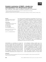
Fig. 1. Conserved surface features of the RbAB domain. Conserva-
tion scores were calculated using the alignment of the RbAB domain
from 46 vertebrate species and
CONSURF [74], and figures were gen-
erated using
PYMOL [75]. Structures correspond to the following com-
plexes: (A) RbAB ⁄ E7 (PDB ID: 1GUX); (B) RbAB ⁄ SV40-LT (PDB ID:
1GH6); (C) RbAB ⁄ E2F-TA (PDB ID: 1N4M); and (D) RbAB ⁄ E1A-CR1
(PDB ID: 2R7G). Asterisk: H549Y missense mutation [54]. Arrows
indicate the rotation of the molecule along the x-axis between two
consecutive images. The color scale indicates the residue conserva-
tion score, as calculated using the
CONSURF algorithm.
L. B. Chemes et al. Viral targeting of the retinoblastoma protein
FEBS Journal 277 (2010) 973–988 ª 2010 The Authors Journal compilation ª 2010 FEBS 975
containing the LxCxE motif [8]. The dissociation con-
stant for this interaction was shown, by isothermal
titration calorimetry, to be 190 nm [8,34], but the con-
tribution of other regions of the E7 protein to the
affinity of the E7–RbAB complex has not been
explored in detail. In order to address this issue, we
developed a solution-based assay that allowed us to
perform quantitative and accurate determinations of
stoichiometry and binding affinity at equilibrium, by
measuring the fluorescence anisotropy change upon the
binding of fluorescein isothiocyanate (FITC)-labeled
E7 fragments to RbAB. This assay was used to mea-
sure the binding of different fragments of E7 (corre-
sponding to well-defined structural and functional
domains and to highly conserved sequence motifs) to
RbAB. Figure 2A shows the E7 regions tested.
A representative example of the assay is presented in
Fig. 2B,C, which show the association of E7N [E7(1-
40)] with RbAB. First, the stoichiometry of the reac-
tions was determined by performing titrations at a
high peptide concentration (Figs 2B and S2). The
anisotropy signal increased linearly up to a 1 : 1 molar
ratio, where it reached a constant value indicating the
saturation of all binding sites. This implies that there
is one binding site for the E7(1-40) peptide per RbAB
monomer and that the stoichiometry of the E7(1-40)–
RbAB interaction is 1 : 1. Far-UV CD spectra of the
complexes formed by binding of the RbAB domain to
full-length E7, and to E7(1-40) and E7(40-98) peptides,
revealed that formation of the complex does not
induce significant structural changes in the secondary
structure of the interacting proteins (data not shown).
Figure 2C shows one representative binding curve per-
formed at substoichiometric concentrations, and the
residuals of the fit from which the K
D
value was calcu-
lated. We tested the association of the RbAB domain
with a 43-residue N-terminal fragment of the BPV1 E7
protein (BPVN), which does not contain an LxCxE
motif. This interaction had marginal affinity, which
was approximately 10
6
times lower than that of the
full-length E7 protein (Table 1). Figure 2D summarizes
all binding curves and shows the dynamic range of the
assay, which allowed us to accurately determine subn-
anomolar to micromolar dissociation constants.
The E7(21-29) peptide, comprising the minimal
LxCxE motif (DLYCYEQLN) [8], associated with
RbAB with a K
D
of 4.7 ± 1.7 nm (Table 1), and the
free energy of binding for this interaction was DG =
A
BCD
Fig. 2. Interaction of different E7 fragments with the RbAB domain. (A) Scheme of HPV16 E7. The positions of conserved regions 1, 2 and
3 (CR1, CR2 and CR3) and the E7 fragments used in this study are shown; the LxCxE motif is underlined. Boxes denote the regions con-
tained in each fragment: black, LxCxE motif; dark grey, CKII ⁄ PEST motif; light grey, CR1 helix-forming residues. Circles denote the position
of FITC moieties. (B) Association of E7(1-40) and RbAB at 200 n
M E7(1-40). (C) Association of E7(1-40) and the RbAB domain at 5 nM E7(1-
40). A fit to a 1 : 1 binding model and residuals are shown. The anisotropy value of the free peptide was 0.054 ± 0.001 and the anisotropy
of the complex was 0.124 ± 0.001, indicating that no oligomerization occurred in this binding regime [76]. (D) Representative normalized
binding curves for the different E7 fragments (symbols are as shown in panel A).
Viral targeting of the retinoblastoma protein L. B. Chemes et al.
976 FEBS Journal 277 (2010) 973–988 ª 2010 The Authors Journal compilation ª 2010 FEBS
)11.2 ± 0.2 kcalÆmol
)1
. The E7(16-31) and E7(16-40)
peptides, which contain the LxCxE motif plus addi-
tional neighboring sequences from the CR2 region,
and the E7(1-40) peptide, which comprises both CR1
and CR2, had the same affinity for the RbAB domain
as the E7(21-29) peptide (Table 1). The full-length
HPV16 E7 protein bound to the RbAB domain with a
ten-fold increased affinity when compared with the
E7(21-29) peptide (K
D
= 0.6 ± 0.3 nm), and the free
energy of binding for the interaction between full-
length E7 and the RbAB domain was DG = )12.4 ±
0.3 kcalÆmol
)1
(Table 1). Therefore, our data show
that the LxCxE motif contributes about 90% of the
total binding energy for the HPV16 E7–RbAB interac-
tion, providing quantitative support to previous results
[47]. The CR1 region does not appear to contribute to
RbAB binding within the context of an E7N mono-
mer, as shown by the fact that the E7(16-40) and
E7(1-40) peptides have the same affinity for the RbAB
domain (Table 1). Finally, we showed that the E7
C-terminal domain contributes 1.0 ± 0.4 kcalÆmol
)1
to
the total free energy of binding, enhancing the affinity
of the E7–RbAB complex by ten-fold.
Previous semiquantitative assays have established
that E7 proteins from HPV types highly associated
with the development of cervical cancer (HPV16 and
HPV18) bind to the full-length Rb protein more
strongly than E7 proteins from HPV types associated
with benign lesions (HPV11 and HPV6) [48]. In order
to explore whether similar regions determine the
affinity for RbAB in E7 proteins from high-risk and
low-risk HPV types, we used a competition assay to
measure the association between the RbAB domain
and the E7 proteins from HPV types 16, 18 and 11.
We assembled a stoichiometric complex of RbAB and
FITC-labeled HPV16 E7 or E7(16-31) and displaced
labeled E7 with each of the different full-length pro-
teins or N-terminal domains (Fig. 3A and Table 2).
The HPV11 E7 protein associated with the RbAB
domain 2.0 kcalÆmol
)1
more weakly than the high-risk
HPV16 and HPV18 type counterparts, providing quan-
titative support to previous reports [48]. The N-termi-
nal domain was the main contributor to the binding
affinity of E7 from HPV11, HPV16 and HPV18 for
the RbAB domain (Fig. 3B), pointing to a conserved
mode of interaction.
Phosphorylation of the conserved CKII sites
within the E7 CR2 region increases affinity for
RbAB
The sequences C-terminal to the LxCxE motif in
HPV16 E7 contain two serine residues, S31 and S32,
which are phosphorylated in vitro and in vivo by CKII
[36,37]. These serine residues are followed by a stretch
of acidic amino acids that constitute an S ⁄ TxxD ⁄ E
CKII consensus site. The PESTfind algorithm suggests
Table 1. Determination of binding affinities for the E7–RbAB
complex. The K
D
was calculated by fitting three to five independent
binding curves to a 1 : 1 binding model, as described in the Materials
and methods.
Fragment K
D
(nM) DG
a
(kcalÆmol
)1
)
E7
b
0.6 ± 0.3 )12.4 ± 0.3
E7(21-29) (LXCXE)
b
4.7 ± 1.7 )11.2 ± 0.2
E7(16-31) (LXCXE)
b
5.1 ± 1.3 )11.1 ± 0.3
E7(16-40) (CR2)
b
6.5 ± 1.0 )11.0 ± 0.3
E7(16-40)PP (CR2PP)
b
1.8 ± 0.4 )11.7 ± 0.1
E7(1-40) (E7N)
b
3.0 ± 1.6 )11.4 ± 0.3
E7(1-20) (CR1) 19000 ± 2000 )6.3 ± 0.1
E7(40-98) (E7C) 2700 ± 600 )7.5 ± 0.1
BPV(1-43) (BPV-N) > 400 000 –
a
DG was calculated as DG = )RT *ln(K
D
), with RT = 0.582
kcalÆmol
)1
.
b
The stoichiometry for these complexes was deter-
mined to be 1 : 1 by titrations performed at peptide concentrations
at least 10 times greater than the determined K
D
.
A
B
Fig. 3. The LxCxE motif is the main determinant of binding affinity
in HPV-E7 proteins. (A) Competition experiments with full-length
E7 proteins and a preformed complex of 5 n
M RbAB and 5 nM
FITC-HPV16-E7 protein. Competitor proteins were: BPV-Nter (s);
HPV11-E7 (
); HPV18-E7 ( ); and HPV16-E7 (d). (B) Comparison
of DG values for different E7 full-length proteins (solid bars) and
N-terminal domains (hatched bars). Data are from Table 2.
L. B. Chemes et al. Viral targeting of the retinoblastoma protein
FEBS Journal 277 (2010) 973–988 ª 2010 The Authors Journal compilation ª 2010 FEBS 977
that this site overlaps with a PEST degradation motif
[49]. Figure 4A shows the sequence of the HPV16 E7
CR2 region, indicating the relative positions of the
LxCxE motif, the phosphorylatable serine residues and
the CKII ⁄ PEST region within CR2. Aligned below this
sequence is a sequence logo created from the alignment
of all 56 E7 proteins from genital HPV types (Fig. S3).
The sequence logo clearly shows that serine residues
are nearly as conserved as the LxCxE motif. Inspection
of individual sequences revealed that all 56 E7 proteins
present at least one CKII consensus site between
positions 30 and 34. This region also contained a high
proportion of negatively charged amino acids (D⁄ E),
with 97% of sequences presenting a net charge that
was equal to or lower than -6.
The striking conservation of sequence features
within the CR2 region of E7 underscores the impor-
tance of this region for E7-mediated transformation.
The CKII ⁄ PEST region of E7 and its phosphorylation
have been postulated to play a role in the E7–Rb
protein interaction. Here, we directly tested this
hypothesis by comparing binding to the RbAB domain
for E7(16-40) and for a synthetic E7(16-40) peptide
phosphorylated at serine residues 31 and 32 [E7(16-
40)PP]. Phosphorylation increased the affinity four-
fold (Table 1). The difference in free energy of binding
of both peptides, DDG = )0.7 ± 0.3 kcalÆmol
)1
, was
significant across repeated assays. We further validated
the data by carrying out competition experiments,
where a stoichiometric complex of FITC-labeled
E7(16-31) and the RbAB domain was titrated with
increasing amounts of unlabeled E7(16-40) or E7(16-
40)PP peptides. Competition experiments (Fig. 4B,C)
confirmed a positive contribution of phosphorylation
to RbAB-binding affinity. The difference in free
A
BC
Fig. 4. Phosphorylation of the E7 CR2 region increases the affinity for the RbAB domain. (A) Conservation of sequence features within E7
CR2. Upper panel: sequence of the HPV16 E7(16-40) peptide. The LxCxE motif is underlined, and the position of phosphoryl serine residues
and the CKII ⁄ PEST consensus are marked. Lower panel: sequence logo of the CR2 region from genital E7 proteins. The height of the stack
of letters at each position denotes the level of conservation (the maximum value is 4.32), while the relative proportions of each residue rep-
resents the relative abundance. (B) Competition experiments with CR2 peptides and a preformed complex of 25 n
M RbAB and the 25 nM
FITC–E7(16-31) peptide. Competitor peptides were: E7(16-31) (d), E7(16-40) (.), E7(16-40)PP (s) and BPVN ()). (C) Comparison of DG val-
ues for the E7(16-40) and E7(16-40)PP peptides with those for the E7(16-31) peptide. Data are from Table 1 and from panel B.
Table 2. The LxCxE motif determines binding affinity in distantly
related HPV E7 proteins.
Fragment K
D
(nM) DG
a
(kcalÆmol
)1
) DDG
b
(kcalÆmol
)1
)
Full-length protein
HPV16 E7 2.4 ± 0.2 )11.6 ± 0.05 –
HPV18 E7 7.8 ± 0.5 )10.9 ± 0.04 0.7 ± 0.06
HPV11 E7 108 ± 5 )9.3 ± 0.03 2.3 ± 0.06
N-terminal domain
HPV16 E7 8.6 ± 1.3 )10.8 ± 0.09 –
HPV18 E7 12.2 ± 0.8 )10.6 ± 0.04 0.2 ± 0.1
HPV11 E7 366 ± 25 )8.6 ± 0.04 2.2 ± 0.1
a
DG = )RT*ln(K
D
), with RT = 0.582 kcalÆmol
)1
.
b
DDG was calcu-
lated as DDG = DG ) DG
E716
.
Viral targeting of the retinoblastoma protein L. B. Chemes et al.
978 FEBS Journal 277 (2010) 973–988 ª 2010 The Authors Journal compilation ª 2010 FEBS
energy of binding from competition experiments was
DDG = )1.4 ± 0.2 kcalÆmol
)1
, in agreement with
the direct binding data. Our data demonstrated that
phosphorylation of the CKII ⁄ PEST region contributes
significantly to the RbAB–E7 interaction, enhancing
the affinity by fourfold to 10-fold.
Structural correlates of E7 phosphorylation at the
CKII sites
We have previously shown that E7N is an extended bona
fide structural domain, with regions of dynamic residual
secondary structure in solution. Far-UV CD analyses
showed that HPV16 E7(1-40) displayed an extended PII
structure, which was stabilized by phosphorylation of
serine residues S31 and S32 [23]. We tested the E7 CR2
region for PII content by measuring the far-UV CD
spectra of the E7(16-40) and the E7(16-40)PP peptides
at 5 °C. Both peptides presented a CD spectrum charac-
teristic for a disordered polypeptide with a positive band
at 218 nm, which is characteristic of the PII conforma-
tion (Fig. 5A). PII conformations are sensitive to tem-
perature, with higher temperatures decreasing the
intensity of the 218 and 198 nm peaks. Increasing the
temperature to 85 °C decreased the intensity of both
peaks for both peptides, characteristic for the disruption
of the PII structure (Fig. 5A). The difference spectra (5–
85 °C) clearly showed the induction at 5 °C of the
218 nm peak (Fig. 5A, inset). The denaturant GdmCl is
known to stabilize PII structures [50]. We have previ-
ously shown that the stability of PII conformations can
be estimated from GdmCl titrations, by validating
changes in the CD spectra with NMR measurements of
PII structure [51]. GdmCl increased the 218 nm band in
the E7(16-40)PP peptide, but not in the E7(16-40) pep-
tide (Fig. 5B), suggesting that the E7(16-40)PP peptide
has a higher propensity for PII structure. The titration
of the E7(16-40)PP peptide with GdmCl is shown in
Fig. 5C, along with a fit of the data to a two-state coil-
PII model. The calculated free energy for the coil-PII
equilibrium in 0 m GdmCl is 1.7 ± 0.7 kcalÆmol
)1
,
which corresponds to 4.6 ± 6% of the PII population
in the absence of denaturant. Although the model used
is a crude estimate of the true conformational equilibria
of the peptide, and the estimated parameters have high
errors as a result of noise in the measurements, the
GdmCl titration data clearly show that the E7(16-40)PP
peptide is in equilibrium between coil and PII conforma-
tions. Overall, our data indicate that both peptides from
the HPV16 E7 CR2 region present residual PII structure
in equilibrium with disordered conformations. GdmCl
titrations strongly suggest that phosphorylation modu-
lates the coil–PII equilibrium, increasing the PII propen-
sity of the E7 CR2 region.
The E7 C-terminal domain binds independently
to RbAB
The increased affinity of the full-length E7 protein
compared with the E7 N-terminal domain suggested
that additional regions within the E7 C-terminal
domain contribute to association with the RbAB
domain. In order to test for a direct interaction
between E7C and RbAB, we performed a pull-down
assay with recombinant purified proteins by forming a
stoichiometric complex of His-tagged RbAB with E7
and E7C (Fig. 6A). Most of the full-length E7 protein
(96%), and a fraction of the E7 C-terminal domain
(23%), bound to RbAB at a concentration of 10 lm.
These results confirmed a direct association of the
RbAB domain with both E7 and E7C, and suggested
that the E7C–RbAB interaction was weaker than the
Fig. 5. Phosphorylation increases the PII content of the E7 CR2 region. (A) Far-UV CD spectra of the E7(16-40) (solid line) and the E7(16-
40)PP (dashed line) peptides, performed at 5 °C and 85 °C. Inset: difference spectra (5–85 °C) for the E7(16-40) (solid line) and the E7(16-
40)PP (dashed line) peptides. (B) CD spectra of E7(16-40) and E7(16-40)PP between 0 and 6
M GdmCl. Titration points graphed are
[GdmCl] = 0, 1.2, 1.9, 2.4, 3.2, 3.7, 4.9 and 5.9
M. The curves corresponding to 0 and 5.9 M GdmCl are shown in bold. (C) GdmCl titration of
the E7(16-40)PP peptide. Data were fit to a two-state coil-PII equilibrium (DG
H2O
E7(16-40)PP
= 1.7 ± 0.7 kcalÆmol
)1
; m = 0.44 ± 0.22 kcal
mol
)1
ÆM
)1
).
L. B. Chemes et al. Viral targeting of the retinoblastoma protein
FEBS Journal 277 (2010) 973–988 ª 2010 The Authors Journal compilation ª 2010 FEBS 979
interaction of the full-length E7 protein with the
RbAB domain. Direct titration showed that the E7C–
RbAB complex had a dissociation constant of
2.7 ± 0.6 lm (Table 1, Fig. 6B). Titration with BPVN
E7 yielded a dissociation constant of 400 lm or higher,
supporting the specificity of the E7C–RbAB interac-
tion. A peptide containing the CR2 region of E7 did
not compete with E7C binding, indicating that E7C
does not bind to the RbAB domain at the LxCxE-
binding cleft (Table 1, Fig. 6B).
The E7 CR1 region can form an alpha helix and
binds independently to RbAB
The CR1 region from E1A binds to the RbAB domain
with micromolar affinity (K
D
=1lm) [43] at the
interface between the A and B subdomains, which is
also the binding site for E2F-TA (Fig. 1C,D). The fact
that the E7 and AdE1A CR1 regions have similar
functional properties [42] suggests that E7 CR1 might
also bind to the RbAB domain at the E2F-TA-binding
site.
E1A CR1 and E2F-TA form a six-residue helix in
the bound conformation (Fig. 1C, residues boxed in
Fig. 7A) [16,18,43]. Four AdE1A residues that estab-
lish intermolecular contacts with the RbAB domain
(P41, L43, H44 and L49), and two residues that stabi-
lize the helix by an intramolecular hydrogen bond
(T42 and E45) [43], are conserved in E7 CR1 (E7
residues 6-10 and 15; Fig. 7A). Furthermore, the
AGADIR algorithm [52] suggests that E7 residues 6 to
15 have local helical propensity (data not shown). We
tested whether the E7 CR1 region could form an
a-helix in solution by measuring the far-UV CD spec-
trum of E7(1-20) in the presence of 1,1,1, trifluoroetha-
nol (TFE), which is known to stabilize helical
conformations in peptides [53]. The addition of 60%
TFE induced an a-helix structure in E7(1-20) (Fig. 7B
and inset). A fit of the TFE titration data to a two-
state coil-helix model yielded a free energy for a-helix
formation in 0% TFE of 1.3 ± 0.2 kcalÆmol
)1
, corre-
sponding to a residual a-helix population of 10 ± 4%
in the absence of cosolvent. These results show that
the E7 and E1A CR1 regions have similar conforma-
tional properties.
We tested for the association between E7 CR1 and
the RbAB domain using three different approaches.
First, we used nondenaturing PAGE and FITC-labeled
E7 peptides to test for complex formation (Fig. 7C).
As a positive control, we tested the association
between FITC-labeled E7(1-40) and the RbAB
domain, and as a test for the specificity of the interac-
tion, we used ovalbumin in place of RbAB. Both
E7(1-40) and E7(1-20) formed a complex with RbAB
but not with the control protein ovalbumin, confirming
the specificity of the interactions (Fig. 7C). A pull-
down assay, similar to that performed with E7C, did
not show significant interaction (data not shown), sug-
gesting that the E7(1-20)–RbAB complex has a lower
affinity than the E7C–RbAB complex. Fluorescence
titration gives a dissociation constant of 19 ± 1 lm
for the E7(1-20)–RbAB complex (Fig. 7D and
Table 1). Titration with BPVN yielded a dissociation
constant of 400 lm or higher. The 20-fold higher affin-
ity for the E7(1-20)–RbAB complex supports the speci-
ficity of the interaction. Peptides containing the CR2
region did not compete for the E7(1-20)–RbAB inter-
action (Fig. 7C), which indicates that E7(1-20) does
not bind RbAB at the LxCxE-binding site.
A
B
Fig. 6. E7C binds independently to the RbAB domain. (A)
Pull-down assay for the RbAB–E7C interaction. His-RbAB was incu-
bated with E7 (lanes 3-4) or with E7C (lanes 7-8). Lanes 1-2 and
5-6: control experiments excluding His-RbAB. The labels to the left
of the gel indicate the position of each protein. % E7: percentage
of E7 or E7C protein in the bound (B) and unbound (U) fractions, as
quantified by densitometry (see the Materials and methods). (B)
Binding of E7C to the RbAB domain in solution. Titrations were per-
formed at 1 l
M FITC-E7C; the titrant was RbAB (d, K
D
= 4.8 ± 0.5
l
M), RbAB-E7(16-40) (s, K
D
= 6.4 ± 0.9 lM). A control experiment
was performed using 5 l
M FITC-BPVN (4, K
D
> 400 lM).
Viral targeting of the retinoblastoma protein L. B. Chemes et al.
980 FEBS Journal 277 (2010) 973–988 ª 2010 The Authors Journal compilation ª 2010 FEBS
Discussion
Despite its vast importance as the guardian of the cell
cycle and its clinical relevance in human cancers, struc-
tural and thermodynamic understanding of the mecha-
nisms of action of the Rb protein is far behind that of
p53, the keeper of the genome, mutated in most can-
cers and targeted by the same DNA tumor viruses that
target the Rb protein [44]. In this work, we set out to
investigate the interaction mechanism of the RbAB
pocket domain with one of the paradigmatic viral
oncoproteins, HPV E7, which targets it for degrada-
tion. Precise quantitative assessment of Rb protein
interactions is fundamental for understanding viral-
mediated subversion of cell cycle control and allows
novel shared features of viral and cellular Rb protein
interaction partners to be uncovered.
We measured the contribution of the LxCxE motif
of E7 to be 90% of the total binding free-energy, and
showed that this motif is also the main determinant of
binding for E7 proteins from three prototypical HPV
types (Figs 2 and 3). The free energy of binding for
full-length HPV16 E7 was 1.0 kcalÆmol
)1
higher than
that of the E7N domain, revealing that the E7C
domain contributes a 10-fold increase in affinity
through a dual-contact mode of interaction. Careful
examination of conserved surface patches in the RbAB
domain suggests a putative binding site for E7C,
located in the RbA domain close to the AB cleft (Rb
residues E492, F514, P515, K548 and H549; Fig. 1A,B,
right). This site is nearly as conserved as the LxCxE
cleft, the lysine-rich patch and the E2F-binding site [8],
and a tumorigenic missense mutation, H549Y, has
been described at this surface (Fig. 1A,B, asterisk)
[3,54], which strongly suggests that this is an important
functional surface in the RbAB domain for which
cellular binding partners are likely to be described in
the future [7]. Mutations in this region affect cell cycle
regulation by E7 [41], suggesting that E7 may bind at
this interaction site and displace Rb protein cellular
targets.
The viral transforming proteins AdE1A and
SV40LT, in addition to nine cellular protein targets of
Rb [17] [histone deacetylase (HDAC)1, HDAC2, tran-
scriptional corepressor CtBP-interacting protein (CtIP),
95kDa retinoblastoma-associated protein (RBP95),
ETS-related transcription factor 1 (Elf1), HMG Box
transcription factor 1 (HBP1), kinetochore protein
Hec1 (Hec1), RBP1 and replication factor C subunit 1
(RFC1)], present a putative serine-phosphorylation site
following the LxCxE motif (Fig. 8). In addition, in vivo
phosphorylation of the AdE1A, SV40-LT and HDAC
sites has functional consequences [55–57]. In HPV E7,
A
B
C
D
Fig. 7. The E7 CR1 region forms an a-helix and interacts with the
RbAB domain. (A) Alignment of the E7 CR1 region with the E1A CR1
and E2F1-TA RbAB-binding sites. Bold: residues in AdE1A involved in
complex formation with RbAB and conserved in E7. Asterisks: resi-
dues of E1A and E2F1 involved in the RbAB-binding a-helix [16,43].
(B) TFE titration of the E7(1-20) peptide. Data were fit to a two-
state helix-coil transition model (DG
H2O
= 1.3 ± 0.2 kcalÆmol
)1
;
m = 25 ± 3 kcal mol
)1
ÆM
)1
). Inset: difference spectrum (60–0% TFE)
showing the conformation induced by TFE addition. (C) Interaction
between E7(1-20) and the RbAB domain, determined using nondena-
turing PAGE. Arrows mark the position of peptides ⁄ complexes:
1 = free peptide, 2 = E7(1-40)–RbAB complex, 3 = E7(1-20)–RbAB
complex. (D) Interaction between E7(1-20) and RbAB in solution.
Experiments were performed at 5 l
M E7(1-20). The titrants
were RbAB (d, K
D
=19±1lM; Table 1), RbAB–E7(16-40) ( , K
D
=
26 ± 1 l
M) and RbAB–E7(1-40) (h, K
D
=30±2lM). A control exper-
iment was performed using 5 l
M FITC-BPVN (4, K
D
> 400 lM).
L. B. Chemes et al. Viral targeting of the retinoblastoma protein
FEBS Journal 277 (2010) 973–988 ª 2010 The Authors Journal compilation ª 2010 FEBS 981
phosphorylation is essential for S-phase re-entry of dif-
ferentiating keratinocytes in organotypic raft models
[39,40] and contributes to E7-mediated transformation
[37]. Our results offer the first molecular insight into
the functional role of E7 phosphorylation, by provid-
ing direct evidence that phosphorylation of serines 31
and 32 of HPV16 E7 increases affinity for the RbAB
domain (Fig. 4). The E7 region surrounding these resi-
dues is natively unfolded [23] and presents a high den-
sity of negative charge, which may interact with a
conserved lysine-rich patch contiguous to the LxCxE
cleft [41,58]. We showed that phosphorylation affects
the local conformation of the E7(16-40) fragment,
increasing the PII content of this region and stabilizing
an extended conformation that optimizes binding to
the LxCxE cleft (Fig. 5). PII-coil transitions induced
by phosphorylation in a similar natively unfolded
PEST region can modulate the stability of a protein to
intracellular degradation [51], which could also be the
case for the E7 oncoprotein.
The isolated E7 CR1 region is able to bind to the
RbAB domain in vitro with measurable affinity, pos-
sibly undergoing a coil-to-helix transition (Fig. 7). In
the AdE1A protein, the 70-residue spacer between
the CR1 and CR2 regions allows for the simulta-
neous binding of both motifs at opposite sides of the
same RbAB molecule (Fig. 1A,D) [59]. Our data
clearly show that the E7(16-40) and E7(1-40) pep-
tides have the same affinity for the RbAB domain
(Table 1). This result implies that the HPV E7 CR1
region does not contribute to binding when CR2 is
present, which is probably because of the short
eight-residue spacer separating both binding motifs.
In a complex between the Rb protein and the weak
E7 dimer [29], the CR2 region of one E7 molecule
may bind to the LxCxE cleft, while the CR1 region
of the other E7 molecule binds to the E2F site of an
RbAB monomer. This mode of interaction may
cooperate in the displacement of E2F, as previously
suggested [25,27].
Our results highlight the modular nature of E7 and
its interaction with the RbAB domain (Fig. 8, top). It
has long been recognized that AdE1A and SV40LT
also present multiple interaction modules that bind to
different Rb protein domains [21,22,59]. This is also a
feature of prototypical Rb protein interacting part-
ners, such as E2F1, HDAC, CtIP and EP300 interact-
ing inhibitor of differentiation 1 (EID-1) (Fig. 8,
bottom). The three secondary sites in E7 (E7C, CR1
and the CKII ⁄ PEST region) contribute far less than
expected from their binding energy in isolation (this
work), which suggests that their main role is to finely
tune affinity and to target multiple interaction sur-
faces of the RbAB domain. It will be interesting to
investigate how the action of these modules is inte-
grated with other known E7 interaction sites within
Fig. 8. Interaction modules and affinities of viral and cellular Rb
protein targets. Proteins and affinities reported are from: HPV-E7
[8,25,34,62] and, from this work, AdE1A [15,43,57], SV40LT
[22,34,56], E2F1 [10,16,18], HDAC [14,34,55], CtIP [13,77] and
EID-1 [9,78,79]. The interaction sites in each protein are marked as
boxes. Linear motifs are marked in color: red (LxCxE motif), dark
blue (CKII site), light blue [cyclin-dependent kinase phosphorylation
(Cdk) site], orange (phosphorylatable serine residues), green (helix
motif), violet (PENF motif) and yellow (FxxxV motif). Dark grey,
interactions mediated by globular domains; light grey, interactions
at unknown sites. Structural domains are indicated above each car-
toon, and the Rb domains targeted, and the affinities, are indicated
below each site. When known, the affinities of the full-length pro-
teins and the effects of phosphorylation are indicated.
Viral targeting of the retinoblastoma protein L. B. Chemes et al.
982 FEBS Journal 277 (2010) 973–988 ª 2010 The Authors Journal compilation ª 2010 FEBS
the Rb protein, such as that of E7C with the RbC
domain [25].
Our data bring together the natively unfolded con-
formation of the N-terminal domain of E7 [23,31] and
its functionality. Although E7N is only 40 residues
long, its natively unfolded conformation provides high
flexibility within a short domain and allows the tight
packing of multiple functional motifs separated by
short linkers [60,61]: the LxCxE motif, the CKII ⁄ PEST
region and the helix-forming CR1, similarly to AdE1A
[21,59]. These functional motifs present dynamic resid-
ual structure in solution, which provides a plausible
mechanism to regulate the affinity of E7 for its inter-
acting partners. The LxCxE motif and the CKII ⁄ PEST
region can evolve independently, as shown by their
presence or absence from the E7 protein depending on
papillomavirus type [62–64]. The different combina-
tions of motifs may give rise to the diverse regulatory
strategies of E7 proteins, with an evolutionary speed
not accessible to globular domains [65].
The E7–RbAB interactions described in this work
present a wide range of affinities, from subnanomolar
to micromolar, which are comparable to those
reported for cellular Rb protein partners (Fig. 8). This
quantitative picture highlights the complex balance
established between physiological and pathological
interactions involving the Rb protein [7], and provides
essential information towards a better understanding
of the Rb network of interactions and the events that
determine normal cell cycle regulation or the progres-
sion to cell transformation [66].
Materials and methods
Cloning and protein expression and purification
The RbAB domain (amino acids 372-787) was cloned into
the BamHI ⁄ HindIII sites of the pRSET-A vector as an
N-terminal 6xHis-tagged fusion protein. Expression of the
pRSET–RbAB construct was performed in Bl21(DE3)-
pLysS cells by addition of 1 mm isopropyl thio-b-d-galacto-
side (IPTG) at 28 °C. The RbAB domain was enriched
from the soluble bacterial fraction using a Ni
2+
-nitrilotri-
acetic acid immobilized metal affinity chromatography
resin, and further purified using a sulfate cation exchange
(SP-Sepharose) resin and by gel filtration (on Superdex 75).
The protein was obtained as a conformationally homoge-
neous monomer, as judged by its gel-filtration profile and
by static light-scattering analysis. The full-length HPV16 E7
protein was expressed and purified as previously described
[32]. The HPV18 E7 and HPV11 E7 proteins were cloned
in pGEX-2T and expressed in Bl21(DE3)pLysS cells. Both
proteins were purified using a combination of glutathione
S-transferase (GST)-sepharose and diethylaminoethane res-
ins, followed by gel filtration. The GST tag was cleaved
with thrombin to yield untagged HPV11 E7 and HPV18
E7. The HPV16 E7 C-terminal domain, spanning residues
40-98, was cloned into the BamHI ⁄ HindIII sites of the
pMalC vector to generate pMal–E7C. The pMal–E7C con-
struct was transformed into TB1 cells, and expression was
induced by the addition of 0.4 mm IPTG at 37 °C.
Maltose-binding protein (MBP)–E7C was enriched from
the soluble bacterial fraction using an amilose resin (New
England Biolabs, Hitchin, UK) and MBP was cleaved by
treatment with thrombin (Sigma-Aldrich, St Louis, MO,
USA) at 0.35 NIH standard units per mg of protein.
E7C was separated from MBP by a Source 15Q resin
(Amersham Biosciences, Uppsala, Sweden), refolded as pre-
viously described for full-length E7 [32] and purified by gel
filtration using a Superdex-75 column. The protein was
obtained as a homogeneous dimer, as judged by its gel-
filtration profile. Protein purity was > 95% in all cases, as
judged by SDS ⁄ PAGE, and protein identity was confirmed
by western blots and by MALDI-TOF MS (Bruker Dalton-
ics, Billerica, MA, USA).
Peptide synthesis and labeling
Peptides were synthesized at the W. M. Keck Facility (Yale
University, New Haven, CT), and purified by RP-HPLC.
The phosphorylated E7(16-40) peptide was obtained by
incorporation of phosphoserine, instead of serine, in the
synthesis. The relative molecular mass of each peptide was
confirmed by MALDI-TOF MS (Bruker, Billerica, MA,
USA). Peptides (1–2 mg) were labeled at their N-terminus
with 1.5 mgÆmL
)1
of FITC in 100 mm sodium carbonate
buffer, pH 8, for 2 h at room temperature, and separated
from labeling reagents by a desalting column (PD-10; GE
Healthcare, Uppsala, Sweden) followed by size-exclusion
chromatography or RP-HPLC. The purity of all prepara-
tions was evaluated using MALDI-TOF spectroscopy and
peptides were quantified by UV absorbance at 220 nm in
HCl. The FITC concentration was determined at pH 7 and
495 nm using a molar extinction coefficient of
75 000 m
)1
Æcm
)1
[67].
Equilibrium and competition binding
experiments
Measurements were performed in an AMINCO-Bowman
fluorescence polarimeter assembled in L geometry. Excita-
tion and emission wavelengths were 495 and 520 nm,
with a 4–16 nm bandwidth. All measurements were per-
formed at 20 ± 0.1 °Cin20mm sodium phosphate buf-
fer, pH 7, 200 mm NaCl, 2 mm dithiothreitol and 0.1%
Tween-20. Five fluorescence anisotropy measurements
were averaged using individual G factors and a measure-
ment time of 3 s.
L. B. Chemes et al. Viral targeting of the retinoblastoma protein
FEBS Journal 277 (2010) 973–988 ª 2010 The Authors Journal compilation ª 2010 FEBS 983
Direct titrations
Increasing amounts of a concentrated solution of RbAB
protein were added to a cuvette containing a fixed amount
of FITC-labeled peptide ⁄ protein. Samples were allowed to
equilibrate for at least 2 min in order to ensure that
measurements were performed at steady state. Maximal
dilution was 20%, and data were corrected accordingly.
The assay conditions were carefully controlled such that
independent measurements were reproducible, and no
oxidation or aggregation of reagents occurred. All
binding reactions were reversible, validating the use of
thermodynamic analysis.
Competition experiments
In the first series (Figs 3 and 4), a pre-assembled complex
of RbAB and FITC-labeled competitor peptide ⁄ protein was
titrated with increasing amounts of unlabeled peptide ⁄ pro-
tein, and disruption of the complex was followed by a
decrease in the anisotropy signal of the FITC-labeled spe-
cies. In the second series (Figs 6 and 7), standard binding
curves were performed in the presence of a fixed concentra-
tion of nonlabeled competitor, and the K
D
obtained was
compared with that in the absence of competitor.
Equilibrium binding data analysis and fitting
Direct titrations
The stoichiometry of each reaction was determined by per-
forming titrations at peptide concentrations 10 to 50 times
larger than the determined K
D
, such that the anisotropy
signal increased linearly with increasing titrant concentra-
tions, until saturation was achieved. The stoichiometry was
determined by extrapolation of two linear fits of the initial
and final anisotropy signals. For all reactions tested, full
saturation was achieved at a 1 : 1 molar ratio of titrant,
indicating a 1 : 1 stoichiometry, which validates the use of
a 1 : 1 binding model (Figs 2B and S2).
Binding curves were fit to a model considering a simple
bimolecular association, and the fluorescence anisotropy
(Y) measured at each point in the titration curve was mod-
eled as a linear combination of the anisotropy of free and
bound peptide (Y
F
and Y
B
). The anisotropy values as a
function of RbAB concentration were fit to the quadratic
equation
Y ¼ Y
F
þ
Y
B
À Y
F
ðÞ
P
o
Ã
x þ P
o
þ K
D
ðÞþ
ffiffiffiffiffiffiffiffiffiffiffiffiffiffiffiffiffiffiffiffiffiffiffiffiffiffiffiffiffiffiffiffiffiffiffiffiffiffiffiffiffiffiffiffiffiffiffiffiffiffiffiffiffiffi
x þ P
o
þ K
D
ðÞ
2
À 4 Ã P
o
à xðÞ
q
2
þ C Ã P
o
;
ð1Þ
where K
D
is the dissociation constant and P
o
is the total
peptide concentration. The last term in the equation takes
into account slight aggregation that may take place at higher
protein concentrations. Data fitting was performed using
profit (Quantumsoft, Zurich, Switzerland). The K
D
values
reported in Tables 1 and 2 were obtained by averaging the
values obtained from three to five independent binding
curves.
Competition experiments
Data were fitted considering the binding at equilibrium of
both the unlabeled and the labeled (competitor) peptides
according to a previously described model [68]. The dissoci-
ation constants obtained by this fitting procedure for the
unlabeled peptides differed from those obtained by direct
titrations by two- to three-fold, confirming that FITC label-
ing does not significantly modify the binding properties of
the peptides.
CD measurements
Far-UV CD spectra
Spectra were recorded on a Jasco J-810 spectropolarimeter
equipped with a Peltier temperature-controlled sample com-
partment with a 0.1 cm path-length. Eight to ten CD scans
were averaged, and buffer spectra were subtracted from all
measurements.
TFE and GdmCl titrations
Titrations were performed in 20 mm sodium phosphate buf-
fer, pH 7, containing 2 mm dithiothreitol. Samples were
allowed to reach equilibrium, after which CD spectra at
each titration point were recorded. TFE (ICN, Biomedicals
Inc.) titrations of 50 lm E7(1-20) peptide were performed
at 20 °C by dissolving the peptide in 0–60% TFE (v ⁄ v).
GdmCl titrations were performed at a peptide concentra-
tion of 25 lm and at a temperature of 5 °C by varying the
denaturant concentration from 0 to 6 m.
Data analysis
We used TFE and GdmCl to stabilize a-helix and PII con-
formations respectively. We assumed that PII populations
(for TFE titrations) and a-helix populations (for GdmCl
titrations) did not change during titrations, and considered
the free energy for a-helix formation to depend linearly on
[TFE] ⁄ [water] and PII structures to depend linearly on
GdmCl molar concentration [51]. Molar ellipticity data
were fitted to the following equation to extract the values
for DG and m:
h½¼
h½
TFE;Gdm:Cl
þ h½
water
 exp À DG
TFE;Gdm:Cl
=RT
ÀÁ
1 þ exp ÀDG
TFE;Gdm:Cl
=RTðÞ
; ð2Þ
where [h]
water
and [h]
TFE,Gdm.Cl
are the mean residue elliptic-
ities in water and at high cosolvent concentration, R is the
Viral targeting of the retinoblastoma protein L. B. Chemes et al.
984 FEBS Journal 277 (2010) 973–988 ª 2010 The Authors Journal compilation ª 2010 FEBS
gas constant and T is the temperature. Data fitting was per-
formed using profit (Quantumsoft, Zurich).
Pull-down and nondenaturing PAGE experiments
Pull-down assays were performed by incubating His-RbAB
and E7 or E7C at a concentration of 10 lm in pull-down
buffer (20 mm sodium phosphate, pH 7.0, 0.2 m NaCl and
50 mm imidazole) for 30 min at room temperature, after
which 20 lLofNi
2+
-nitrilotriacetic acid resin was added
and the complexes were spun down. Free proteins were
recovered from the supernatant (unbound fraction) and
complexes were eluted from the resin by incubation in
500 mm imidazole (bound fraction). Fractions were
resolved by 12.5% PAGE, and the percentage of E7 protein
recovered in each fraction was quantified by densitometry
and expressed as a percentage of the total E7 protein. Non-
denaturing PAGE experiments were carried out by incubat-
ing FITC-labeled peptides (50 or 200 lm) with 50 lm
RbAB for 30 min at room temperature, after which com-
plexes were resolved under non-denaturing conditions by
6% PAGE. Free peptides were included to mark the posi-
tion of the free peptide in the gel.
Sequence logos and analysis of conserved RbAB
surfaces
Sequence logos were generated with WebLogo (http://weblo-
go.berkeley.edu/logo.cgi) [69] by using the manually aligned
protein sequences corresponding to the CR2 region of 56 E7
proteins from all genital HPV types (Fig. S3). The height of
the stack of letters at each position reflects sequence conser-
vation and reaches a maximum value of 4.32 for proteins.
The height of each letter within a stack is proportional to its
abundance. Calculation of conservation scores for surface
residues of the RbAB domain was carried out using an
alignment of the RbAB domain from 46 vertebrate species,
performed using ClustalW [70] and manual curation.
Sequences were obtained from PFAM and ENSEMBL
[71,72] and from a Psi-Blast search of UNIPROT [73] using
the human Rb protein as seed (UniProt ID: P06400). Uni-
Prot and Ensembl accession numbers and the alignment are
provided in Fig. S1. This alignment and the PDB files from
RbAB complexes (PDB ID, 1GUX; PDB ID, 1GH6; PDB
ID, 1N4M; PDB ID, 2R7G) were provided to the consurf
server () [74] to calculate conservation
scores, and figures were made using pymol [75].
Acknowledgements
L.B.C is a recipient of a Jose
´
A. Estenssoro predoctoral
fellowship from Fundacio
´
n YPF. C.S is a recipient of a
University of Buenos Aires predoctoral fellowship.
I.E.S. is the recipient of a CONICET postdoctoral
fellowship. G.d.P.G. is a career investigator from CON-
ICET. We acknowledge Diana Wetzler for assistance
with CD data analysis and Guillermo Solovey for assis-
tance with ProFit programming. We thank Liliana
Alonso for careful corrections of the manuscript.
References
1 Friend SH, Bernards R, Rogelj S, Weinberg RA,
Rapaport JM, Albert DM & Dryja TP (1986) A human
DNA segment with properties of the gene that predis-
poses to retinoblastoma and osteosarcoma. Nature 323,
643–646.
2 Lohmann DR (1999) RB1 gene mutations in retinoblas-
toma. Hum Mutat 14, 283–288.
3 Valverde JR, Alonso J, Palacios I & Pestana A (2005)
RB1 gene mutation up-date, a meta-analysis based on
932 reported mutations available in a searchable data-
base. BMC Genet 6, 53.
4 Burkhart DL & Sage J (2008) Cellular mechanisms of
tumour suppression by the retinoblastoma gene. Nat
Rev Cancer 8, 671–682.
5 Hiebert SW, Chellappan SP, Horowitz JM & Nevins
JR (1992) The interaction of RB with E2F coincides
with an inhibition of the transcriptional activity of E2F.
Genes Dev 6, 177–185.
6 Khidr L & Chen PL (2006) RB, the conductor that
orchestrates life, death and differentiation. Oncogene 25,
5210–5219.
7 Morris EJ & Dyson NJ (2001) Retinoblastoma protein
partners. Adv Cancer Res 82, 1–54.
8 Lee JO, Russo AA and Pavletich NP (1998) Structure
of the retinoblastoma tumour-suppressor pocket
domain bound to a peptide from HPV E7. Nature 391,
859–865.
9 Hassler M, Singh S, Yue WW, Luczynski M, Lakbir R,
Sanchez-Sanchez F, Bader T, Pearl LH & Mittnacht S
(2007) Crystal structure of the retinoblastoma protein N
domain provides insight into tumor suppression, ligand
interaction, and holoprotein architecture. Mol Cell 28,
371–385.
10 Rubin SM, Gall AL, Zheng N & Pavletich NP (2005)
Structure of the Rb C-terminal domain bound to E2F1-
DP1: a mechanism for phosphorylation-induced E2F
release. Cell 123, 1093–1106.
11 Chow KN & Dean DC (1996) Domains A and B in the
Rb pocket interact to form a transcriptional repressor
motif. Mol Cell Biol 16, 4862–4868.
12 Sellers WR, Rodgers JW & Kaelin WG Jr (1995) A
potent transrepression domain in the retinoblastoma
protein induces a cell cycle arrest when bound to E2F
sites. Proc Natl Acad Sci USA 92, 11544–11548.
13 Stokes PH, Thompson LS, Marianayagam NJ &
Matthews JM (2007) Dimerization of CtIP may stabilize
L. B. Chemes et al. Viral targeting of the retinoblastoma protein
FEBS Journal 277 (2010) 973–988 ª 2010 The Authors Journal compilation ª 2010 FEBS 985
in vivo interactions with the Retinoblastoma-pocket
domain. Biochem Biophys Res Commun 354, 197–202.
14 Magnaghi-Jaulin L, Groisman R, Naguibneva I, Robin
P, Lorain S, Le Villain JP, Troalen F, Trouche D &
Harel-Bellan A (1998) Retinoblastoma protein represses
transcription by recruiting a histone deacetylase. Nature
391, 601–605.
15 Dyson N, Guida P, McCall C & Harlow E (1992)
Adenovirus E1A makes two distinct contacts with the
retinoblastoma protein. J Virol 66, 4606–4611.
16 Xiao B, Spencer J, Clements A, Ali-Khan N, Mittnacht
S, Broceno C, Burghammer M, Perrakis A,
Marmorstein R & Gamblin SJ (2003) Crystal structure
of the retinoblastoma tumor suppressor protein bound
to E2F and the molecular basis of its regulation. Proc
Natl Acad Sci USA 100, 2363–2368.
17 Dick FA (2007) Structure-function analysis of the reti-
noblastoma tumor suppressor protein – is the whole a
sum of its parts? Cell Div 2, 26.
18 Lee C, Chang JH, Lee HS & Cho Y (2002) Structural
basis for the recognition of the E2F transactivation
domain by the retinoblastoma tumor suppressor. Genes
Dev 16, 3199–3212.
19 McLaughlin-Drubin ME & Munger K (2009) The
human papillomavirus E7 oncoprotein. Virology 384,
335–344.
20 Helt AM, Funk JO & Galloway DA (2002) Inactivation
of both the retinoblastoma tumor suppressor and p21
by the human papillomavirus type 16 E7 oncoprotein is
necessary to inhibit cell cycle arrest in human epithelial
cells. J Virol 76, 10559–10568.
21 Pelka P, Ablack JN, Fonseca GJ, Yousef AF &
Mymryk JS (2008) Intrinsic structural disorder in adeno-
virus E1A: a viral molecular hub linking multiple diverse
processes. J Virol 82, 7252–7263.
22 Sullivan CS & Pipas JM (2002) T antigens of simian
virus 40: molecular chaperones for viral replication and
tumorigenesis. Microbiol Mol Biol Rev 66, 179–202.
23 Garcia-Alai MM, Alonso LG & de Prat-Gay G (2007)
The N-terminal module of HPV16 E7 is an intrinsically
disordered domain that confers conformational and rec-
ognition plasticity to the oncoprotein. Biochemistry 46,
10405–10412.
24 Ohlenschlager O, Seiboth T, Zengerling H, Briese L,
Marchanka A, Ramachandran R, Baum M, Korbas
M, Meyer-Klaucke W, Durst M et al. (2006) Solution
structure of the partially folded high-risk human pap-
illoma virus 45 oncoprotein E7. Oncogene 25, 5953–
5959.
25 Liu X, Clements A, Zhao K & Marmorstein R (2006)
Structure of the human Papillomavirus E7 oncoprotein
and its mechanism for inactivation of the retinoblas-
toma tumor suppressor. J Biol Chem 281, 578–586.
26 Phelps WC, Munger K, Yee CL, Barnes JA & Howley
PM (1992) Structure-function analysis of the human
papillomavirus type 16 E7 oncoprotein. J Virol 66,
2418–2427.
27 Helt AM & Galloway DA (2001) Destabilization of
the retinoblastoma tumor suppressor by human papil-
lomavirus type 16 E7 is not sufficient to overcome
cell cycle arrest in human keratinocytes. J Virol 75,
6737–6747.
28 Alonso LG, Garcia-Alai MM, Smal C, Centeno JM,
Iacono R, Castano E, Gualfetti P & de Prat-Gay G
(2004) The HPV16 E7 viral oncoprotein self-assembles
into defined spherical oligomers. Biochemistry 43, 3310–
3317.
29 Clements A, Johnston K, Mazzarelli JM, Ricciardi RP
& Marmorstein R (2000) Oligomerization properties of
the viral oncoproteins adenovirus E1A and human pap-
illomavirus E7 and their complexes with the retinoblas-
toma protein.
Biochemistry 39, 16033–16045.
30 Dantur K, Alonso L, Castano E, Morelli L, Centeno-
Crowley JM, Vighi S & de Prat-Gay G (2009) Cytosolic
accumulation of HPV16 E7 oligomers supports different
transformation routes for the prototypic viral oncopro-
tein: the amyloid-cancer connection. Int J Cancer 125,
1902–1911.
31 Uversky VN, Roman A, Oldfield CJ & Dunker AK
(2006) Protein intrinsic disorder and human papillom-
aviruses: increased amount of disorder in E6 and E7
oncoproteins from high risk HPVs. J Proteome Res 5,
1829–1842.
32 Alonso LG, Garcia-Alai MM, Nadra AD, Lapena AN,
Almeida FL, Gualfetti P & Prat-Gay GD (2002) High-
risk (HPV16) human papillomavirus E7 oncoprotein is
highly stable and extended, with conformational transi-
tions that could explain its multiple cellular binding
partners. Biochemistry 41, 10510–10518.
33 Huang PS, Patrick DR, Edwards G, Goodhart PJ,
Huber HE, Miles L, Garsky VM, Oliff A & Heimbrook
DC (1993) Protein domains governing interactions
between E2F, the retinoblastoma gene product, and
human papillomavirus type 16 E7 protein. Mol Cell Biol
13, 953–960.
34 Singh M, Krajewski M, Mikolajka A & Holak TA
(2005) Molecular determinants for the complex forma-
tion between the retinoblastoma protein and LXCXE
sequences. J Biol Chem 280, 37868–37876.
35 Patrick DR, Oliff A & Heimbrook DC (1994) Identifi-
cation of a novel retinoblastoma gene product binding
site on human papillomavirus type 16 E7 protein. J Biol
Chem 269, 6842–6850.
36 Smotkin D & Wettstein FO (1987) The major human
papillomavirus protein in cervical cancers is a cytoplas-
mic phosphoprotein. J Virol 61, 1686–1689.
37 Barbosa MS, Edmonds C, Fisher C, Schiller JT, Lowy
DR & Vousden KH (1990) The region of the HPV E7
oncoprotein homologous to adenovirus E1a and Sv40
large T antigen contains separate domains for Rb binding
Viral targeting of the retinoblastoma protein L. B. Chemes et al.
986 FEBS Journal 277 (2010) 973–988 ª 2010 The Authors Journal compilation ª 2010 FEBS
and casein kinase II phosphorylation. EMBO J 9, 153–
160.
38 Storey A, Almond N, Osborn K & Crawford L (1990)
Mutations of the human papillomavirus type 16 E7
gene that affect transformation, transactivation and
phosphorylation by the E7 protein. J Gen Virol 71,
965–970.
39 Chien WM, Parker JN, Schmidt-Grimminger DC,
Broker TR & Chow LT (2000) Casein kinase II phos-
phorylation of the human papillomavirus-18 E7 protein
is critical for promoting S-phase entry. Cell Growth Dif-
fer 11, 425–435.
40 Genovese NJ, Banerjee NS, Broker TR & Chow LT
(2008) Casein kinase II motif-dependent phosphoryla-
tion of human papillomavirus E7 protein promotes
p130 degradation and S-phase induction in differenti-
ated human keratinocytes. J Virol 82, 4862–4873.
41 Dick FA & Dyson NJ (2002) Three regions of the pRB
pocket domain affect its inactivation by human papillo-
mavirus E7 proteins. J Virol 76, 6224–6234.
42 Davies RC & Vousden KH (1992) Functional analysis
of human papillomavirus type 16 E7 by complementa-
tion with adenovirus E1A mutants. J Gen Virol 73,
2135–2139.
43 Liu X & Marmorstein R (2007) Structure of the retino-
blastoma protein bound to adenovirus E1A reveals the
molecular basis for viral oncoprotein inactivation of a
tumor suppressor. Genes Dev 21, 2711–2716.
44 Joerger AC & Fersht AR (2008) Structural biology of the
tumor suppressor p53. Annu Rev Biochem 77, 557–582.
45 Buday L & Downward J (2008) Many faces of Ras acti-
vation. Biochim Biophys Acta 1786 , 178–187.
46 Seeliger MA, Breward SE, Friedler A, Schon O &
Itzhaki LS (2003) Cooperative organization in a macro-
molecular complex. Nat Struct Biol 10, 718–724.
47 Jones RE, Wegrzyn RJ, Patrick DR, Balishin NL,
Vuocolo GA, Riemen MW, Defeo-Jones D, Garsky
VM, Heimbrook DC & Oliff A (1990) Identification of
HPV-16 E7 peptides that are potent antagonists of E7
binding to the retinoblastoma suppressor protein. J Biol
Chem 265, 12782–12785.
48 Munger K, Werness BA, Dyson N, Phelps WC, Harlow
E & Howley PM (1989) Complex formation of human
papillomavirus E7 proteins with the retinoblastoma
tumor suppressor gene product. EMBO J 8, 4099–4105.
49 Rechsteiner M & Rogers SW (1996) PEST sequences
and regulation by proteolysis. Trends Biochem Sci 21,
267–271.
50 Liu Z, Chen K, Ng A, Shi Z, Woody RW &
Kallenbach NR (2004) Solvent dependence of PII confor-
mation in model alanine peptides. J Am Chem Soc 126,
15141–15150.
51 Garcia-Alai MM, Gallo M, Salame M, Wetzler DE,
McBride AA, Paci M, Cicero DO & de Prat-Gay G
(2006) Molecular basis for phosphorylation-dependent,
PEST-mediated protein turnover. Structure 14,
309–319.
52 Munoz V & Serrano L (1994) Elucidating the folding
problem of helical peptides using empirical parameters.
Nat Struct Biol 1, 399–409.
53 Jasanoff A & Fersht AR (1994) Quantitative determina-
tion of helical propensities from trifluoroethanol titra-
tion curves. Biochemistry 33, 2129–2135.
54 Liu Z, Song Y, Bia B & Cowell JK (1995) Germline
mutations in the RB1 gene in patients with hereditary
retinoblastoma. Genes Chromosomes Cancer 14, 277–284.
55 Pflum MK, Tong JK, Lane WS & Schreiber SL (2001)
Histone deacetylase 1 phosphorylation promotes enzy-
matic activity and complex formation. J Biol Chem 276,
47733–47741.
56 Rihs HP, Jans DA, Fan H & Peters R (1991) The rate
of nuclear cytoplasmic protein transport is determined
by the casein kinase II site flanking the nuclear localiza-
tion sequence of the SV40 T-antigen. EMBO J 10, 633–
639.
57 Whalen SG, Marcellus RC, Barbeau D & Branton
PE (1996) Importance of the Ser-132 phosphorylation
site in cell transformation and apoptosis induced by
the adenovirus type 5 E1A protein. J Virol 70, 5373–
5383.
58 Kim HY, Ahn BY & Cho Y (2001) Structural basis for
the inactivation of retinoblastoma tumor suppressor by
SV40 large T antigen. EMBO J 20, 295–304.
59 Ferreon JC, Martinez-Yamout MA, Dyson HJ &
Wright PE (2009) Structural basis for subversion of
cellular control mechanisms by the adenoviral E1A
oncoprotein. Proc Natl Acad Sci USA 106, 13260–
13265.
60 Tokuriki N, Oldfield CJ, Uversky VN, Berezovsky IN
& Tawfik DS (2009) Do viral proteins possess
unique biophysical features? Trends Biochem Sci 34,
53–59.
61 Tompa P, Fuxreiter M, Oldfield CJ, Simon I, Dunker
AK & Uversky VN (2009) Close encounters of the third
kind: disordered domains and the interactions of pro-
teins. Bioessays 31, 328–335.
62 Dong WL, Caldeira S, Sehr P, Pawlita M & Tommasin-
o M (2001) Determination of the binding affinity of
different human papillomavirus E7 proteins for the
tumour suppressor pRb by a plate-binding assay.
J Virol Methods 98, 91–98.
63 Narechania A, Terai M, Chen Z, DeSalle R & Burk
RD (2004) Lack of the canonical pRB-binding domain
in the E7 ORF of artiodactyl papillomaviruses is associ-
ated with the development of fibropapillomas. J Gen
Virol 85, 1243–1250.
64 Fuchs PG, Iftner T, Weninger J & Pfister H (1986)
Epidermodysplasia verruciformis-associated human
papillomavirus 8: genomic sequence and comparative
analysis. J Virol 58, 626–634.
L. B. Chemes et al. Viral targeting of the retinoblastoma protein
FEBS Journal 277 (2010) 973–988 ª 2010 The Authors Journal compilation ª 2010 FEBS 987
65 Beltrao P & Serrano L (2007) Specificity and evolvabili-
ty in eukaryotic protein interaction networks. PLoS
Comput Biol 3, e25.
66 Ferrari R, Pellegrini M, Horwitz GA, Xie W, Berk AJ
& Kurdistani SK (2008) Epigenetic reprogramming by
adenovirus e1a. Science 321, 1086–1088.
67 Hermanson GT (1996) Bioconjugate Techniques.
Academic Press, San Diego, CA.
68 Kuzmic P, Moss ML, Kofron JL & Rich DH (1992)
Fluorescence displacement method for the determina-
tion of receptor-ligand binding constants. Anal Biochem
205, 65–69.
69 Schneider TD & Stephens RM (1990) Sequence logos: a
new way to display consensus sequences. Nucleic Acids
Res 18, 6097–6100.
70 Larkin MA, Blackshields G, Brown NP, Chenna R,
McGettigan PA, McWilliam H, Valentin F, Wallace
IM, Wilm A, Lopez R et al. (2007) Clustal W and
Clustal X version 2.0. Bioinformatics 23, 2947–2948.
71 Hubbard TJ, Aken BL, Ayling S, Ballester B, Beal K,
Bragin E, Brent S, Chen Y, Clapham P, Clarke L et al.
(2009) Ensembl 2009. Nucleic Acids Res 37, D690–D697.
72 Finn RD, Tate J, Mistry J, Coggill PC, Sammut SJ,
Hotz HR, Ceric G, Forslund K, Eddy SR, Sonnham-
mer EL et al. (2008) The Pfam protein families data-
base. Nucleic Acids Res 36, D281–D288.
73 (2009) The Universal Protein Resource (UniProt) 2009.
Nucleic Acids Res 37, D169–D174.
74 Armon A, Graur D & Ben-Tal N (2001) ConSurf: an
algorithmic tool for the identification of functional
regions in proteins by surface mapping of phylogenetic
information. J Mol Biol 307, 447–463.
75 DeLano WL (2002) The PyMOL Molecular Graphics
System. DeLano Scientific, San Carlos, CA, USA.
76 Ferreiro DU, Lima LM, Nadra AD, Alonso LG,
Goldbaum FA & de Prat-Gay G (2000) Distinctive
cognate sequence discrimination, bound DNA confor-
mation, and binding modes in the E2 C-terminal
domains from prototype human and bovine papillom-
aviruses. Biochemistry 39, 14692–14701.
77 Dick FA, Sailhamer E & Dyson NJ (2000) Mutagenesis
of the pRB pocket reveals that cell cycle arrest func-
tions are separable from binding to viral oncoproteins.
Mol Cell Biol 20, 3715–3727.
78 MacLellan WR, Xiao G, Abdellatif M & Schneider
MD (2000) A novel Rb- and p300-binding protein
inhibits transactivation by MyoD. Mol Cell Biol 20,
8903–8915.
79 Miyake S, Sellers WR, Safran M, Li X, Zhao W,
Grossman SR, Gan J, DeCaprio JA, Adams PD &
Kaelin WG Jr (2000) Cells degrade a novel inhibitor of
differentiation with E1A-like properties upon exiting
the cell cycle. Mol Cell Biol 20, 8889–8902.
Supporting information
The following supplementary material is available:
Fig. S1. Alignment of the RbAB domain from 46 ver-
tebrate species.
Fig. S2. Determination of stoichiometry for the
RbAB ⁄ E721-29 (A), RbAB ⁄ E716-31 (B), RbAB ⁄ E71-
98 (C) RbAB ⁄ E716-40 (D) and RbAB ⁄ E716-40PP (E)
interactions.
Fig. S3. Alignment of the E7 CR2 region for 56 E7
proteins from genital HPV types.
This supplementary material can be found in the
online version of this article.
Please note: As a service to our authors and readers,
this journal provides supporting information supplied
by the authors. Such materials are peer-reviewed and
may be re-organized for online delivery, but are not
copy-edited or typeset. Technical support issues arising
from supporting information (other than missing files)
should be addressed to the authors.
Viral targeting of the retinoblastoma protein L. B. Chemes et al.
988 FEBS Journal 277 (2010) 973–988 ª 2010 The Authors Journal compilation ª 2010 FEBS