Báo cáo khoa học: Antimicrobial and conformational studies of the active and inactive analogues of the protegrin-1 peptide docx
Bạn đang xem bản rút gọn của tài liệu. Xem và tải ngay bản đầy đủ của tài liệu tại đây (386.36 KB, 13 trang )
Antimicrobial and conformational studies of the active
and inactive analogues of the protegrin-1 peptide
Sylwia Rodziewicz-Motowidło
1
, Beata Mickiewicz
1
, Katarzyna Greber
2
, Emilia Sikorska
1
, Łukasz
Szultka
2
,El
_
zbieta Kamysz
1
and Wojciech Kamysz
2
1 Faculty of Chemistry, University of Gdan
´
sk, Poland
2 Faculty of Pharmacy, Medical University of Gdan
´
sk, Poland
Introduction
The search for new drugs and target sites has gener-
ated interest in a group of short polypeptides, antimi-
crobial peptides (AMPs), compounds that can combat
bacterial infections [1], and have a broad spectrum of
activity against bacteria, fungi and protozoa [2,3].
AMPs are positively charged molecules (there are also
a few negatively charged ones [4,5]) isolated from a
variety of animals and plants, where they participate
in natural defence mechanisms. AMPs are usually
highly amphipathic molecules with hydrophobic and
hydrophilic moieties segregated into distinct patches
on the molecular surface. Topologically, they can be
grouped into linear and cysteine-bridged peptides.
Further subdivided according to the number of disul-
phide bridges in their structure, cysteine-bridged AMPs
include the protegrins, first isolated in 1993 from
porcine leucocytes [6]. Protegrins are active against
bacteria (Escherichia coli, Staphylococcus aureus [7],
Pseudomonas aeruginosa, Chlamydia trachomatis, Nei-
sseria gonorrhoeae [8]), yeasts (Candida albicans [6])
and viruses (HIV-1 [9]). The protegrin family contains
the following peptides: protegrin-1, protegrin-2, prote-
grin-3, protegrin-4 and protegrin-5 (PG-1–PG-5)
[10]. They are produced from a family of antimicrobial
Keywords
antimicrobial peptides; IB-367; NMR;
protegrin-1 analogues; three-dimensional
structure
Correspondence
S. Rodziewicz-Motowidło, Faculty of
Chemistry, University of Gdan
´
sk,
Sobieskiego 18, 80-952 Gdan
´
sk, Poland
Fax: (+48 58) 523 54 72
Tel: (+48 58) 52 35 430
E-mail:
(Received 11 August 2009, revised 6
November 2009, accepted 9 December
2009)
doi:10.1111/j.1742-4658.2009.07544.x
The natural antimicrobial cationic peptide protegrin-1 displays a broad
spectrum of antimicrobial activity and rapidly kills pathogens by interacting
with their cell membrane. We investigated the structure–activity relation-
ships of three protegrin-1 analogues: IB-367 (RGGLCYCRGRFCVCVGR-
NH
2
), BM-1 (RGLCYCRGRFCVCVG-NH
2
) and BM-2 (RGLCYRPRFV
CVG-NH
2
). Our antimicrobial and antifungal activity studies of these
peptides showed that BM-1 was much more active than IB-367 against
Gram-positive bacteria and fungi, whereas BM-2 was inactive. The BM-1
peptide showed fourfold reduced haemolysis relative to IB-367, an addi-
tional advantage of this peptide. In addition, BM-1 was about 15% cheaper
than IB-367 to synthesize. The absence of two cysteine residues in the BM-2
sequence could be the main reason for its unstable conformation and
antimicrobial inactivity. The solution structures of these peptides were
determined in dimethyl sulphoxide using two-dimensional NMR and
restrained molecular dynamics calculations. IB-367 and BM-1 formed short,
antiparallel, b-hairpin structures connected by a type II¢ b-turn. The
shorter, inactive BM-2 analogue exhibited major and minor conformations
(predominantly unordered) in the NMR spectra and was much more
flexible.
Abbreviations
AMP, antimicrobial peptide; CSI, chemical shift index; MIC, minimal inhibitory concentration; NOESY, nuclear Overhauser effect
spectroscopy; PG-1, protegrin-1; ROESY, rotating frame Overhauser effect spectroscopy.
1010 FEBS Journal 277 (2010) 1010–1022 ª 2010 The Authors Journal compilation ª 2010 FEBS
peptide precursors known as cathelicidins [11], which
are synthesized as the C-terminal portion of a cathelin-
containing proregion. The N-terminal cathelin domain
of the precursor is highly conserved at both the amino
acid and nucleotide sequence levels; this conservatism
is emphasized by both inter- and intraspecific compari-
sons. Conversely, the sequence of the C-terminal pep-
tide carrying the antimicrobial activity is highly
variable. It has been shown that activated porcine neu-
trophils release intact pro-protegrin, which is inactive
as an antimicrobial. It is then processed extracellularly
by elastase to form antimicrobial protegrin [12].
PG-1 is an 18-amino-acid peptide with an amidated
C-terminus. It is thought to form an antiparallel
b-sheet constrained by two disulphide bridges, Cys6–
Cys15 and Cys8–Cys13 (1PG1 [13] and 1ZY6 [14] in
the Protein Data Bank) [7,15]. Containing six posi-
tively charged arginine residues, the native sequence of
PG-1 is highly cationic. The distribution of hydropho-
bic and hydrophilic residues at the peptide surface is a
structural feature required for the cytolytic activity of
PG-1. Structure–activity relationship studies of several
hundred PG-1 analogues were analysed to determine
the role of individual hydrophobic and hydrophilic
residues in antimicrobial activity, i.e. to gain an under-
standing of the relationship between the primary and
secondary structure of protegrins and their microbial
activities, and to identify a protegrin analogue for clin-
ical development [16]. The presence of the b-hairpin
structure was found to be crucial to the antimicrobial
activity of the protegrin. The analogues – linearized or
with amino acid substitutions eliminating hydrogen
bonding across the b-sheet – showed a reduced biologi-
cal activity, especially in the presence of physiological
concentrations of NaCl [17,18]. However, Tam et al.
[19] reported that the activity of nondisulphide-bonded
analogues could be restored by the cyclization of the
peptide backbone. In addition, Harwig et al. [17]
found that, in peptides containing one disulphide
bond, the ‘bullet’ analogue (cysteines one and four
linked by a disulphide bond) had an activity compara-
ble with that of PG-1, whereas the ‘kite’ analogue
(cysteines two and three linked by a disulphide bond)
was less active. In addition, the maintenance of the
amphiphilicity of the b-sheet is essential. The cationic
and hydrophobic clusters in PG-1 have been shown to
be the structural features required for antibacterial
activity [16,20]. Analogues with reduced positive
charge tend to be less active, which may imply that the
binding of a cationic surface to a lipopolysaccharide
is a key mechanistic step in the killing of bacteria
[16,20]. The conformations of the structural features
determining the antimicrobial activity of protegrins
were calculated for 62 peptides and correlated with
their experimental activity against six microbe species
(E. coli, N. gonorrhoeae (Strain F-62), N. gonorrhoeae
(Strain FA-19), Listeria monocytogenes, C. albicans,
P. aeruginosa), as well as their haemolytic and cyto-
toxic activities [20]. Based on broad structure–activity
relationship studies, only one analogue of PG-1 – IB-
367 – was selected for clinical development as a topical
agent to prevent the oral mucositis associated with
cancer therapy [16,21,22]. It displays a broad spectrum
of activity, rapid microbicidal action and limited
ability to induce resistance. IB-367 kills a broad spec-
trum of bacteria and fungi, including those resistant to
conventional antimicrobial drugs, by attaching to and
destroying the integrity of the lipid cell membrane [23].
In addition, IB-367 demonstrates enhanced bactericidal
and fungicidal activity compared with that of native
protegrins [16,24]. It could therefore be an interesting
compound for the inhibition of bacterial translocation
and endotoxin release in obstructive jaundice [25].
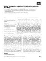
IB-367 is a 17-amino acid peptide with an amidated
C-terminus (Fig. 1): a peptide with such C-terminus
displays greater biological activity than an analogue
without one [16]. Compared with other protegrin pep-
tides of comparable activity, IB-367 has three advanta-
ges in chemical synthesis: (a) most importantly, it
contains an achiral amino acid residue (glycine) at
position 9 in the b-turn – the problem of racemisation
is thus avoided [16]; (b) it has only four arginine resi-
dues compared to six in PG-1 – this is significant, as
arginine is expensive to purchase; (c) it contains 17
amino acids compared to 18 in PG-1.
This paper elucidates the structure-activity relation-
ship in IB-367 and two other analogues of PG-1
Fig. 1. Amino acid sequences of PG-1 and its analogues IB-367,
BM-1 and BM-2.
S. Rodziewicz-Motowidło et al. Conformational studies of protegrin-1 analogues
FEBS Journal 277 (2010) 1010–1022 ª 2010 The Authors Journal compilation ª 2010 FEBS 1011
(BM-1 and BM-2) (see the sequences in Fig. 1) using
2D-NMR spectroscopy and molecular dynamics. The
results of these investigations are compared with pub-
lished structural information about PG-1 and other
antimicrobial peptides in an attempt to understand
which structural features are responsible for the high
biological activity of AMPs.
Results
Design of the new BM-1 and BM-2 analogues
Our aim was to design new analogues of PG-1 with
biological activity comparable with or better than that
of PG-1, but cheaper (about 15% less) to synthesize
chemically on a large scale. On the basis of the struc-
ture–activity relationship studies of other PG-1 ana-
logues (see introductory paragraphs), we designed two
PG-1 analogue sequences. The first analogue, BM-1,
contains four cysteines occupying the nonhydrogen-
bonded sites of the natural b-hairpin core; Gly3, Arg4
and Arg18 were removed from the amino acid
sequence. Although previous studies on protegrin ana-
logues have shown that a positive charge in the loop is
essential for activity [16], we also replaced Arg10 with
a glycine residue (see Fig. 1). We reasoned that, by
removing these residues, we would retain the cationic
nature of the peptide (+4 under physiological condi-
tions), but only in the loop (Arg7, Arg9) and in the
N-terminal fragment (Arg1), not in the C-terminal
fragment. In PG-1, the cationic arginine residues
occupy the loop, N- and C-termini. In this way, we
endeavoured to reduce the cost of synthesis (arginine is
very expensive).
The second analogue, BM-2, was designed in such a
way that the relative importance of the rigidity and
charge at the turn of the b-hairpin in the context of a
single disulphide bridge could be assessed. In BM-2,
we removed Gly3 and Arg18 as in BM-1. We also
removed the two cysteines forming the disulphide bond
proximal to the turn. Although the turn structure of
single disulphide variants of PG-1 is less well defined
and their activity is intermediate relative to that of
PG-1 [16], we wanted to see how removal of the two
cysteines (shortening the amino acid sequence) would
affect their structure and activity. We also wanted to
promote b-hairpin formation by inducing a b-turn,
and so we replaced Arg10 with Pro10 (see Fig. 1).
Although the proline residue is known to have high
frequencies in b-turn formation [26,27], the incorpora-
tion of l-proline at position i + 1 of the reverse
turn prevents b-hairpin formation as a result of
incompatibilities with the intrinsic right-handed twist
of b-strands [28–30]. Presumably, therefore, the BM-2
analogue would have an undefined structure and
reduced antimicrobial activity.
An analogue similar to the BM-2 amino acid
sequence has been suggested previously by Lai et al.
[18]. They designed a peptide in which two proximal
cysteines were replaced with branched residues (threo-
nine) with a high intrinsic preference for b-sheet
conformation, and with a d-proline residue instead of
an arginine residue incorporated at position i +1of
the reverse turn (see peptide 10 in [18]). The amino acid
sequence of BM-2 also has a similar profile to the bullet
variants studied by Harwig et al. [17].
Antimicrobial and haemolytic activity
PG-1, IB-367, BM-1 and BM-2 were characterized
with regard to their antibacterial activity against
Gram-positive bacteria, Gram-negative bacteria and
fungi (see Table 1). All the test organisms were human
pathogens, good selections for the initial screening of
antimicrobial ⁄ antifungal activity. IB-367 and BM-1
exhibited antimicrobial activity against all the bacteria,
but were less active against the fungi (see Table 1). In
contrast, BM-2 showed a marked decrease in activity
against all the bacteria. Surprisingly, BM-1 was far
more active than IB-367 against Gram-positive bacte-
ria and fungi, showing better inhibition than IB-367 of
S. aureus, S. epidermidis and fungi. The antimicrobial
activity of IB-367 and BM-1 against Gram-negative
bacteria was similar, however. Interestingly, BM-2,
inactive against bacteria, displayed a better antifungal
activity than either IB-367 or BM-1 against Aspergil-
lus niger. Comparison of the PG-1 and BM-1 peptides
showed that BM-1 exhibited slightly increased activity
against Gram-positive bacteria and slightly decreased
activity against Gram-negative bacteria and fungi than
did PG-1.
The comparison of the C. albicans minimal inhibi-
tory concentrations (MICs) obtained in this work with
the values found by Barchiesi et al. [31] showed much
lower values found by Barchiesi et al. [31] (2.0–
32 lgÆmL
)1
compared with 256 lgÆmL
)1
in our work).
The differences between these MICs resulted from the
fact that Barchiesi et al. [31] used different experimen-
tal conditions and another strain of C. albicans.
Barchiesi et al. [31] used RPMI 1640 medium, Mops
buffer (pH 7.2) and 50% reduction of the initial inocu-
lum. They also used another strain of C. albicans
ATCC 90029. In our work, we used Sabouraud 5%
glucose medium, pH 7.4 and 99% reduction of the
initial inoculum. In addition, we used the reference
strain of C. albicans (C. albicans ATCC 10231).
Conformational studies of protegrin-1 analogues S. Rodziewicz-Motowidło et al.
1012 FEBS Journal 277 (2010) 1010–1022 ª 2010 The Authors Journal compilation ª 2010 FEBS
The haemolytic activity (see Table 2), using human
red blood cells as targets, was measured for PG-1, IB-
367, BM-1 and BM-2 peptides. The IC
50
results
showed fourfold reduced haemolysis of the BM-1 pep-
tide (32 lgÆmL
)1
) relative to IB-367 (8 lgÆmL
)1
). BM-2
was not cytotoxic when tested against human red
blood cells (> 256 lgÆmL
)1
).
NMR results
The two-dimensional NMR spectra of the peptides,
obtained via the standard sequential assignment meth-
ods developed by Wu
¨
thrich [32], were assigned
(Figs S1 and S2, see Supporting information). The
proton and carbon aC chemical shifts,
3
J
NH–aH
cou-
pling constants and amide-proton temperature coeffi-
cients are given in Tables S1–S4 (see Supporting
information). The
1
H and
13
C NMR chemical shifts of
IB-367 and BM-1 were well dispersed, a property char-
acteristic of b-sheet structures [32]. Moreover, the good
dispersion of the
1
H chemical shifts permitted the iden-
tification of all the protons in the amino acid side-
chains, which was of further help in obtaining the
v torsion angles and in precise structural calculations.
One distinct set of residual proton resonances in all
spectra was displayed for IB-367 and BM-1. The
chemical shifts of IB-367 and BM-1 were very similar
(Tables S1 and S2, see Supporting information), except
for Phe10, which indicates that this phenylalanine resi-
due in BM-1 points in the opposite direction to that in
IB-367. The chemical shifts of IB-367 and BM-1 were
also similar to the previously published data for PG-1
acquired at room temperature in water and in deuter-
ated dimethyl sulphoxide [7,13]. The dispersion of
chemical shifts in BM-2 was not as good as in IB-367
and BM-1; there were also two sets of signals (major
and minor) in the NMR spectra. Analysis of rotating
frame Overhauser effect spectroscopy (ROESY) and
nuclear Overhauser effect spectroscopy (NOESY)
revealed the trans geometry of all the peptide bonds in
IB-367 and BM-1; the geometry of the Arg6–Pro7 pep-
tide bond (major conformation) in BM-2 was cis. The
d
Ha–Ha
(i,i + 1) connectivity characteristic of the cis
peptide bond was seen in the NOESY spectrum, and
the relevant chemical exchange cross-peaks of this
bond were present in the ROESY spectrum, indicating
its cis–trans isomerization (trans geometry in the minor
species). The cis–trans isomerization of the Xaa–Pro
peptide bonds located in the turns is a common
feature of peptides [33]; it is hardly surprising to find
this conformational equilibrium in BM-2. As men-
tioned above, at least two sets of proton resonances
were present in the NMR spectrum of BM-2. Com-
plete analysis, however, would require a separate
conformational analysis for each set of resonances; too
few proton resonances were obtained for the minor
species to perform reliable three-dimensional structural
calculations.
The chemical shift index (CSI), defined as the devia-
tion of the random-coil chemical shift from the experi-
mental value, is a very sensitive indicator of the
secondary structure of peptides and proteins [34]. In
Table 2. Haemolytic activity (lgÆmL
)1
) (IC
50
values – concentra-
tions that cause 50% haemolysis) of the PG-1, IB-367, BM-1 and
BM-2 peptides.
Cells PG-1 IB-367 BM-1 BM-2
Human red blood cells 32 8 32 > 256
Table 1. Antimicrobial activity of the PG-1, IB-367, BM-1 and BM-2 peptides. MBC ⁄ MFC, minimal bactericidal ⁄ fungicidal concentration;
MIC, minimal inhibitory concentration.
Organism
MIC (lgÆmL
)1
) MBC ⁄ MFC (lgÆmL
)1
)
PG-1 IB-367 BM-1 BM-2 PG-1 IB-367 BM-1 BM-2
Gram-positive bacteria
Bacillus subtilis ATCC 6633 2 8 2 128 2 8 2 128
Enterococcus faecalis ATCC 29212 8 8 2 128 8 8 2 256
Rhodococcus equi ATCC 6939 4 8 2 128 8 8 4 256
Staphylococcus aureus ATCC 25923 4 32 8 256 8 64 16 256
Staphylococcus epidermidis ATCC 14990 1 4 1 128 2 4 2 256
Gram-negative bacteria
Escherichia coli ATCC 25922 8 16 32 256 16 16 128 256
Pseudomonas aeruginosa ATCC 27853 4 32 16 > 256 16 64 64 > 256
Fungi
Aspergillus niger ATCC 16404 64 256 128 64 256 256 256 128
Candida albicans ATCC 10231 32 256 128 256 64 256 256 256
S. Rodziewicz-Motowidło et al. Conformational studies of protegrin-1 analogues
FEBS Journal 277 (2010) 1010–1022 ª 2010 The Authors Journal compilation ª 2010 FEBS 1013
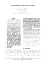
the case of IB-367, most of the amino acids had CSIs
of –1 (Leu4–Arg8 and Arg10–Arg17), a value charac-
teristic of b-sheet structure formation (see Fig. 2). The
CSI pattern of IB-367 resembled that of PG-1, where
the Leu5–Arg10 and Cys13–Gly17 regions exhibited
typical b-strand chemical shifts, whereas the Arg9–
Arg11 region exhibited deviations from the b-strand
chemical shifts [7,13]. The CSIs of the aC atoms in
BM-1 and BM-2 displayed no regularity, indicating a
predominantly random structure.
Figure 2 summarizes the NOE pattern, vicinal cou-
plings
3
J
NH–aH
and temperature coefficients of the
amide protons in the investigated peptides. In IB-367
and BM-1, the presence of strong d
aN
(i,i + 1) and
weak (or absence of) d
MN
(i,i + 1) and d
bN
(i,i +1)
NOE connectivities in the Gly3–Arg8 and Cys14–
Gly16 regions of IB-367 and in the Gly2–Cys6 and
Val12–Val14 regions of BM-1 indicates a significant
population of conformers with dihedral angles in
the b-strand region of (/,w) space [35–37]. In both
peptides, the weak or absent d
NN
(i,i + 1) NOE effects
indicate an unordered structure in the N-terminal frag-
ments and bend structures in the Arg10–Val13 region
of IB-367 and the Cys6–Cys11 region of BM-1. The
d
aa
(5–14; 7–12) of IB-367 and d
aa
(4–13; 6–11) of BM-1
NOEs strongly suggest a disulphide pattern in both
peptides; this results from subsequent calculations.
Several long-range NOEs for IB-367 and BM-1
(Fig. 2) were found, which involved residues from
N- and C-termini, in agreement with a two-stranded
antiparallel b-structure. Moreover, the high values of
the vicinal coupling constants
3
J
NH–aH
(> 9.0 Hz)
in whole peptide sequences and the temperature
coefficients of many amide protons higher than –
3.0 ppbÆK
)1
(Fig. S2A, B, Tables S1 and S2, see
Supporting information) strongly confirmed the pres-
ence of a b-sheet structure in IB-367 and BM-1.
Inspection of the NOE pattern of BM-2 (major con-
formation, Fig. 2C) showed a lack of diagnostic
d
MN
(i,i + 1) NOEs and provided evidence for the
unstructured conformation of this peptide in the mid-
dle part of the peptide (Tyr5–Val10). Some strong
d
aN
(i,i + 1) NOEs in the Gly2–Cys4 and Val10–Gy13
regions suggested a b-structure. In addition, the
lack of hydrogen bonds in BM-2 indicated a flexible
structure.
Finally, no NOE cross-peaks, indicative of oligo-
meric association in solution, could be detected, consis-
tent with the high abundance of positively charged
residues (four arginines in IB-367 and three arginines
in BM-1 and BM-2) in the primary structure of the
peptides.
Structural analysis
Conformational analysis was performed for the three
PG-1 analogues, in an attempt to correlate their struc-
ture and activity in comparison with native PG-1, which
has been shown previously to form a highly stable, rigid
b-hairpin [7,13,18]. IB-367 also adopts a well-defined
b-hairpin structure, as expected from the sequence simi-
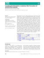
larities with PG-1 (Fig. 3A, B). The 300 structures of
IB-367 were well defined, with an rmsd value of the
Ca atoms of all residues of 2.57 A
˚
, falling to 1.30 A
˚
in
the Cys5–Cys14 region (Table S5, see Supporting
information). The solution structure of IB-367 consisted
of two antiparallel b-strands in the Tyr6–Arg8 and
Phe11–Val13 regions linked by two residues – Gly9 and
Arg10. The Arg8–Phe11 fragment formed a well-defined
type II¢ b-turn, stabilized by a hydrogen bond between
Fig. 2. CSIs relative to sodium 3-(trimethylsilyl)-(2,2,3,3-
2
H
4
)-propionate, summary of intra- and inter-residual NOEs among the backbone NH,
aH and bH, vicinal coupling constants
3
J
HN–Ha
measured in deuterated dimethyl sulphoxide at 22 °C, and temperature coefficients of amide
protons measured in deuterated dimethyl sulphoxide at 22, 25, 27, 30, 32, 35 and 37 °C for IB-367 (A), BM-1 (B), BM-2 major (C) and BM-2
minor (D). CSIs were equal to zero or were not calculated for amino acid residues with open squares. Bar height indicates the strength of
the NOE correlation as strong, medium or weak. Filled squares show
3
J
NH–Ha
coupling constants > 9.0 Hz, and filled circles the temperature
coefficients of amide protons higher than –3.0 ppbÆdeg
)1
.
Conformational studies of protegrin-1 analogues S. Rodziewicz-Motowidło et al.
1014 FEBS Journal 277 (2010) 1010–1022 ª 2010 The Authors Journal compilation ª 2010 FEBS
the CO group of Arg8 and the NH group of Phe11,
found in all the calculated structures. The b-sheet was
strongly stabilized by five regular backbone–backbone
hydrogen bonds – NH(Tyr6)–O(Val13), NH(Arg8)–
O(Phe11), NH(Arg10)–O(Arg8), NH(Phe11)–O(Arg8)
and NH(Val13)–O(Tyr6) – found between the antiparal-
lel strands in most of the calculated structures. All
of these structures adopted a very similar b-hairpin
structure in the middle region of the molecule with
unordered N- and C-termini. In addition, the N- and
C-terminal fragments pointed in opposite directions as a
result of the electrostatic repulsions of Arg1 and Arg17
(see Fig. 3). The side-chains in all the structures were
well defined, particularly in the middle region of the
peptide (Fig. 3A), owing to the presence of numerous
interstrand NOEs. In contrast, the side-chains at the
N- and C-termini displayed large conformational
variability. Two interstrand disulphide bridges adopted
a well-defined, right-handed conformation. IB-367
formed a characteristic amphipathic structure, display-
ing a hydrophobic face formed by the bulked,
hydrophobic residues (Leu4, Tyr6, Phe11, Val13, Val15)
located on the concave surface of the peptide. Two
apolar disulphide bridges and charged Arg17 side-chains
formed the second face of the peptide. Two hydrophilic
regions were located at the two spatial tips of the
molecule, at both termini with Arg1 and Arg17, and in
the turn in the presence of Arg8 and Arg10 (see Figs 3
and 4).
Our conformational studies showed that BM-1
formed a twisted b-sheet structure, similar to that of
native PG-1 and IB-367 (Fig. 3C, D). The structures of
BM-1 were well defined in the backbone, with rmsd
values of the Ca atoms of all residues of 2.60 A
˚
and
1.82 A
˚
in the Cys4–Cys13 region (Table S6, see
Supporting information). The BM-1 structures con-
sisted of two antiparallel b-strands in the Leu3–Cys6
and Cys11–Cys13 regions, linked by Arg7–Phe10
residues. The turn region was formed by a type II¢
b-turn, as in IB-367. b-Hairpin stabilization was guar-
anteed by three regular backbone–backbone hydrogen
bonds – NH(Leu3)–O(Cys13), NH(Cys13)–O(Leu3)
and NH(Cys13)–O(Cys4) – between the disulphide
bridges in most calculated structures. Although the
backbone of BM-1 was well defined, the side-chains
were much less clearly defined than in IB-367
(cf. Fig. 3A, C). Two interstrand disulphide bridges
adopted a right-handed or extended conformation. In
most of the calculated structures, the disulphide bonds
were located at the same peptide face, but, in some, the
disulphide bonds were on the opposite side of the pep-
tide face. Thus, it was difficult to state unequivocally
which residues were located on any given face of the
peptide. BM-1 also formed a characteristic amphipathic
structure, which displayed a hydrophobic face formed
by the hydrophobic residues and the disulphide bridges,
and a second polar face formed by charged Arg1, Arg7
and Arg9 side-chains (see Figs 3 and 4). In general,
BM-1 was more flexible than PG-1 and IB-367, but, as
AB
CD
EF
Fig. 3. Superimposed conformations of the aC atoms of residues
Cys5–Cys13 of IB-367 (130 conformations) (A), Cys4–Cys13 of BM-
1 (93 conformations) (C) and Cys4–Cys11 of BM-2 (95 conforma-
tions) (E). The most populated families of conformations are
shown. Averaged structures of IB-367 (B), BM-1 (D) and BM-2 (F)
peptides. The backbone is shown in ribbon representation, the
side-chains in stick representation. Arginine residues are shown in
blue, disulphide bonds in yellow.
S. Rodziewicz-Motowidło et al. Conformational studies of protegrin-1 analogues
FEBS Journal 277 (2010) 1010–1022 ª 2010 The Authors Journal compilation ª 2010 FEBS 1015
in the last two peptides, two hydrophilic regions were
located at the two spatial tips of the molecule, at the
N-terminus with Arg1 and in the turn in the presence
of Arg7 and Arg9 (see Figs 3 and 4).
The shorter analogue, BM-2, was the most flexible of
all the peptides studied in this work. BM-2 formed
major and minor conformations – this was easily read
from the NMR spectra. The calculated structures of the
major BM-2 conformation were predominantly unor-
dered, especially in the turn region of the peptide with
the cis peptide bond between Arg6 and Pro7 (Fig. 3E,
F). There were no hydrogen bonds in the calculated
structures and no regularities in the secondary structure.
The rmsd values of all the aC atoms in the 300 calcu-
lated structures were 2.71 A
˚
and 1.54 A
˚
in the Cys4–
Cys11 region (Table S7, see Supporting information).
The conformational ensemble of peptides, deter-
mined by molecular dynamics simulation restraints
from NMR experiments, was clustered into families.
Ten families were found for IB-367 and BM-2, and six
for BM-1, at an rmsd cut-off of 5.0 A
˚
, one of which
was dominant (130 molecules) for IB-367, one (93 mol-
ecules) for BM-1 and two (95 and 65 molecules) for
BM-2. The conformations in all the families for IB-367
and BM-1 had one feature in common: the central part
of the structure was better defined than the C- and
N-terminal parts. The conformational differences
between the structural families of these peptides
applied mainly to the varied structures of the N- and
C-termini. All the features of the structures calculated
for all three peptides were in very good agreement with
the experimental NMR data.
Discussion
The presence of a cationic, amphiphilic b-sheet is key
to maintaining the biological activity and stability of
PG-1-like peptides (IB-367 and BM-1). The highly
flexible analogue without a b-sheet structure (BM-2)
has no antimicrobial activity. Previous studies on prote-
grin variants have also shown that the antimicrobial
activity is highly dependent on b-hairpin stabilization
by disulphide bonds or backbone cyclization [16–
19,38,39]. IB-367 and BM-1 share characteristic physi-
cochemical properties with most antimicrobial peptides,
adopting a b-hairpin-like structure with two disulphide
bridges [39,40]. Sequence alignments revealed great sim-
ilarities between IB-367 or BM-1 and PG-1 from por-
cine leucocytes [6], gomesin from mygalomorph spider
haemocytes [41] and androctonin from scorpions [42].
All have a molecular mass of approximately 2 kDa,
including a rather high percentage (> 20%) of basic
residues. Their three-dimensional structures are stabi-
lized by two internal disulphide bridges. Most have
a broad spectrum of antimicrobial activity against
various microorganisms.
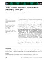
Comparison of the PG-1 structure from the Protein
Data Bank (1PG1 [13] in water and 1ZY6 [14] in
dodecylphosphocholine micelles) with the structures of
IB-367 and BM-1 obtained here reveals several charac-
teristic differences (see Fig. 4). The b-sheet structures
of our peptides are shorter than that of PG-1. The
positive charge in PG-1 in water turns almost the
whole of one side of its structure into a cationic
surface, whereas, in PG-1 in dodecylphosphocholine
micelles, IB-367 and BM-1, the positive charge is
distributed separately. The b-sheet structures of IB-367
and BM-1, as in all the PG-1 peptide family, are char-
acteristically amphipathic, with one surface hydrophilic
and one hydrophobic. Such a structure is essential to
both Gram-positive and Gram-negative antimicrobial
activity [16]. PG-1, IB-367 and BM-1 form four-resi-
due b-turns, but, in PG-1, there is an atypical b-turn,
possibly caused by the presence of Arg10 in position
i + 1 of the turn, whereas, in IB-367 and BM-1, a
type II¢ b-turn is formed. In PG-4, IB-367 and BM-1
analogues, the Arg10 residue is replaced by a glycine
and, in PG-5, by a proline residue. Glycine and proline
residues are better suited than arginine to induce a
canonical b-turn conformation [43]. In addition, there
is only one cationic Arg1 residue on the N-terminus
in BM-1, rather than two arginines (Arg1 on the
N-terminus and Arg18 in PG-1 or Arg17 in IB-367 on
the C-terminus), one at the N- and the other at the
C-terminus; this is sufficient to ensure the cationic
nature of the peptide at its terminus. The structure of
BM-1 is very compact, like the PG-1 structure,
whereas that of IB-367 is more expanded. These struc-
tural features could be responsible for the better
antimicrobial activity of BM-1 than IB-367 against
Gram-positive bacteria.
Natural b-hairpin-like antimicrobial peptides other
than PG-1 (gomesin [41], tachyplesin I [44], polyphemu-
sin I [45] and androctonin [46]), with two disulphide
bridges, are structurally similar to IB-367 and BM-1.
Like PG-1, gomesin and polyphemusin contain 18
amino acids, but tachyplesin contains 17 residues and
androctonin is significantly longer with 25 residues.
Although the spacing of the cysteine residues differs in
these peptides from those studied here, all the molecules
adopt a similar rigid plated b-sheet structure. Androc-
tonin has an unequal number of residues on each
strand between the two bridges – five in the N-terminal
strand and three in the C-terminal strand. This causes
a greater twist in the b-sheet of androctonin com-
pared with the other peptides. Despite such differences,
Conformational studies of protegrin-1 analogues S. Rodziewicz-Motowidło et al.
1016 FEBS Journal 277 (2010) 1010–1022 ª 2010 The Authors Journal compilation ª 2010 FEBS
the rmsd value of the coordinates of the peptide
b-strands (IB-367, BM-1, PG-1, gomesin, tachyplesin I,
polyphemusin I and androctonin), when superimposed
on the backbone atoms, is approximately 6 A
˚
.
Comparison of the hydrophilic ⁄ hydrophobic proper-
ties on the molecule surfaces shows that the structures
of IB-367, BM-1, PG-1, gomesin, tachyplesin I and
polyphemusin I share two highly hydrophilic and
positively charged poles in the N- and C-terminal
regions and in the turn. Androctonin also has a highly
hydrophilic and positively charged turn and N-termi-
nus, but, in contrast, the C-terminus containing
Pro24–Tyr25 is hydrophobic [46]. There is a large
difference in the distribution of hydrophobic ⁄ hydro-
philic potentials on the b-sheet surface between the
tails and the turn. The central portion of IB-367,
BM-1 and PG-1 is particularly hydrophobic, as it con-
tains only apolar residues distributed on either side of
the b-sheet. The b-sheet in gomesin is divided into two
nonequivalent faces: the hydrophobic side-chains are
clustered on the concave face, whereas the two polar
side-chains flanked by the apolar disulphide bridges
are located on the other face [41]. In tachyplesin I and
polyphemusin I, the cationic arginine and lysine
Fig. 4. Structures of PG-1 (Protein Data
Bank code 1PG1 [13]), PG-1 (Protein Data
Bank code 1ZY6 [14]), IB-367, BM-1 and
BM-2. The backbone is shown in ribbon
representation, the side-chains of arginine
(blue) and cysteine (yellow) residues in stick
representation. The electrostatic potential
was calculated on the van der Waals’
surface. Positive potential is shown in blue,
neutral in grey.
S. Rodziewicz-Motowidło et al. Conformational studies of protegrin-1 analogues
FEBS Journal 277 (2010) 1010–1022 ª 2010 The Authors Journal compilation ª 2010 FEBS 1017
residues are also located in the central part of the
b-hairpin structures. In androctonin, the highly twisted
character of the b-sheet does not suggest a clear
dichotomy in the distribution of polar and apolar
residues [46]. Differences in the distribution of hydro-
philic and hydrophobic residues at the surface of the
peptides may indicate different modes of action on the
membrane. This may also account for differences in
the haemolytic activity of the peptides. A better under-
standing of the mode of action of these peptides is
crucial for the development of a new generation of
antibiotics.
It is known that, in the absence of both disulphide
bonds, or even one disulphide bond, the b-sheet struc-
ture is less stable, and the antimicrobial activity is
much reduced [16–19,47,48]. This was also found in
BM-2, which has no b-hairpin structure and is inactive
against bacteria. The single disulphide bond and the
proline residue in position i + 1 of the reverse b-turn
prevent b-hairpin formation and are responsible for
the great flexibility of BM-2 in solution. Lai et al. [18]
obtained similar results with their analogue 12.
Interestingly, BM-2 is more active than IB-367 and
BM-1 against A. niger; the considerable plasticity of
the BM-2 structure may permit better activity against
this fungus.
The current study shows that, with its broad spectrum
of antimicrobial activity, especially against Gram-
positive bacteria, the BM-1 analogue could be a good
molecule for clinical development. Moreover, the BM-1
peptide shows fourfold reduced haemolysis relative
to IB-367, an additional advantage of this peptide.
This peptide is easy to synthesize; it is 15% cheaper to
produce than IB-367.
Materials and methods
Peptide synthesis
The peptides were solid-phase synthesized on Polystyrene
AM-RAM resin (0.76 mmolÆg
)1
, Rapp Polymere, Tu
¨
bingen,
Germany) using 9-fluorenylmethoxycarbonyl chemistry [49],
all the relevant reagents being obtained from Sigma-Aldrich
(Poznan
´
, Poland). The procedure was as follows: (a) 2 and
20 min deprotection steps using 20% piperidine in dimethyl-
formamide in the presence of 1% Triton X-100; (b) the cou-
pling reactions were carried out with the protected amino
acid diluted in dimethylformamide in the presence of 1%
Triton X using diisopropylcarbodiimide as coupling reagent
in 1-hydroxybenzotriazole for 2 h. The completeness of each
coupling reaction was monitored by the chloranil test [50].
If positive, the coupling reaction was repeated using
O-(benzotriazol-1-yl)-N,N,N¢,N¢-tetramethyluronium tetra-
fluoroborate and 1-hydroxybenzotriazole in diisopropyleth-
ylamine and mixed for 2 h. The protected peptidyl resin was
treated with a mixture of 90% trifluoroacetic acid, 2.5%
ethanedithiol, 2.5% phenol, 2.5% water and 2.5% triiso-
propylsilane for 2 h. After cleavage, the solid support was
removed by filtration, and the filtrate was concentrated
under reduced pressure. The cleaved peptide was precipi-
tated with diethyl ether. The linear product was oxidized
with 0.1 m I
2
in CH
3
OH. The peptide was purified by HPLC
on a Knauer K501 two-pump system with a Kromasil C8
column [10 · 250 mm; particle diameter, 5 lm; pore size,
100 A
˚
; flow rate, 5 mLÆmin
)1
; gradient, 10–60% A ⁄ 120 min
(A, 0.1% trifluoroacetic acid in acetonitrile; B, 0.1% aque-
ous trifluoroacetic acid), absorbance at 226 nm]. The result-
ing fractions with a purity of better than 96–98% were
tested by HPLC (Hypersil C18 column, 4.6 · 250 mm). The
peptides were analysed by matrix-assisted laser desorption
ionization-time of flight mass spectrometry.
Organism and antimicrobial assay
The reference strains were supplied by the Polish Collection
of Microorganisms (Polish Academy of Sciences, Institute
of Immunology and Experimental Therapy, Wrocaw,
Poland).
MICs were determined using a broth microdilution
method with Mueller–Hinton broth (Becton Dickinson, Le
Pont de Claix, France) at initial inocula of 5 · 10
5
colony-
forming units (cfu)ÆmL
)1
for bacteria, and Sabouraud 5%
dextrose broth pH 7.4 (Sigma-Aldrich) at initial inocula of
5 · 10
3
cfuÆmL
)1
for fungi, according to the procedures of
the Clinical and Laboratory Standards Institute (formerly
National Committee on Clinical Laboratory Standards).
Polystyrene 96-well plates (Sigma-Aldrich) were incubated
in air at 37 °C for 18 h (bacteria) and at 25 °C for 72 h
(fungi). MIC was taken as the lowest drug concentration at
which observable growth was inhibited. The minimum bac-
tericidal concentration was taken as the lowest concentra-
tion of each drug resulting in > 99.9% reduction of the
initial inoculum. Experiments were performed in triplicate
on three different days.
Haemolytic activity
Freshly collected human blood was washed with NaCl ⁄ P
i
(pH 7.4) until the supernatant became colourless. A suspen-
sion was made of 0.5 mL packed cells in 10 mL of
NaCl ⁄ P
i
. Peptides were dissolved in NaCl ⁄ P
i
and serially
diluted on a 96-well polystyrene microtiter plate. The final
concentration of peptides ranged from 256 to 0.5 lgÆmL
)1
.
Twenty microlitres of cell suspension were added. As a
positive control (100% lysis), a 10% solution of Triton X
was used. After incubation for 4 h at 37 ° C, haemolysis
was observed and compared with the positive control
Conformational studies of protegrin-1 analogues S. Rodziewicz-Motowidło et al.
1018 FEBS Journal 277 (2010) 1010–1022 ª 2010 The Authors Journal compilation ª 2010 FEBS
(Triton X). A positive result (IC
50
) was defined when 50%
of haemolysed red blood cells were taken.
NMR experiments
The NMR samples were prepared by dissolving 6 mg peptide
in 0.5 mL deuterated dimethyl sulphoxide (peptide
concentration, 3mm). The same solvent was used in
conformational studies of native PG-1 [7] to enable a further
three-dimensional structural comparison of the studied
peptides with PG-1. Deuterated dimethyl sulphoxide was
also used in conformational studies of other bioactive
peptides. A sample contained a small amount of trifluoroace-
tic acid in order to downfield shift the vestigial water signal
and to retard amide-proton exchange. Chemical shifts
are given relative to sodium 3-(trimethylsilyl)-(2,2,3,3-
2
H
4
)-
propionate, the internal chemical shift standard.
1
H-NMR spectra (Varian Unity+ 500 NMR spectro-
meter, Varian Inc., Palo Alto, CA, USA) were obtained at
a proton frequency of 500 MHz and the following temper-
atures: 22, 25, 27, 30, 32, 35 and 37 °C. Two-dimensional
spectra, including double-quantum filtered correlation spec-
troscopy, total correlation spectroscopy (60 ms), NOESY
(100 and 200 ms), ROESY (200 ms) and
1
H–
13
C heteronucle-
ar single quantum coherence spectroscopy, were obtained at
22 °C. NMR data were processed with vnmr [51] and analy-
sed with xeasy software [52]. Both ROESY spectra (200 ms)
were compared with the NOESY spectra (200 ms) and no
additional peaks demonstrating the appearance of a spin-
diffusion effect were found. The NOESY spectra showed
better quality and, for this reason, were used for further
calculations. Assignments were carried out according to
standard procedures, including spin-system identification
and sequential assignment [32]. In the case of the one-dimen-
sional NMR spectra, 16 000 data points were collected and a
spectral width of 6 kHz was used. The two-dimensional
homonuclear experiments were measured using a proton
spectral width of 4.5 kHz, collecting 2000 data points.
3
J
NH-aH
vicinal coupling constants were determined by
two-dimensional double-quantum filtered correlation spec-
troscopy experiments. Distance constraints and coupling
constants were used in the habas program [53] of the
dyana package [53] to generate /, w and v
1
dihedral angle
constraints and stereospecific assignments. Dihedral angle
constraints were calculated from the Karplus equation with
A = 6.4, B = )1.4 and C = 1.9 [54].
NOE intensities were determined from the NOESY
(200 ms) spectra of the PG-1 analogues. NOE volumes
were integrated and calibrated with xeasy software [52].
After internal calibration, the cross-peaks from the NOESY
experiments were converted into upper distance limits with
the caliba program of the dyana package [53].
Based on the experimental chemical shifts of aC nuclei,
CSIs were calculated relative to the sodium 3-(trimethylsi-
lyl)-(2,2,3,3-
2
H
4
)-propionate reference [55]. For cysteines,
the reference random-coil chemical shift was not reported
in [55]; hence, CSIs of cysteine amino acid residues were
not calculated.
The temperature dependence of the amide proton chemi-
cal shifts was measured in order to determine whether any
of the amide protons were involved in hydrogen-bonding
interactions. The temperature coefficients (Dd ⁄ DT) of the
amide proton chemical shifts were measured from one-
dimensional NMR spectra for the following temperatures:
22, 25, 27, 30, 32, 35 and 37 °C. About 80% of all hydro-
gen-bonded amides in proteins occur in the range )5to
0 ppbÆdeg
)1
, and their average value is )3.2 ± 2.0
ppbÆdeg
)1
[56]. In our studies, we used the criterion of
hydrogen bond formation by amide protons as a value
higher than )3.0 ppbÆdeg
)1
, and all values more negative
than )3.0 ppbÆdeg
)1
indicated a lack of hydrogen bond
formation.
Structures of the peptides studied
The structures of the peptides studied were determined
with xplor software, Version 3.1 [57], each structure being
produced using distance and torsion angle restraints.
For the xplor three-dimensional structure calculations, the
NOESY experiments provided 558 distance restraints for
IB-367, 427 for BM-1 and 213 for the major conformation
of BM-2. The habas program provided 36, 24 and 21
torsion angles for IB-367, BM-1 and BM-2 (major confor-
mation), respectively. The structures were calculated with
the standard xplor program modules, as well as the
charmm force field [58] in vacuo, starting from a random
structure. For all three molecules, 300 cycles of simulated
annealing were carried out, each with 27 000 iterations of
80 ps with 3 fs steps. The molecule was maintained at
1000 K for 50 ps and annealed at 100 K for 29 ps. In the
last 200 iterations (1 ps), energy was minimized with
Powell’s algorithm [59]. During simulated annealing refine-
ment, the molecule was slowly cooled from 1000 to 100 K
over 30 ps. Finally, 300 energy-minimized conformations
were obtained.
The set of final conformations was clustered using the
graphic molmol program [60]; this was also used to draw,
analyse and display the electrostatic potential on the van
der Waals’ surface. All conformations for each peptide were
divided into families at an rmsd cut-off of 5.0 A
˚
.
Acknowledgements
The authors wish to thank the State Committee for
Scientific Research for grants DS 8440-4-0172-10 and
DS 8452-4-0135-9. This research was conducted using
the resources of the Linux cluster at the Informatics
Centre of the Metropolitan Academic Network (IC
MAN) in Gdan
´
sk, Poland. Beata Mickiewicz expresses
S. Rodziewicz-Motowidło et al. Conformational studies of protegrin-1 analogues
FEBS Journal 277 (2010) 1010–1022 ª 2010 The Authors Journal compilation ª 2010 FEBS 1019
her gratitude to EFS, project No ZPORR ⁄ 2.22 ⁄ II ⁄ 2.6 ⁄
APR ⁄ U ⁄ 2 ⁄ 05.
References
1 Toke O (2005) Antimicrobial peptides: new candidates
in the fight against bacterial infections. Biopolymers 80,
717–735.
2 Hancock REW & Chapple DS (1999) Peptide antibiot-
ics. Antimicrob Agents Chemother 43, 1317–1323.
3 Cho Y, Turner JS, Dinh NN & Lehrer RI (1998)
Activity of protegrins against yeast-phase Candida
albicans. Infect Immun 66, 2486–2493.
4 Brodgen KA, Ackermann M, McCray PB & Tack BF
(2003) Antimicrobial peptides in animals and their role
in host defences. Int J Antimicrob Agents 22, 465–478.
5 Lai R, Liu H, Lee WH & Zhang Y (2002) A novel
proline rich bombesin-related peptide (PR-bombesin)
from toad Bombina maxima . Peptides 23, 437–442.
6 Kokryakov VN, Harwig SS, Panyutich EA, Shevchenko
AA, Aleshina GM, Shamova OV, Korneva HA &
Lehrer RI (1993) Protegrins: leukocyte antimicrobial
peptides that combine features of corticostatic defensins
and tachyplesins. FEBS Lett 327, 231–236.
7 Aumelas AA, Mangoni M, Roumestand C, Chiche L,
Despaux E, Grassy G, Calas B & Chavanieu A (1996)
Synthesis and solution structure of the antimicrobial
peptide protegrin-1. Eur J Biochem 237, 575–583.
8 Qu XD, Harwig SS, Oren A, Shafer WM & Lehrer RI
(1996) Susceptibility of Neisseria gonorrhoeae to
protegrins. Infect Immun 64, 1240–1245.
9 Tamamura H, Murakami T, Horiuchi S, Sugihara K,
Otaka A, Takada W, Ibuka T, Waki M, Yamamoto N
& Fujii N (1995) Synthesis of protegrin-related peptides
and their antibacterial and anti-human immunodefi-
ciency virus activity. Chem Pharm Bull 43, 853–858.
10 Zhao C, Ganz T & Lehrer RI (1995) The structure of
porcine protegrin genes. FEBS Lett 368, 197–202.
11 Gennaro R & Zanetti M (2000) Structural features and
biological activities of the cathelicidin-derived antimi-
crobial peptides. Biopolymers 55 , 31–49.
12 Panyutich A, Shi J, Boutz PL, Zhao C & Ganz T
(1997) Porcine polymorphonuclear leukocytes generate
extracellular microbicidal activity by elastase-mediated
activation of secreted proprotegrins. Infect Immunol 65,
978–985.
13 Fahrner RL, Dieckmann T, Harwig SS, Lehrer RI,
Eisenberg D & Feigon J (1996) Solution structure of
protegrin-1, a broad-spectrum antimicrobial peptide
from porcine leukocytes. J Chem Biol 3, 543–550.
14 Mani R, Tang M, Wu X, Buffy JJ, Waring AJ,
Sherman MA & Hong M (2006) Membrane-bound
dimer structure of a beta-hairpin antimicrobial peptide
from rotational-echo double-resonance solid-state
NMR. Biochemistry 45, 8341–8349.
15 Harwig SS, Swiderek KM, Lee TD & Lehrer RI (1995)
Determination of disulphide bridges in PG-2, an antimi-
crobial peptide from porcine leukocytes. J Pept Sci 1,
207–215.
16 Chen J, Falla TJ, Liu H, Hurst MA, Fujii CA, Mosca
DA, Embree JR, Loury DJ, Radel PA, Cheng Chang C
et al. (2000) Development of protegrins for the
treatment and prevention of oral mucositis: structure–
activity relationships of synthetic protegrin analogues.
Biopolymers 55, 88–98.
17 Harwig SS, Waring A, Yang HJ, Cho Y, Tan L &
Lehrer RI (1996) Intramolecular disulfide bonds
enhance the antimicrobial and lytic activities of
protegrins at physiological sodium chloride
concentrations. Eur J Biochem 240, 352–357.
18 Lai JR, Huck BR, Weisblum B & Gellman SH (2002)
Design of non-cysteine-containing antimicrobial
b-hair-
pins: structure–activity relationship studies with linear
protegrin-1 analogues. Biochemistry 41, 12835–12842.
19 Tam JP, Wu C & Yang JL (2000) Membranolytic
selectivity of cystine-stabilized cyclic protegrins. Eur J
Biochem 267, 3289–3300.
20 Ostberg N & Kaznessis Y (2005) Protegrin structure–
activity relationships: using homology models of syn-
thetic sequences to determine structural characteristics
important for activity. Peptides 26, 197–206.
21 Cole AM & Waring AJ (2002) The role of defensins in
lung biology and therapy. Am J Respir Med 1, 249–259.
22 Bellm L, Giles FJ, Redman R & Yazji S (2002)
Iseganan HCl: a novel antimicrobial agent. Expert Opin
Investig Drugs 11, 1161–1170.
23 Toney JH (2002) Iseganan (IntraBiotics pharmaceuti-
cals). Curr Opin Investig Drugs 3, 225–228.
24 Mosca DA, Hurst MA, So W, Viajar BS, Fuji CA &
Falla TJ (2000) IB-367, a protegrin peptide with in vitro
and in vivo activities against the microflora associated
with oral mucositis. Antimicrob Agents Chemother 44,
1803–1808.
25 Giacometti A, Cirioni O, Ghiselli R, Mocchegiani F,
D’Amato G, Simona Del Prete M, Orlando F,
Kamysz W, Lukasiak J, Saba V et al. (2003)
Administration of protegrin peptide IB-367 to prevent
endotoxin induced mortality in bile duct ligated rats.
Gut 52, 874–878.
26 Wilmot CM & Thornton JM (1988) Analysis and
prediction of the different types of beta-turn in proteins.
J Mol Biol 203 , 221–232.
27 Levitt M (1978) Conformational preferences of amino
acids in globular proteins. Biochemistry 17, 4277–4285.
28 Haque TS, Little JC & Gellman SH (1996) Stereochemi-
cal requirements for b-hairpin formation: model studies
with four-residue peptides and depsipeptides. JAm
Chem Soc 118, 6975–6985.
29 Haque TS & Gellman SH (1997) Insights on b-hairpin
stability in aqueous solution from peptides with
Conformational studies of protegrin-1 analogues S. Rodziewicz-Motowidło et al.
1020 FEBS Journal 277 (2010) 1010–1022 ª 2010 The Authors Journal compilation ª 2010 FEBS
enforced type I¢ and type II¢ b-turns. J Am Chem Soc
119, 2303–2304.
30 Stanger HE & Gellman SH (1998) Rules for antiparallel
b-sheet design: d-Pro-Gly is superior to l-Asn-Gly for
b-hairpin nucleation. J Am Chem Soc 120, 4236–4237.
31 Barchiesi F, Giacometti A, Cirioni O, Arzeni D,
Kamysz W, Silvestri C, Licci A, Marigliano A, Della
Vittoria A, Nadolski P et al. (2007) In-vitro activity of
the synthetic protegrin IB-367 alone and in combination
with antifungal agents against clinical isolates of
Candida spp. J Chemother 19, 514–518.
32 Wu
¨
thrich K (1986) NMR of Proteins and Nucleic Acids.
Wiley Interscience, New York.
33 Tsikaris V, Sakarellos-Daitsiotis M, Tzovaras D &
Sakarellos C (1996) Isomerization of the Xaa–Pro
peptide bond induced by ionic interactions of arginine.
Biopolymers 38, 673–682.
34 Wishart DS, Sykes BD & Richards FM (1992) The
chemical shift index: a fast and simple method for the
assignment of protein secondary structure through
NMR spectroscopy. Biochemistry 31, 1647–1651.
35 Wu
¨
thrich K, Billeter M & Braun W (1984) Polypeptide
secondary structure determination by nuclear
magnetic-resonance observation of short proton
distances. J Mol Biol 180, 715–740.
36 Billeter M, Braun W & Wu
¨
thrich K (1982) Sequential
resonance assignments in protein
1
H nuclear mag-
netic-resonance spectra – computation of sterically
allowed proton distances and statistical-analysis of
proton distances in single-crystal protein conformations.
J Mol Biol 155 , 321–346.
37 Dyson HJ & Wright PE (1991) Defining solution
conformations of small linear peptides. Annu Rev
Biophys Biophys Chem 20, 519–538.
38 Mangoni ME, Aumelas A, Charnet P, Roumestand C,
Chiche L, Despaux E, Grassy G, Calas B & Chavanieu
A (1996) Change in membrane permeability induced by
protegrin 1: implication of disulphide bridges for pore
formation. FEBS Lett 383, 93–98.
39 Mani R, Waring AJ & Hong M (2007) Conformation,
dynamics, and insertion of a noncysteine-containing
protegrin-1 analogue in lipid membranes from solid-
state NMR spectroscopy. Chembiochem 8, 1877–1884.
40 Dimarcq JL, Bulet P, Hetru C & Hoffmann JA (1998)
Cysteine-rich antimicrobial peptides in invertebrates.
Biopolymers 47, 465–477.
41 Mandard N, Bulet P, Caille A, Daffre S & Vovelle F
(2002) The solution structure of gomesin, an
antimicrobial cysteine-rich peptide from the spider.
Eur J Biochem 269, 1190–1198.
42 Ehret-Sabatier L, Loew D, Goyffon M, Fehlbaum P,
Hoffmann JA, van Dorsselaer A & Bulet P (1996)
Characterization of novel cysteine-rich antimicrobial
peptides from scorpion blood. J Biol Chem 271, 29537–
29544.
43 Smith JA & Pease LG (1980) Reverse turns in peptides
and proteins. CRC Crit Rev Biochem 8, 315–399.
44 Laederach A, Andreotti AH & Fulton DB (2002) Solu-
tion and micelle-bound structures of tachyplesin I and
its active aromatic linear derivatives. Biochemistry 41,
12359–12368.
45 Powers JP, Rozek A & Hancock RE (2004) Structure–
activity relationships for the beta-hairpin cationic anti-
microbial peptide polyphemusin I. Biochim Biophys
Acta 1698, 239–250.
46 Mandard N, Sy D, Maufrais C, Bonmatin JM, Bulet P,
Hetru C & Vovelle F (1999) Androctonin, a novel anti-
microbial peptide from scorpion Androctonus australis:
solution structure and molecular dynamics simulations
in the presence of a lipid monolayer. J Biomol Struct
Dyn 17, 367–380.
47 Muhle SA & Tam JP (2001) Design of gram-negative
selective antimicrobial peptides. Biochemistry 40,
5777–5785.
48 Lai JR, Epand RF, Weisblum B, Epand RM &
Gellman SH (2006) Roles of salt and conformation in
the biological and physicochemical behavior of
protegrin-1 and designed analogues: correlation of
antimicrobial, hemolytic, and lipid bilayer-perturbing
activities. Biochemistry 45, 15718–15730.
49 Fields GB & Noble RL (1990) Solid phase peptide
synthesis utilizing 9-fluorenylmethoxycarbonyl amino
acids. Int J Pept Protein Res 35, 161–214.
50 Kaiser E, Colescott RL, Bossinger CD & Cook PI
(1970) Color test for detection of free terminal amino
groups in the solid-phase synthesis of peptides. Anal
Biochem 34, 595–598.
51 Varian (2002). Nuclear Magnetic Resonance Instruments,
VnmrTM Software, Revision 5.3B 1 ⁄ 97. Varian Inc.,
Palo Alto, CA.
52 Bartles C, Xia T, Billeter M, Gu
¨
nter P & Wu
¨
thrich K
(1995) The program XEASY for computer-supported
NMR spectral analysis of biological macromolecules.
J Biomol NMR 4, 1–10.
53 Gu
¨
ntert P, Braun W & Wu
¨
thrich K (1991) Efficient
computation of 3-dimensional protein structures in
solution from nuclear-magnetic-resonance data using
the program DIANA and the supporting programs
CALIBA, HABAS and GLOMSA. J Mol Biol 217,
517–530.
54 Pardi A, Billeter M & Wu
¨
thrich K (1984) Calibration
of the angular dependence of the amide proton-C
alpha proton coupling constants, 3JHN alpha, in a
globular protein. Use of 3JHN alpha for identification
of helical secondary structure. J Mol Biol 180,
741–751.
55 Spera S & Bax A (1991) Empirical correlation between
protein backbone conformation and C.alpha. and
C.beta.
13
C nuclear magnetic resonance chemical shifts.
J Am Chem Soc 113, 5490–5492.
S. Rodziewicz-Motowidło et al. Conformational studies of protegrin-1 analogues
FEBS Journal 277 (2010) 1010–1022 ª 2010 The Authors Journal compilation ª 2010 FEBS 1021
56 Cierpicki T & Otlewski J (2001) Amide proton tempera-
ture coefficients as hydrogen bond indicators in pro-
teins. J Biomol NMR 21, 249–261.
57 Bru
¨
nger AT (1992) The X-PLOR Software Manual,
Version 3.1. Yale University Press, New Haven, CT.
58 Brooks BR, Bruccoleri RE, Olafson BD, States DJ,
Swaminathan S & Karplus M (1983) CHARMM:
a program for macromolecular energy, minimiza-
tion, and dynamics calculations. J Comput Chem 4,
187–217.
59 Powell MJD (1977) Restart procedures for conjugate
gradient method. Math Prog 12, 241–254.
60 Koradi R, Billeter M & Wu
¨
thrich K (1996) MOLMOL:
a program for display and analysis of macromolecular
structures. J Mol Graph 15, 51–55.
Supporting information
The following supplementary material is available:
Fig. S1. The fingerprint region of a NOESY spectrum
(100 ms) of c. 3.0 mm IB-367, BM-1 and BM-2 in deu-
terated dimethyl sulphoxide at 22 °C.
Fig. S2. Amide-a region of an 80 ms mixing time total
correlation spectrum of IB-367, BM-1 and BM-2 in
deuterated dimethyl sulphoxide at 22 °C.
Table S1. Proton and aC chemical shifts and
3
J
NH–Ha
coupling constants of IB-367 measured in deuterated
dimethyl sulphoxide at 22 °C.
Table S2. Proton and aC chemical shifts and
3
J
NH–Ha
coupling constants of BM-1 measured in deuterated
dimethyl sulphoxide at 22 °C.
Table S3. Proton and aC chemical shifts and
3
J
NH–Ha
coupling constants of BM-2 (major conformation)
measured in deuterated dimethyl sulphoxide at 22 °C.
Table S4. Proton and aC chemical shifts and
3
J
NH–Ha
coupling constants of BM-2 (minor conformation)
measured in deuterated dimethyl sulphoxide at 22 °C.
Table S5. Structural statistics for the bundle of all cal-
culated IB-367 structures.
Table S6. Structural statistics for the bundle of all cal-
culated BM-1 structures.
Table S7. Structural statistics for the bundle of all cal-
culated BM-2 (major conformation) structures.
This supplementary material can be found in the
online version of this article.
Please note: As a service to our authors and readers,
this journal provides supporting information supplied
by the authors. Such materials are peer-reviewed and
may be re-organized for online delivery, but are not
copy-edited or typeset. Technical support issues arising
from supporting information (other than missing files)
should be addressed to the authors.
Conformational studies of protegrin-1 analogues S. Rodziewicz-Motowidło et al.
1022 FEBS Journal 277 (2010) 1010–1022 ª 2010 The Authors Journal compilation ª 2010 FEBS