Báo cáo khoa học: AdipoR2 is transcriptionally regulated by ER stress-inducible ATF3 in HepG2 human hepatocyte cells pdf
Bạn đang xem bản rút gọn của tài liệu. Xem và tải ngay bản đầy đủ của tài liệu tại đây (473.8 KB, 14 trang )
AdipoR2 is transcriptionally regulated by ER
stress-inducible ATF3 in HepG2 human hepatocyte cells
In-uk Koh1,2, Joo H. Lim1, Myung K. Joe1, Won H. Kim1, Myeong H. Jung3, Jong B. Yoon2 and
Jihyun Song1
1 Division of Metabolic Disease, Department of Biomedical Science, National Institutes of Health, Seoul, South Korea
2 Department of Biochemistry, College of Science, Yonsei University, Seodaemoon-Gu, Seoul, South Korea
3 School of Korean Medicine, Pusan National University, Yangsan-si, Gyeongnam, South Korea
Keywords
AdipoR2; ATF3; ER stress; insulin
resistance; obesity
Correspondence
M. H. Jung, School of Korean Medicine,
Pusan National University, 30 Beom-eo ri,
Mulguem-eup, Yangsan-si, Gyeongnam
609-735, South Korea
Fax: +82 51 510 8437
Tel: +82 51 510 8468
E-mail:
J. B. Yoon, Department of Biochemistry,
College of Science, Yonsei University, 134
Shinchon-Dong, Seodaemoon-Gu, Seoul
120-749, South Korea
Fax: +82 2 392 3488
Tel: +82 2 2123 2704
E-mail:
J. Song, Division of Metabolic Disease,
Department of Biomedical Science, National
Institutes of Health, 5 Nokbun-dong,
Eunpyung-gu, Seoul 122-701, South Korea
Fax: +82 2 354 1057
Tel: +82 2 380 1530
E-mail:
(Received 22 September 2009, revised 22
February 2010, accepted 9 March 2010)
Adiponectin acts as an insulin-sensitizing adipokine that protects against
obesity-linked metabolic disease, which is generally associated with endoplasmic reticulum (ER) stress. The physiological effects of adiponectin on
energy metabolism in the liver are mediated by its receptors. We found that
the hepatic expression of adiponectin receptor 2 (AdipoR2) was lower, but
the expression of markers of the ER stress pathway, 78 kDa glucose-regulated protein (GRP78) and activating transcription factor 3 (ATF3), was
higher in the liver of ob/ob mice compared with control mice. To investigate the regulation of AdipoR2 by ER stress, we added thapsigargin, an
ER stress inducer, to a human hepatocyte cell line, HepG2. Addition of
the ER stress inducer increased the levels of GRP78 and ATF3, and
decreased that of AdipoR2, whereas addition of a chemical chaperone, 4phenyl butyric acid (PBA), could reverse them. Up- or down-regulation of
ATF3 modulated the AdipoR2 protein levels and AdipoR2 promoter activities. Reporter gene assays using a series of 5¢-deleted AdipoR2 promoter
constructs revealed the location of the repressor element responding to ER
stress and ATF3. In addition, using electrophoretic mobility shift and chromatin immunoprecipitation assays, we identified a region between nucleotides )94 and )86 of the AdipoR2 promoter that functions as a putative
ATF3-binding site in vitro and in vivo. Thus, our findings suggest that the
ER stress-induced decrease in both protein and RNA of AdipoR2 results
from a concomitant increase in expression of ATF3, which may play a role
in the development of obesity-induced insulin resistance and related ER
stress in hepatocytes.
doi:10.1111/j.1742-4658.2010.07646.x
Introduction
Obesity and/or obesity-linked insulin resistance, one of
the key features of type 2 diabetes, are regarded as risk
factors for metabolic syndrome and atherosclerosis [1].
Adiponectin, which is abundantly expressed in adipose
tissue, is a circulating peptide hormone with direct
insulin-sensitizing activity. This adipokine has ameliorative effects on insulin resistance in peripheral tissues,
and plays a central role in the regulation of energy
Abbreviations
AdipoR2, adiponection receptor 2; ATF3, activating transcription factor 3; GRP78, 78 kDa glucose-regulated protein; PBA, 4-phenyl butyric
acid.
2304
FEBS Journal 277 (2010) 2304–2317 ª 2010 The Authors Journal compilation ª 2010 FEBS
In-uk Koh et al.
homeostasis [2–4]. In obesity, adiponectin activity
declines as a result of decreased adiponectin expression
and/or a defect in downstream adiponectin signalling.
The combined actions of genetic factors such as singlenucleotide polymorphisms in the adiponectin gene
and environmental factors such as a high-fat diet and
sedentary lifestyle promoting obesity are thought to be
one of the mechanisms leading to insulin resistance [5].
Several drugs are known to affect adiponectin levels
in the plasma and expression in tissues. Plasma
adiponectin levels increase in response to the PPAR
(peroxisome proliferator-activated receptor) agonists,
thiazolidinediones, but decrease in response to antiHIV drugs and the well-known endoplasmic reticulum
(ER) stressor, thapsigargin. ER stress is a malfunction
of the organelle caused by the influx of immature proteins and/or depletion of calcium ions [6–8], and this
perturbation of ER homeostasis occurs in diet-induced
and genetic models of obesity [9]. Studies of the regulation of obesity-linked insulin resistance have led to
the suggestion that ER stress plays a role in diabetic
insulin resistance [10].
ER dysfunction and the integrated stress response
could lead to abnormal activation of c-Jun N-terminal
kinase (JNK) and/or activating transcription factor 3
(ATF3), which in turn modify the lipogenic pathway,
insulin signaling and the expression levels of genes
related to insulin action, such as insulin receptor substrates and adiponectin [8,9], resulting in obesity-related
insulin resistance [11–13]. In an in vitro model of ER
stress induced by proteasome inhibition, the stress
induced transcription of the transcription factors
GADD153, ATF4 and ATF3, and regulation of lipogenic pathways by the ER stress response was also
shown in hepatocytes [14]. ER stressors such as thapsigargin or tunicamycin reduced insulin signaling by serine phosphorylation of insulin receptor substrate 1
[9,15]. We recently demonstrated that an agent causing
ER stress activated JNK and consequently induced
ATF3, with a reduction in adiponectin transcription [8].
Nakatani et al. [16] identified a molecular chaperone
that protects cells from ER stress and its effect on
insulin sensitivity in the liver. The chemical chaperones
4-phenyl butyric acid (PBA) and tauroursodeoxycholic
acid, which have the ability to decrease ER stress, act
as potent anti-diabetic agents [10]. Because ER is
abundant in hepatocytes, and the liver is a primary
target organ of insulin and adiponectin [3,14,17], we
focused on regulation of the adiponectin receptor
under ER stress-induced conditions in liver cells.
In humans, adiponectin receptors 1 and 2 (AdipoR1
and AdipoR2, respectively) serve as receptors for globular and/or full-length adiponectin. AdipoR1 is ubiqui-
Transcriptional regulation of AdipoR2 by ATF3
tously expressed and is particularly abundant in skeletal
muscle, whereas AdipoR2 is expressed primarily in liver
[17]. These receptors have seven transmembrane
domains, and share 67% amino acid homology. In contrast to G protein-coupled receptors, their N-terminus is
intracellular [5]. Although the intracellular adaptor protein APPL1 (adaptor protein, phosphotyrosine interaction, PH domain and leucine zipper containing 1) has
recently been proposed as a modulator of insulin action
by binding to adiponectin receptors [18], the overall
mechanism of adiponectin signaling is largely unknown.
As expression of adiponectin receptors, as well as adiponectin itself, is known to be decreased in obesity-related
insulin resistance but increased by PPAR agonists [1,19–
21], both adiponectin and its receptors are regarded as
potential therapeutic targets for control of obesitylinked insulin resistance [22,23]. However, there are very
few studies that have examined a specific agent or the
mechanisms responsible for regulating adiponectin
receptor expression [17,24–26].
In this study, we demonstrate that AdipoR2 is negatively regulated in liver cells in response to ER stress or
induced expression of ATF3. In addition, by analyzing
the promoter region of the AdipoR2 gene, we have
identified a putative ATF3-binding site in the 5’ flanking
region, suggesting that direct binding of this transcription factor might negatively regulate AdipoR2
expression. We hypothesize that ER stress-inducible
ATF3 plays an important role in regulating AdipoR2 in
the liver.
Results
Down-regulation of AdipoR2 with up-regulation
of ATF3 expression under increased ER stress
conditions
As shown in previous studies of obesity and ER stress
[9,10], the expression of the ER stress marker proteins
GRP78 and ATF3 was increased 1.4- and 2.0-fold,
respectively, in ob/ob mice compared with lean controls, whereas that of AdipoR2 was decreased 0.8-fold
(Fig. 1A). AdipoR2 is the major hepatic receptor for
adiponectin [5]. ER stress-induced disruption of adiponectin action and increased insulin resistance in hepatocytes could be attributed to down-regulation of
the AdipoR2 level, and thus we studied the effect of
ER stress on the AdipoR2 level in human hepatocytes.
When HepG2 cells were exposed to 1.0 lm thapsigargin, a well-known inducer of ER stress, for 24 h,
both the ER molecular chaperone GRP78 and also
ATF3, which has been shown to repress transcription
of the adiponectin gene in adipocytes [8], were induced.
FEBS Journal 277 (2010) 2304–2317 ª 2010 The Authors Journal compilation ª 2010 FEBS
2305
Transcriptional regulation of AdipoR2 by ATF3
A
In-uk Koh et al.
C57BL6/J
1
2
ob/ob
3
1
2
2.5
3
C57BL6/J
ob/ob
*
Arbitrary units
GRP78
ATF3
AdipoR2
2
1.5
*
1
0.5
Actin
0
GRP78
B
ATF3
C
+
–
+
+
GRP78
ATF3
C
*
4
Thap Thap/PBA
atf3
adipor2
3
* *
gapdh
2
*
2
*
1
AdipoR2
* *
1.5
(AU)
–
–
Arbitrary units
Thap (1.0 µM)
PBA (20 mM)
AdipoR2
1
0
GRP78
Actin
Control
ATF3
Thap
0.5
AdipoR2
0
Thap/PBA
atf3
D
adipor2
E
Adv-ATF3
(MOI)
–
2
5
10
Adv-YFP
Ad YFP
5
–
–
–
(MOI)
ATF3
Thap
siATF3
Neg.RNAi
ATF3
–
–
–
+
–
–
+
+
–
+
–
+
Control
Thap
Thap + siATF3
Thap + Neg.RNAi
2.5
* *
2
* *
1.5
AdipoR2
AdipoR2
Actin
Actin
1
0.5
0
ATF3
AdipoR2
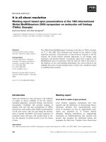
Fig. 1. Changes in expression of AdipoR2 under conditions of ER stress and ATF3 over-expression. (A) Relative levels of GRP78, ATF3 and
AdipoR2 protein in C57BL6/J and ob/ob mice (n = 3 for each group; 30 lg protein per lane). (B) Relative levels of GRP78, ATF3 and AdipoR2
protein in HepG2 cells treated with or without pre-incubation in 20 mM PBA for 24 h prior to treatment with 1.0 lM thapsigargin (30 lg protein per lane; b-actin as control). (C) Relative levels of ATF3 and AdipoR2 mRNA in treated cells. Semi-quantitative RT-PCR analysis was performed using GAPDH as the internal control and the values were normalized to control (untreated). (D) Changes in expression of AdipoR2
after infection with ATF3-expressing adenovirus. HepG2 cells were infected with an adenoviral vector expressing human ATF3 (Adv-ATF3) at
a multiplicity of infection of 2–10 and incubated for 48 h. HepG2 cells infected with Adv-YFP at a multiplicity of infection of 5 were used as
control. (E) Changes in expression of endogenous AdipoR2 upon thapsigargin-induced ER stress with or without silencing of ATF3. siATF3
or Neg.RNAi was introduced to the cells 24 h prior to treatment with 1.0 lM thapsigargin. For western blot analysis, b-actin was used as a
protein loading control. The asterisks indicate a P value < 0.05 for the bracketed comparisons.
Interestingly, the level of AdipoR2 protein was
decreased coincidentally with the increase of ATF3
(Fig. 1B and Table S1).
To determine whether the observed changes in
protein expression were caused by thapsigargin-induced
2306
ER stress, cells were pre-incubated with 20 mm PBA
for 24 h prior to thapsigargin exposure. In cells
pre-incubated with PBA, thapsigargin-induced increases
in GRP78 and ATF3 protein levels did not occur, and
the ER stress-induced decrease in AdipoR2 level was
FEBS Journal 277 (2010) 2304–2317 ª 2010 The Authors Journal compilation ª 2010 FEBS
In-uk Koh et al.
specifically rescued (Fig. 1B, lane 3). In addition, we
measured the mRNA levels for ATF3 and AdipoR2
in HepG2 cells with or without thapsigargin or PBA
treatment, and the results showed a trend similar to the
protein level changes (Fig. 1C).
We next examined the effect of ATF3 over-expression in hepatocytes on changes in the AdipoR2 protein
level. We introduced an adenoviral vector carrying
recombinant ATF3 (Adv-ATF3) into HepG2 cells,
and analyzed the resulting protein expression
using western blotting. As expected, transduction of
Adv-ATF3 resulted in a dose-dependent increase in
the ATF3 protein level. The increase in ATF3 protein
level resulted in a decrease in the AdipoR2 protein
level, but in a non-dose-dependent way (Fig. 1D).
The absence of dose dependence for the reduction of
AdipoR2 may be due to cellular systemic utilization of
the proteins.
In order to further investigate whether ATF3 plays
an important role in ER stress-induced down-regulation of AdipoR2, we assessed the effect of knocking
down ATF3 on the AdipoR2 level. Within 48 h after
introducing siRNA against ATF3 (siATF3) to HepG2
cells, ATF3 was mostly repressed, but the endogenous
AdipoR2 level was relatively increased. The expected
changes in ATF3 and AdipoR2 levels as a result of
thapsigargin treatment were significantly ameliorated
by siATF3 (Fig. 1E and Table S2). These changes were
not observed in cells treated with control siRNA
(Neg.RNAi). These data confirm the negative regulatory effect of transcription factor ATF3 on AdipoR2
levels.
Localization of a repressor element in the
AdipoR2 promoter
To further investigate the changes in AdipoR2 expression as a result of increased ER stress in hepatocytes,
we examined the AdipoR2 promoter activities in
HepG2 cells using the reporter gene construct
AR2P()1974), comprising nucleotides )1974 to +0.
Exposure of cells transfected with AR2P()1974) to
1.0 lm thapsigargin caused an approximately 80%
repression of transcription from the promoter region
of AdipoR2 in 24 h (Fig. 2A).
In addition, to assess the effect of ATF3, a known ER
stress-induced transcriptional repressor in adipocytes
[8], on AdipoR2 regulation in hepatocytes, we measured
the transcriptional activity of the AdipoR2 promoter
when AR2P()1974) was co-transfected with an ATF3expressing vector (ATF3/pcDNA3.1). As for thapsigargin exposure (Fig. 2A), ATF3 expression in HepG2 cells
down-regulated the promoter activity in a dose-depen-
Transcriptional regulation of AdipoR2 by ATF3
dent manner (Fig. 2B). Compared with Neg.RNAi
treatment, silencing of ATF3 reduced the repressive
effect of thapsigargin on the promoter activity of
AdipoR2 (Fig. 2C). To investigate whether ATF3 affects
AdipoR2 expression directly, in other words to locate
the repressor element in the AR2P()1974) promoter
region, as suggested by the above results, we analyzed
the promoter activity of 5¢ serially deleted human
AdipoR2 promoter constructs in pGL3-Basic vector
(Fig. 2D,E). Four plasmid constructs containing
portions of the promoter region of various lengths were
transfected into HepG2 cells with or without co-transfection of the ATF3-expressing vector (ATF3/
pcDNA3.1). As shown in Fig. 2D, ATF3 co-transfection repressed the promoter activities of the transfected
AdipoR2 reporter constructs AR2P()1974), AR2P
()870) and AR2P()343). However, the activity of the
shortest construct AR2P()72) was as low as that in the
control (pGL3) group. In another experiment, various
amounts of ATF3/pcDNA3.1 (0, 0.2 and 0.4 lg) were
co-transfected with AR2P()343) or AR2P()72), and
significant dose-dependent repression by ATF3 was
observed in cells transfected with AR2P()343) but not
in those transfected with AR2P()72) (Fig. 2E). ATF3
co-transfection with this shortest construct AR2P()72)
showed a tendency to decrease the reporter activity
(approximately 50%) but without statistical significance
(P = 0.15) (Table S3 and Fig. S1).
Given that AR2P()72) was not responsive to ATF3,
we presume that more than 72 nucleotides of promoter
region are required for the expression of AdipoR2,
and that at least one of the repressive elements of
AdipoR2 is located between nucleotides )343 and )72.
ATF3 binds to the AdipoR2 promoter in vitro and
in vivo
To confirm that the above results are an effect of
ATF3 on AdipoR2 expression, we searched for a putative repressor binding site by observing sequences without the aid of computer software between nucleotides
)343 and )72 of the human AdipoR2 gene and using
TESS analysis ( />tess) with TRANSFAC database version 6.0 (available
online; ). We isolated
the sequence 5¢-TGCGCGTCA-3¢ located at nucleotides )94 to )86 (Fig. 3A), which is similar to the consensus palindromic ATF/CRE site (TGACGTCA) to
which members of the ATF3 family are known to
homo- or heterodimerize for DNA binding and transcriptional regulation [27].
We performed electrophoretic mobility shift assays
(EMSAs) using nuclear extracts from HepG2 cells and
FEBS Journal 277 (2010) 2304–2317 ª 2010 The Authors Journal compilation ª 2010 FEBS
2307
Transcriptional regulation of AdipoR2 by ATF3
A
B
*
120
80
60
40
20
C
*
*
100
80
60
40
20
0
D
150
+
–
+
–
+
–
*
–
+
+
AR2P(–1974) –
ATF3 –
*
75
50
NS
0
AR2P(–343) –
AR2P(–72) –
ATF3 –
+
–
+
+
Luciferase activity (%)
100
AR2P(–870) –
80
60
40
20
+ AR2P(–1974) +
++ Thap (1 µM) –
siATF3 –
Neg.RNAi –
+
+
–
–
*
120
125
AR2P(
AR2P(–1974) +
100
E
*
25
*
*
0
0
pGL3 +
AR2P(–1974) –
Thap –
*
120
Luciferase activity (%)
100
Luciferase activity (%)
*
120
Luciferase activity (%)
Luciferase activity (%)
In-uk Koh et al.
+
+
+
–
*
+
+
–
+
*
100
80
60
40
NS
NS
20
NS
0
+
–
–
–
+
–
+
–
–
–
–
+
–
–
+
–
–
+
–
–
–
–
+
–
+
–
–
–
+
–
–
–
–
+
+
AR2P(–72) +
(
+
+
–
–
–
AR2P(–343) –
–
–
+
+
+
ATF3
0
0.4
0
0.4
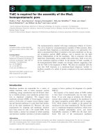
Fig. 2. Changes in the promoter activity of AdipoR2 under conditions of ER stress and ATF3 over-expression. (A) Activity of the AdipoR2 promoter in HepG2 human liver cells with ER stress induction by 1.0 lM thapsigargin for 24 h. The pGL3-Basic-derived reporter construct comprising nucleotides )1974 to +0 of the AdipoR2 promoter [AR2P(–1974)] was transfected into HepG2 cells, followed by treatment with
thapsigargin. (B) Activity of the AdipoR2 promoter in HepG2 cells with ATF3 over-expression by co-transfection of 0.2 or 0.4 lg of ATF3 expression vector. (C) Activity of the AdipoR2 promoter in HepG2 cells upon thapsigargin-induced ER stress with or without silencing of ATF3. siATF3
or Neg.RNAi was introduced to the cells 24 h prior to treatment with 1.0 lM thapsigargin. Luciferase activity values were measured in triplicate
and expressed as arbitrary units. (D) Promoter activities of reporter gene constructs containing 0.6 lg of various lengths of 5¢ deleted fragments
of the promoter region subcloned into the pGL3-Basic plasmid vector and transfected with or without 0.4 lg of ATF3-expressing vector. (E)
ATF3-dose-dependent repression of the promoter activity upon co-transfection of 0, 0.2 or 0.4 lg of ATF3-expressing plasmids with 0.6 lg of
reporter plasmid into HepG2 cells. The asterisks indicate a P value < 0.05 for the bracketed comparisons. NS, not significant.
22 bp radiolabeled DNA probes (nucleotides )79 to
)100) containing the putative ATF3-binding site
5¢-TGCGCGTCA-3¢ to determine whether ATF3
directly interacts with the AdipoR2 promoter. The
EMSA results revealed that this oligonucleotide formed
a DNA–protein complex with the hepatocyte nuclear
extracts (Fig. 3B, C). A specific interaction between
the putative ATF3-binding site and the repressor ATF3
was confirmed by competition with unlabeled oligonu2308
cleotides (Fig. 3C) and by dose-dependent inhibition
by antibody against ATF3 (Fig. 3D). As a negative control for binding of the bZIP (basic leucine zipper) transcription factor ATF3 to the putative binding site, we
used probe ‘X’, containing a sequence that recruits one
of the zinc-finger DNA-binding transcription factor,
also known to interact with the CREB-binding
protein. In the competition EMSA shown in Fig. 3C, a
100 x excess of non-specific probe ‘X’ did not showed
FEBS Journal 277 (2010) 2304–2317 ª 2010 The Authors Journal compilation ª 2010 FEBS
In-uk Koh et al.
Transcriptional regulation of AdipoR2 by ATF3
A
: –94/–86 of promoter
Consensus ATF/CRE
NE: HepG2 cells
Competitor
Non-specific Ab
N
4μg
2μg
1μg
μ
w/o Ab
w
NE: HepG2 cells
o
No Ext.
x100 ATF/CRE
x
x100 Cold
x
Competitor
o
No Comp.
o
No Ext.
x100
x10
No Comp.
No Ext.
NE: HepG2 cells
D
ATF3
A
C
x100 Non-specific
c
x
B
ATF3 Ab
+ Ab
*
Labeled probes
Labeled probes
*
Labeled probes
*
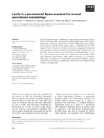
Fig. 3. ATF3 binds to the promoter of AdipoR2 in vitro. (A) Comparison of the sequence of the EMSA probe containing the putative ATF3/
CRE-binding site (TGCGCGTCA) from the promoter of AdipoR2 with that of the palindromic consensus ATF3-binding sequence (TGACGTCA).
(B–D). The putative ATF3-binding site exhibited specific binding with the HepG2 nuclear extract. Nuclear extracts were prepared from HepG2
cells, and 5.0 mg of extract was used in EMSA reactions with 100 ng of radiolabeled double-stranded probe containing the putative ATF3binding site between nucleotides )94 and )86 (B). Competition EMSAs were performed with a 10- or 100-fold excess of the unlabeled wildtype nucleotide )94/)86 probe, a 100-fold excess of the consensus ATF/CRE-binding site sequence, or a 100-fold excess of the non-specific
probe (B,C). Competition assays with ATF-specific antibody (1–4 lg) were also performed (D).
competition in binding to ATF3, but assays using a
100 x excess of cold/unlabeled )94/)86 or the ATF/
CRE positive control probe did show competition with
tested probes containing the putative )94/)86 site, indicating the specificity of this binding assay.
To determine the physiological relevance of ER
stress and/or stress-related expression of ATF3 on
formation of the protein–DNA complex in vitro, we
increased the expression of ATF3 in HepG2 cells by
treatment with 1.0 lm thapsigargin or transduction
with
ATF3-expressing
adenovirus,
Adv-ATF3
(Fig. 4A,B, upper panel). More protein–DNA complex
was formed between radiolabeled oligonucleotides
containing the putative ATF3-binding site, or the
ATF/CRE consensus sequence, and nuclear extracts of
cells when the cells were thapsigargin-treated. Nuclear
extracts of the cells adenovirally over-expressing ATF3
also formed more DNA–protein complex with both
the ATF/CRE consensus sequence and the )94/)86
oligonucleotide probe (Fig. 4A,B).
To further investigate this interaction in vivo, we
validated the predicted ATF3-binding site in the
regulatory region of the human AdipoR2 gene using
chromatin immunoprecipitation (ChIP). This showed
specific in vivo binding of ATF3 to the putative
ATF3-binding element at nucleotides )94/)86 of the
promoter region of AdipoR2 (Fig. 4C). In addition to
the EMSA results (Figs 3 and 4A,B), showing that
recruitment of ATF3 was increased by treatment with
thapsigargin in a time-dependent manner, these results
confirm that the transcription factor ATF3 binds to
the promoter of AdipoR2 both in vitro and in vivo.
Decreased responsiveness as a result of deletion
of the putative ATF3-responsive repressor
element in the nucleotide )343/)72 region of the
promoter
As the putative ATF3-binding site 5¢-TGCGCGTCA-3¢
from the promoter region of human AdipoR2 showed
FEBS Journal 277 (2010) 2304–2317 ª 2010 The Authors Journal compilation ª 2010 FEBS
2309
Transcriptional regulation of AdipoR2 by ATF3
Thap (1.0 µM)
+
Adv-ATF3 Mock
B
–
ATF3
ATF3
β-Actin
β-Actin
ATF/CRE
–94 / –86
Mock
k
Adv-ATF3
No Ext.
E
No Ext.
E
ap
– Tha
ap
+ Tha
No Ext.
E
– Tha
ap
ap
+ Tha
No Ext.
E
ATF/CRE
Mock
k
NE: HepG2 cells
NE: HepG2 cells
Adv-ATF3
A
In-uk Koh et al.
–94 / –86
*
Labeled probes
Labeled probes
*
(–94/–86)
C
Exon1
Thapsigargin (1.0 μM)
–
2 h 4 h 6 h 12 h
–
2 h 4 h 6 h 12 h
IgG
Input
ATF3
Promoter, 323 bp
Exon1, 328 bp
binding ability in EMSA and ChIP experiments, we
generated a mutant promoter construct lacking a
22 bp fragment of the promoter between nucleotides
)94 and )86 (Fig. 5A), to investigate whether this
region responds to ATF3 and ER stress and plays a
role in the transcriptional regulation of AdipoR2.
2310
Fig. 4. Binding of ATF3 to the AdipoR2
promoter region increases under conditions
of ER stress and/or ATF3 over-expression
in vitro and in vivo. (A) ATF3 expression was
increased in HepG2 cells exposed to 1.0 lM
thapsigargin for 24 h compared to control.
The amount of protein–DNA complex
formed with the ATF/CRE consensus
sequence and nucleotide )94/)86 doublestranded oligonucleotide probes was higher
for thapsigargin-exposed samples.
(B) Transfection of an ATF3-expressing
adenoviral vector (Adv-ATF3) resulted in
over-expression of recombinant human
ATF3 in HepG2 cells compared to control
(Adv-YFP). Adenoviral vectors were infected
at a multiplicity of infection of 5 for each
sample. Nuclear extracts from cells overexpressing ATF3 from Adv-ATF3 showed
increased binding affinity for both the
putative ATF3-binding site and the consensus ATF/CRE-binding site. (C) ChIP assays
were performed with anti-ATF3 antibody
(ATF3) or without it (IgG). PCR was used to
amplify immunoprecipitated DNA fragments
from HepG2 cells exposed to 1.0 lM
thapsigargin for 0–12 h, showing a timedependent increase in recruitment of ATF3
to the putative binding element as a result
of ER stress.
The activity of this construct, AR2P()343D), was
then analyzed with or without co-transfection of
ATF3/pcDNA3.1 (Fig. 5B). Co-transfection with
ATF3 dramatically decreased the promoter activity of
the wild-type promoter construct [AR2P()343)] to
one-tenth that of untreated cells, but attenuated the
FEBS Journal 277 (2010) 2304–2317 ª 2010 The Authors Journal compilation ª 2010 FEBS
In-uk Koh et al.
Transcriptional regulation of AdipoR2 by ATF3
(–343 bp)
A
Luc
AR2P(–343Δ)
Luc
AR2P(–343)
Luc
AR2P(–72)
(–343 bp)
(–72 bp)
(–94/–86)
Luciferase activity (%)
120
*
C
*
100
80
60
40
20
AR2P(–343Δ)
AR2P(–343)
ATF3
*
100
80
60
40
20
0
pGL3
*
120
Luciferase activity (%)
B
0
+
–
–
–
+
–
–
+
–
+
–
–
–
+
–
+
–
–
+
–
–
–
+
+
AR2P(–343Δ)
+
+
AR2P(–343)
–
–
+
+
Thapsigargin
–
+
–
+
–
–
Fig. 5. Decreased responsiveness by deletion of the putative ATF3-responsive repressor element in the nucleotide )343/)72 region of the
promoter. (A) Sequences of the deletion mutant construct used in the luciferase reporter assay. In the AR2P()343D) reporter construct, the
putative ATF3/CRE-binding sequence (nucleotides )94/)86: TGCGCGTCA) and six 5¢ and seven 3¢ flanking nucleotides are deleted. (B)
Reduced ATF3-induced repression of the promoter activity was observed for the deletion mutant promoter construct AR2P()343D) lacking
the putative ATF3/CRE-binding site at nucleotides )94/)86. Reporter construct derivatives (0.6 lg) were transfected into HepG2 cells with
or without 0.4 lg of ATF3-expressing vector. (C) Rescued ER stress-induced repression of promoter activity for AR2P()343D). Cells were
treated with 1.0 lM thapsigargin for 24 h to induce ER stress. The asterisks indicate a P value < 0.05 for the bracketed comparisons. All
luciferase assays were performed in triplicate, and error bars indicate the SEM of 3 or 6 experiments.
activity of the mutant construct without the )94/)86
putative binding element [AR2P()343D)] to only half
that of untreated cells (Fig. 5B). As shown in Fig. 5C,
the ER stress inducer thapsigargin had less of a repressor effect on the mutant reporter construct ()56%)
than on the wild-type construct ()85%).
Discussion
Many groups have confirmed the anti-diabetic/insulinsensitizing effect of adiponectin, and thus plasma
adiponectin and its receptors in peripheral organs have
been proposed as therapeutic targets for the treatment
of diabetes and obesity-linked insulin resistance
[2,28,29]. The action of adiponectin is known to be
transduced via regulation of AMP-activated protein
kinase (AMPK) function, and, given the report of a
putative adaptor protein that interacts with adiponectin
receptors, insulin and adiponectin signaling are now
considered to be linked in the peripheral organs of
insulin action, such as the liver and skeletal muscle
[16,30,31]. Despite the fact that the action and plasma
level of adiponectin have been reported to be reduced
in diseases associated with obesity, including peripheral
insulin resistance and related ER stress cascades
[2,3,8,9], the relationship between obesity-related ER
stress and the consequent reduction in adiponectin
action is obscure. We found that hepatic expression of
AdipoR2 was lower but expression of the markers of
the ER stress pathway, GRP78 and ATF3, was higher
in the liver of ob/ob mice compared with control mice.
To determine the molecular mechanisms of this relationship, we studied the regulation of human AdipoR2
in ER stress-induced hepatocytes. ATF3, a member of
the ATF/CREB family of transcription factors, is
known to be a transcriptional repressor that is induced
by many stress signals, including ER stress [31,32], and
has also been proposed to play a role in liver dysfunction involving defects in glucose homeostasis [33]. We
and other investigators have also reported that adiponectin is negatively regulated by ATF3 and by the ER
stress-mediated protein CHOP (C/EBP homologous
protein) under obesity-related hypoxic conditions in
adipocytes [8,34]. In particular, in a transcriptional
context, ATF3 functioned in response to thapsigargininduced ER stress as a negative regulator of adiponectin expression by direct binding to the promoter [8].
These reports imply that a relationship exists between
the decreased transcriptional activity of AdipoR2 and
subsequently-induced ATF3 in ER stress (Fig. 1B).
In thapsigargin-treated hepatocytes, AdipoR2 expression was inversely correlated with the induction of
GRP78 and/or ATF3 by ER stress (Fig. 1). Meanwhile, in cells pre-treated with PBA, the rescued
FEBS Journal 277 (2010) 2304–2317 ª 2010 The Authors Journal compilation ª 2010 FEBS
2311
Transcriptional regulation of AdipoR2 by ATF3
In-uk Koh et al.
AdipoR2 level showed strong support for an ER
stress-based mechanism of AdipoR2 decrease in
human hepatocytes (Fig. 1B). To determine the mechanism of changes in AdipoR2 expression resulting from
ER stress and ATF3 over-expression or silencing in
the liver (Fig. 1B–E), we measured the promoter activity of the AdipoR2 gene. Up- or down-regulation of
ATF3 modulated the AdipoR2 promoter activity
(Fig. 2B,C). By analysing the promoter region of the
AdipoR2 gene, we identified a putative ATF3-binding
sequence (Fig. 3). As shown in Figs 3 and 4, the
decrease in AdipoR2 expression by ATF3 in ER stress
was mediated by this putative sequence which recruited
ATF3 in vitro and in vivo. This result provides an
explanation for the role of ER stress and induced
ATF3 in obesity-linked insulin resistance through regulation of adiponectin action.
These transcription-repressing mechanisms of ER
stress-induced ATF3 have been shown to contribute to
the development of insulin resistance and type 2 diabetes. Insulin receptor substrates 1 and 2 were found to be
repressed by ATF3 in myocytes [35] and pancreatic bcells [11], respectively. The level of the insulin-sensitizing
hormone adiponectin was decreased by ATF3 in adipocytes [8], and the major receptor in hepatocytes, AdipoR2, was also negatively regulated. The above effects
of ATF3 on insulin signaling and glucose homeostasis
involve the action of adiponectin in peripheral tissues.
In particular, given that the cause and effect relationship
between adiponectin and insulin action is not fully
understood, the inappropriate actions of adiponectin in
obesity-linked insulin resistance are described as a
‘vicious cycle’ of adiponectin and insulin resistance [36].
For example, insulin receptor transgenic/knockout mice
exhibit decreased AdipoR2 levels in liver and muscle, as
well as decreased expression of the peroxisome proliferator-activated receptor gamma (PPARc) target genes of
fatty acid oxidation, showing that AdipoR2 defects are
relevant to diabetes susceptibility [37]. In addition,
a decrease in levels of expression of adiponectin receptors was reported to be associated with type 2 diabetes
[21], as well as reductions in plasma adiponectin levels in
various cases associated with insulin resistance [21, 38,
39] and alterations in the adiponectin gene [40–42].
On the other hand, despite decreased responsiveness
to thapsigargin and induced ATF3, a mutant AdipoR2
reporter construct lacking the putative ATF3-binding
site still showed repression of transcriptional activity
to some extent. In addition, absence of the putative
ATF3-binding site reduced expression of the reporter
gene itself (Fig. 5B,C). Co-transfection of ATF3
reduced the promoter activity of wild-type AdipoR2
dose-dependently, and mutant AdipoR2 to a lesser
2312
degree (Fig. S2 and Table S4), but co-transfection of
ATF3 had a non-specific effect on the activity of the
pGL3-basic control vector (Fig. S3 and Table S5).
Thus the decrease in promoter activity itself (Table S4)
and the smaller but remaining responsiveness to ATF3
for the mutant reporter gene suggests that, in addition
to the ATF3-binding site ()94/)86), an indirect effect
of ATF3 on the promoter region of AdipoR2 may
exist through an unidentified binding site. In addition,
this putative ATF3-binding site could recruit the
transcription factor complex for dichotomous actions,
possibly through the action of uncharacterized dimerization partner(s) of ATF3, such as ATF2, c-Jun,
JunB, JunD, etc. [43]. Given the nature of the deleted
‘semi-palindromic’ sequence )94/)86 (TGCGCGTCA)
and bZip transcription factors including ATF3 [27],
these dimerization partner(s) of ATF3 may have very
complicated transcription factor/co-factor relationships. These possibilities must be studied further to
clarify the ATF3-mediated negative effect on transcription of AdipoR2 under ER stress in the liver.
In this study, exposure to the ER stress inducer
thapsigargin and the accompanying induction of ATF3
were inversely correlated with changes in the expression
level of AdipoR2 in human HepG2 cells, and this correlation was the result of direct transcriptional regulation
of AdipoR2 by the repressor ATF3 via the putative binding site between nucleotides )94 and )86 of the promoter region. This finding of decreased AdipoR2 levels
as a result of the regulation by ATF3 is noteworthy, and
suggests that obesity-related ER stress may affect the
development of hepatic insulin resistance, at least in part
by transcriptional repressing activity of ATF3.
Experimental procedures
Animals and materials
To compare the expression levels of ER stress markers and
the adiponectin receptor in animals of various genetic backgrounds, ob/ob mice and age-matched lean control
C57BL6/J mice (10 weeks, three mice per group) were
purchased from the Jackson Laboratory (Bar Harbor, ME,
USA). After overnight fasting, the mice were killed and
liver was collected for further analysis. The Animal Care
and Use Committee of the National Institutes of Health
and the Korean Food and Drug Administration approved
all animal protocols. The expression plasmid encoding
ATF3 was kindly provided by Dr T. Hai (Department of
Molecular and Cellular Biochemistry, Ohio State University, Columbus, OH, USA). Rabbit polyclonal antibodies
against GRP78, ATF3 and AdipoR2 (sc-13968, sc-188 and
sc-46754, respectively) and siRNA for ATF3 (sc-29758)
FEBS Journal 277 (2010) 2304–2317 ª 2010 The Authors Journal compilation ª 2010 FEBS
In-uk Koh et al.
were purchased from Santa Cruz Biotechnology Inc. (Santa
Cruz, CA, USA).
Cell culture and treatments
Human hepatocyte HepG2 cells and human embryonic
kidney HEK 293 cells (both American Type Culture Collection, Manassas, VA, USA) were cultured in Dulbecco’s
modified Eagle’s medium containing 4.5 gỈL)1 glucose (Invitrogen, Carlsbad, CA, USA) and supplemented with 10%
fetal bovine serum (GibcoBRL, Gaithersburg, MD, USA).
To investigate the effect of ER stress, cells were treated
with 1.0 lm thapsigargin (Sigma, St Louis, MO, USA) in
Dulbecco’s modified Eagle’s medium supplemented with
10% fetal bovine serum for 24 h. To reduce the effect of
ER stress, cells were pre-incubated for 24 h in culture medium containing 20 mm 4-phenyl butyric acid (PBA) (Calbiochem, San Diego, CA, USA) prior to treatment with
1.0 lm thapsigargin.
Over-expression of adenoviral ATF3
After PCR amplification, the ATF3 gene was ligated into
the adenovirus shuttle vector pShuttle-CMV (Stratagene,
La Jolla, CA, USA), which includes GFP (green fluorescent
protein) tagged to the C-terminus of the ATF3 protein.
Recombinant adenoviral genomes were produced by recombination between the shuttle vector constructed above and
the pAdEasy vector (Stratagene), according to the manufacturer’s protocol [44]. The genomes were subsequently transfected into HEK 293 cells using Lipofectamine reagent
(Invitrogen). ATF3-expressing adenovirus particles (AdvATF3) were obtained as a viral mixture in culture medium
7–9 days after transfection, with the viral particle number
of the adenoviral mixture ranging between 1.0 and
2.0 · 1010 IFU (inclusion-forming units)ỈmL)1 depending
on the sample. The recombinant virus was propagated in
HEK 293 cells before transduction into HepG2 cells. Control adenovirus (mock, Adv-YFP) was generated by the
same method using an empty adenoviral shuttle plasmid.
HepG2 cells were infected with the adenoviral mixture at a
multiplicity of infection between 2 and 10 for over-expression of recombinant ATF3, while Adv-YFP was infected
into HepG2 at a multiplicity of infection of 5 as a control
(Figs 1C and 5B). To maximize ATF3 expression, cells
were lysed 48 h after infection.
Knock-down of ATF3
Commercially available siRNA against ATF3 (siATF3,
Santa Cruz Biotechnology) was used. HepG2 cells grown
in six-well plates were transfected with siATF3 using
LipofectAMINE reagent according to the manufacturer’s
protocol. Briefly, the transfection reaction included
Transcriptional regulation of AdipoR2 by ATF3
optimized amount of siATF3 (100 pm), 2 · 106 cells and
4 lL of Lipofectamine reagent. A possible non-specific gene
silencing effect was assessed using a non-targeting negative
control siRNA (46-2001; Invitrogen).
Promoter region constructs
Portions of the AdipoR2 promoter region (approximately
2 kb) were amplified using PCR with human genomic
DNA as the template. The AR2P()1974) primer pair
sequences
were
5¢-AGCACACGGTGAACTGTTCCA
GAGG-3¢ and 5¢-ACTTCTTGGGAGCCACCGCTGAG3¢. A series of deletion constructs of the AdipoR2 promoter
were PCR-generated using pairwise combinations of the
antisense primer 5¢-ACTGGCGGCCGCTCGAG-3¢ with
one of the sense primers AR2P()870), AR2P()343) or
AR2P()72) (5¢-GGTACCTTCCCCCTCCTACTGAATGT-3¢,
5¢-GGTACCCCTCCTCCTCAGCTCCAAAT-3¢ and
5¢-GGTACCTCGTGGGGGCGGGGAGA-3¢, respectively).
Plasmids were constructed as derivatives of pGL3-Basic
luciferase reporter vectors (Promega, Madison, WI, USA)
using the KpnI and XhoI restriction sites. AR2P()343D),
a deletion mutant lacking the putative ATF3-binding site,
was PCR-generated from the AR2P()343) plasmid using
the additional internal primers 5¢-GAGGCGGTTCGAG
CCAATA-3¢ and 5¢-CGTGCGGTCGTGGGGG-3¢, which
hybridized upstream and downstream, respectively, of the
22 bp promoter region containing the putative ATF3-binding site at nucleotide positions )94 to )86.
Luciferase activity assay
HepG2 cells were grown in six-well plates to 70% confluence and then transfected with pGL3-Basic-derived reporter
constructs containing the AdipoR2 promoter region and a
pcDNA3.1-derived ATF3 expression plasmid using LipofectAMINE reagent (Invitrogen) according to the manufacturer’s instructions [45]. b-galactosidase (CMV-b-gal)
expression vectors were used to correct differences in transfection efficiency. The cells were lysed 24 h after transfection, and their luciferase activity was measured using a
luciferase assay system (Promega).
Semi-quantitative RT-PCR
We used the following primers for RT-PCR: atf3-sense,
5¢-GGTTTGCCATCCAGAACAAG-3¢; atf3-antisense, 5¢-CC
TCCCAGGAGAAGGTAAGC-3¢; adipor2-sense, 5¢-TAGC
CTTTGGTTTGCTTTGG-3¢; adipor2-antisense, 5¢-CATAT
CTCCAGGCGTCAACC-3¢; gapdh-sense, 5¢-ATGACATC
AAGAAGGTGGTG-3¢; gapdh-antisense, 5¢-CCAAATTC
GTTGTCATACCA-3¢. Total RNA was obtained from
HepG2 cells using an RNeasy kit (Qiagen GmbH, Hilden,
Germany) according to the manufacturer’s instructions.
FEBS Journal 277 (2010) 2304–2317 ª 2010 The Authors Journal compilation ª 2010 FEBS
2313
Transcriptional regulation of AdipoR2 by ATF3
In-uk Koh et al.
We obtained first-strand cDNA using the SuperScriptÔ
first-strand synthesis system for RT-PCR according to the
manufacturer’s protocol (Invitrogen), and then performed
PCR using the cDNA as a template and Taq polymerase.
The intensity of ethidium bromide-stained bands was
analyzed using an i-MAX gel image analysis system (CoreBioSystem, Seoul, Korea) and Alpha EasyÔ FC software
(Alpha Innotech, San Leandro, CA, USA). The relationship between the inverse of band intensity and the number
of PCR cycles was linear. The number of PCR cycles was
45 for ATF3, AdipoR2 and GAPDH.
Western blot analysis
Mouse liver extract was obtained by homogenizing the
same amount of liver tissue from each of two groups of
mice, and HepG2 cells were lysed in PRO-PREP lysis
reagent according to the manufacturer’s protocol (Intron
Biotechnology, Sungnam, Korea). Lysed samples were centrifuged at 12 000 g for 10 min, and equal amounts of
protein were separated by 12% SDS/PAGE, transferred to
polyvinylidene difluoride membranes, and incubated with
primary antibodies in blocking solution (5% skim milk in
phosphate buffer, pH 7.2). The immune complexes were
identified using enhanced chemiluminescence detection
reagents (Amersham Biosciences, Uppsala, Sweden) with
appropriate secondary antibodies. Each blot was probed
with an anti-actin antibody to verify equal loading of
extracted protein. The band intensity, i.e. the expression of
each protein (GRP78, ATF3 or AdipoR2), was measured
densitometrically, and was normalized to the level of
b-actin. Then the protein level for ob/ob mice was compared with that of C57BL6/J mice to obtain the relative
ratio value versus the mean of the control group.
Electrophoretic mobility shift assay (EMSA)
Nuclear extracts of HepG2 cell were prepared as described
previously [46]. Probes corresponding to the putative ATF3/
CRE-binding site on the AdipoR2 promoter region were synthesized and radiolabeled with [c-32P]dATP (sense 5¢GTGCGATGCGCGTCACGGCGA-3¢; antisense 5¢-TC
GCCGTGACGCGCATCGCAC-3¢). Labeled probes were
then incubated with 5 mg of nuclear extract protein in the
presence or absence of competitor DNA or antibodies. The
resulting complexes were electrophoresed on a 5% non-denaturing polyacrylamide gel in 0.5· Tris borate/EDTA electrophoresis buffer (45 mm Tris borate, 1 mm EDTA, pH 8.0).
After drying, gels were visualized using autoradiography.
Chromatin immunoprecipitation
Chromatin immunoprecipitation (ChIP) was performed
with a ChIP assay kit (Upstate Biotechnology, Lake Placid,
2314
NY, USA) according to the manufacturer’s protocol, modified as previously described [47]. After 0–12 h of thapsigargin-induced ER stress, 1 · 106 HepG2 cells in a 100 mm
plate were cross-linked with 1% formaldehyde in
Dulbecco’s modified Eagle’s medium for 10 min at room
temperature. The cells were collected, and the chromatin
was sheared into fragments averaging 300–500 bp. The
DNA fragments immunoprecipitated with ATF3 polyclonal
antibody or normal IgG were detected using PCR with
primers specific for the AdipoR2 promoter (nucleotides
)323 to )1) (forward 5¢-TGCTTCCTTTTTCGGTGGG
A-3¢, reverse 5¢-ATGCCGCTTCTGGAATCGC-3¢), with
the exon 1 region (forward 5¢-GAGATTGCACCACTGC
GCTCTA-3¢, reverse 5¢-AGCCAGAATGTCCCGTCAA
AAA-3¢) as a negative control.
Statistical analysis
All values for the luciferase activity assay are means ±
SEM. Data were analyzed by Student’s t-test, with
P < 0.05 being statistically significant.
Acknowledgements
This work was supported by an intramural grant from
the National Institute of Health, Korea (4845-300-21013).
References
1 Kadowaki T, Yamauchi T, Kubota N, Hara K, Ueki K
& Tobe K (2006) Adiponectin and adiponectin receptors in insulin resistance, diabetes, and the metabolic
syndrome. J Clin Invest 116, 1784–1792.
2 Yamauchi T, Kamon J, Waki H, Terauchi Y, Kubota
N, Hara K, Mori Y, Ide T, Murakami K,
Tsuboyama-Kasaoka N et al. (2001) The fat-derived
hormone adiponectin reverses insulin resistance associated with both lipoatrophy and obesity. Nat Med 7,
941–946.
3 Berg AH, Combs TP, Du X, Brownlee M & Scherer PE
(2001) The adipocyte-secreted protein Acrp30 enhances
hepatic insulin action. Nat Med 7, 947–953.
4 Yamauchi T, Kamon J, Waki H, Imai Y, Shimozawa
N, Hioki K, Uchida S, Ito Y, Takakuwa K, Matsui J
et al. (2003) Globular adiponectin protected ob/ob mice
from diabetes and ApoE-deficient mice from atherosclerosis. J Biol Chem 278, 2461–2468.
5 Kadowaki T & Yamauchi T (2005) Adiponectin and
adiponectin receptors. Endocr Rev 26, 439–451.
6 Xu C, Ma H, Inesi G, Al-Shawi MK & Toyoshima C
(2004) Specific structural requirements for the inhibitory
effect of thapsigargin on the Ca2+ ATPase SERCA.
J Biol Chem 279, 17973–17979.
FEBS Journal 277 (2010) 2304–2317 ª 2010 The Authors Journal compilation ª 2010 FEBS
In-uk Koh et al.
7 Addy CL, Gavrila A, Tsiodras S, Brodovicz K,
Karchmer AW & Mantzoros CS (2003) Hypoadiponectinemia is associated with insulin resistance,
hypertriglyceridemia, and fat redistribution in human
immunodeficiency virus-infected patients treated with
highly active antiretroviral therapy. J Clin Endocrinol
Metab 88, 627–636.
8 Kim HB, Kong M, Kim TM, Suh YH, Kim WH, Lim
JH, Song JH & Jung MH (2006) NFATc4 and ATF3
negatively regulate adiponectin gene expression in 3T3L1 adipocytes. Diabetes 55, 1342–1352.
9 Ozcan U, Cao Q, Yilmaz E, Lee AH, Iwakoshi NN,
Ozdelen E, Tuncman G, Gorgun C, Glimcher LH &
ă ă
Hotamisligil GS (2004) Endoplasmic reticulum stress
links obesity, insulin action, and type 2 diabetes.
Science 306, 457–461.
10 Ozcan U, Yilmaz E, Ozcan L, Furuhashi M,
Vaillancourt E, Smith RO, Gorgun CZ & Hotamisligil
ă ă
GS (2006) Chemical chaperones reduce ER stress and
restore glucose homeostasis in a mouse model of type 2
diabetes. Science 313, 1137–1140.
11 Li D, Yin X, Zmuda EJ, Wolford CC, Dong X, White
MF & Hai T (2008) The repression of IRS2 gene by
ATF3, a stress-inducible gene, contributes to pancreatic
b-cell apoptosis. Diabetes 57, 635–644.
12 Urano F, Wang X, Bertolotti A, Zhang Y, Chung P,
Harding HP & Ron D (2000) Coupling of stress in the
ER to activation of JNK protein kinases by transmembrane protein kinase IRE1. Science 302, 664–666.
13 Cai Y, Zhang C, Nawa T, Aso T, Tanaka M, Oshiro S,
Ichijo H & Kitajima S (2000) Homocysteine-responsive
ATF3 gene expression in human vascular endothelial
cells: activation of c-Jun NH2-terminal kinase and promoter response element. Blood 96, 2140–2148.
14 Parker RA, Flint OP, Mulvey R, Elosua C, Wang F,
Fenderson W, Wang S, Yang WP & Noor MA
(2005) Endoplasmic reticulum stress links dyslipidemia
to inhibition of proteasome activity and glucose transport by HIV protease inhibitors. Mol Pharmacol 67,
1909–1919.
15 Aguirre V, Davis R & White MF (2000) The c-Jun
NH2-terminal kinase promotes insulin resistance
during association with insulin receptor substrate-1
and phosphorylation of Ser307. J Biol Chem 275,
9047–9054.
16 Nakatani Y, Kaneto H, Kawamori D, Yoshiuchi K,
Hatazaki M, Matsuoka TA, Ozawa K, Ogawa S, Hori
M, Yamasaki Y et al. (2005) Involvement of endoplasmic reticulum stress in insulin resistance and diabetes.
J Biol Chem 280, 847–851.
17 Rauchenzauner M, Laimer M, Luef G, Kaser S, Engl J,
Tatarczyk T, Ciardi C, Tschoner A, Lechleitner M,
Patsch J et al. (2008) Adiponectin receptor R1 is upregulated by valproic acid but not by topiramate in human
hepatoma cell line, HepG2. Seizure 17, 723–726.
Transcriptional regulation of AdipoR2 by ATF3
18 Mao X, Kikani CK, Riojas RA, Langlais P, Wang L,
Ramos FJ, Fang Q, Christ-Roberts CY, Hong JY, Kim
RY et al. (2006) APPL1 binds to adiponectin receptors
and mediates adiponectin signalling and function. Nat
Cell Biol 8, 516–523.
19 Hu E, Liang P & Spiegelman BM (1996) AdipoQ is a
novel adipose-specific gene dysregulated in obesity.
J Biol Chem 271, 10697–10703.
20 Arita Y, Kihara S, Ouchi N, Takahashi M, Maeda K,
Miyagawa J, Hotta K, Shimomura I, Nakamura T,
Miyaoka K et al. (1999) Paradoxical decrease of an
adipose-specific protein, adiponectin, in obesity.
Biochem Biophys Res Commun 257, 79–83.
21 Tsuchida A, Yamauchi T, Ito Y, Hada Y, Maki T,
Takekawa S, Kamon J, Kobayashi M, Suzuki R, Hara
K et al. (2004) Insulin/Foxo1 pathway regulates expression levels of adiponectin receptors and adiponectin sensitivity. J Biol Chem 279, 30817–30822.
22 Sharabi Y, Oron-Herman M, Kamari Y, Avni I, Peleg
E, Shabtay Z, Grossman E & Shamiss A (2007) Effect
of PPAR-c agonist on adiponectin levels in the metabolic syndrome: lessons from the high fructose fed rat
model. Am J Hypertens 20, 206–210.
23 Sun X, Han R, Wang Z & Chen Y (2006) Regulation
of adiponectin receptors in hepatocytes by the peroxisome proliferator-activated receptor-c agonist rosiglitazone. Diabetologia 49, 1303–1310.
24 Inukai K, Nakashima Y, Watanabe M, Takata N,
Sawa T, Kurihara S, Awata T & Katayama S (2005)
Regulation of adiponectin receptor gene expression in
diabetic mice. Am J Physiol 288, E876–E882.
25 Bauche IB, Ait S, Mkadem El, Rezsohazy R, Funahashi
T, Maeda N, Miranda LM & Brichard SM (2006)
Adiponectin downregulates its own production and the
expression of its AdipoR2 receptor in transgenic mice.
Biochem Biophys Res Commun 345, 1414–1424.
26 Oana F, Takeda H, Matsuzawa A, Akahane S, Isaji M
& Akahane M (2005) Adiponectin receptor 2 expression
in liver and insulin resistance in db/db mice given a b3adrenoceptor agonist. Eur J Pharmacol 518, 71–76.
27 Liang G, Wolfgang CD, Chen BPC, Chen T & Hai T
(1996) ATF3 gene. Genomic organization, promoter,
and regulation. J Biol Chem 271, 1695–1701.
28 Chinetti G, Zawadski C, Fruchart JC & Staels B (2004)
Expression of adiponectin receptors in human macrophages and regulation by agonists of the nuclear receptors PPARa, PPARc, and LXR. Biochem Biophys Res
Commun 314, 151–158.
29 Yamauchi T, Kamon J, Minokoshi Y, Ito Y, Waki H,
Uchida S, Yamashita S, Noda M, Kita S, Ueki K et al.
(2002) Adiponectin stimulates glucose utilization and
fatty-acid oxidation by activating AMP-activated protein kinase. Nat Med 8, 1288–1295.
30 Wu X, Motoshima H, Mahadev K, Stalker TJ, Scalia R
& Goldstein BJ (2003) Involvement of AMP-activated
FEBS Journal 277 (2010) 2304–2317 ª 2010 The Authors Journal compilation ª 2010 FEBS
2315
Transcriptional regulation of AdipoR2 by ATF3
31
32
33
34
35
36
37
38
39
40
41
In-uk Koh et al.
protein kinase in glucose uptake stimulated by the globular domain of adiponectin in primary rat adipocytes.
Diabetes 52, 1355–1363.
Hai T, Wolfgang CD, Marsee DK, Allen AE &
Sivaprasad U (1999) ATF3 and stress responses. Gene
Expr 7, 321–335.
Jiang HY, Wek SA, McGrath BC, Lu D, Hai T,
Harding HP, Wang X, Ron D, Cavener DR & Wek
RC (2004) Activating transcription factor 3 is integral
to the eukaryotic initiation factor 2 kinase stress
response. Mol Cell Biol 24, 1365–1377.
Allen-Jennings AE, Hartman MG, Kociba GJ & Hai T
(2002) The roles of ATF3 in liver dysfunction and the
regulation of phosphoenolpyruvate carboxykinase gene
expression. J Biol Chem 277, 20020–20025.
Hosogai N, Fukuhara A, Oshima K, Miyata Y, Tanaka
S, Segawa K, Furukawa S, Tochino Y, Komuro R,
Matsuda M et al. (2007) Adipose tissue hypoxia in
obesity and its impact on adipocytokine dysregulation.
Diabetes 56, 901–911.
Lim JH, Lee JI, Suh YH, Kim W, Song JH & Jung
MH (2006) Mitochondrial dysfunction induces
aberrant insulin signalling and glucose utilisation
in murine C2C12 myotube cells. Diabetologia 49,
1924–1936.
Ouchi N, Kihara S, Arita Y, Okamoto Y, Maeda K,
Kuriyama H, Hotta K, Nishida M, Takahashi M,
Muraguchi M et al. (2000) Adiponectin, an adipocytederived plasma protein, inhibits endothelial NF-jB
signaling through a cAMP-dependent pathway. Circulation 102, 1296–1301.
Lin HV, Kim JY, Pocai A, Rossetti L, Shapiro L,
Scherer PE & Accili D (2007) Adiponectin resistance
exacerbates insulin resistance in insulin receptor transgenic/knockout mice. Diabetes 56, 1969–1976.
Hotta K, Funahashi T, Arita Y, Takahashi M,
Matsuda M, Okamoto Y, Iwahashi H, Kuriyama H,
Ouchi N, Maeda K et al. (2000) Plasma concentrations
of a novel, adipose-specific protein, adiponectin, in
type 2 diabetic patients. Arterioscler Thromb Vasc Biol
20, 1595–1599.
Trujillo ME & Scherer PE (2005) Adiponectin: journey
from an adipocyte secretory protein to biomarker of the
metabolic syndrome. J Intern Med 257, 167–175.
Vasseur F, Helbecque N, Dina C, Lobbens S, Delannoy
V, Gaget S, Boutin P, Vaxillaire M, Lepretre F, Dupont
ˆ
S et al. (2002) Single-nucleotide polymorphism haplotypes in the both proximal promoter and exon 3 of the
APM1 gene modulate adipocyte-secreted adiponectin
hormone levels and contribute to the genetic risk for
type 2 diabetes in French Caucasians. Hum Mol Genet
11, 2607–2614.
Mori Y, Otabe S, Dina C, Yasuda K, Populaire C,
Lecoeur C, Vatin V, Durand E, Hara K, Okada T et al.
(2002) Genome-wide search for type 2 diabetes in
2316
42
43
44
45
46
47
Japanese affected sib-pairs confirms susceptibility
genes on 3q, 15q, and 20q and identifies two new
candidate loci on 7p and 11p. Diabetes 51, 1247–
1255.
Menzaghi C, Ercolino T, Di PaolaR, Berg AH, Warram
JH, Scherer PE, Trischitta V & Doria A (2002)
A haplotype at the adiponectin locus is associated with
obesity and other features of the insulin resistance
syndrome. Diabetes 51, 2306–2312.
Yin X, DeWille JW & Hai T (2008) A potential
dichotomous role of ATF3, an adaptive-response
gene, in cancer development. Oncogene 27, 2118–
2127.
Benihoud K, Yeh P & Perricaudet M (1999) Adenovirus
vectors for gene delivery. Curr Opin Biotechnol 10,
440–447.
Hawley-Nelson P, Ciccarone V, Gebeyhu G, Jessee J &
Feigner PL (1993) Lipofectamine reagent: a new higher
efficacy polycationic liposome transfection reagent.
Focus Mol Biol 15, 73–79.
Crabtree GR (2001) Calcium, calcineurin, and
the control of transcription. J Biol Chem 276, 2313–
2316.
Latasa MJ, Griffin MJ, Moon YS, Kang C & Sul HS
(2003) Occupancy and function of the –150 sterol regulatory element and –65 E-box in nutritional regulation
of the fatty acid synthase gene in living animals. Mol
Cell Biol 23, 5896–5907.
Supporting information
The following supplementary material is available:
Fig. S1. Arbitrary values corresponding to the luciferase activities of AR2P(–72) with or without ATF3
induction.
Fig. S2. Determination of ATF3-dose-dependent repression of promoter activity by co-transfection of various
amounts of ATF3-expressing plasmids in HepG2 cells.
Fig. S3. Comparison of reporter activity of the pGL3basic vector with or without ATF3 co-transfection.
Table S1. Densitometric values of proteins analyzed in
ER stress-induced HepG2 cells determined by western
blot.
Table S2. Densitometric values of proteins analyzed in
HepG2 cells by western blot.
Table S3. Statistical analysis of arbitrary values corresponding to the luciferase activities of AR2P(–72) with
or without ATF3 induction.
Table S4. Arbitrary values of the reporter activities of
WT AR2P(-343) and its mutant and the changes by
ATF3 co-transfection.
Table S5. Arbitrary values for the reporter activity of
pGL3-basic vector with or without ATF3 co-transfection.
FEBS Journal 277 (2010) 2304–2317 ª 2010 The Authors Journal compilation ª 2010 FEBS
In-uk Koh et al.
This supplementary material can be found in the
online version of this article.
Please note: As a service to our authors and
readers, this journal provides supporting information
supplied by the authors. Such materials are peer-
Transcriptional regulation of AdipoR2 by ATF3
reviewed and may be re-organized for online delivery, but are not copy-edited or typeset. Technical
support issues arising from supporting information
(other than missing files) should be addressed to the
authors.
FEBS Journal 277 (2010) 2304–2317 ª 2010 The Authors Journal compilation ª 2010 FEBS
2317