Báo cáo khoa học: Modulation of the Arabidopsis KAT1 channel by an activator of protein kinase C in Xenopus laevis oocytes potx
Bạn đang xem bản rút gọn của tài liệu. Xem và tải ngay bản đầy đủ của tài liệu tại đây (534.01 KB, 11 trang )
Modulation of the Arabidopsis KAT1 channel by an
activator of protein kinase C in Xenopus laevis oocytes
Aiko Sato
1
, Franco Gambale
2
, Ingo Dreyer
3
and Nobuyuki Uozumi
1
1 Department of Biomolecular Engineering, Graduate School of Engineering, Tohoku University, Sendai, Japan
2 Istituto di Biofisica, Consiglio Nazionale delle Ricerche, Genova, Italy
3 Heisenberg Group of Biophysics and Molecular Plant Biology, Institute for Biochemistry and Biology, University of Potsdam, Potsdam-
Golm, Germany
Introduction
Plants possess guard cells in leaves to control gas
exchange and water loss. Guard cells control stomatal
aperture by osmotic swelling and shrinking in response
to, for example, carbon dioxide concentration, humidity
and light irradiation. The volume change in guard
cells is regulated by fluxes of K
+
,Cl
)
and organic
compounds via diverse transport systems. The hyper-
polarization-activated (inward-rectifying) K
+
channel
KAT1 expressed in guard cells is of great interest as it
has been suggested to play a key role in controlling
Keywords
K
+
channel; KAT1; kinase; phosphorylation;
protein kinase C
Correspondence
N. Uozumi, Department of Biomolecular
Engineering, Graduate School of
Engineering, Tohoku University, Aobayama
6-6-07, Sendai 980-8579, Japan
Fax: +81 22 795 7293
Tel: +81 22 795 7280
E-mail:
(Received 19 November 2009, revised
17 February 2010, accepted 10 March
2010)
doi:10.1111/j.1742-4658.2010.07647.x
The Arabidopsis thaliana K
+
channel KAT1 has been suggested to play a
key role in the regulation of the aperture of stomatal pores on the surface
of plant leaves. Calcium-dependent and calcium-independent signaling
pathways are involved in abscisic acid-mediated regulation of guard cell
turgidity. Although the activity of the KAT1 channel is thought to be regu-
lated by calcium-dependent protein kinases, the effect of phosphorylation
on KAT1 and the phosphorylated target sites remain elusive. Because it
has been proposed that the phosphorylation recognition sequence of plant
calcium-dependent protein kinases resembles that of animal protein
kinases C, in this study, we used the Xenopus laevis oocyte protein kinase C
to identify the target sites of calcium-dependent protein kinases. KAT1
expressed in Xenopus oocytes was inhibited by the protein kinase C activa-
tor phorbol 12-myristate 13-acetate. On the basis of an in silico search, we
selected S ⁄ T-X-K ⁄ R motifs facing the cytosol, as it has been reported that
protein kinase C and calcium-dependent protein kinase share a common
consensus sequence. Mutagenesis analyses revealed that six Ser ⁄ Thr
residues were responsible for the reduction in activity after phorbol
12-myristate 13-acetate application. Simultaneous mutation of the five
residues located in the carboxyl-terminus region of KAT1 led to a
K
+
channel mutant that was insensitive to protein kinase C. These results
indicate that, in plant cells, a kinase analogous to protein kinase C might
exist that may modulate KAT1 channel activity through calcium-dependent
phosphorylation at some of the pinpointed residues in the cytosolic region
of KAT1.
Abbreviations
AAPK ⁄ ABR kinase, ABA-activated protein kinase ⁄ ABA-responsive kinase; ABA, abscisic acid; CDPK, calcium-dependent protein kinase;
DAG, diacylglycerol; InsP
3,
inositol 1,4,5-trisphosphate; Kv, voltage-activated K
+
channel; PAs, phosphatidic acids; PI, phosphatidylinositol;
PI-PLC, PI-specific phospholipase C; PKC, protein kinase C; PMA, phorbol 12-myristate 13-acetate; SnRK, SNF1-related protein kinase;
WT, wild-type.
2318 FEBS Journal 277 (2010) 2318–2328 ª 2010 The Authors Journal compilation ª 2010 FEBS
the volume of guard cells in Arabidopsis thaliana leaves
[1–3]. KAT1 has been proposed to be involved in the
mediation of K
+
uptake during stomatal opening. The
plasma membrane H
+
-ATPase establishes a negative
membrane voltage which, in turn, results in the open-
ing of inward-rectifying K
+
channels, allowing the
influx of K
+
ions [4]. For stomatal closure, increased
levels of cytosolic Ca
2+
inhibit plasma membrane pro-
ton pumps [5], leading to a depolarization of the mem-
brane. This activates anion efflux channels and inhibits
inward K
+
uptake channels [6] to reduce the turgor
pressure of the cells. The involvement of KAT1 in
these regulatory processes has been suggested in sev-
eral reports. For example, the co-injection of KAT1
cRNA into oocytes with transcripts extracted from
Vicia faba guard cells decreases KAT1 channel
activity, unlike that with transcripts from mesophyll
cells [7]. In the same heterologous system, KAT1 cur-
rent amplitudes decrease in the presence of a soybean
calcium-dependent protein kinase (CDPK) [8]. Consis-
tent with this, CDPKs in guard cells are involved in
Ca
2+
and anion channel activation and stomatal
closure [9,10]. In line with these data is the finding
that Ca
2+
channels are activated by abscisic acid
(ABA) [11] and, as a consequence of cytosolic Ca
2+
elevation, inward K
+
channel activity is reduced,
resulting in stomatal closure [12]. In addition to
these Ca
2+
-dependent reactions, a calcium-indepen-
dent pathway also contributes to the control of guard
cell volume. The calcium-independent, ABA-activated
protein kinase ⁄ ABA-responsive kinase (AAPK ⁄ ABR
kinase) from Vicia faba has been found to be present
in guard cells and to control stomatal response to
ABA [13–15]. In an in vitro phosphorylation assay, the
Vicia AAPK ⁄ ABR kinase has been shown to phos-
phorylate the C-terminal region of KAT1 [16]. One of
the 10 members of the SNF1-related protein kinase 2
in A. thaliana, SnRK2.6, is an ortholog of AAPK and
shares 79% amino acid identity [15]. SnRK2.6 has
been identified as an essential element of the ABA
signaling pathway that mediates stomatal regulation
[17–20]. Recently, it has been shown that SnRK2.6,
after heterologous expression and purification from
Escherichia coli, can phosphorylate the residues T306
and T308 in KAT1. Modification of T306 abolished
KAT1 activity in oocyte recordings, whereas modifica-
tion of T308 did not cause a loss of function [21].
In animal cells, one type of Ser ⁄ Thr protein kinase,
protein kinase C (PKC), is involved in signal transduc-
tion pathways that govern a wide range of physiologi-
cal processes, such as proliferation, apoptosis, cell
survival and migration [22,23]. The animal Shaker
superfamily comprises the so-called voltage-activated
K
+
channel (Kv), Kv long QT, small-conductance cal-
cium-activated K
+
channel, large-conductance Ca
2+
-
and voltage-regulated K
+
channel, hyperpolarization-
activated cyclic nucleotide gated channel, ether-a-go-go
and cyclic nucleotide-gated channel members. It is well
known that some of these are modulated by PKC [24–
27]. In addition, G-protein-coupled inward rectifier
K
+
channels are inhibited by PKC phosphorylation
[28].
Diacylglycerol (DAG), a natural degradation prod-
uct of phosphatidylinositol (PI), allosterically activates
PKC and regulates the activity of other proteins
involved in carcinogenesis and metastasis, as well as in
cell growth, development, survival and apoptosis [29–
33]. DAG, generated from PI in the PI-specific phos-
pholipase C (PI-PLC) pathway, and elevated Ca
2+
induce the activation of conventional animal PKCs.
Although canonical PKC-encoding genes have not
been found in plants, a large family of CDPKs, some
of them being activated by phospholipids (e.g. CPK1
in A. thaliana), has been documented [34–36]. PKC
can be classified as conventional PKCs (cPKC; a, bI,
bII and c), which contain a putative Ca
2+
-binding
site, novel PKCs (nPKC; d, h, g and e), which lack
Ca
2+
-binding sites, and atypical PKCs (aPKC; f, k ⁄ i
and l), which are Ca
2+
-insensitive and are not acti-
vated by phorbol esters [37]. Both cPKCs and nPKCs
are activated by phorbol esters, such as phorbol 12-
myristate 13-acetate (PMA). In Xenopus oocytes, the
presence of all 11 PKC isozymes in mammals (a, bI,
bII, c, d, f, e, h, g, k ⁄ i and l) has been reported [38].
In Arabidopsis, the existence of a PI-PLC pathway
has been reported [39,40]. DAG has been considered
to be rapidly converted to phosphatidic acids (PAs) by
DAG kinases in plant cells [41]. Therefore, it may be
possible that the other downstream events uncovered
in animal cells may also have an equivalent in plant
cells. Ca
2+
plays an important role as an intracellular
signal in both plants and animals, including its
involvement in the regulation of CDPK activity [42].
Despite the absence of PKC in plant cells, PKC-like
enzymes have been reported to be present in protein
extracts from various plant species. For example, an
enzyme (ZmcPKC70) has been extensively purified and
characterized in leaf protein extracts from the C4 plant
maize [43], which belongs to the cPKC family, because
it is activated by both PMA, a well-known agonist of
animal PKC, and Ca
2+
. In addition, a PKC homolog,
which can be detected with the PKC antibody in
Brassica juncea, is activated by PMA and inhibited by
the general kinase inhibitor H-7 and the PKC-specific
inhibitor staurosporine [44]. Moreover, a large family
of CDPKs, including some showing cPKC-like charac-
A. Sato et al. Phosphorylation of KAT1 channel
FEBS Journal 277 (2010) 2318–2328 ª 2010 The Authors Journal compilation ª 2010 FEBS 2319
teristics, is present in plant genomes [34–36]. Maize
CDPK-1 phosphorylates in vitro sequence motifs simi-
lar to those recognized by animal PKCs [45].
On the basis of these facts, we examined the effect
of PKC activation on KAT1 channel activity by the
PKC activator PMA in Xenopus oocytes. We also pin-
pointed the phosphorylation target sites which regulate
KAT1 channel activity. We uncovered a complex pat-
tern of sites that are involved in channel regulation,
indicating that phospho-regulation of plant K
+
chan-
nels should not be considered as a ‘single switch’, but
rather as the result of a multistage process.
Results
Reduction of KAT1 currents by PKC activation
Earlier studies have reported the phosphorylation of
the KAT1 channel expressed in guard cells by CDPK.
On co-expression of KAT1 with a CDPK from soy-
bean in oocytes, a decrease in the current amplitude
was monitored [8,46]. To further evaluate whether
KAT1 channel activity is regulated by phosphoryla-
tion, we expressed KAT1 in Xenopus laevis oocytes
and applied PMA, which is known to activate endoge-
nous PKC. The recognition sequence of PKC for
phosphorylation resembles that of plant CDPKs
[45,47,48]. In KAT1-expressing oocytes, we measured
inward-rectifying K
+
currents as reported previously
[2,49]. After the addition of 1 lm PMA to the bath
solution [50], the current amplitude apparently began
to decrease. At 30 min after PMA application, currents
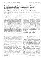
were inhibited by about 45.0 ± 5.6% (Fig. 1A). The
normalized current–voltage characteristics were almost
identical before and after PMA application (Fig. 1B).
Likewise, the normalized cord conductance was
not altered as a result of PMA treatment (Fig. 1C).
These results suggest that KAT1 is inhibited by PKC
in oocytes without affecting its voltage-dependent
properties.
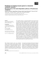
To confirm the regulation of KAT1 channel activity
by PKC, we applied a different voltage pulse protocol
to record changes in KAT1 currents over time. For
this purpose, we applied a voltage pulse to –150 mV
every 30 s. The current amplitude decreased, and the
inhibition appeared to be saturated at 30 min after
PMA application (Fig. 2A). In addition, we measured
the current–voltage characteristics of KAT1 every
5.5 min with or without pre-incubation of oocytes
in 2 lm calphostin C, a PKC-specific inhibitor, for
12–24 h (Fig 2B, C). KAT1 currents measured in
calphostin-pre-incubated oocytes were less susceptible
to PMA than those in nontreated oocytes. Taken
together, these results demonstrate that KAT1 is
inhibited by PMA-stimulated activation of the oocyte
intrinsic PKC.
Identification of Ser
⁄
Thr PKC phosphorylation
sites influencing KAT1 channel activity
Several groups have reported different sequence
motifs that are recognized by PKC [51,52]. We used
2 µA
200 ms
0 min
15 min
30 min
–0.5
0
–200 –150 –100 –50 0
V (mV)
0 min
–2
–1.5
–1
Normalized current
15.5 min
32 min
A
B
C
PMA
No addition
Fig. 1. Effects of PMA on KAT1 WT chan-
nel activity. (A) Representative current
profile of KAT1 expressed in Xenopus
oocytes before (0 min) and 15 and 30 min
after the addition of 1 l
M PMA (a PKC
activator in oocytes). (B) Current–voltage
relationship of the current at 0, 15.5 and
32 min after PMA application. Currents
were normalized with respect to the current
at )150 mV. (C) Normalized KAT1 conduc-
tance G
Nor
before (full line; squares) and
after (broken line; circles) PMA application.
Phosphorylation of KAT1 channel A. Sato et al.
2320 FEBS Journal 277 (2010) 2318–2328 ª 2010 The Authors Journal compilation ª 2010 FEBS
the program prosite ( to
predict residues of KAT1 that might be phosphory-
lated by PKC. All of the resultant sequences con-
tained the classical S ⁄ T-X-K ⁄ R motif, which can be
recognized by PKCs in Xenopus laevis oocytes [38,53].
Moreover, ‘S ⁄ T-X-K ⁄ R’ matches with the consensus
sequences recognized by plant CDPKs [54,55]. In
addition, we employed the prediction program Netph-
osK 1.0 ( />using the Phospho.ELM database (http://phospho.
elm.eu.org/) containing experimentally verified phos-
phorylation patterns of Ser ⁄ Thr ⁄ Tyr residues in
eukaryotic proteins. The analysis for 11 Ser ⁄ Thr resi-
dues in the cytosolic C-terminus and for Thr at posi-
tion 45 resulted in a relatively high score (more than
0.7). Consequently, we selected these 12 Ser ⁄ Thr resi-
dues as possible phosphorylation sites for PKC within
the N- and C-terminal cytosolic regions of KAT1
(Fig. 3A). We systematically screened all of them by
replacing the Ser ⁄ Thr residues by an Ala residue
which mimics the dephosphorylated form. All vari-
ants, except KAT1 T303A, showed detectable
inwardly rectifying currents. We also tested the K
+
transport activities of KAT1 T303D. However, as for
the mutant T303A, we could not obtain K
+
currents
in oocytes (data not shown). The remaining mutants
could be subdivided into three datasets: (a) the muta-
tions T45A, T308A, S312A, S589A, S590A and
S641A displayed a decrease in PMA-induced channel
inhibition (27.1 ± 2.9%, 20.6 ± 3.2%, 17.0 ± 6.5%,
28.4 ± 6.0%, 23.9 ± 5.0% and 24.9 ± 6.6%, respec-
tively; Fig. 3B, top panel); (b) the mutants T22A,
S44A, S125A and S529A behaved similarly to the
wild-type (WT) (43.2 ± 7.2%, 36.9 ± 3.3%, 34.3 ±
5.6% and 44.5 ± 0.7%, respectively; Fig. 3B, middle
panel); (c) T458A showed slightly greater inhibition
by PMA-induced PKC activation compared with WT
(52.5 ± 2.3%; Fig. 3B, bottom panel). The data sug-
gested that six Ser ⁄ Thr residues – one in the cytosolic
N terminus (T45) and five in the cytosolic C-terminus
(T308, S312, S589, S590 and S641) – were candidates
for PKC phosphorylation target sites altering KAT1
channel activity.
Quintuple mutation renders KAT1 PKC-insensitive
The data in Fig. 3B show that the single mutations do
not completely abolish the inhibitory effect of PMA
application. This may indicate that PKC stimulates
simultaneously multiple phosphorylation events on
KAT1. To confirm this, we constructed the quintuple
mutant KAT1-T308A-S312A-S589A-S590A-S641A
eliminating all putative PKC target sites in the cyto-
solic C-terminus. After expression in oocytes, the quin-
tuple mutant was no longer sensitive to PMA-induced
PKC activation. Even after PMA application, the
–3
–4
–2
–1
0
PMA
A
B
C
–6
–5
–4.5 0 10 20 30 37.5
Time (min)
Current (µA)
0.8
0.6
1
1.2
0
0.2
0.4
0102030
Time (min)
No addition
PMA
PMA + calphostin C
60
37.5
10
20
30
40
50
0
No addition
PMA
PMA +
calphostin C
Current inhibition by PMA (%) I/I
control
Fig. 2. Inhibition of WT KAT1 activity by PKC activation. (A) Time
course of a representative WT KAT1 current amplitude at –150 mV
after the addition of 1 l
M PMA to the bath solution. A black bar
indicates the addition and removal of PMA. (B) Changes in the cur-
rent at –150 mV in response to 1 l
M PMA. PMA was added imme-
diately after the recording of currents at t = 0 min. The current
amplitude was normalized to the value measured before PMA
application (mean ± SEM, n = 3–4). Calphostin C indicates that
KAT1-expressing oocytes were pre-incubated for 12–18 h with
2 l
M calphostin C, a specific PKC inhibitor. (C) Percentage of cur-
rent inhibition 32 min after the addition of PMA.
A. Sato et al. Phosphorylation of KAT1 channel
FEBS Journal 277 (2010) 2318–2328 ª 2010 The Authors Journal compilation ª 2010 FEBS 2321
current amplitude behaved similarly to that of
WT KAT1 in the absence of PMA (Fig. 4A, B and
Table 1). These results suggest that PMA-induced
phosphorylation at some or all of the five residues
determines an inhibition of the K
+
currents.
Discussion
Phosphorylation and dephosphorylation events are
critical for the modulation of the activity of the guard
cell-expressed K
+
uptake channel KAT1. Nevertheless,
information on the kinase-mediated phosphorylation
of KAT1 and the target sites involved in the regulation
of channel activity is scarce. In this study, we investi-
gated the effect of the PKC-mediated phosphorylation
of KAT1 expressed in Xenopus oocytes. On stimulation
of the oocyte endogenous PKC by the application of
Putative cyclic
A
B
nucleotide
binding domain
(CNBD)
22
458
529
45
303
308
312
589
590
641
S
S
S
T
T
S
44
125
T
S
T
T
S
S
T45A
T308A
S312A
S589A
S590A
S641A
T22A
S44A
S125A
S529A
1
1.2
WT (no addition)
0
0.2
0.4
0.6
0.8
1
I/I
control
1
1.2
0
0.2
0.4
0.6
0.8
1
I/I
control
1.2
0
0.2
0.4
0.6
0.8
1
I/I
control
T458A
WT (PMA)
10 20 30 37.50
Time (min)
10 20 30 37.50
Time (min)
10 20 30 37.50
Time (min)
Fig. 3. Effects of mutations on possible phosphorylation sites on
KAT1 currents. (A) Schematic representation of the consensus
PKC phosphorylation sites at the cytosolic face of KAT1. (B)
Changes in the current amplitudes of the different KAT1 mutants
after PMA application. The characteristics of WT KAT1 in the
presence and absence of PMA are displayed as broken lines.
The mutants are divided into three groups: top panel, smaller
degree of inhibition compared with WT; middle panel, WT-like
behavior; bottom panel, larger degree of inhibition compared with
WT.
0.2
0.4
0.6
0.8
1
1.2
I/I
control
Quintuple mutant
WT (no addition)
WT (PMA)
0
0102030
Time (min)
40
50
60
37.5
N.D.
0
10
20
30
T22A
S44A
T45A
S125A
T303A
T308A
S312A
T458A
S529A
S589A
S590A
S641A
WT
Current inhibition
by PMA (%)
B
A
Quintuple mutant
No addition
Fig. 4. Quintuple mutations confer insensitivity to PKC activation.
(A) Change in the current amplitudes of the quintuple KAT1 mutant
KAT1-T308A-S312A-S589A-S590A-S641A after PMA application.
The characteristics of WT KAT1 in the presence and absence of
PMA are displayed as broken lines. (B) Inhibition of individual
mutants by PMA application at 32 min (mean ± SEM, n = 3–5).
*Student’s t-test, P < 0.05. N.D., no detectable currents.
Phosphorylation of KAT1 channel A. Sato et al.
2322 FEBS Journal 277 (2010) 2318–2328 ª 2010 The Authors Journal compilation ª 2010 FEBS
PMA to the bath medium, the KAT1 current ampli-
tude decreased more strongly than under control con-
ditions. This indicates that phosphorylation by PKC
has a downregulatory effect on KAT1. Subsequently,
we pinpointed by in silico analyses 12 putative target
sites for PKC in KAT1 and evaluated their role on
channel regulation. Among the 12 Ser ⁄ Thr sites in
KAT1, we identified experimentally six residues that
were involved in the regulation by PKC (Figs 3 and
5). The behavior of the different channel mutants on
PMA application could be divided into four groups:
(a) loss of function; (b) increase in the inhibitory
effect; (c) decrease in the inhibitory effect; (d) inhibi-
tion comparable with WT. In the first case, the
replacements T303A and T303D abolished the KAT1
current. It is possible that the mutation of Thr at
position 303 immediately after the S6 segment may
interfere with KAT1 channel gating, as illustrated
for residues a few positions upstream [56]. In addi-
tion, for other K
+
channels, it has been shown that
the region immediately after the last transmembrane
segment is strongly involved in channel gating [57–
61].
The inhibition of the KAT1-mediated K
+
current
on PMA application may depend on a decline in the
number of active channels or on a lower single-channel
conductance, as the voltage-dependent characteristics
were not affected by PMA application in oocytes
injected with the WT KAT1 channel (as shown in
Fig. 1B, C); notably, almost identical I–V characteris-
tics were also observed in oocytes injected with
mutants before and after the addition of PMA (data
not shown).
As the A. thaliana genome does not comprise a gene
encoding a protein that is homologous to animal
PKC, there is no evidence that the reduction in
KAT1 by phosphorylation suggested in this study
actually occurs in vivo in plant cells. Instead of PKC,
in the genome sequence of A. thaliana, 34 different
genes encoding CDPKs are present, which is currently
recognized as a major group of Ca
2+
-stimulated pro-
tein kinases [35]. A calcium-dependent kinase from
Vicia faba was found to phosphorylate the KAT1
protein translated in vitro [46], and a CDPK from
soybean decreased KAT1-mediated K
+
current ampli-
tudes in Xenopus oocytes [8]. To date, several CDPK
phosphorylation target sequences have been reported
[62–64]. The two most classical motifs are S-X-R ⁄ K
and R ⁄ K-X-X-S ⁄ T [54,55]. The S-X-R ⁄ K sequence is
included in the optimal oligopeptide which may be
phosphorylated by PKCs [53]. On the other hand,
PKC may also recognize Ser ⁄ Thr in the sequence, as
PKC recognizes preferentially substrates with a basic
residue at position )3 [53]. KAT1 comprises 10 R ⁄ K-
X-X-S ⁄ T motifs in the cytosolic N- and C-terminal
regions. Among them, T308 and S641 are matching
both motifs S ⁄ T-X-R ⁄ K as well as R-X-X-S ⁄ T (Figs 3
and 4).
Interestingly, in a recent study, residue T308 was
also identified as a target site for the Ca
2+
-indepen-
dent ABA-activated SnRK2.6 kinase [21]. In addition,
the SnRK2.6 kinase could phosphorylate T306 in an
in vitro kinase assay. This evidence suggests that multi-
ple protein kinases may participate in the regulation
of KAT1 channel activity to respond to various
Table 1. Percentage of current inhibition by PKC activation. Current
inhibition of KAT1 and its mutants by PKC activation at 32 min after
PMA (mean ± SEM, n = 3–6). Replacement of T303 by Ala or Asp
led to a loss of KAT1 activity.
Mutant Inhibition (%) n
T22A 43.2 ± 7.2 4
S44A 36.9 ± 3.3 4
T45A 27.1 ± 2.9 4
a
S125A 34.3 ± 5.6 3
T308A 20.6 ± 3.2 5
a
S312A 17.0 ± 6.5 3
a
T458A 52.5 ± 2.3 4
S529A 44.5 ± 0.7 3
S589A 28.4 ± 6.0 3
a
S590A 23.9 ± 5.0 3
a
S641A 24.9 ± 6.6 3
a
T308A ⁄ S312A ⁄ S589A ⁄ S590A ⁄ S641A 14.9 ± 5.2 4
a
WT 45.0 ± 5.6 3
Control 10.6 ± 4.5 3
WT + calphostin C 19.5 ± 4.8 4
a
P < 0.05.
K
+
KAT1 inhibition
45
308
589
590
641
S
S
S
T
T
S
Phosphorylation
306
303
T
T
312
Fig. 5. Possible regulation of KAT1 channel activity by phosphoryla-
tion via Ca
2+
-dependent ⁄ independent pathways. The target sites of
PKC (CDPK) and ABA-activated SnRK2.6 identified in heterologous
expression systems are indicated. T306 and T308 were possible
target sites for the Ca
2+
-independent, ABA-activated SnRK2.6
kinase [21]. T45, T308, S312, S589, S590 and S641 were possible
targets for PKC in Xenopus oocytes performed in this study.
Replacement of T303 by Ala or Asp led to a loss of KAT1 activity.
A. Sato et al. Phosphorylation of KAT1 channel
FEBS Journal 277 (2010) 2318–2328 ª 2010 The Authors Journal compilation ª 2010 FEBS 2323
physiological signals (Fig. 5). Indeed, the conversion of
T306 to Ala or Asp resulted in a loss of KAT1 activity
in Xenopus oocyte and yeast expression systems [21].
In addition, after the replacement of T303 by Asp, no
K
+
transport activity could be measured in oocytes.
The C-terminal region after the last transmembrane
region of S6 in plant K
+
channels is involved in chan-
nel gating [56,65,66]. If the residue at position 303 can
be recognized as a phosphorylation target site, the
modification of T303 may lead to a loss of K
+
trans-
port activity.
Although, in this study, we took advantage of a sig-
naling pathway in Xenopus oocytes to stimulate (by
PMA treatment), an animal-specific PKC, the results
obtained may have implications on signaling in plants.
The application of PMA to plant tissues has been
shown to alter the expression level of some genes
[67–70]. This fact may indicate that, in plants, similar
signaling pathways exist which connect the application
of phorbol esters to the activation of certain kinases
analogous to PKC. This is in line with other observa-
tions. Inositol 1,4,5-trisphosphate (InsP
3
) and Ca
2+
induce stomatal closure [71]. InsP
3
does not affect out-
wardly rectifying K
+
channels [72,73], but inhibits
only inwardly rectifying K
+
channels [72]. DAG is
rapidly converted to PAs by DAG kinases in plants
[41], and carrot CDPK, DcCPK1, and maize CDPK,
ZmCK11, are activated by PAs and Ca
2+
[48,74].
The phosphoinositide-dependent protein kinase-1 spe-
cifically binds PA [75]. In guard cell protoplasts, PA
inhibits the activity of inwardly rectifying K
+
channels and also induces stomatal closure and inhib-
its stomatal opening [76]. Through the serial trans-
duction pathway, the end result is the alteration of
the activity of inwardly rectifying K
+
channels by
phosphorylation.
It has been reported that exogenously supple-
mented animal protein kinase A greatly retards the
rundown rate of KAT1 [77,78]. In the same studies,
it was also shown that PKC application was not
effective in preventing rundown. This result is in
line with our study demonstrating that PKC appli-
cation has the inverse effect, and may be different
from the phosphorylation mechanism involved in
rundown.
Taken together, our results suggest the existence of
several PKC phosphorylation sites in the cytosolic
region of KAT1. K
+
channel modulation, e.g. K
+
-
uptake channel inhibition during stomatal closure,
may occur via protein kinases which have PKC-
like characteristics, such as CDPKs. Phospholipid
signaling may be involved in the preceding signaling
cascades.
Materials and methods
Channel expression in oocytes and electrical
recordings
The cDNAs encoding full-length KAT1 WT or its variants
were amplified by a two-step PCR using HindIII site-con-
taining sense primer and BamHI site-containing antisense
primer (Table S1, see Supporting information) [49]. The
HindIII-BamHI DNA fragments were ligated into the same
sites of a modified pYES2 vector (Invitrogen, Carlsbad,
CA, USA) for expression in oocytes and yeast [79]. Capped
cRNAs were synthesized in vitro from NotI-linearized plas-
mids using an in vitro transcription kit (Ambion, Austin,
TX, USA). Xenopus oocytes were defolliculated using colla-
genase and microinjected with either 1 or 2 ng of cRNAs
after a 1–3 day incubation in Barth’s buffer containing
88 mm NaCl, 1 mm KCl, 0.41 mm CaCl
2
, 0.33 mm
Ca(NO
3
)
2
,1mm MgSO
4
, 2.4 mm NaHCO
3
,5mm Hepes
and 50 lgÆmL
)1
gentamicin sulfate (pH 7.3) at 18 °C. The
two-electrode voltage clamp experiments were performed
using a voltage clamp amplifier (AxoClamp 2B, Axon
Instruments, Foster City, CA, USA) at room temperature
in Xenopus laevis oocytes [49]. Microelectrodes contained
3 m KCl with a resistance of 0.3–1.0 MX. The bath solu-
tion was 120 mm KCl, 1 mm MgCl
2
,1mm CaCl
2
and
10 mm Hepes (pH 7.3). Time-dependent changes in current
were recorded at –150 mV in single-step pulses every 30 s
and in step voltage pulses ()30 to )170 mV with a 20 mV
decrement) every 5.5 min. Step voltage pulses were applied
from a holding potential of –40 mV, the duration of
each pulse being 500 ms. Data acquisition and analysis
were performed using pclamp 9.2 (Molecular Devices,
Sunnyvale, CA, USA) and origin 5.0 software (Axon
Instruments).
Drug treatment and application
PMA (Alexis Biochemicals, Lausen, Switzerland) and
calphostin C (Alexis Biochemicals) were dissolved in dim-
ethylsulfoxide as stocks and mixed with the recording solu-
tion, reaching the final concentrations indicated in the
figures [50]. PMA was applied to oocytes after initial
measurements in its absence had been carried out.
Acknowledgements
This work was supported in part by Grants-in-Aid for
Scientific Research (17078005, 20246044 and 20-08103
to N.U.) from MEXT Japan Ministry of Education,
Culture, Sports, Science & Technology and JSPS
(Japan Society for the Promotion of Science) as well as
by the JSPS-CNR (National Research Council of
Italy) Bilateral Program to N.U. and F.G.
Phosphorylation of KAT1 channel A. Sato et al.
2324 FEBS Journal 277 (2010) 2318–2328 ª 2010 The Authors Journal compilation ª 2010 FEBS
References
1 Anderson JA, Huprikar SS, Kochian LV, Lucas WJ &
Gaber RF (1992) Functional expression of a probable
Arabidopsis thaliana potassium channel in Saccharomy-
ces cerevisiae. Proc Natl Acad Sci USA 89, 3736–3740.
2 Schachtman DP, Schroeder JI, Lucas WJ, Anderson JA
& Gaber RF (1992) Expression of an inward-rectifying
potassium channel by the Arabidopsis KAT1 cDNA.
Science, 258(cDNA), 1654–1658.
3 Nakamura RL, McKendree WL Jr, Hirsch RE, Sed-
brook JC, Gaber RF & Sussman MR (1995) Expression
of an Arabidopsis potassium channel gene in guard
cells. Plant Physiol 109, 371–374.
4 Lebaudy A, Vavasseur A, Hosy E, Dreyer I, Leonhardt
N, Thibaud JB, Very AA, Simonneau T & Sentenac H
(2008) Plant adaptation to fluctuating environment and
biomass production are strongly dependent on guard
cell potassium channels. Proc Natl Acad Sci USA 105,
5271–5276.
5 Kinoshita T, Nishimura M & Shimazaki K (1995)
Cytosolic concentration of Ca
2+
regulates the plasma
membrane H
+
-ATPase in guard cells of fava bean.
Plant Cell 7, 1333–1342.
6 Schroeder JI & Hagiwara S (1989) Cytosolic calcium
regulates ion channels in the plasma membrane of Vicia
faba guard cells. Nature 338, 427–430.
7 Sutton F, Paul SS, Wang XQ & Assmann SM (2000)
Distinct abscisic acid signaling pathways for modulation
of guard cell versus mesophyll cell potassium channels
revealed by expression studies in Xenopus laevis oocytes.
Plant Physiol 124, 223–230.
8 Berkowitz G, Zhang X, Mercie R, Leng Q & Lawton
M (2000) Co-expression of calcium-dependent protein
kinase with the inward rectified guard cell K
+
channel
KAT1 alters current parameters in Xenopus laevis
oocytes. Plant Cell Physiol 41, 785–790.
9 Mori IC, Murata Y, Yang Y, Munemasa S, Wang
YF, Andreoli S, Tiriac H, Alonso JM, Harper JF,
Ecker JR et al. (2006) CDPKs CPK6 and CPK3
function in ABA regulation of guard cell S-type
anion- and Ca
2+
-permeable channels and stomatal
closure. PLoS Biol 4, e327.
10 Zhu SY, Yu XC, Wang XJ, Zhao R, Li Y, Fan RC,
Shang Y, Du SY, Wang XF, Wu FQ et al. (2007) Two
calcium-dependent protein kinases, CPK4 and CPK11,
regulate abscisic acid signal transduction in Arabidop-
sis. Plant Cell 19, 3019–3036.
11 Hamilton DW, Hills A, Kohler B & Blatt MR (2000)
Ca
2+
channels at the plasma membrane of stomatal
guard cells are activated by hyperpolarization and
abscisic acid. Proc Natl Acad Sci USA 97, 4967–4972.
12 Grabov A & Blatt MR (1999) A steep dependence of
inward-rectifying potassium channels on cytosolic free
calcium concentration increase evoked by hyperpo-
larization in guard cells. Plant Physiol 119, 277–
288.
13 Li J & Assmann SM (1996) An abscisic acid-activated
and calcium-independent protein kinase from guard
cells of fava bean. Plant Cell 8, 2359–2368.
14 Mori IC & Muto S (1997) Abscisic acid activates a 48-
kilodalton protein kinase in guard cell protoplasts.
Plant Physiol 113, 833–839.
15 Li J, Wang XQ, Watson MB & Assmann SM (2000)
Regulation of abscisic acid-induced stomatal closure
and anion channels by guard cell AAPK kinase. Science
287, 300–303.
16 Mori IC, Uozumi N & Muto S (2000) Phosphorylation
of the inward-rectifying potassium channel KAT1 by
ABR kinase in Vicia guard cells. Plant Cell Physiol 41,
850–856.
17 Yoshida R, Hobo T, Ichimura K, Mizoguchi T,
Takahashi F, Aronso J, Ecker JR & Shinozaki K
(2002) ABA-activated SnRK2 protein kinase is required
for dehydration stress signaling in Arabidopsis. Plant
Cell Physiol 43, 1473–1483.
18 Mustilli AC, Merlot S, Vavasseur A, Fenzi F &
Giraudat J (2002) Arabidopsis OST1 protein kinase
mediates the regulation of stomatal aperture by abscisic
acid and acts upstream of reactive oxygen species
production. Plant Cell 14, 3089–3099.
19 Merlot S, Mustilli AC, Genty B, North H, Lefebvre
V, Sotta B, Vavasseur A & Giraudat J (2002) Use of
infrared thermal imaging to isolate Arabidopsis
mutants defective in stomatal regulation. Plant J 30,
601–609.
20 Yoshida R, Umezawa T, Mizoguchi T, Takahashi S,
Takahashi F & Shinozaki K (2006) The regulatory
domain of SRK2E ⁄ OST1 ⁄ SnRK2.6 interacts with ABI1
and integrates abscisic acid (ABA) and osmotic stress
signals controlling stomatal closure in Arabidopsis.
J Biol Chem 281, 5310–5318.
21 Sato A, Sato Y, Fukao Y, Fujiwara M, Umezawa T,
Shinozaki K, Hibi T, Taniguchi M, Miyake H, Goto
DB et al. (2009) Threonine at position 306 of the
KAT1 potassium channel is essential for channel
activity and is a target site for ABA-activated
SnRK2 ⁄ OST1 ⁄ SnRK2.6 protein kinase. Biochem J 424,
439–448.
22 Nishizuka Y (1989) The Albert Lasker medical awards.
the family of protein kinase c for signal transduction.
J Am Med Assoc 262, 1826–1833.
23 Nishizuka Y (1995) Protein kinase C and lipid signaling
for sustained cellular responses. FASEB J 9, 484–496.
24 Busch AE, Varnum MD, North RA & Adelman JP
(1992) An amino acid mutation in a potassium channel
that prevents inhibition by protein kinase C. Science
255, 1705–1707.
25 Covarrubias M, Wei A, Salkoff L & Vyas TB (1994)
Elimination of rapid potassium channel inactivation by
A. Sato et al. Phosphorylation of KAT1 channel
FEBS Journal 277 (2010) 2318–2328 ª 2010 The Authors Journal compilation ª 2010 FEBS 2325
phosphorylation of the inactivation gate. Neuron 13,
1403–1412.
26 Barros F, Gomez-Varela D, Viloria CG, Palomero T,
Giraldez T & de la Pena P (1998) Modulation of
human erg K
+
channel gating by activation of a
G protein-coupled receptor and protein kinase C.
J Physiol 511 (Pt 2) 333–346.
27 Thomas D, Zhang W, Wu K, Wimmer AB, Gut B,
Wendt-Nordahl G, Kathofer S, Kreye VA, Katus HA,
Schoels W et al. (2003) Regulation of HERG potassium
channel activation by protein kinase C independent of
direct phosphorylation of the channel protein. Cardio-
vasc Res 59, 14–26.
28 Mao J, Wang X, Chen F, Wang R, Rojas A, Shi Y,
Piao H & Jiang C (2004) Molecular basis for the inhibi-
tion of G protein-coupled inward rectifier K
+
channels
by protein kinase C. Proc Natl Acad Sci USA 101,
1087–1092.
29 Asaoka Y, Nakamura S, Yoshida K & Nishizuka Y
(1992) Protein kinase C, calcium and phospholipid deg-
radation. Trends Biochem Sci 17, 414–417.
30 Ebinu JO, Bottorff DA, Chan EY, Stang SL, Dunn RJ
& Stone JC (1998) RasGRP, a Ras guanyl nucleotide-
releasing protein with calcium- and diacylglycerol-
binding motifs. Science 280, 1082–1086.
31 Ron D & Kazanietz MG (1999) New insights into the
regulation of protein kinase C and novel phorbol ester
receptors. FASEB J 13, 1658–1676.
32 Sakane F, Imai S, Yamada K, Murakami T, Tsushima
S & Kanoh H (2002) Alternative splicing of the human
diacylglycerol kinase delta gene generates two isoforms
differing in their expression patterns and in regulatory
functions. J Biol Chem 277, 43519–43526.
33 Tognon CE, Kirk HE, Passmore LA, Whitehead IP,
Der CJ & Kay RJ (1998) Regulation of RasGRP via a
phorbol ester-responsive C1 domain. Mol Cell Biol 18,
6995–7008.
34 Harper JF, Binder BM & Sussman MR (1993) Calcium
and lipid regulation of an Arabidopsis protein kinase
expressed in Escherichia coli. Biochemistry 32, 3282–
3290.
35 Cheng SH, Willmann MR, Chen HC & Sheen J (2002)
Calcium signaling through protein kinases. The Arabid-
opsis calcium-dependent protein kinase gene family.
Plant Physiol 129, 469–485.
36 Hrabak EM, Chan CW, Gribskov M, Harper JF,
Choi JH, Halford N, Kudla J, Luan S, Nimmo HG,
Sussman MR et al. (2003) The Arabidopsis CDPK-
SnRK superfamily of protein kinases. Plant Physiol
132, 666–680.
37 Spitaler M & Cantrell DA (2004) Protein kinase C and
beyond. Nat Immunol 5 , 785–790.
38 Rajagopal S, Fang H, Patanavanich S, Sando JJ &
Kamatchi GL (2008) Protein kinase C isozyme-specific
potentiation of expressed Cav2.3 currents by acetyl-
beta-methylcholine and phorbol-12-myristate, 13-ace-
tate. Brain Res 1210, 1–10.
39 Tasma IM, Brendel V, Whitham SA & Bhattacharyya
MK (2008) Expression and evolution of the phosphoino-
sitide-specific phospholipase C gene family in Arabidopsis
thaliana. Plant Physiol Biochem 46, 627–637.
40 Gomez-Merino FC, Brearley CA, Ornatowska M,
Abdel-Haliem ME, Zanor MI & Mueller-Roeber B
(2004) AtDGK2, a novel diacylglycerol kinase from
Arabidopsis thaliana, phosphorylates 1-stearoyl-2-arachi-
donoyl-sn-glycerol and 1,2-dioleoyl-sn-glycerol and
exhibits cold-inducible gene expression. J Biol Chem
279, 8230–8241.
41 Laxalt AM & Munnik T (2002) Phospholipid signalling
in plant defence. Curr Opin Plant Biol 5, 332–338.
42 Harper JF, Breton G & Harmon A (2004) Decoding
Ca
2+
signals through plant protein kinases. Annu Rev
Plant Biol 55, 263–288.
43 Chandok MR & Sopory SK (1998) ZmcPKC70, a pro-
tein kinase C-type enzyme from maize. Biochemical
characterization, regulation by phorbol 12-myristate
13-acetate and its possible involvement in nitrate
reductase gene expression. J Biol Chem 273, 19235–
19242.
44 Deswal R, Chowdhary GK & Sopory SK (2004) Purifi-
cation and characterization of a PMA-stimulated kinase
and identification of PMA-induced phosphorylation of
a polypeptide that is dephosphorylated by low tempera-
ture in Brassica juncea. Biochem Biophys Res Commun
322, 420–427.
45 Loog M, Toomik R, Sak K, Muszynska G, Jarv J &
Ek P (2000) Peptide phosphorylation by calcium-depen-
dent protein kinase from maize seedlings. Eur J Bio-
chem 267, 337–343.
46 Li J, Lee YR & Assmann SM (1998) Guard cells pos-
sess a calcium-dependent protein kinase that phospho-
rylates the KAT1 potassium channel. Plant Physiol 116 ,
785–795.
47 Pearson RB & Kemp BE (1991) Protein kinase phos-
phorylation site sequences and consensus specificity
motifs: tabulations. Methods Enzymol 200, 62–81.
48 Farmer PK & Choi JH (1999) Calcium and phospho-
lipid activation of a recombinant calcium-dependent
protein kinase (DcCPK1) from carrot (Daucus carota
L.). Biochim Biophys Acta 1434, 6–17.
49 Uozumi N, Gassmann W, Cao Y & Schroeder JI (1995)
Identification of strong modifications in cation selectiv-
ity in an Arabidopsis inward rectifying potassium chan-
nel by mutant selection in yeast. J Biol Chem 270,
24276–24281.
50 Zhang L, Lee JK, John SA, Uozumi N & Kodama I
(2004) Mechanosensitivity of GIRK channels is medi-
ated by protein kinase C-dependent channel–phosphati-
dylinositol 4,5-bisphosphate interaction. J Biol Chem
279, 7037–7047.
Phosphorylation of KAT1 channel A. Sato et al.
2326 FEBS Journal 277 (2010) 2318–2328 ª 2010 The Authors Journal compilation ª 2010 FEBS
51 Kishimoto A, Nishiyama K, Nakanishi H, Uratsuji Y,
Nomura H, Takeyama Y & Nishizuka Y (1985) Studies
on the phosphorylation of myelin basic protein by
protein kinase C and adenosine 3¢:5¢-monophosphate-
dependent protein kinase. J Biol Chem 260, 12492–
12499.
52 Woodgett JR, Gould KL & Hunter T (1986) Substrate
specificity of protein kinase C. Use of synthetic peptides
corresponding to physiological sites as probes for sub-
strate recognition requirements. Eur J Biochem 161,
177–184.
53 Nishikawa K, Toker A, Johannes FJ, Songyang Z &
Cantley LC (1997) Determination of the specific sub-
strate sequence motifs of protein kinase C isozymes.
J Biol Chem 272, 952–960.
54 Roberts DM & Harmon AC (1992) Calcium-modulated
proteins: targets of intracellular calcium signals in
higher plants. Annu Rev Plant Physiol Plant Mol Biol
43, 375–414.
55 Neumann GM, Thomas I & Polya GM (1996) Identifi-
cation of the site on potato carboxypeptidase inhibitor
that is phosphorylated by plant calcium-dependent
protein kinase. Plant Sci 114, 45–51.
56 Gajdanowicz P, Garcia-Mata C, Gonzalez W, Morales-
Navarro SE, Sharma T, Gonzalez-Nilo FD, Gutowicz
J, Mueller-Roeber B, Blatt MR & Dreyer I (2009) Dis-
tinct roles of the last transmembrane domain in control-
ling Arabidopsis K
+
channel activity. New Phytol 182,
380–391.
57 Jiang Y, Lee A, Chen J, Cadene M, Chait BT &
MacKinnon R (2002) The open pore conformation of
potassium channels. Nature 417, 523–526.
58 Sansom MS, Shrivastava IH, Bright JN, Tate J,
Capener CE & Biggin PC (2002) Potassium channels:
structures, models, simulations. Biochim Biophys Acta
1565, 294–307.
59 Kuo A, Gulbis JM, Antcliff JF, Rahman T, Lowe ED,
Zimmer J, Cuthbertson J, Ashcroft FM, Ezaki T &
Doyle DA (2003) Crystal structure of the potassium
channel KirBac1.1 in the closed state. Science 300,
1922–1926.
60 Long SB, Campbell EB & Mackinnon R (2005) Crystal
structure of a mammalian voltage-dependent Shaker
family K
+
channel. Science 309, 897–903.
61 Riedelsberger J, Sharma T, Gonzalez W, Gajdanowicz
P, Morales-Navarro SE, Garcia-Mata C, Mueller-Roe-
ber B, Gonza
´
lez-Nilo FD, Blatt MR & Dreyer I (2010)
Distributed structures underlie gating differences
between the K
in
channel KAT1 and the K
out
channel
SKOR. Mol Plant 3, 236–245.
62 Sebastia
`
CH, Hardin SC, Clouse SD, Kieber JJ &
Huber SC (2004) Identification of a new motif for
CDPK phosphorylation in vitro that suggests ACC
synthase may be a CDPK substrate. Arch Biochem
Biophys 428, 81–91.
63 Klimecka M & Muszynska G (2007) Structure and
functions of plant calcium-dependent protein kinases.
Acta Biochim Pol 54, 219–233.
64 Vlad F, Turk BE, Peynot P, Leung J & Merlot S (2008)
A versatile strategy to define the phosphorylation pref-
erences of plant protein kinases and screen for putative
substrates. Plant J 55, 104–117.
65 Cherel I, Michard E, Platet N, Mouline K, Alcon C,
Sentenac H & Thibaud JB (2002) Physical and func-
tional interaction of the Arabidopsis K
+
channel AKT2
and phosphatase AtPP2CA. Plant Cell 14, 1133–1146.
66 Michard E, Dreyer I, Lacombe B, Sentenac H &
Thibaud JB (2005) Inward rectification of the AKT2
channel abolished by voltage-dependent phosphoryla-
tion. Plant J 44 , 783–797.
67 Baudouin E, Charpenteau M, Ranjeva R & Ranty B
(1999) Involvement of active oxygen species in the regu-
lation of a tobacco defence gene by phorbol ester. Plant
Sci 142, 67–72.
68 Chandok MR, Sopory SK & Oelmuller R (2001)
Cytoplasmic kinase and phosphatase activities can
induce PsaF gene expression in the absence of func-
tional plastids: evidence that phosphorylation ⁄ dephos-
phorylation events are involved in interorganellar
crosstalk. Mol Gen Genet 264, 819–826.
69 Wostemeyer A & Oelmuller R (2003) The promoter of
the spinach PsaF gene for the subunit III of the pho-
tosystem I reaction center directs beta-glucuronidase
gene expression in transgenic tobacco roots. Implica-
tion of the involvement of phospholipases and protein
kinase C in PsaF gene expression. J Plant Physiol 160,
503–508.
70 Chandok MR & Sopory SK (1996) Phosphoryla-
tion ⁄ dephosphorylation steps are key events in the phy-
tochrome-mediated enhancement of nitrate reductase
mRNA levels and enzyme activity in maize. Mol Gen
Genet 251, 599–608.
71 Blatt MR, Thiel G & Trentham DR (1990) Reversible
inactivation of K
+
channels of Vicia stomatal guard
cells following the photolysis of caged inositol 1,4,5-tris-
phosphate. Nature 346, 766–769.
72 Gilroy S, Read ND & Trewavas AJ (1990) Elevation of
cytoplasmic calcium by caged calcium or caged inositol
triphosphate initiates stomatal closure. Nature 346, 769–
771.
73 Schroeder JI (1988) K
+
transport properties of K
+
channels in the plasma membrane of Vicia faba guard
cells. J Gen Physiol 92, 667–683.
74 Szczegielniak J, Klimecka M, Liwosz A, Ciesielski A,
Kaczanowski S, Dobrowolska G, Harmon AC &
Muszynska G (2005) A wound-responsive and phospho-
lipid-regulated maize calcium-dependent protein kinase.
Plant Physiol 139, 1970–1983.
75 Deak M, Casamayor A, Currie RA, Downes CP &
Alessi DR (1999) Characterisation of a plant 3-phos-
A. Sato et al. Phosphorylation of KAT1 channel
FEBS Journal 277 (2010) 2318–2328 ª 2010 The Authors Journal compilation ª 2010 FEBS 2327
phoinositide-dependent protein kinase-1 homologue
which contains a pleckstrin homology domain. FEBS
Lett 451, 220–226.
76 Jacob T, Ritchie S, Assmann SM & Gilroy S (1999)
Abscisic acid signal transduction in guard cells is medi-
ated by phospholipase D activity. Proc Natl Acad Sci
USA 96, 12192–12197.
77 Hoshi T (1995) Regulation of voltage dependence of the
KAT1 channel by intracellular factors. J Gen Physiol
105, 309–328.
78 Tang XD & Hoshi T (1999) Rundown of the hyperpo-
larization-activated KAT1 channel involves slowing of
the opening transitions regulated by phosphorylation.
Biophys J 76, 3089–3098.
79 Kato Y, Sakaguchi M, Mori Y, Saito K, Nakamura T,
Bakker EP, Sato Y, Goshima S & Uozumi N (2001)
Evidence in support of a four transmembrane-pore-
transmembrane topology model for the Arabidopsis
thaliana Na
+
⁄ K
+
translocating AtHKT1 protein, a
member of the superfamily of K
+
transporters. Proc
Natl Acad Sci USA 98, 6488–6493.
Supporting information
The following supplementary material is available:
Table S1. Primer sequences for plasmid construction.
This supplementary material can be found in the
online version of this article.
Please note: As a service to our authors and readers,
this journal provides supporting information supplied
by the authors. Such materials are peer-reviewed and
may be re-organized for online delivery, but are not
copy-edited or typeset. Technical support issues arising
from supporting information (other than missing files)
should be addressed to the authors.
Phosphorylation of KAT1 channel A. Sato et al.
2328 FEBS Journal 277 (2010) 2318–2328 ª 2010 The Authors Journal compilation ª 2010 FEBS