Báo cáo khoa học: Transgenic Cdx2 induces endogenous Cdx1 in intestinal metaplasia of Cdx2-transgenic mouse stomach pot
Bạn đang xem bản rút gọn của tài liệu. Xem và tải ngay bản đầy đủ của tài liệu tại đây (382.04 KB, 11 trang )
Transgenic Cdx2 induces endogenous Cdx1 in intestinal
metaplasia of Cdx2-transgenic mouse stomach
Hiroyuki Mutoh, Hiroko Hayakawa, Hirotsugu Sakamoto, Miho Sashikawa and Kentaro Sugano
Department of Medicine, Division of Gastroenterology, Jichi Medical University, Tochigi, Japan
Introduction
In intestinal metaplasia of the human stomach, normal
gastric mucosa is replaced by an intestinalized epithe-
lium, and is mainly induced together with the progres-
sion of Helicobacter pylori-infected chronic gastritis.
Intestinal metaplasia of the human stomach has been
extensively studied as a premalignant condition of
gastric carcinoma [1]. The intestine-specific homeo-
box genes Cdx1 and Cdx2 have been shown to be
Keywords
chromatin immunoprecipitation; luciferase
reporter assay; methylation; RT-PCR; siRNA
Correspondence
H. Mutoh, Department of Medicine, Division
of Gastroenterology, Jichi Medical
University, Yakushiji 3311-1, Shimotsuke,
Tochigi 329-0498, Japan
Fax: +81 285 44 8297
Tel: +81 285 58 7348
E-mail:
(Received 12 April 2009, revised 1 August
2009, accepted 6 August 2009)
doi:10.1111/j.1742-4658.2009.07263.x
Cdx1 and Cdx2, which are transcription factors regulating normal intesti-
nal development, have been studied as potential key molecules in the
pathogenesis of the precancerous intestinal metaplasia of the human
stomach. However, the regulation of Cdx1 expression in the intestinal
metaplasia is poorly understood. Cdx2-expressing gastric mucosa of Cdx2-
transgenic mouse stomach was replaced by intestinal metaplastic mucosa.
The aim of this study was to investigate the following: (a) Cdx1 expres-
sion in the intestinal metaplastic mucosa of the Cdx2-transgenic mouse
stomach; and (b) the relationship between Cdx1 and Cdx2. A mouse
model of intestinal metaplasia, the Cdx2-transgenic mouse, was used to
investigate Cdx1 gene expression by RT-PCR. DNA methylation profile
analysis was performed by bisulfite sequencing, and the interaction of
Cdx2 with the Cdx1 promoter was examined by chromatin immunoprecip-
itation assay, electrophoretic mobility shift assay, and luciferase reporter
assays. Cdx2 mRNA was expressed in the Cdx2-transgenic mouse stom-
ach. However, endogenous Cdx2 mRNA was not expressed in the intesti-
nal metaplasia of the Cdx2-transgenic mouse stomach. On the other hand,
endogenous Cdx1 mRNA and protein were expressed in the intestinal
metaplasia of the Cdx2-transgenic mouse stomach. The Cdx1 promoter
was unmethylated in the intestinal metaplasia of the Cdx2-transgenic
mouse stomach. Chromatin immunoprecipitation assay and electrophoretic
mobility shift assay showed that Cdx2 was bound to the Cdx1 promoter
region in the intestinal metaplasia and the normal intestine. Cdx2 upregu-
lated and siRNA-Cdx2 downregulated the transcriptional activity of the
Cdx1 gene in the human gastric carcinoma cell lines AGS, MKN45, and
MKN74. In conclusion, transgenic Cdx2 induced endogenous Cdx1
through the binding of Cdx2 to the unmethylated Cdx1 promoter region
in the intestinal metaplasia of the Cdx2-transgenic mouse stomach.
Abbreviations
ChIP, chromatin immunoprecipitation; EMSA, electrophoretic mobility shift assay; GAPDH, glyceraldehyde-3-phosphate dehydrogenase; RA,
retinoic acid; si, small interfering.
FEBS Journal 276 (2009) 5821–5831 ª 2009 The Authors Journal compilation ª 2009 FEBS 5821
aberrantly expressed in human intestinal metaplasia.
Cdx1 and Cdx2 are mammalian members of the cau-
dal-related homeobox gene family. In adult mice and
humans, expression is strictly confined to the gut, from
the duodenum to the rectum. Normal stomach does
not express the transcription factors Cdx1 and Cdx2.
We and others have reported the presence of Cdx1
and Cdx2 in the intestinal metaplasia of the H. pylori-
infected human stomach [2–4].
We have previously generated Cdx2-transgenic mice
as model mice for intestinal metaplasia [5,6]. Cdx2-
transgenic mice specifically express Cdx2 in the gastric
mucosa, and develop intestinal metaplasia in the stom-
ach [5,6]. Gastric carcinoma spontaneously developed
from intestinal metaplasia in all stomachs of Cdx2-
transgenic mice examined [7].
In Barrett’s esophagus, normal squamous esopha-
geal mucosa is also replaced by an intestinalized
columnar epithelium in which Cdx2 is expressed [8].
Exposure to acid and ⁄ or bile acids has been reported
to activate Cdx2 expression in human esophageal epi-
thelial cells through promoter demethylation [9–11].
However, it is still unclear how Cdx1 is induced in
intestinal metaplasia. Furthermore, the relationship
between Cdx1 and Cdx2 in intestinal metaplasia has
not been clarified as yet. To investigate these ques-
tions, we focused on the induction of endogenous
Cdx1 in Cdx2-induced intestinal metaplasia using
Cdx2-transgenic mice.
Results
Expression of Cdx1 and Cdx2 in the intestinal
metaplasia of the Cdx2-transgenic mouse
stomach
Cdx2-transgenic mice we generated showed intestinal
metaplasia in the stomach [5,6]. First, Cdx2 expression
in the intestinal metaplasia of Cdx2-transgenic mouse
stomachs was examined, using RT-PCR. Cdx2 mRNA
was detected in normal intestine and in all of the intes-
tinal metaplasia of the Cdx2-transgenic mouse stom-
ach, but not in the normal mouse stomach (Fig. 1B).
Cdx2 expression was detected using a primer pair for
the Cdx2 coding region (Cdx2 coding-fw and Cdx2
coding-rv; Fig. 1A and Table 1). When Cdx2-trans-
genic mice were generated, only the Cdx2 coding
region, without the noncoding region, was used. To
investigate whether endogenous Cdx2 was expressed in
Cdx2-induced intestinal metaplasia, endogenous Cdx2
expression was detected using a primer pair for the
coding region and the 3¢-noncoding region (Cdx2 cod-
ing-fw and Cdx2 non-coding-rv; Fig. 1A and Table 1).
Endogenous Cdx2 was expressed in the normal intes-
tine, but in none of the intestinal metaplasia of the
Cx2-transgenic mouse stomachs (Fig. 1C). Transgenic
Cdx2 did not induce endogenous Cdx2 expression,
indicating that Cdx2 is not autoregulated in intestinal
metaplasia.
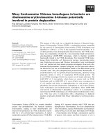
Next, whether endogenous Cdx1 was expressed in
Cdx2-induced intestinal metaplasia was investigated.
Endogenous Cdx1 was detected in the normal intestine
and in all of the Cdx2-induced intestinal metaplasia,
but not in the normal stomach (Fig. 2A).
Cdx1 gene expression was characterized by quantita-
tive real-time RT-PCR (Fig. 2B). The Cdx1 mRNA
level in the Cdx2-transgenic mouse stomach was
almost same as that in the normal mouse small intes-
tine (Fig. 2B).
Cdx1 expression in the intestinal metaplasia of the
Cdx2-transgenic mouse stomach was also investigated,
using immunohistochemistry. Cdx1 was expressed in
the intestinal metaplasia of the Cdx2-transgenic mouse
stomach (Fig. 2E) and normal intestine (Fig. 2D), but
not in the normal stomach (Fig. 2C). The expression
of Cdx1 mRNA and protein in the intestinal meta-
plasia of the Cdx2-transgenic mouse stomach indicates
that Cdx1 might be induced by Cdx2 in intestinal
metaplasia.
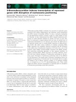
Cdx1 promoter methylation status
We focused on epigenetic regulation of Cdx1 gene
expression as a possible cause of Cdx1 activation in
the intestinal metaplasia of the Cdx2-transgenic mouse
stomach. To investigate whether the differences in
Cdx1 expression were under promoter methylation
control, bisulfite sequencing was performed on DNA
extracted from five normal stomachs, five normal intes-
tines, and five intestinal metaplasias of Cdx2-transgenic
mouse stomachs. All of the CpGs including the CpGs
(located around the TATA box and indicated by the
box in Fig. 3A) that appear to be critical for the con-
trol of Cdx1 expression in colorectal carcinoma [12]
were unmethylated in the Cdx1 promoter sequences
from the five intestinal metaplasias, the five normal
intestines and the five normal stomachs (Fig. 3A).
These results made it clear that Cdx1 promoter meth-
ylation status does not determine the expression of
Cdx1 in the normal intestine and in the intestinal
metaplasia of the Cdx2-transgenic mouse stomach.
Next, the methylation status of the Cdx2 promoter
region was examined. All of the CpGs (shown in red
in Fig. 3B) in the Cdx2 promoter sequences from five
intestinal metaplasias, five normal intestines and five
normal stomachs were unmethylated, except for one
Cdx1 expression in intestinal metaplasia H. Mutoh et al.
5822 FEBS Journal 276 (2009) 5821–5831 ª 2009 The Authors Journal compilation ª 2009 FEBS
CpG, indicated by the box in Fig. 3B, that was methy-
lated in five normal intestines and five intestinal meta-
plasias. These results indicate that Cdx2 promoter
methylation status does not determine the expression
of endogenous Cdx2 in the normal intestine and in the
intestinal metaplasia of the Cdx2-transgenic mouse
stomach.
Cdx2 binds directly to the Cdx1 promoter region
in vivo
The putative TATA-box (TATAAA) sequence at posi-
tions )51 to )46 (relating to the transcription start
site; GenBank number NM_009880) exhibits obvious
sequence similarity with the consensus Cdx-binding site
(C ⁄ TATAAAG ⁄ T) (Fig. 4A), whereas no additional
putative Cdx-binding site could be found elsewhere in
the Cdx1 promoter (at position )2000 from the tran-
scription start site). To examine whether the expression
of Cdx1 mRNA in the intestinal metaplasia of the
Cdx2-transgenic mouse stomach is associated with the
binding of Cdx2 to this TATAAA region, we per-
formed chromatin immunoprecipitation (ChIP) assays,
using an antibody against Cdx2. We cross-linked the
protein and DNA in the intestinal metaplasia of Cdx2-
transgenic mouse stomach as well as in the stomach
and intestine of normal mice. The Cdx1 promoter
region encompassing the TATAAA sequence at )51 to
)46 was amplified by PCR with two sets of primers
(Fig. 4B, Cdx1 promoter fw1 and Cdx1 promoter rv1;
A
Terminal codon
Intron
B
C
345678
1
Stomach
2
Intestine
Cdx2 stomach
Marker
β-actin
Stomach
Intestine
Cdx2 stomach
Marker
3456712
β-actin
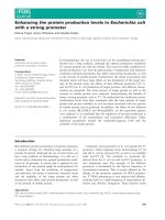
Fig. 1. RT-PCR analysis of Cdx2 expression.
(A) Scheme of a part of the mouse Cdx2
mRNA, including the stop codon ‘tga’,
which is shown in red. The primers used for
detecting Cdx2 transcript are indicated by
underlining and yellow shading. The exo-
n 2–exon 3 boundary site is indicated by an
arrow. (B) RT-PCR analysis of Cdx2 mRNA
transcripts (primer pair; Cdx2 coding-fw and
Cdx2 coding-rv) in normal mouse stomach
(lane 1), normal mouse small intestine
(lane 2), and Cdx2-transgenic mouse stom-
ach (lanes 3–8). (C) RT-PCR analyses of
endogenous Cdx2 mRNA transcripts (primer
pair; Cdx2 coding-fw and Cdx2 non-coding-
rv) in normal mouse stomach (lane 1),
normal mouse small intestine (lane 2), and
Cdx2-transgenic mouse stomach
(lanes 3–7). The lower panels in (B) and (C)
show standard RT-PCR conducted with
primers designed to detect b-actin mRNA.
H. Mutoh et al. Cdx1 expression in intestinal metaplasia
FEBS Journal 276 (2009) 5821–5831 ª 2009 The Authors Journal compilation ª 2009 FEBS 5823
Fig. 4C, Cdx1 promoter fw2 and Cdx1 promoter rv1).
Binding of Cdx2 to the promoter region of the Cdx1
gene, including the TATAAA sequence, was detected
in the intestinal metaplasia of the Cdx2-transgenic
mouse stomach and the normal intestine, but not in
the normal stomach (Fig. 4B,C).
Cdx2 binds to the TATAAA sequence
We investigated Cdx2 binding to the TATAAA
sequence, using the nuclear fractions extracted from
Cdx2-expressing AGS cells (Fig. 4D). We found that
nuclear extracts from AGS cells formed the Cdx2–
DNA complex (Fig. 4D). The presence of Cdx2 in
DNA–protein complexes was eliminated by using
monoclonal antibody specific to Cdx2 (Fig. 4D,
lane 3). With the use of a mutant probe, DNA–protein
complexes were not formed (Fig. 4D, lane 1). These
results indicate that Cdx2 binds to the TATAAA
sequence.
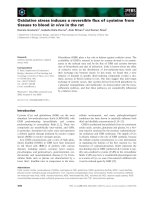
The Cdx1 promoter was activated in
Cdx2-expressing human gastric carcinoma
AGS, MKN45 and MKN74 cells
The expression of Cdx1 in the intestinal metaplasia of
the Cdx2-transgenic mouse stomach supports the
hypothesis that Cdx2 could regulate Cdx1 transcrip-
tion. Supporting this, the ChIP assay indicated that
Cdx2 is bound to the region between )191 and +112
(relating to the transcription start site). The region
between )191 and +112 contains the Cdx consensus
sequence TATAAA ()51 to )46) (Fig. 4A). Further-
more, electrophoretic mobility shift assay (EMSA)
indicated that Cdx2 binds to the TATAAA sequence.
We examined the Cdx1 transcriptional activity in
Cdx2-expressing AGS, MKN45 and MKN74 cells
(Fig. 5A), using pGL4.10[luc2]–Cdx1 deletion and
mutation constructs. These cell lines (AGS, MKN45,
and MKN74) also expressed Cdx1, which was detected
by RT-PCR (Fig. 5A). The Cdx1 promoter reporter
gene containing the region between )365 and +12 was
activated, whereas the Cdx1 promoter reporter gene
containing the region between )365 and )78 was not
activated, in Cdx2-expressing AGS, MKN45 and
MKN74 cells (Fig. 5B). This result suggests that the
element between )77 and +12 in the Cdx1 promoter
may be critical for Cdx1 gene transcriptional activity
in Cdx2-expressing AGS, MKN45 and MKN74 cells.
The sequence between )77 and +12 contains a poten-
tial Cdx2-binding site (TATAAA, )51 and )46). Anal-
ysis of a reporter construct with mutation of the Cdx2
consensus-binding element at )51 and )46 revealed
that the element was critical for transcriptional activity
of the Cdx1 reporter gene construct in AGS, MKN45
and MKN74 cells (Fig. 5B).
Furthermore, we examined the effects of the transfec-
tion of the Cdx2 expression plasmid or small interfering
RNA targeting Cdx2 (siRNA-Cdx2) on the trans-
criptional activities of the Cdx1 promoter luciferase
Table 1. The sequences of oligonucleotide primers used in this
study.
Primers Sequence (5¢-to3¢)
Primers used for mouse Cdx2 detection
Cdx2-fw CGGCTGGAGCTGGAGAAGG
Cdx2 coding-rv GACAGTGGAGTTTAAAACCC
Cdx2 noncoding-rv GCCTGGGATTGCTGTGCCG
Primers used for mouse Cdx1 detection
Cdx1cDNAfw CCGAACCAAGGACAAGTACC
Cdx1cDNArv GTTTACTTTGCGCTCCTTGG
Primers used for mouse b-actin detection
b-Actin-fw ATCTACGAGGGCTATGCTCT
b-Actin-rv TACTCCTGCTTGCTGATCCA
Primers used for human Cdx2 detection
Cdx2-human-fw AGCCAAGTGAAAACCAGGAC
Cdx2-human-rv ATTTCTTGAGGCCCCAAATC
Primers used for human Cdx1 detection
Cdx1-human-fw TCGGACCAAGGACAAGTACC
Cdx1-human-rv TGTTGCTGCTGCTGTTTCTT
Primers used for human GAPDH detection
GAPDH-fw ACGGATTTGGTCGTATTGGG
GAPDH-rv TGATTTTGGAGGGATCTCGC
Primers used for Cdx1 methylation
CpG-Cdx1-fw1
[)331 ⁄ )305]
GAG
TTAGTTTTTTTATTTGT
AA
TTTAG
CpG-Cdx1-fw2
[)312 ⁄ )293]
TAA
TTTAGGGGTGGGTGGTG
CpG-Cdx1-rv
[+114 ⁄ +89]
AAAAAATCCTTATCCAACAC
ATA
ACC
Primers used for Cdx2 methylation
CpG-Cdx2-fw1
[)234 ⁄ )212]
AGTG
TATTTAGGTTGGAAGGAG
CpG-Cdx2-fw2
[)206 ⁄ )185]
GTAG
TTAGTAAGAAGGGTTTGA
CpG-Cdx2-rv
[+194 ⁄ +173]
TA
ACTAACTACACCTCAACCCA
Primers used for ChIP assay
Cdx1 promoter-fw1 CTAGGGTCATGCCACCACTC
Cdx1 promoter-fw2 ATCCACCTCCCGCTTAGG
Cdx1 promoter-rv2 GGAGTCCTTGTCCAGCACAT
Primers used for Cdx1 promoter
Cdx1 promoter-fw1-XhoI
[)365 ⁄ )345]
CTCGAGCTAGGGTCATGCCACCACTC
Cdx1 promoter-rv1-HindIII
[+12 ⁄ )7]
AAGCTTACCAGCGACTGCTCACCT
Cdx1 promoter-rv2-HindIII
[)78 ⁄ )95]
AAGCTTAAGCTTGGGCGGCTTTGC
ATTTCA
Cdx1-Csp45I-fw
TTCGAAAGGCCGGGGTGGGGC
Cdx1-Csp45I-rv
TTCGAAGCCGCGGGCCGTCCGC
Cdx1 expression in intestinal metaplasia H. Mutoh et al.
5824 FEBS Journal 276 (2009) 5821–5831 ª 2009 The Authors Journal compilation ª 2009 FEBS
construct containing the region between )365 and +12
or the mutant Cdx1 reporter luciferase construct.
Cotransfection with the Cdx2 expression plasmid
increased the transcriptional activities of the intact Cdx1
reporter gene, but did not affect the transcriptional
activities of the mutant Cdx1 reporter gene, in AGS,
MKN45 and MKN74 cells (Fig. 5B). Cotransfection
with siRNA-Cdx2 decreased the transcriptional activi-
ties of the intact Cdx1 reporter gene, but did not affect
the transcriptional activities of the mutant Cdx1 repor-
ter gene, in AGS, MKN45 and MKN74 cells (Fig. 5B).
Next, after transfection of Cdx2 expression plasmid
or siRNA-Cdx2 into AGS, MKN45 and MKN74
cells, Cdx1 mRNA levels were measured using quanti-
tative real-time RT-PCR. As compared with the
transfection of a negative control, the transfection of
the Cdx2 expression plasmid resulted in an increase
in Cdx1 mRNA (Fig. 5C). As compared with the
transfection of a negative control, the transfection of
siRNA-Cdx2 resulted in a decrease in Cdx1 mRNA
(Fig. 5C).
Discussion
Intestinal metaplasia has been extensively studied as a
putative preneoplastic lesion in the human stomach [1].
In the present study, endogenous Cdx1, but not Cdx2,
was induced by transgenic Cdx2 in the intestinal meta-
plasia of the Cdx2-transgenic mouse stomach.
Cdx1 is essential for anterior–posterior vertebral
patterning of the body axis in the early embryonic per-
iod [13], and its expression persists selectively in the
intestinal epithelium from the later embryonic period
to the adult [14]. In addition to its physiological
expression, Cdx1 is ectopically expressed in the precan-
cerous intestinal metaplasia of the stomach and
Barrett’s esophagus. The regulatory mechanisms that
modulate Cdx1 gene expression during development
A
Normal Normal
Cdx2
3456781
stomach
2
intestine
stomach
Marker
9
β-actin
B
1
0.6
0.8
0.2
0.4
Normal
intestine
Normal
stomach
Cdx2
stomach
0
CDE
Cdx2 stomachNormal stomach Normal intestine
Fig. 2. RT-PCR and immunohistochemical
analysis of Cdx1 expression. (A) RT-PCR
analysis of Cdx1 expression. RT-PCR analy-
ses of Cdx1 mRNA transcripts in normal
mouse stomach (lanes 1 and 2), normal
mouse intestine (lanes 3 and 4) and Cdx2-
transgenic mouse stomach (lanes 5–9) are
shown. The lower panel in (A) shows
standard RT-PCR conducted with primers
designed to detect b-actin mRNA. (B) Cdx1
gene expression characterized by quantita-
tive real-time RT-PCR. The Cdx1 mRNA
level in Cdx2-transgenic mouse stomach
was almost the same as that in normal
mouse small intestine (B). (C–E) Immunohis-
tochemical staining for Cdx1 in the normal
stomach (C), the normal intestine (D) and
the intestinal metaplasia of the Cdx2-trans-
genic mouse stomach (E).
H. Mutoh et al. Cdx1 expression in intestinal metaplasia
FEBS Journal 276 (2009) 5821–5831 ª 2009 The Authors Journal compilation ª 2009 FEBS 5825
and in the normal intestinal epithelium have been
gradually clarified. Cdx1 is a direct transcriptional tar-
get of both retinoic acid (RA) and the Wnt ⁄ b-catenin
signaling pathway during early embryogenesis [15,16].
The Wnt ⁄ b-catenin signaling pathway is also active in
the crypt compartment [17]. Cdx1 regulation by RA
and Wnt3a is mediated, respectively, through the RA
response element and two LEF ⁄ TCF response ele-
ments present on the Cdx1 promoter [17]. However,
very little is known about the molecular mechanisms
for induction of the ectopic expression of the Cdx1
gene in the intestinal metaplasia of the H. pylori-
infected human stomach. In the present study, we
focused on the initiation of Cdx1 gene transcription in
the intestinal metaplasia through Cdx2-transgenic
mouse studies. Unlike in normal regulation, ectopic
expression of Cdx1 was upregulated by Cdx2. Cdx2
mRNA and protein were absent in the gastric-like
heteroplasias arising spontaneously in the pericecal
region and proximal colon of Cdx2
+ ⁄ )
mice, and, in
common with that of Cdx2, Cdx1 expression was also
absent in the gastric-like heteroplasias [18]. The finding
that the gastric-like heteroplasia, which does not
express Cdx2, also shows a lack of Cdx1 expression
is consistent with our present data showing that
the stomach expressing Cdx2 generated endogenous
Cdx1.
Epigenetic inactivation, in particular aberrant DNA
hypermethylation, is an important mechanism for
gene silencing. In the majority of human colon cancer
specimens and colorectal cancer cell lines, Cdx1
expression is lost due to active Cdx1 gene silencing
by promoter hypermethylation [12,19]. However, in
this study, we demonstrated that the Cdx1 promoter
is unmethylated in the normal stomach, the normal
intestine, and the intestinal metaplasia, indicating that
loss of Cdx1 expression in the normal stomach is not
associated with promoter hypermethylation. Cdx1 and
Cdx2 proteins bind to a binding site in an AT-rich
motif whose consensus sequence is C ⁄ TATAAAT ⁄ G
in direct or reverse orientation [20]. In some instances,
the Cdx-binding site presents high homology with the
A Cdx1 promoter
B Cdx2 promoter
Fig. 3. Cdx1 (A) and Cdx2 (B) promoter bisulfite sequencing of the stomach and intestine of normal mice and the intestinal metaplasia of
the Cdx2-transgenic mouse stomach. (A) A sequence of the 5¢-flanking region for the mouse Cdx1 gene, including the TATA box, transcrip-
tion start site and initiation codon (ATG). The TATA box is highlighted in green, the transcription start site in blue, and the initiation codon
(ATG) in red. Cdx1 promoter CpGs are shown in red. Base positions relative to the Cdx1 transcription start site are shown on the left of each
line of sequence. All CpGs were unmethylated. CpGs enclosed by the box ()54 to )68) represent those suggested to be crucial for tran-
scriptional control [12]. (B) A sequence of the 5¢-flanking region for the mouse Cdx2 gene, including the TATA box, transcription start site,
and initiation codon (ATG). The TATA box and another AT-rich motif, designated DBS (downstream binding site) [28], are highlighted in
green, the transcription start site in blue, and the initiation codon (ATG) in red. Cdx2 promoter CpGs are shown in red. Base positions rela-
tive to the Cdx2 transcription start site are shown on the left of each line of sequence. All CpGs were unmethylated, except for the CpG
enclosed by the box, which was methylated in the normal intestine and the intestinal metaplasia.
Cdx1 expression in intestinal metaplasia H. Mutoh et al.
5826 FEBS Journal 276 (2009) 5821–5831 ª 2009 The Authors Journal compilation ª 2009 FEBS
canonical TATA-box sequence, and, indeed, the Cdx1
and ⁄ or Cdx2 homeoproteins were found to be able to
bind to the TATA-boxes of some intestinal genes,
such as those of the calbindin-D9 gene [21], the clus-
terin gene [22], and the glucose-6-phosphatase gene
[23]. In the present study, CpGs in the 5 ¢-region of
the TATAAAA sequence located )51 ⁄ )45 upstream
of the transcription start site were also found to be
unmethylated in the normal stomach, the normal
intestine, and the intestinal metaplasia. The present
results, including those from ChIP, EMSA and
reporter gene analysis, indicate that Cdx2 is present
on the Cdx1 promoter region containing the
TATAAAA sequence located at )51 ⁄ )45. On the
other hand, endogenous Cdx2 was not expressed in
the intestinal metaplasia of the Cdx2-transgenic mouse
stomach, indicating that endogenous Cdx2 was not
autoregulated.
In the present study, we demonstrated that Cdx1 is
expressed in the Cdx2-induced intestinal metaplasia
of Cdx2-transgenic mice. This may coincide with our
previous clinical data why the expression of Cdx2 pre-
cedes that of Cdx1 during the progression of intestinal
metaplasia [3]. These clinical data also suggest that
Cdx2 might induce Cdx1 expression.
In conclusion, we propose that the ectopic expres-
sion of Cdx2 in the gastric epithelium is triggered first,
and in turn Cdx1 is directly induced by Cdx2 in the
intestinal metaplasia. The present results indicate that
Cdx2 induces Cdx1 expression by directly binding to
the Cdx2-consensus cis-regulatory element of the
unmethylated Cdx1 promoter region.
Experimental procedures
Cdx2-transgenic mice
The Cdx2-transgenic mice we generated had free access to
standard food and drinking water and were maintained on
a 12 h light ⁄ dark cycle. All experiments in this study were
performed in accordance with the Jichi Medical University
Guide for Laboratory Animals.
A
Cdx1 promoter-fw1
–400
–341
–281
Cdx1 promoter-fw2
TATA box
–221
–161
–101
Cdx1 promoter-rv1
Initiation codon
Transcription start site (+1)
+20
+80
–41
B
312 45
C
312 45
D
312
Fig. 4. Cdx2 is present on the Cdx1 promoter region in vivo. (A) A sequence of the 5¢-flanking region for the mouse Cdx1 gene, including
the TATA box, transcription start site, and initiation codon. The TATA box is highlighted in green, the transcription start site in blue, and the
initiation codon in red. PCR fragments corresponding to the DNA sequences including the TATA box were designed for ChIP analysis. The
sequences for the primers used for ChIP assays are underlined and highlighted in yellow. The base positions relative to the transcription
start site for the mouse Cdx1 gene are shown on the left of each line of sequence. (B, C) ChIP assays that were performed using a Cdx2
antibody [26] or control IgG. The region of the Cdx1 promoter encompassing the TATA box sequence was amplified by PCR with the follow-
ing primer pairs: (B) Cdx1 promoter-fw1 and Cdx1 promoter-rv1; (C) Cdx1 promoter-fw2 and Cdx1 promoter-rv1. Lane 1: normal stomach.
Lane 2: normal intestine. Lane 3: Cdx2-transgenic mouse stomach. Lane 4: input. Lane 5: control IgG. (D) EMSA. A radiolabeled dsDNA
probe (CCCGCGGCTATAAAAGGCCGGGGTGGGG) containing the TATAAA sequence in the Cdx1 promoter was incubated with nuclear
extracts from AGS cells and separated on a 5% polyacrylamide gel (lane 2). Specificity was determined by addition of antibody for supershift
(lane 3) and mutant probe (CCCGCGGCTTCGAAAGGCCGGGGTGGGG) (lane 1).
H. Mutoh et al. Cdx1 expression in intestinal metaplasia
FEBS Journal 276 (2009) 5821–5831 ª 2009 The Authors Journal compilation ª 2009 FEBS 5827
RNA isolation and RT-PCR
Total RNA was extracted from the stomach (normal mice),
small intestine (normal mice), intestinal metaplasia (Cdx2-
transgenic mice), and human gastric cancer cell lines AGS,
MKN45 and MKN74, using the guanidinium isothiocya-
nate ⁄ phenol method (Isogen; Nippon Gene, Tokyo, Japan),
according to the manufacturer’s instructions. Total RNA
(1 lg) was reverse-transcribed as previously described [24].
To compare endogenous Cdx1 expression, endogenous Cdx2
expression and total (endogenous and transgenic) Cdx2
expression in the stomach (normal mice), small intestine
(normal mice), and intestinal metaplasia (Cdx2-transgenic
mice), PCR amplification was performed using the primer
pairs Cdx1cDNAfw and Cdx1cDNArv (for endogenous
Cdx1), Cdx2-fw and Cdx2 coding-rv (for total Cdx2), and
Cdx2-fw and Cdx2 noncoding-rv (for endogenous Cdx2)
(Table 1), by incubation at 94 °C for 2 min, followed by 35
cycles of 94 °C for 30 s, 60 °C for 30 s and 72 °C for 30 s,
and a final extension at 72 °C for 10 min. The PCR products
were separated in 2% agarose gels. As an internal standard,
RT-PCR was performed with primers hybridizing to the
mRNA encoding b-actin or glyceraldehyde-3-phosphate
dehydrogenase (GAPDH) (Table 1).
Real-time RT-PCR
One hundred nanograms of cDNA was used in each real-
time PCR reaction. Expression levels for the Cdx1 gene
were determined by real-time PCR using ready-to use
Assay-on-Demand gene expression product (Applied Bio-
systems, Foster City, CA, USA): Mm00438172_m1 for
mouse Cdx1, and Hs00156451_m1 for human Cdx1. Each
Assay-on-Demand gene expression product contains tar-
get-specific primers and probes and a Taqman Gene
Expression Master Mix containing AmpErase uracil-N-gly-
cosylase (Applied Biosystems) to prevent reamplification of
carryover PCR products. PCR amplification and fluores-
cence data collection were performed with the ABI
PRISM 7900 HT Sequence Detection System (Applied
Biosystems), using the following conditions: 50 °C for
A
AGS
MKN45
MKN74
Cdx2
GAPDH
Cdx1
B
Luciferase activity
(Ratio of firefly to renilla luciferase)
024681012
1814 16
Luciferase
(–)
(–)
(–)
+Cdx2
+siCdx2
+Cdx2
(–)
(–)
–365 +12
+siCdx2
(–)
+Cdx2
+siCdx2
+Cdx2
+siCdx2
+Cdx2
(–)
(–)
+siCdx2
(–)
+Cdx2
+siCdx2
(–)
(–)
(–)
–78–365
–365 +12
AGS
MKN45
MKN74
TATAAA
TTCGAA
–51 –46
C
1
2
Relative Cdx1 expression
0
Cdx2
siRNA
+
+
+
+
+
+
AGS MKN45 MKN74
Fig. 5. Activation of the Cdx1 promoter in Cdx2-expressing AGS,
MKN45 and MKN74 cells. (A) Cdx2 and Cdx1 expression deter-
mined by RT-PCR. Human gastric carcinoma AGS, MKN45 and
MKN74 cells expressed both Cdx2 and Cdx1. The lower panel in
(A) shows standard RT-PCR conducted with primers designed to
detect GAPDH mRNA. (B) Cdx1 promoter reporter gene activities.
AGS, MKN45 and MKN74 cells were transiently transfected with
the different fragments of Cdx1 promoter fused to a luciferase
reporter vector, pGL4.10[luc2], and pGL4.70[hRluc] vector. Lucifer-
ase activities were normalized relative to the level of Renilla lucifer-
ase activities. The lengths of the promoter fragments tested are
indicated. The numbers correspond to the relative positions with
respect to the transcription start site. The sequence of the pre-
sumptive Cdx2-binding site (TATAAA) was changed to TTCGAA.
Cdx1 promoter reporter plasmids were added to each plate with or
without Cdx2 expression vector (pRC ⁄ CMV–Cdx2) or siRNA
(Applied Biosystems, Silencer Select Pre-designed siRNA, #s2878;
UUCUUGUUGAUUUUCCUCUcc). The luciferase activities of empty
pGL4.10[luc2], which does not contain any Cdx1 promoter, were
used as controls for AGS, MKN45 and MKN74 cells, respectively.
Each bar represents the mean ± standard error. Transfections were
performed in triplicate and repeated three times. (C) Cdx1 mRNA
levels of AGS, MKN45 and MKN74 cells transfected with Cdx2
expression plasmid, Cdx2 siRNA, or negative control. At 24 h after
transfection, total RNA was extracted.
Cdx1 expression in intestinal metaplasia H. Mutoh et al.
5828 FEBS Journal 276 (2009) 5821–5831 ª 2009 The Authors Journal compilation ª 2009 FEBS
2 min, 95 ° C for 10 min, and 40 cycles for amplification
(95 °C for 15 s, and 60 °C for 1 min). PCR reactions were
performed in 96-well plates, using a final volume of 20 lL,
and the Cdx1 gene was studied in triplicate. In order to
normalize RNA transcript abundance for the Cdx1 gene, a
housekeeping gene (the b-actin gene) (Pre-Developed Taq-
man Assay Reagents; Applied biosystems) was used to cal-
culate the DC
T
(DC
T
= C
T target
⁄ C
T actin
). The C
t
values
for the b-actin gene for the normal stomach, the normal
intestine and Cdx2-transgenic mouse stomach tissues fell
in a close range, with no specific pattern of spatial or tem-
poral variation (data not shown). A relative quantification
approach was used in this study to describe the change in
expression of the target gene in a test sample relative to a
calibrator sample (reference). The relative RNA transcript
abundance value was calculated as follows. First, the DC
T
for the normal stomach, normal small intestine and Cdx2-
transgenic mouse stomach tissues was calculated. In the
second step, differences between the normal and Cdx2-
transgenic mouse stomach tissues were calculated as DDC
T
(DC
T target
⁄ DC
T reference
). The normal mouse small intestine
was used as reference for Cdx1 expression. Finally, the
fold difference (relative abundance) was calculated using
the formula 2
)DDCT
[25], and was plotted as means
(n = 6).
Immunohistochemistry
Murine tissue sections were stained with the antibody for
Cdx1 (1 : 40, rabbit polyclonal; Abcam, Cambridge, UK)
after antigenicity was enhanced by autoclaving the sections,
as previously described [24].
Bisulfite sequencing for Cdx1 and Cdx2
promoters
The methylation status of gene promoter CpGs is best
analyzed by using direct sequencing after sodium bisulfite
modification of target DNA (bisulfite sequencing). DNA
(1 lg of DNA per sample) was sodium bisulfite modified
with the DNA modification kit (Zymo Research Intergen,
Purchase, NY, USA), according to the manufacturer’s
instructions. A 426 bp region of Cdx1 was amplified from
bisulfite-modified genomic DNA by nested PCR using two
sets of primers. Genomic DNAs were extracted from five
stomachs and five intestines of five normal mice and five
stomachs of five Cdx2-transgenic mice. The first PCR
reaction was performed using the forward primer CpG-
Cdx1-fw1[)331 ⁄ )305] and the reverse primer CpG-Cdx1-
rv[+114 ⁄ +89] (Table 1). A second, nested, PCR was then
performed on 1 lL of the amplificate, using the upstream
(CpG-Cdx1-fw2[)312 ⁄ )293]) and downstream (CpG-Cdx1-
rv[+114 ⁄ +89]) primers (Table 1). A 400 bp region of
Cdx2 was amplified from bisulfite-modified genomic DNA
by nested PCR, using two sets of primers. The first PCR
reaction was performed using the forward primer
CpG-Cdx2-fw1[)234 ⁄ )212] and reverse primer CpG-Cdx2-
rv[+194 ⁄ +173] (Table 1). A second, nested, PCR was
then performed on 1 lL of the amplificate, using the
upstream (CpG-Cdx2-fw2[)206 ⁄ )185]) and downstream
(CpG-Cdx2-rv[+194 ⁄ +173]) primers (Table 1). The pri-
mer pairs were designed to bind sequences lacking any
CpGs, therefore avoiding any preferential amplification of
methylated or unmethylated DNA strands. The PCR
products were purified (GenElute agarose spin column;
Sigma, St Louis, MO, USA), and the purified product was
used for cloning (Topo TA Cloning kit; Invitrogen, Carls-
bad, CA, USA) and sequencing by using the Big Dye
Terminator Cycle Sequencing kit (Applied Biosystems).
ChIP assay
The mucosae removed from the stomach (normal mice), the
small intestine (normal mice) and the intestinal metaplasia
(Cdx2-transgenic mice) were incubated with fixation solu-
tion (1% formaldehyde, 4.5 mm Hepes, pH 8.0, 9 mm
NaCl, 0.09 mm EDTA, 0.04 mm EGTA) in NaCl ⁄ P
i
for
30 min at 37 °C. The reaction was terminated by the addi-
tion of glycine to a final concentration of 150 mm. After
being washed in NaCl/P
i
containing protease inhibitors
(Protease inhibitor cocktail; Sigma), the samples were soni-
cated in SDS lysis buffer (50 mm Tris ⁄ HCl, pH 8.0, 10 mm
EDTA, pH 8.0, 1% SDS, 0.5 mm phenylmethanesulfonyl
fluoride), when the DNA size of samples was 200–500 bp.
The solubilized chromatin was incubated with anti-Cdx2
IgG (BioGenex, San Ramon, CA, USA) [26] or control
IgG for 90 min at 4 °C. Beads were washed five times with
IP buffer (50 mm Hepes, pH 7.5, 150 mm KCl, 5 mm
MgCl
2
,10lm ZnSO
4
, 1% Triton X-100, 0.05% SDS), and
then incubated with elution buffer (50 mm Tris ⁄ HCl,
pH 8.0, 1% SDS, 10 mm EDTA) for 30 min at 65 °C. The
supernatant was collected and coimmunoprecipitated DNA
was recovered. Primer sequences used for the ChIP assays
are listed in Table 1. All ChIP assays were repeated at least
twice, and representative data are presented.
EMSA
Nuclear fractions were extracted for EMSA from AGS
cells. To extract nuclear fractions for EMSA studies, AGS
cells were washed in NaCl ⁄ P
i
, and subjected to swelling in
400 lL of hypotonic buffer A (10 mm Hepes, pH 7.9,
10 mm KCl, 0.1 mm EDTA, 0.1 mm EGTA, 1 mm dith-
iothreitol) supplemented with protease inhibitor cocktail
(Sigma Chemical Co.), and lysed [27]. Then, 25 lL of 10%
Nonidet P-40 solution were added, and nuclear fractions
were collected by sedimentation for 5 min at 500 g. Super-
natants were discarded, and precipitated nuclei were resus-
pended in 100 lL of buffer C (20 mm Hepes, pH 7.9,
400 mm NaCl, 1 mm dithiothreitol, 1 mm EDTA, 1 mm
H. Mutoh et al. Cdx1 expression in intestinal metaplasia
FEBS Journal 276 (2009) 5821–5831 ª 2009 The Authors Journal compilation ª 2009 FEBS 5829
EGTA, and protease inhibitor cocktail) and centrifuged for
5 min at 14 000 g. Supernatants containing nuclear proteins
were collected, and tested for their ability to bind labeled
nucleotides corresponding to the Cdx1 promoter. All
DNA–protein binding reaction protocols were those of the
manufacturer (Promega, Madison, WI, USA). The dsDNA
probes used in the gel mobility shift assays were as follows:
wild-type sequence, CCCGCGGCTATAAAAGGCCGGG
GTGGGG; mutant sequence, CCCGCGGCTTCGAAAG
GCCGGGGTGGGG. Briefly, 0.5 ng of
32
P-labeled probe
was incubated for 20 min at 4 °C with 5 lg of nuclear
extracts in the presence of 1 · gel shift buffer (Promega).
Subsequently, 1.5 lLof10· loading buffer were added to
the reaction, and this was followed by separation by elec-
trophoresis on 5% nondenaturing polyacrylamide gel until
free probe was close to the bottom of the gel.
Luciferase assays
To construct the luciferase reporter vector pGL4.10[luc2]–
Cdx1, 377 bp ()365 to +12) and 288 bp ()365 to )78)
fragments, located at 5¢-region of the mouse Cdx1 coding
sequence, were amplified by PCR with specific primers
(Table 1) from 500 ng of mouse genomic DNA. The ampli-
fied fragments for the Cdx1 promoter were directly cloned
into the TA cloning vector pCRII (Invitrogen), to yield the
plasmid pCRII ⁄ Cdx1 promoter. Each pCRII ⁄ Cdx1 pro-
moter was digested with XhoI and HindIII (sites underlined
in the primers in Table 1), and the resulting fragments were
subcloned into the XhoI and HindIII restriction sites of the
pGL4.10[luc2] vector (Promega) and confirmed by sequence
analysis. The sequence of the presumptive Cdx2-binding
site (TATAAA) was changed to TTCGAA (underlined in
the primers) by using Cdx1-Csp45I-fw and Cdx1-Csp45I-rv
primers (Table 1).
AGS, MKN45 and MKN74 cells were seeded at
2 · 10
5
cells per well in Nunc 24-well dishes 18–24 h before
transfection. Transient transfections were performed using
Lipofectamine 2000 (Invitrogen). One hundred nanograms
of a Cdx1 promoter reporter plasmid with or without
800 ng of Cdx2 expression vector (pRC ⁄ CMV–Cdx2) or
2.5 pmol of siRNA (Applied Biosystems, Silencer Select
Pre-designed siRNA, #s2878; UUCUUGUUGAUUUUC
CUCUcc) were added to each plate, together with 50 ng of
the Renilla luciferase control reporter plasmid
(pGL4.70[hRluc]; Promega) as a control for the transfection
efficiency. At 24 h after transfection, the cells were lysed in
lysis buffer (Promega), and the firefly and Renilla luciferase
activities were measured, using the Dual-Luciferase Repor-
ter Assay System (Promega) in a luminometer. The relative
firefly luciferase activities were calculated by normalizing
the transfection efficiency according to the Renilla luciferase
activities produced by the internal control plasmid
pGL4.70[hRluc]. Three separate experiments were per-
formed in triplicate.
Transfections of Cdx2 expression plasmid or
Cdx2 siRNA
AGS, MKN45 and MKN74 cells were plated in 10 cm
plates 24 h before transfection. Transfections were
performed using Lipofectamine 2000, following the
manufacturer’s protocol (Invitrogen). Six micrograms of
Cdx2 expression plasmid and 25 pmol of siRNA or nega-
tive control were used for the transfection. siRNA (Applied
Biosystems, Silencer Select Pre-designed siRNA, #s2878;
UUCUUGUUGAUUUUCCUCUcc) was used. At 24 h
after transfection, total RNA was extracted.
References
1 Correa P (1992) Human gastric carcinogenesis: a multi-
step and multifactorial process – First American Cancer
Society Award Lecture on Cancer Epidemiology and
Prevention. Cancer Res 52, 6735–6740.
2 Silberg DG, Furth EE, Taylor JK, Schuck T, Chiou T
& Traber PG (1997) CDX1 protein expression in nor-
mal, metaplastic, and neoplastic human alimentary tract
epithelium. Gastroenterology 113, 478–486.
3 Eda A, Osawa H, Yanaka I, Satoh K, Mutoh H, Kihira
K & Sugano K (2002) Expression of homeobox gene
CDX2 precedes that of CDX1 during the progression
of intestinal metaplasia. J Gastroenterol 37, 94–100.
4 Satoh K, Mutoh H, Eda A, Yanaka I, Osawa H,
Honda S, Kawata H, Kihira K & Sugano K (2002)
Aberrant expression of CDX2 in the gastric mucosa
with and without intestinal metaplasia: effect of eradica-
tion of Helicobacter pylori. Helicobacter 7, 192–198.
5 Mutoh H, Hakamata Y, Sato K, Eda A, Yanaka I,
Honda S, Osawa H, Kaneko Y & Sugano K (2002)
Conversion of gastric mucosa to intestinal metaplasia in
Cdx2-expressing transgenic mice. Biochem Biophys Res
Commun 294, 470–479.
6 Mutoh H, Satoh K, Kita H, Sakamoto H, Hayakawa
H, Yamamoto H, Isoda N, Tamada K, Ido K &
Sugano K (2005) Cdx2 specifies the differentiation of
morphological as well as functional absorptive entero-
cytes of the small intestine. Int J Dev Biol 49, 867–871.
7 Mutoh H, Sakurai S, Satoh K, Tamada K, Kita H,
Osawa H, Tomiyama T, Sato Y, Yamamoto H, Isoda
N et al. (2004) Development of gastric carcinoma from
intestinal metaplasia in Cdx2-transgenic mice. Cancer
Res 64, 7740–7747.
8 Eda A, Osawa H, Satoh K, Yanaka I, Kihira K, Ishino
Y, Mutoh H & Sugano K (2003) Aberrant expression
of CDX2 in Barrett’s epithelium and inflammatory
esophageal mucosa. J Gastroenterol 38, 14–22.
9 Kazumori H, Ishihara S, Rumi MA, Kadowaki Y &
Kinoshita Y (2006) Bile acids directly augment caudal
related homeobox gene Cdx2 expression in oesophageal
keratinocytes in Barrett’s epithelium. Gut 55, 16–25.
Cdx1 expression in intestinal metaplasia H. Mutoh et al.
5830 FEBS Journal 276 (2009) 5821–5831 ª 2009 The Authors Journal compilation ª 2009 FEBS
10 Debruyne PR, Witek M, Gong L, Birbe R, Chervoneva
I, Jin T, Domon-Cell C, Palazzo JP, Freund JN, Li P
et al. (2006) Bile acids induce ectopic expression of
intestinal guanylyl cyclase C through nuclear factor-
kappaB and Cdx2 in human esophageal cells. Gastroen-
terology 130, 1191–1206.
11 Liu T, Zhang X, So CK, Wang S, Wang P, Yan L,
Myers R, Chen Z, Patterson AP, Yang CS et al. (2007)
Regulation of Cdx2 expression by promoter methyla-
tion, and effects of Cdx2 transfection on morphology
and gene expression of human esophageal epithelial
cells. Carcinogenesis 28, 488–496.
12 Wong NA, Britton MP, Choi GS, Stanton TK,
Bicknell DC, Wilding JL & Bodmer WF (2004) Loss
of CDX1 expression in colorectal carcinoma: promoter
methylation, mutation, and loss of heterozygosity
analyses of 37 cell lines. Proc Natl Acad Sci USA 101,
574–579.
13 Subramanian V, Meyer BI & Gruss P (1995) Disruption
of the murine homeobox gene Cdx1 affects axial skele-
tal identities by altering the mesodermal expression
domains of Hox genes. Cell 83, 641–653.
14 Silberg DG, Swain GP, Suh ER & Traber PG (2000)
Cdx1 and cdx2 expression during intestinal develop-
ment. Gastroenterology 119, 961–971.
15 Houle M, Prinos P, Iulianella A, Bouchard N & Lohnes
D (2000) Retinoic acid regulation of Cdx1: an indirect
mechanism for retinoids and vertebral specification.
Mol Cell Biol 20, 6579–6586.
16 Ikeya M & Takada S (2001) Wnt-3a is required for
somite specification along the anteroposterior axis of
the mouse embryo and for regulation of cdx-1 expres-
sion. Mech Dev 103, 27–33.
17 Lickert H, Domon C, Huls G, Wehrle C, Duluc I,
Clevers H, Meyer BI, Freund JN & Kemler R (2000)
Wnt ⁄ (beta)-catenin signaling regulates the expression of
the homeobox gene Cdx1 in embryonic intestine. Devel-
opment 127, 3805–3813.
18 Bonhomme C, Duluc I, Martin E, Chawengsaksophak
K, Chenard MP, Kedinger M, Beck F, Freund JN &
Domon-Dell C (2003) The Cdx2 homeobox gene has a
tumour suppressor function in the distal colon in addi-
tion to a homeotic role during gut development. Gut 52,
1465–1471.
19 Suh ER, Ha CS, Rankin EB, Toyota M & Traber PG
(2002) DNA methylation down-regulates CDX1 gene
expression in colorectal cancer cell lines. J Biol Chem
277, 35795–35800.
20 Margalit Y, Yarus S, Shapira E, Gruenbaum Y & Fain-
sod A (1993) Isolation and characterization of target
sequences of the chicken CdxA homeobox gene. Nucleic
Acids Res 21, 4915–4922.
21 Lambert M, Colnot S, Suh E, L’Horset F, Blin C, Cal-
liot ME, Raymondjean M, Thomasset M, Traber PG &
Perret C (1996) cis-Acting elements and transcription
factors involved in the intestinal specific expression of
the rat calbindin-D9K gene: binding of the intestine-
specific transcription factor Cdx-2 to the TATA box.
Eur J Biochem 236, 778–788.
22 Suh E, Wang Z, Swain GP, Tenniswood M & Traber PG
(2001) Clusterin gene transcription is activated by cau-
dal-related homeobox genes in intestinal epithelium. Am
J Physiol Gastrointest Liver Physiol 280, G149–G156.
23 Gautier-Stein A, Domon-Dell C, Calon A, Bady I,
Freund JN, Mithieux G & Rajas F (2003) Differential
regulation of the glucose-6-phosphatase TATA box by
intestine-specific homeodomain proteins CDX1 and
CDX2. Nucleic Acids Res 31, 5238–5246.
24 Mutoh H, Sakurai S, Satoh K, Osawa H, Hakamata Y,
Takeuchi T & Sugano K (2004) Cdx1 induced intestinal
metaplasia in the transgenic mouse stomach: compara-
tive study with Cdx2 transgenic mice. Gut 53, 1416–
1423.
25 Livak KJ & Schmittgen TD (2001) Analysis of relative
gene expression data using real-time quantitative PCR
and the 2(-Delta Delta C(T)) method. Methods 25, 402–
408.
26 Uesaka T & Kageyama N (2004) Cdx2 homeodomain
protein regulates the expression of MOK, a member of
the mitogen-activated protein kinase superfamily, in the
intestinal epithelial cells. FEBS Lett 573, 147–154.
27 Schreiber E, Matthias P, Muller MM & Schaffner W
(1989) Rapid detection of octamer binding proteins with
‘mini-extracts’, prepared from a small number of cells.
Nucleic Acids Res 17, 6419.
28 Xu F, Li H & Jin T (1999) Cell type-specific autoregula-
tion of the caudal-related homeobox gene Cdx-2 ⁄ 3.
J Biol Chem 274, 34310–34316.
H. Mutoh et al. Cdx1 expression in intestinal metaplasia
FEBS Journal 276 (2009) 5821–5831 ª 2009 The Authors Journal compilation ª 2009 FEBS 5831