Báo cáo khoa học: LRRK2 in Parkinson’s disease: in vivo models and approaches for understanding pathogenic roles pdf
Bạn đang xem bản rút gọn của tài liệu. Xem và tải ngay bản đầy đủ của tài liệu tại đây (215.68 KB, 10 trang )
MINIREVIEW
LRRK2 in Parkinson’s disease: in vivo models and
approaches for understanding pathogenic roles
Zhenyu Yue
Department of Neurology and Neuroscience, Mount Sinai School of Medicine, New York, NY, USA
Introduction
Clinical symptoms of patients carrying Parkinson’s dis-
ease (PD)-associated mutations of leucine-rich repeat
kinase 2 (LRRK2) are indistinguishable from typical
sporadic PD. The spectra of neuropathological features
of PARK8 (LRRK2) patients is broad and appears to
encompass those associated with other familial PD
cases such as PARK1 (a-synuclein) and PARK2
(Parkin). However, the neuropathology of PARK8 is
variable and is not always associated with the presence
of intracellular inclusions (e.g. Lewy body, tau tangles
and ubiquitin inclusions) [1,2]. Recent studies also
suggest that the penetrance of LRRK2 pathogenic
mutations is incomplete [3,4].
LRRK2 encodes a large complex protein consisting
of 2527 amino acids (285 kDa). It belongs to the
ROCO family, which is defined by the presence of a
Ras of complex proteins (ROC) domain followed by a
C-terminal of ROC (COR) domain of unknown
Keywords
animal models; BAC transgenics; dopamine;
GTPase; kinase; leucine-rich repeat
kinase 2 (LRRK2); Parkinson’s disease;
pathogenesis; ROCO
Correspondence
Z. Yue, Department of Neurology and
Neuroscience, Mount Sinai School of
Medicine, New York, NY 10029, USA
Fax: +1 212 241 3869
Tel: +1 212 241 3155
E-mail:
(Received 30 May 2009, revised 30 July
2009, accepted 18 August 2009)
doi:10.1111/j.1742-4658.2009.07343.x
The recent discovery of the genetic causes for Parkinson’s disease (PD) is
fruitful; however, the continuing revelation of PD-related genes is rapidly
outpacing the functional characterization of the gene products. Although
the discovery of multiple PD-related genes places PD as one of the most
complex multigenetic diseases of the brain, it will undoubtedly facilitate the
unfolding of a central pathogenic pathway and an understanding of the eti-
ology of PD. Recent findings of pathogenic mutations in leucine-rich repeat
kinase 2 (LRRK2) (PARK8) that are linked to the most common familial
forms and some sporadic forms of PD provide a unique opportunity to
gain insight into the pathogenesis of PD. Despite rapid growth in biochem-
ical, structural and in vitro cell culture studies of LRRK2, the in vivo char-
acterizations of LRRK2 function generally fall short and are largely
limited to invertebrates. The investigation of LRRK2 or homologs of
LRRK2 in nonmammalian models provides important clues with respect
to the cellular functions of LRRK2, but an elucidation of the physiology
and pathophysiology of LRRK2 relevant to PD would still depend on
mammalian models established by multiple genetic approaches, followed by
rigorous examination of the models for pathological process. This minire-
view summarizes previous studies of genes for ROCO and LRRK2 homo-
logs in slime mold, nematode worms and fruit flies. It also discusses the
results obtained from available mouse models of LRRK2 that begin to
provide information for understanding LRRK2-mediated pathogenesis in
PD.
Abbreviations
BAC, bacterial artificial chromosome; COR, C-terminal of ROC; KO, knockout; LRCK, LRR-ROC-COR-kinase; LRRK2, leucine-rich repeat
kinase 2; PD, Parkinson’s disease; ROC, Ras of complex proteins; TH, tyrosine hydroxylase.
FEBS Journal 276 (2009) 6445–6454 ª 2009 The Author Journal compilation ª 2009 FEBS 6445
function [5]. LRRK2 also contains armadillo-like
repeats, LRR, kinase and WD40 domains [6]. In vitro
biochemical analysis demonstrates that LRRK2 con-
tains kinase and GTPase activities that are apparently
altered by pathogenic mutations of LRRK2 [7–12]. In
addition, studies using cultured cells or neurons show
that enhancement of kinase activity in PD-related
mutants of LRRK2 is correlated with increased neuro-
toxicity, thus implicating a causal role of aberrant
enzymatic activity of LRRK2 in neuropathogenesis
[9,13–15]. However, whether this possible gain-of-func-
tion in the kinase activity of LRRK2 contributes to
the the pathological process of PD has yet to be shown
in mammalian models.
Understanding the physiological function of LRRK2
under normal conditions and in the context of PD
remains a daunting task, especially given the complex-
ity of LRRK2 protein structure, which consists of mul-
tiple functional domains that are likely to be involved
in numerous cellular pathways. There is clearly a need
to investigate LRRK2 structure ⁄ function-related pro-
teins (e.g. ROCO family proteins) in various model
systems, including lower eukaryotes and invertebrates,
in order to obtain clues for building important hypoth-
eses. The rapidity and efficiency of in vivo studies in
many nonmammal models have already provided
timely information about molecular mechanisms of
many disease processes and will continue to impact
our understanding of disease pathogenesis. This mini-
review will examine the previous studies of genes for
ROCO or LRRK2 homologs in slime mold Dictyoste-
lium discoideum, nematode worms Caenorhabditis ele-
gans and fruit flies Drosophila melanogaster. It will
also discuss the available information reported in the
literature (albeit limited), as well as ongoing studies in
several laboratories that have created LRRK2 rodent
models.
ROCO proteins in slime mold
D. discoideum
The finding of the conserved ROC and COR domains
in LRRK2 has stirred particular interest with respect
to studying the functions of the known ROCO pro-
teins. The first ROCO protein was identified in slime
mold Dictyostelium [5] and, so far, at least two ROCO
proteins, GbpC and Pats1, have been characterized
in vivo in this species [16,17]. Similar to LRRK2,
GbpC and Pats1 both have LRR and kinase domains
flanking the central ROCO sequence. The sequence
arrangement of these functional motifs ‘LRR-ROC-
COR-kinase’ (LRCK) is also found in LRRK2
homologs of nematode worms and fruit flies. The inves-
tigation of ROCO structure of a prokaryotic protein in
Chlorobium tepidum revealed mechanistic insight into
protein dimerization and the regulation of ROC GTPase
activity [18], which may be involved in intramolecular
control of the kinase activity [9,19]. It is possible that
the subgroup of ROCO proteins (including mammalian
LRRK2), which contain the conserved functional motifs
‘LRCK’, may adopt a similar structural mechanism to
regulate enzymatic activities and their cellular functions
(Fig. 1).
A series of in vivo studies have revealed that the
ROCO proteins, GbpC and Pats1, are involved in mul-
tiple cellular processes: chemotaxis, cell division and
development. Deletion of GbpC in D. discoideum was
shown to cause a reduction of chemotactic reaction
towards cAMP [20]. In addition, loss of GbpC was
associated with a decrease in phosphorylation of myo-
sin II and a change in subcellular localization of myo-
sin heavy chain [21]. The chemotactic ‘rescue’
experiment showed that the kinase domain alone is
insufficient to complement the chemotactic defect in
the GbpC-deletion mutant. Furthermore, the study
indicated that the LRR, ROC and kinase domains are
all required for chemotaxis [16]. These results thus sug-
gest that the functional integrity of GbpC protein
requires all subdomains in the core ‘LRCK’ sequence.
ROCO protein Pats1 was originally found in a
genetic screen of cellular defect in cytokinesis of D. dis-
coideum [17]. Mutant cells with Pats1 deletion exhibit
abnormal cell morphology and division deficits. It was
shown that the WD40 domain of Pats1 interacted with
myosin heavy chain, whereas the deletion of Pats1
caused an alteration in the localization of myosin heavy
chain [17]. Moreover, over-expression of the kinase
domain alone resulted in a similar phenotype to that of
the deletion mutants, suggesting that deregulation of
kinase activity underlies the mechanism of cytokinesis
impairment. Taken together, the studies of mutant phe-
notypes for the two ROCO proteins in D. discoideum
indicate their roles in regulating cytoskeleton structures.
Interestingly, it was previously noted that human
LRRK2 binds to cytoskeleton-related proteins [22],
microtubules [23] and phosphorylates moesin, a protein
that anchors the actin cytoskeleton to the plasma mem-
brane [24]. Although cytoskeleton proteins are known
as frequent ‘contaminants’ in the process of searching
binding proteins, the in vivo evidence for the relation-
ship between ROCO proteins and cytoskeletons in
D. discoideum suggests a need to further investigate
the possibility that cytoskeleton proteins are the
physiological targets of LRRK2. It is possible that
LRRK2 regulates cytoskeletal mobility, which is linked
to various cellular vesicle trafficking events.
Approaches for understanding pathogenic roles Z. Yue
6446 FEBS Journal 276 (2009) 6445–6454 ª 2009 The Author Journal compilation ª 2009 FEBS
The nematode worm models
Despite the fact that nematode worms are used exten-
sively as an in vivo model to study disease gene func-
tion, there are only a few reports on LRRK2 or LRK-1
(LRRK2 ortholog in C. elegans) available to date.
LRK-1 is the only ortholog of human LRRK2 found
in C. elegans. The product of LRK-1 shares a con-
served ‘LRCK’ core sequence with LRRK2 (Fig. 1).
The first study characterized the phenotype of mutant
worms containing deletion of LRK-1. It provided
important evidence implicating a role for LRK-1 in
synaptic vesicle localization [25]. It showed that synap-
tic vesicles and their associated proteins are exclusively
localized in the pre-synaptic regions but not in
dendrites. By contrast, in mutant worms carrying
truncated LRK-1, synaptic vesicle proteins are located
in axons (pre-synaptic), as well as dendritic terminals
(post-synaptic). The localization of the synaptic vesicle
proteins in both pre- and post-synaptic regions as a
result of the lack of LRK-1 apparently is not a random
event because the mislocalization of the synaptic pro-
teins in dendrites depends on AP-1 clathrin adaptor,
which is known to be involved in dendritic transport,
but not on Unc104 kinesin, a motor protein required
for axonal transport. Therefore, this study suggests
that LRK-1 protein, as a resident of Golgi apparatus,
controls the directionality of synaptic vesicle proteins
by restricting these proteins from going to the
dendrites. This result further indicates a critical func-
tion of LRK-1 in establishing the polarity of synaptic
vesicle proteins and perhaps in regulating synaptic ves-
icle life cycle in the axons. However, this study did not
reveal any results regarding the viability of neurons,
especially dopaminergic neurons, or any functional
consequence of the mislocalization of synaptic vesicle
proteins.
The above study also made reference to the partial
defect of chemotaxis to volatile odorants in mutant
worms carrying LRK-1 deletion [25]. Although it is
unknown whether the chemotaxis deficiency involves a
dysfunctional cytoskeletal system (as found in mutant
slime mold carrying an ROCO proteins deletion), it
would be interesting to investigate the underlying
mechanism associated with the loss of LRK-1 that
could be related to the pre-motor symptom of hypo-
smia in PD.
The second study investigated how ectopic expres-
sion of human LRRK2 wild-type or G2019S mutant
in worms modifies cellular responses to rotenone, a
mitochondrial toxin in nematode worms [26]. The
results showed that over-expression of wild-type
LRRK2 offers the transgenic worms a strong
protection against rotenone toxicity, whereas over-
expression of G2019S LRRK2 also protects, but to a
lesser degree. Furthermore, reduced endogenous
LRK1-1 expression potentiates rotenone toxicity. This
report implicates a role for LRRK2 in cellular protec-
tion against mitochondria-related stress. This function
may be partially impaired by the PD mutation of
G2019S. Interestingly, over-expression of LRRK2
wild-type, but not the G2019S mutant, extended the
Human LRRK2
Human LRRK1
C elegans LRK-1
Drosophila dLRRK
Dictyostelium Gbpc
Dictyostelium Pats1
ANK LRR ROC COR Kinase WD40
2631
2527
2014
2393
3184
2351
RasGEFn RasGEF
cNB cNB
GRAM
DEP
LRCK
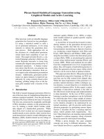
Fig. 1. Schematic illustration of domain structures and alignment for LRRK2 and LRRK2 structure-related proteins. ANK, N-terminal ankyrin
repeat domain; DEP, Dishevelled, EGL-10, pleckstrin domain; GEF, guanine-exchange factor; GRAM, glucosyltransferases, Rab-like GTPase
activators and myotublarins domain.
Z. Yue Approaches for understanding pathogenic roles
FEBS Journal 276 (2009) 6445–6454 ª 2009 The Author Journal compilation ª 2009 FEBS 6447
lifespan of worms, indicating a beneficial role of
LRRK2 in the ageing process. This result suggests that
overproduction of LRRK2 (wild-type or G2019S
mutant) in worms, unlike in mammalian cell cultures
[9,19], does not cause toxicity [26].
The third report in nematode worms, however,
shows that over-expression of worm green flourescent
protein-tagged LRK-1 wild-type or LRK-1 G1876S
(corresponding to human G2019S) leads to an early
larval arrest [27]. Although this result may suggest that
over-expresssion of worm LRK-1 is much more toxic
than human LRRK2, it also raises the question of
whether or not LRK-1 is a true ortholog of human
LRRK2 [6]. Importantly, this study suggests a genetic
link between Lrk-1 and Pink-1,aC. elegans homolog
of human PINK-1 that is associated with a recessive
form of PD [28]. Mutant worms that lack Lrk-1 were
shown to have enhanced sensitivity to endoplasmic
reticulum stress induced by tunicamycin, a specific
inhibitor for N-linked glycosylation. Interestingly, this
enhanced sensitivity is suppressed in mutant worms
with deletion of both Lrk-1 and Pink-1 genes. On the
other hand, although Pink-1 mutant worms exhibit
increased vulnerability to paraquat, defects in mito-
chondrial cristae and impairment of axonal guidance,
a lack of Lrk-1 appeared to reverse the Pink-1 dele-
tion-associated defects in double mutant Lrk-1 and
Pink-1. This study suggests an antagonistic role of
Lrk-1 and Pink-1 in stress response and neuronal
activities [27].
Fruit fly models of LRRK2
The fruit fly homolog of LRRK2 is dLRRK, which also
contains the conserved ‘LRCK’ core sequence (Fig. 1).
To date, at least four studies have reported using fruit
fly D. melanogaster to investigate the in vivo functions
of human LRRK2 or dLRRK. The first study showed
that the mutant flies lacking dLRRK exhibited
impaired locomotive activity and a significant reduc-
tion of tyrosine hydroxylase (TH) immunostaining in
dopaminergic neurons. Although the number of dopa-
minergic neurons appears unaltered, they display
abnormal morphology, suggesting that they are under
pathogenic stress or undergoing slow degeneration
[29]. Two other studies, however, did not reproduce
the behavioral and TH deficits in mutant flies carrying
deletion of dLRRK. Instead, they observed unchanged
numbers of TH+ neurons in these mutants, indicating
that dLRRK is dispensable for the survival of dopami-
nergic neurons [30,31]. In addition, Wang et al. [30]
showed that mutant flies containing C-terminal kinase
domain truncated dLRRK are selectively sensitive to
H
2
O
2
, but not to paraquat, rotenone or b-mercapto-
ethanol. By contrast, Imai et al. [31] showed that
dLRRK null flies are relatively resistant to general oxi-
dative stress, such as paraquat and H
2
O
2
treatment,
compared to wild-type flies [31]. Furthermore, dLRRK
null flies have significant reduced levels of 4-hydroxy-
2-nonenal of lipid peroxidation, an indication of oxida-
tive damage. Although the exact role of dLRRK in
oxidative stress remains unclear, all studies in fly mod-
els reported to date consistently demonstrate that
dLRRK is not essential for the early development and
viability of dopaminergic neurons.
The results obtained from studies of transgenic flies
over-expressing dLRRK or human LRRK2 have been
somewhat inconsistent between the different groups.
Although Lee et al. [29] indicated that over-expression
of a pathogenic mutant or wild-type dLRRK did not
cause any significant defects in transgenic flies, two
other independent reports demonstrated that express-
ing mutants of dLRRK or LRRK2 in flies causes selec-
tive degeneration of dopaminergic neurons as well as
motor function deficits [31,32]. Of these two reports,
however, one showed that even over-expressing wild-
type human LRRK2 led to the toxicity of dopaminer-
gic neurons and impairment of motor function
(although to a lesser degree than LRRK2 G2019S)
[32], whereas the other indicated that over-expressing
wild-type dLRRK did not affect the number of dopa-
minergic neurons or motor function [31].
Interestingly, two studies have shown a relationship
of LRRK2 or dLRRK to the dopamine physiology. Liu
et al. [32] found that treatment of l-DOPA improved
the motor impairment of transgenic flies caused by
LRRK-G2019S but not the degeneration of TH+ neu-
rons. The results obtained by Imai et al. [31] suggested
that dLRRK is involved in negatively regulating
homeostatic levels of dopamine. They demonstrated
that the over-expression of a PD-pathogenic mutant of
dLRRK (but not wild-type dLRRK) resulted in a
reduction in brain dopamine levels compared to that
of nontransgenic flies. Conversely, dopamine content
was elevated in mutant flies with a dLRRK deletion.
This increase in dopamine content is likely to be a
result of dopamine release, uptake or metabolism, but
not to an alteration of TH+ neuron numbers [31].
Finally, Imai et al. [31] provided evidence that both
dLRRK and LRRK2 kinase can phosphorylate
eukaryotic initiation factor 4E-binding protein, a nega-
tive regulator of eukaryotic initiation factor 4E-medi-
ated protein translation and a key mediator of various
stress responses. They proposed a model in which
LRRK2 mediates the pathological effect in part
through modulating translation initiation [31].
Approaches for understanding pathogenic roles Z. Yue
6448 FEBS Journal 276 (2009) 6445–6454 ª 2009 The Author Journal compilation ª 2009 FEBS
In summary, these studies in fruit flies have provided
important in vivo information regarding the potential
function of LRRK2 (Table 1). Indeed, certain observa-
tions reported in fly models may appear to be conflict-
ing. However, it is possible that the different genetic
backgrounds, the genomic locus of insertion for gene
disruption, transgenic expression levels and nutrient
conditions are responsible for the divergent results.
These issues need to be resolved in the future in order
to understand better the physiological function of
LRRK2, as well as the pathogenic effect of PD muta-
tions of LRRK2. Once validated in mammalian
models, selected models could serve as a robust system
for revealing the genetic pathways of LRRK2 in PD
and for screening chemical compounds to intervene
with the LRRK2-mediated pathogenesis.
Rodent models of LRRK2 and bacterial artificial
chromosome (BAC)-mediated LRRK2 transgenic
mice
Although there have been several studies reporting the
generation of genetically engineered LRRK2 mice
(including targeted deletion and transgenic expression),
no systematic investigation of these mice has been
described to date (one report was published recently
during the preparation of this manuscript [33]). There-
fore, the physiological role of LRRK2 in the mamma-
lian central nervous system remains largely elusive.
Analysis of LRRK2 expression in mouse brain shows
that it is broadly distributed in many regions, including
the cerebral cortex, hippocampus, striatum, amygdala,
cerebelluam and olfactory bulb, as well as in ventral
tegamental area and substantia nigra (albeit at low lev-
els) [11,34–40]. Analysis of LRRK2 expression levels
during pre- and post-natal stages reveals that the
LRRK2 protein appears at embryonic day 17 (E17)
and is increasingly produced over the early post-natal
stage [11,34], reaching peak levels by 2 months [11].
LRRK2 knockout (KO) mice
Biskup et al. [34] were the first to report the generation
of LRRK2 KO mice. Taking advantage of the lack of
LRRK2 expression in these mice, they performed the
comprehensive evaluation of a panel of commercial
antibodies against LRRK2 for their staining specificity.
However, no characterization of these mice was shown
[34]. Although without showing any experimental data,
a study by Wang et al. [41] indicated that LRRK2 KO
mice survive normally, and that they do not develop
any obvious neuropathological abnormalities or motor
dysfunctions up to 12 months of age. Indeed, no loss
of dopaminergic neurons or motor behavioral deficits
was observed even at 24 months of age in LRRK2 KO
mice (Dr H. Cai, personal communication). This
result, along with the study showing the developmental
expression levels of LRRK2, suggests that the role of
LRRK2 in early embryonic development is negligible,
but may be important for cellular function at the adult
stage. Furthermore, although it is possible that the
lack of LRRK2 function can be compensated for by
LRRK2 function-related molecules (e.g. LRRK1), this
observation in LRRK2 KO mice is consistent with the
findings in nematode worms and fruit flies that
the deletion of the single homolog of LRRK2 in either
species has no effect on the viability of dopaminergic
neurons. Therefore, we propose that LRRK2 (as well
as LRRK2 homologs dLRRK and LRK-1) does not
play a major role in a cellular pathway that is critical
for neuronal survival. Rather, it is involved perhaps in
specific neuronal functions that can only distantly
modulate neuronal survival or death in an age-depen-
dent manner.
It is also not surprising that the deletion of the
LRRK2 gene does not lead to degeneration of dopami-
nergic neurons in mice, given that disruption of all
known PD-related genes, such as a-synuclein, Parkin,
DJ-1 and PINK-1, has not been associated with any
obvious loss of dopaminergic neurons in mice. It is
intriguing to note that none of these PD-related genes
are essential for neural development and differentia-
tion, which is also in support of the hypothesis that
dysfunction of these genes only leads to disruption of
neuronal functions mostly at the adult stage in PD.
BAC transgenic mice of LRRK2
To date, three laboratories have reported the availabil-
ity of LRRK2 transgenic mice without providing
details of the characterization of these mice. Two labo-
ratories, including ours, generated BAC-transgenic
mice expressing murine FLAG-tagged LRRK2 [11]
and human LRRK2 [42], whereas the third indicated
the usage of a tetracyline-regulated system for the
transgenic expression of human G2019S LRRK2 [41].
The application of BAC-transgenic mice was initially
described in 1997 [43] and has grown significantly over
the past decade because of its usefulness in studying
gene function in vivo, particularly in the central ner-
vous system [44]. Growing evidence demonstrates the
power of this transgenic approach in conferring correct
transgene expression under endogenous promoter con-
trol with little concern about positional effect [45]. The
BAC transgenic approach has been successfully used
in establishing mouse models for neurodegenerative
Z. Yue Approaches for understanding pathogenic roles
FEBS Journal 276 (2009) 6445–6454 ª 2009 The Author Journal compilation ª 2009 FEBS 6449
diseases [46] and is expected to contribute to an under-
standing of the disease mechanisms in vivo.
The application of BAC transgenics is especially
advantageous over conventional transgenics for study-
ing LRRK2. The main reasons are: (1) generation of
LRRK2 BAC transgenic mice does not involve the
synthesis of full-length LRRK2 cDNA, which is a
> 7 kb nucleotide and technically difficult to manipu-
late as a result of the large size; (2) the entire genomic
sequence of mouse or human LRRK2 is approximately
180 kb, which is the average length of BAC clones that
are readily available in public domains; and (3)
LRRK2 BAC transgenes with introduced PD muta-
tions are suitable for modeling the LRRK2-mediated
pathological process as a result of the dominant
disease transmission for LRRK2 mutations. Our
laboratory has previously generated numerous BAC
transgenic lines expressing FLAG-tagged LRRK2
wild-type. Examination of the transgene expression in
the brain shows a similar distribution pattern in all
Table 1. In vivo models for LRRK2 and LRRK2 homologs.
Transgene Species Phenotype Reference
Truncation of endogenous
LRK-1
Worm Subtle defects in movement; partially defective in chemotaxis to volatile
odorants; impairment of polarized synaptic vesicle localization
[25]
Transgenic expression of
human LRRK2
Worm Over-expression of LRRK2 wild-type protects against rotenone toxicity and
extend life span; over-expression of LRRK2 G2019S also protects but to a
lesser extent
[26]
Transgenic expression of
green flourescent protein-
tagged LRK-1 wild-type or
G1876S mutant
Worm Over-expression of either LRK-1 wild-type or G1876S (corresponding to
human G2019S) leads to an early larval arrest
[27]
Disruption of endogenous
LRK-1
Worm Antagonistic action of worm Lrk-1 versus Pink-1 in stress response and
neuronal activities
[27]
Transgenic expression and
disruption of endogenous
dLRRK
Fly No obvious behavioral abnormality associated with transgenic over-expression
of dLRRK wild-type or mutant; deletion mutant shows impaired locomotive
activity and a significant reduction of TH immunostaining in dopaminergic
neurons
[29]
Disruption of endogenous
dLRRK
Fly No obvious behavioral deficits; unchanged TH+ neurons; enhanced sensitivity
to H
2
O
2
[30]
Disruption of endogenous
dLRRK
Fly Relatively resistant to general oxidative stress; reduced oxidative damage;
unchanged TH+ neurons; increased dopamine content
[31]
Transgenic expression of
dLRRK
Fly Over-expression of ‘pathogenic’ dLRRK mutant caused loss of TH+ neurons
in aged mice and reduced dopamine content; over-expression of wild-type
dLRRK2 or kinase-dead mutant had no effect on viability of TH+ neurons
[31]
Transgenic expression of
human LRRK2
Fly Over-expression of LRRK2 wild-type or G2019S mutant causes loss of TH+
neurons and impairment of motor function (with worse phenotype in
G2019S mutant flies); treatment of
L-DOPA improves motor function but not
neurodegeneration
[32]
LRRK2 KO Mouse No description of characterization [34]
LRRK2 KO Mouse Survived normally; display no overt behavioral abnormality; unaltered number
of dopaminergic neurons for up to 24 months
[41, and
unpublished
results]
BAC transgenics ⁄ murine
LRRK2
Mouse One line expressing FLAG tagged LRRK2 wild-type (> 20-fold) shows regulated
expression pattern and unaltered TH+ neuron morphology or number
[11]
BAC transgenics ⁄ human
LRRK2
Mouse Human BAC mice show very similar expression pattern to mouse BAC
transgenic lines [11]; over-expression of LRRK2 wild-type (> 20-fold),
G2019S or Y1699C (seven- to 11-fold and up to 12 months) did not cause
overt behavioral abnormalities
[42,47]
Tetracycline-regulated
transgenics ⁄ human LRRK2
G2019S
Mouse No obvious neuropathologies or motor abnormalities at 12 months and older [41]
BAC transgenics ⁄ human
LRRK2 R1441C
Mouse BAC mice expressing LRRK2 R1441C develop typical motor function
abnormality related to PD; no obvious loss of midbrain TH+ cells; age-
dependent and levodopa-responsive slowness of movement associated with
reduced dopamine release and axonal pathology of nigrostriatal
dopaminergic projection
[33]
Approaches for understanding pathogenic roles Z. Yue
6450 FEBS Journal 276 (2009) 6445–6454 ª 2009 The Author Journal compilation ª 2009 FEBS
transgenic lines. We identified one BAC line that pro-
duces FLAG-LRRK2 wild-type protein at a level
twenty-fold greater than the endogenous LRRK2 pro-
tein [11]. Unexpectedly, the transgenic mice overloaded
with the exogenous LRRK2 did not show obvious
neurotoxicity or motor function abnormalities over
20 months (X. Li and Z. Yue, unpublished results),
despite FLAG-LRRK2 purified from transgenic brain
displaying robust kinase and GTPase activity [11].
Melrose et al. [42] previously reported the generation
of BAC transgenic mice producing human LRRK2
wild-type or mutants. Although no information was
given about the viability of TH+ neurons, it was indi-
cated that BAC transgenic mice over-expressing
LRRK2 wild-type [20-fold for up to 24 months),
mutant G2019S or Y1699C (seven- to 11-fold for up
to 12 months) did not show an overt behavioral phe-
notype [47]. Consistent with these observations, tetra-
cycline-regulated transgenic mice producing LRRK2
G2019S were also reported to be spared of obvious
neuropathologies or motor abnormalities at 12 months
and older [41].
Interestingly, a more recent study by Li et al. [33]
suggests that BAC transgenic mice expressing the
human LRRK2 R1441C mutant develop typical motor
function deficit related to PD. These mice are associ-
ated with the degeneration of TH+ axons and tauopa-
thy, as well as TH+ cell atrophy, despite lacking
obvious loss of midbrain TH+ cells. Furthermore,
these BAC models develop an age-dependent and levo-
dopa-responsive slowness of movement associated with
diminished dopamine release and axonal pathology of
nigrostriatal dopaminergic projection [33]. Although
this transgenic line provides a promising model for fur-
ther dissection of LRRK2-associated PD pathogenesis,
future experiments will be needed to resolve the differ-
ence in behavioral as well as possible pathological
phenotypes observed among different BAC transgenic
models. Although it remains unclear at present, the dif-
ferent PD-related mutations examined and the distinct
genetic background of the host mice, as well as the
varied transgene expression levels, may be responsible
for the differential phenotype of these BAC models.
It is mysterious that none of the reported LRRK2
transgenic mice show the loss of dopaminergic neurons
or the accumulation of a-synuclein at substantia nigra,
the hallmarks of PD pathology. Alhough the physio-
logical function of LRRK2 has yet to be formally dem-
onstrated in these transgenic models, current evidence
suggests that the pathological consequence of over-
expressing only LRRK2 wild-type or PD-related muta-
tions in rodent model is mostly neuron dysfunction,
rather than degeneration. One of the intriguing findings
reported by Li et al. [47] is that the LRRK2-R1441C
BAC mice show reduced dopamine release, which is
consistent with previous studies conducted in fruit flies
showing the connection of dLRRK to dopamine and
movement control [31,32]. In addition, we found that
BAC transgenic mice expressing LRRK2-G2019S also
displayed a decrease in dopamine release and striatal
dopamine levels in the absence of obvious neuropathol-
ogy (X. Li, J. C. Patel and coworkers, unpublished
results). These results suggest a pathogenic role of
LRRK2 mutants in the deregulation of the striatal
dopamine system. Whether other PD-related mutants
of LRRK2 also have the same effect, and whether the
normal function of LRRK2 is involved in striatal dopa-
mine transmission, remains to be shown.
The above observation, therefore, is in line with the
previous evidence indicating that single genetic alter-
ation of PD-related genes, such as the over-expression
of a dominant gene a-synuclein or the deletion of a
recessive gene (DJ-1, Parkin or PINK1), is unlikely to
recapitulate the full spectrum of PD. The lack of mani-
festation of the most important PD hallmarks in
LRRK2 transgenic mice (e.g. dopaminergic neuron
loss and deposits of a-synuclein in Lewy body) is also
not surprising considering that LRRK2 PD-mutations
are not fully penetrant and that LRRK2 patients dis-
play a broad range of clinical phenotypes [1–4]. There-
fore, the current challenges facing us are not only to
‘tease out’ LRRK2-associated neuronal functions that
are perturbed as a result of PD-related mutations, but
also to identify the cellular pathways or factors that
cross-talk with and thus can significantly modify
LRRK2-mediated phenotypic expressions.
Concluding remarks
The invertebrate models including nematode worms
and fruit flies have begun to unveil the functions of
the orthologs of LRRK2 in vivo (Table 1). Although
rapid analysis of these models will undoubtedly facili-
tate an understanding of the function of LRRK2 func-
tion in PD, the lack of a sophisticated structural and
functional equivalent of the human central nervous
system in these organisms limits their application when
understanding the in vivo function of LRRK2 in
humans. The ultimate comprehension of LRRK2 phys-
iology and pathophysiology in PD will still depend on
the establishment and detailed characterization of
mammalian models of LRRK2. The collective data
obtained from both KO and transgenic mouse models
(albeit in preliminary form) suggest that LRRK2 is
not essential for neural development and differentia-
tion, and that it does not play a primary role in cell
Z. Yue Approaches for understanding pathogenic roles
FEBS Journal 276 (2009) 6445–6454 ª 2009 The Author Journal compilation ª 2009 FEBS 6451
death pathways. However, these LRRK2 rodent mod-
els should provide valuable tools for dissecting the
specific neuronal functions of LRRK2 (e.g. dopamine
transmission) and likely pre-symptomatic (or early)
events of the disease process. They should also be use-
ful in testing the ‘two-hit’ or ‘multiple-hit’ hypothesis
proposing that LRRK2 and other genetic or environ-
mental factors are required to work together and
facilitate the pathological process of PD.
Acknowledgements
I wish to thank Drs Chenjian Li, Huaibin Cai,
Xianting Li and Sarah Funderburk for their critical
comments, and Dr Huaibin Cai for sharing unpub-
lished results. I am also grateful to Dr Nina Pan for
assisting in the preparation of Fig. 1 and Table 1. This
work was supported by grants to Z.Y. from the US
NIH ⁄ NINDS NS061152, NS060809, RNS055683A,
the Michael J. Fox Foundation, and the Bachmann-
Strauss Dystonia & Parkinson Foundation.
References
1 Zimprich A, Biskup S, Leitner P, Lichtner P, Farrer M,
Lincoln S, Kachergus J, Hulihan M, Uitti RJ, Calne
DB et al. (2004) Mutations in LRRK2 cause autoso-
mal-dominant parkinsonism with pleomorphic
pathology. Neuron 44, 601–607.
2 Paisan-Ruiz C, Jain S, Evans EW, Gilks WP, Simon J,
van der Brug M, Lopez de Munain A, Aparicio S, Gil
AM, Khan N et al. (2004) Cloning of the gene contain-
ing mutations that cause PARK8-linked Parkinson’s
disease. Neuron 44, 595–600.
3 Healy DG, Falchi M, O’Sullivan SS, Bonifati V, Durr
A, Bressman S, Brice A, Aasly J, Zabetian CP,
Goldwurm S et al. (2008) Phenotype, genotype, and
worldwide genetic penetrance of LRRK2-associated
Parkinson’s disease: a case-control study. Lancet Neurol
7, 583–590.
4 Ozelius LJ, Senthil G, Saunders-Pullman R, Ohmann E,
Deligtisch A, Tagliati M, Hunt AL, Klein C, Henick B,
Hailpern SM et al. (2006) LRRK2 G2019S as a cause
of Parkinson’s disease in Ashkenazi Jews. N Engl J
Med 354, 424–425.
5 Bosgraaf L & Van Haastert PJ (2003) Roc, a Ras ⁄ GT-
Pase domain in complex proteins. Biochim Biophys Acta
1643, 5–10.
6 Marin I (2006) The Parkinson disease gene LRRK2:
evolutionary and structural insights. Mol Biol Evol 23,
2423–2433.
7 West AB, Moore DJ, Biskup S, Bugayenko A, Smith
WW, Ross CA, Dawson VL & Dawson TM (2005)
Parkinson’s disease-associated mutations in leucine-rich
repeat kinase 2 augment kinase activity. Proc Natl Acad
Sci USA 102, 16842–16847.
8 Gloeckner CJ, Kinkl N, Schumacher A, Braun RJ,
O’Neill E, Meitinger T, Kolch W, Prokisch H &
Ueffing M (2006) The Parkinson disease causing
LRRK2 mutation I2020T is associated with increased
kinase activity. Hum Mol Genet 15, 223–232.
9 Smith WW, Pei Z, Jiang H, Dawson VL, Dawson TM
& Ross CA (2006) Kinase activity of mutant
LRRK2 mediates neuronal toxicity. Nat Neurosci 9,
1231–1233.
10 Ito G, Okai T, Fujino G, Takeda K, Ichijo H, Katada
T & Iwatsubo T (2007) GTP binding is essential to the
protein kinase activity of LRRK2, a causative gene
product for familial Parkinson’s disease. Biochemistry
46, 1380–1388.
11 Li X, Tan YC, Poulose S, Olanow CW, Huang XY &
Yue Z (2007) Leucine-rich repeat kinase 2
(LRRK2) ⁄ PARK8 possesses GTPase activity that is
altered in familial Parkinson’s disease R1441C ⁄ G
mutants. J Neurochem 103, 238–247.
12 Guo L, Gandhi PN, Wang W, Petersen RB, Wilson-
Delfosse AL & Chen SG (2007) The Parkinson’s dis-
ease-associated protein, leucine-rich repeat kinase 2
(LRRK2), is an authentic GTPase that stimulates
kinase activity. Exp Cell Res 313, 3658–3670.
13 Smith WW, Pei Z, Jiang H, Moore DJ, Liang Y, West
AB, Dawson VL, Dawson TM & Ross CA (2005)
Leucine-rich repeat kinase 2 (LRRK2) interacts with
parkin, and mutant LRRK2 induces neuronal
degeneration. Proc Natl Acad Sci USA 102, 18676–
18681.
14 Greggio E, Jain S, Kingsbury A, Bandopadhyay R,
Lewis P, Kaganovich A, van der Brug MP, Beilina A,
Blackinton J, Thomas KJ et al. (2006) Kinase activity is
required for the toxic effects of mutant LRRK2 ⁄ darda-
rin. Neurobiol Dis 23, 329–341.
15 MacLeod D, Dowman J, Hammond R, Leete T, Inoue
K & Abeliovich A (2006) The familial Parkinsonism
gene LRRK2 regulates neurite process morphology.
Neuron 52, 587–593.
16 van Egmond WN, Kortholt A, Plak K, Bosgraaf L,
Bosgraaf S, Keizer-Gunnink I & van Haastert PJ (2008)
Intramolecular activation mechanism of the Dictyosteli-
um LRRK2 homolog Roco protein GbpC. J Biol Chem
283, 30412–30420.
17 Abysalh JC, Kuchnicki LL & Larochelle DA (2003)
The identification of pats1, a novel gene locus required
for cytokinesis in Dictyostelium discoideum. Mol Biol
Cell 14, 14–25.
18 Gotthardt K, Weyand M, Kortholt A, Van Haastert PJ
& Wittinghofer A (2008) Structure of the Roc-COR
domain tandem of C. tepidum, a prokaryotic homo-
logue of the human LRRK2 Parkinson kinase. EMBO
J 27, 2352.
Approaches for understanding pathogenic roles Z. Yue
6452 FEBS Journal 276 (2009) 6445–6454 ª 2009 The Author Journal compilation ª 2009 FEBS
19 West AB, Moore DJ, Choi C, Andrabi SA, Li X,
Dikeman D, Biskup S, Zhang Z, Lim KL, Dawson VL
et al. (2007) Parkinson’s disease-associated mutations in
LRRK2 link enhanced GTP-binding and kinase
activities to neuronal toxicity. Hum Mol Genet 16,
223–232.
20 Bosgraaf L, Russcher H, Smith JL, Wessels D, Soll DR
& Van Haastert PJ (2002) A novel cGMP signalling
pathway mediating myosin phosphorylation and chemo-
taxis in Dictyostelium. EMBO J 21, 4560–4570.
21 Bosgraaf L, Waijer A, Engel R, Visser AJ, Wessels D,
Soll D & van Haastert PJ (2005) RasGEF-containing
proteins GbpC and GbpD have differential effects on
cell polarity and chemotaxis in Dictyostelium. J Cell Sci
118, 1899–1910.
22 Dachsel JC, Taylor JP, Mok SS, Ross OA, Hinkle
KM, Bailey RM, Hines JH, Szutu J, Madden B,
Petrucelli L et al. (2007) Identification of potential
protein interactors of Lrrk2. Parkinsonism Relat
Disord 13, 382–385.
23 Gandhi PN, Wang X, Zhu X, Chen SG & Wilson-Delf-
osse AL (2008) The Roc domain of leucine-rich repeat
kinase 2 is sufficient for interaction with microtubules.
J Neurosci Res 86, 1711–1720.
24 Jaleel M, Nichols RJ, Deak M, Campbell DG,
Gillardon F, Knebel A & Alessi DR (2007) LRRK2
phosphorylates moesin at threonine-558: characteriza-
tion of how Parkinson’s disease mutants affect kinase
activity. Biochem J 405, 307–317.
25 Sakaguchi-Nakashima A, Meir JY, Jin Y, Matsumoto
K & Hisamoto N (2007) LRK-1, a C. elegans PARK8-
related kinase, regulates axonal-dendritic polarity of SV
proteins. Curr Biol 17, 592–598.
26 Wolozin B, Saha S, Guillily M, Ferree A & Riley M
(2008) Investigating convergent actions of genes linked
to familial Parkinson’s disease. Neurodegener Dis 5,
182–185.
27 Samann J, Hegermann J, von Gromoff E, Eimer S,
Baumeister R & Schmidt E (2009) Caenorhabditits ele-
gans LRK-1 and PINK-1 act antagonistically in stress
response and neurite outgrowth. J Biol Chem 284,
16482–16491.
28 Valente EM, Abou-Sleiman PM, Caputo V, Muqit
MM, Harvey K, Gispert S, Ali Z, Del Turco D,
Bentivoglio AR, Healy DG et al. (2004) Hereditary
early-onset Parkinson’s disease caused by mutations in
PINK1. Science 304, 1158–1160.
29 Lee SB, Kim W, Lee S & Chung J (2007) Loss of
LRRK2 ⁄ PARK8 induces degeneration of dopaminergic
neurons in Drosophila. Biochem Biophys Res Commun
358, 534–539.
30 Wang D, Tang B, Zhao G, Pan Q, Xia K, Bodmer R &
Zhang Z (2008) Dispensable role of Drosophila ortho-
log of LRRK2 kinase activity in survival of dopaminer-
gic neurons. Mol Neurodegener 3,3.
31 Imai Y, Gehrke S, Wang HQ, Takahashi R, Hasegawa
K, Oota E & Lu B (2008) Phosphorylation of 4E-BP
by LRRK2 affects the maintenance of dopaminergic
neurons in Drosophila. EMBO J 27, 2432–2443.
32 Liu Z, Wang X, Yu Y, Li X, Wang T, Jiang H, Ren Q,
Jiao Y, Sawa A, Moran T et al. (2008) A Drosophila
model for LRRK2-linked parkinsonism. Proc Natl Acad
Sci USA 105, 2693–2698.
33 Li Y, Liu W, Oo TF, Wang L, Tang Y, Jackson-Lewis
V, Zhou C, Geghman K, Bogdanov M, Przedborski S
et al. (2009) Mutant LRRK2(R1441G) BAC transgenic
mice recapitulate cardinal features of Parkinson’s
disease. Nat Neurosci 12, 826–828.
34 Biskup S, Moore DJ, Rea A, Lorenz-Deperieux B,
Coombes CE, Dawson VL, Dawson TM & West AB
(2007) Dynamic and redundant regulation of LRRK2
and LRRK1 expression. BMC Neurosci 8, 102.
35 Westerlund M, Ran C, Borgkvist A, Sterky FH, Lindq-
vist E, Lundstromer K, Pernold K, Brene S, Kallunki
P, Fisone G et al. (2008) Lrrk2 and alpha-synuclein are
co-regulated in rodent striatum. Mol Cell Neurosci 39,
586–591.
36 Galter D, Westerlund M, Carmine A, Lindqvist E,
Sydow O & Olson L (2006) LRRK2 expression
linked to dopamine-innervated areas. Ann Neurol 59,
714–719.
37 Melrose H, Lincoln S, Tyndall G, Dickson D & Farrer
M (2006) Anatomical localization of leucine-rich repeat
kinase 2 in mouse brain. Neuroscience 139, 791–794.
38 Higashi S, Biskup S, West AB, Trinkaus D, Dawson
VL, Faull RL, Waldvogel HJ, Arai H, Dawson TM,
Moore DJ et al. (2007) Localization of Parkinson’s dis-
ease-associated LRRK2 in normal and pathological
human brain. Brain Res 1155, 208–219.
39 Simon-Sanchez J, Herranz-Perez V, Olucha-Bordonau
F & Perez-Tur J (2006) LRRK2 is expressed in areas
affected by Parkinson’s disease in the adult mouse
brain. Eur J Neurosci 23, 659–666.
40 Taymans JM, Van den Haute C & Baekelandt V (2006)
Distribution of PINK1 and LRRK2 in rat and mouse
brain. J Neurochem 98, 951–961.
41 Wang L, Xie C, Greggio E, Parisiadou L, Shim H, Sun
L, Chandran J, Lin X, Lai C, Yang WJ et al. (2008)
The chaperone activity of heat shock protein 90 is criti-
cal for maintaining the stability of leucine-rich repeat
kinase 2. J Neurosci 28, 3384–3391.
42 Melrose HL, Kent CB, Taylor JP, Dachsel JC, Hinkle
KM, Lincoln SJ, Mok SS, Culvenor JG, Masters CL,
Tyndall GM et al. (2007) A comparative analysis of
leucine-rich repeat kinase 2 (Lrrk2) expression in mouse
brain and Lewy body disease. Neuroscience 147, 1047–
1058.
43 Antoch MP, Song EJ, Chang AM, Vitaterna MH, Zhao
Y, Wilsbacher LD, Sangoram AM, King DP, Pinto LH
& Takahashi JS (1997) Functional identification of the
Z. Yue Approaches for understanding pathogenic roles
FEBS Journal 276 (2009) 6445–6454 ª 2009 The Author Journal compilation ª 2009 FEBS 6453
mouse circadian Clock gene by transgenic BAC rescue.
Cell 89, 655–667.
44 Heintz N (2001) BAC to the future: the use of bac
transgenic mice for neuroscience research. Nat Rev
Neurosci 2, 861–870.
45 Gong S, Zheng C, Doughty ML, Losos K, Didkovsky
N, Schambra UB, Nowak NJ, Joyner A, Leblanc G,
Hatten ME et al. (2003) A gene expression atlas of the
central nervous system based on bacterial artificial
chromosomes. Nature 425, 917–925.
46 Gray M, Shirasaki DI, Cepeda C, Andre VM, Wilburn
B, Lu XH, Tao J, Yamazaki I, Li SH, Sun YE et al.
(2008) Full-length human mutant huntingtin with a
stable polyglutamine repeat can elicit progressive and
selective neuropathogenesis in BACHD mice. J Neurosci
28, 6182–6195.
47 Melrose H (2008) Update on the functional biology of
Lrrk2. Future Neurol 3, 669–681.
Approaches for understanding pathogenic roles Z. Yue
6454 FEBS Journal 276 (2009) 6445–6454 ª 2009 The Author Journal compilation ª 2009 FEBS