Báo cáo khoa học: Viral entry mechanisms: human papillomavirus and a long journey from extracellular matrix to the nucleus docx
Bạn đang xem bản rút gọn của tài liệu. Xem và tải ngay bản đầy đủ của tài liệu tại đây (529.81 KB, 11 trang )
MINIREVIEW
Viral entry mechanisms: human papillomavirus and a long
journey from extracellular matrix to the nucleus
Martin Sapp and Malgorzata Bienkowska-Haba
Department of Microbiology and Immunology, Feist Weiller-Cancer Center, Louisiana State University Health Sciences Center, Shreveport,
LA, USA
Introduction
Papillomaviruses (PV) are epitheliotropic non-envel-
oped small DNA viruses with icosahedral symmetry.
Their strict dependence on terminally differentiating
keratinocytes for completion of the replication cycle
initially made the study of entry processes difficult for
two reasons. First, it was impossible to produce virions
until the development of organotypic raft cultures
based on keratinocytes harboring human papillomavi-
rus (HPV) genomes [1]. Because these culture systems
produced only very limited amounts of virions, they
provided only partial relief. The limitation was
partially overcome by the use of DNA-free virus-like
particles and, subsequently, by pseudovirions harboring
marker plasmids, which were generated using hetero-
logous expression systems [2–4]. The observation that
codon optimization of capsid genes yielded high level
expression of capsid proteins [5,6] and the development
of packaging cell lines harboring high copy numbers of
packaging plasmids finally allowed the large-scale pro-
duction of pseudovirions [7] as well as quasivirions [8].
This advance further facilitated the investigation of
early events of PV infection. Second, until very recently
[9], it was not possible to infect either organotypic
raft cultures or primary keratinocytes in vitro unless
Keywords
attachment; capsid protein; conformational
change; endocytosis; endosomal escape;
heparan sulfate; papillomavirus; PML
nuclear body; receptor; uncoating
Correspondence
M. Sapp, Department of Microbiology and
Immunology, Feist Weiller-Cancer Center,
Louisiana State University Health Sciences
Center, Shreveport, LA 71130-3932,
USA
Fax: +1 318 675 5764
Tel: +1 318 675 5760
E-mail:
(Received 16 June 2009, revised 2
September 2009, accepted 17 September
2009)
doi:10.1111/j.1742-4658.2009.07400.x
Papillomaviruses are epitheliotropic non-enveloped double-stranded DNA
viruses, whose replication is strictly dependent on the terminally differenti-
ating tissue of the epidermis. They induce self-limiting benign tumors of
skin and mucosa, which may progress to malignancy (e.g. cervical carci-
noma). Prior to entry into basal cells, virions attach to heparan sulfate
moieties of the basement membrane. This triggers conformational changes,
which affect both capsid proteins, L1 and L2, and such changes are a pre-
requisite for interaction with the elusive uptake receptor. These processes
are very slow, resulting in an uptake half-time of up to 14 h. This mini-
review summarizes recent advances in our understanding of cell surface
events, internalization and the subsequent intracellular trafficking of papil-
lomaviruses.
Abbreviations
BPV1, bovine PV type 1; CyPB, cyclophilin B; ECM, extracellular matrix; EEA, early endosomal antigen; ER, endoplasmic reticulum; HPV,
human papillomavirus; HSPG, heparan sulfate proteoglycan; PV, papillomaviruses; si, small interfering.
7206 FEBS Journal 276 (2009) 7206–7216 ª 2009 The Authors Journal compilation ª 2009 FEBS
pseudovirions had been activated (see below) [10]. The
reason for this deficiency is unknown, although it
suggests that taking primary keratinocytes into culture
induces sufficient changes to make them refractory to
HPV infection. Therefore, studies have had to rely on
established cell lines (with the most commonly used
being the HaCaT cell line) to investigate PV binding
and uptake. However, the recent development of an
in vivo mouse model by the Schiller group will allow
for the testing of observations made in vitro [11]. In
this minireview, we focus on the entry of HPV type 16
(HPV16) and closely-related viruses, which are the
main cause of various cancers, including cervical carci-
noma. In vitro data backed by recent in vivo studies
suggest the existence of an elaborate sequence of cell
surface events that may explain the extremely slow
uptake of viral particles with reported half-times of up
to 14 h.
Capsid structure
To fully appreciate viral entry strategies, their surface
structure must be considered. The outer shell of PV is
composed of 360 molecules of the major capsid protein,
L1 [12]. They are organized into 72 capsomeres, each
comprised of a pentameric L1 assembly forming a
T = 7 icosahedral lattice (Fig. 1A). Twelve and sixty
capsomeres are pentavalent and hexavalent, respec-
tively (i.e. they have five and six nearest neighbors).
Initial structural information for HPV16 was derived
from T = 1 capsids composed of only 12 pentamers
[13], which was later modified using cryoelectron
microscopy and image reconstruction [14]. The core of
the capsomeres is mainly composed of an antiparallel
b-sandwich to which eight b-strands, labeled B through
I, contribute. The outwards facing BC, DE, FG and HI
loops, which connect the b-strands, contain the major
neutralizing epitopes [15–19] (Fig. 1B). These loops
show the highest sequence variations among different
HPV types, which translate into characteristic struc-
tural differences and are most likely responsible for the
type-specificity of neutralizing antibodies [20]. The five
L1 molecules within a capsomere are intimately associ-
ated, even displaying an interlock of their secondary
structures (Fig. 1C). The initial structural information
suggested that the C-terminal arm folds back into the
core structure from which it emanates. However, cryo-
electron microscopy-based image reconstruction [14]
points rather to an invading C-terminal arms model
similar to that of polyomaviruses, which form the prin-
cipal interpentamer contacts (Fig. 1D). This model
implies that a flexible hinge (amino acids 403–413)
bridges the gap between capsomeres forming the base
of the protein shell in the intercapsomeric region. The
a-helix h4 (amino acids 419–429) reaches halfway up
the wall of the invaded capsomere and brings Cys428
into close contact with Cys175, thus allowing disulfide
bond formation [14,21,22], which is not essential for
AB
CD
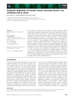
Fig. 1. Structure of the HPV16 L1 protein.
(A) Structure of a T = 7 HPV16 capsid as
previously described [13,14]. (B) L1 mono-
mer; a-helices are highlighted in pink; all five
surface loops are marked in addition to the
internal C–D loop. (C) Top view of a L1
pentamer (spacefill); individual L1 molecules
are displayed in different colors to highlight
the intertwining of the molecules. (D) L1
invading C-terminal arm model as previously
proposed [14]. Side view of a pentamer in
addition to a single L1 molecule from the
neighboring capsomere shown in spacefill.
The arrow points to the intercapsomeric
disulfide bond. Images were downloaded
from the RCSB Protein Data Bank (http://
www.rcsb.org) and modified using
RASMOL
(A,C) () and JMOL
(B,D) ( />software.
M. Sapp and M. Bienkowska-Haba HPV entry
FEBS Journal 276 (2009) 7206–7216 ª 2009 The Authors Journal compilation ª 2009 FEBS 7207
virion formation but strongly stabilizes virions [23,24].
Finally, the C-terminus extends further around the cir-
cumference of the targeted capsomere (amino acids
430–446) and inserts between two L1 molecules of the
invaded pentamer to firmly link capsomeres (amino
acids 447–474). This model suggests that the majority
of the C-terminal arm is surface-exposed, although
located within the intercapsomeric cleft. Therefore, it
may provide surfaces for receptor binding and for the
induction of neutralizing antibodies. Indeed, binding
sites of some neutralizing antibodies have been mapped
to the C-terminal arm [15].
Under forced expression, up to 72 molecules of the
minor capsid protein, L2, are incorporated into a vir-
ion, suggesting that it requires the pentameric L1
structure for interaction [25]. The observation that L2
can occupy binding sites in adjacent capsomeres raises
the possibility of homotypic L2 interactions. L2 is
mainly hidden inside the capsid and only portions of
the N-terminus including residues 60–120 are accessible
on the capsid surface [26,27]. Additional evidence sug-
gests that the extreme N-terminus folds back into the
capsid, thus rendering it inaccessible to antibody bind-
ing and proteolytic cleavage [28,29]. As discussed sub-
sequently, these regions undergo conformational
changes after cell attachment. The N-terminus also
contains two highly conserved cysteine residues, which,
in HPV16, form an intramolecular disulfide bond [30].
L2 density was located at the central internal cavity of
each capsomere by cryoelectron microscopy, although
the majority of the L2 chain was not discernable [25].
L2 residues 396–439 (HPV11) probably mediate this
likely hydrophobic interaction [31]. However, other
regions of L2 also contribute to interaction with L1, as
shown for bovine PV type 1 (BPV1) and HPV33
[32,33]. The central cavity of capsomeres is not large
enough to allow passage of polypeptide chains. Thus,
the L2 N-terminus likely extends to the capsid surface
between neighboring capsomeres. This notion is
supported by observations that L2 protein stabilizes
capsomere interactions under reducing conditions [33].
Receptors
The majority of PV types that have been examined to
date use heparan sulfate proteoglycans (HSPGs) as the
primary attachment receptors [34,35] (Fig. 2). HSPGs
contain unbranched oligosaccharides composed of
alternating disaccharide units of uronic acid and gluco-
samine, which are sulfated and acetylated to various
degrees. O-sulfation occurs at the 2-O, 3-O, and 6-O
position of the uronic acid and at the 3-O and 6-O
position of the amino sugar. The amino group of the
glucosamine may be either acetylated or sulfated. The
two major families of cell surface HSPGs are the syn-
decans and glypicans [36,37]. In addition, secreted per-
lecans are abundant in the extracellular matrix (ECM).
In vitro studies have shown that infectious entry of
HPV33 requires N- as well as O-sulfation. However,
O-sulfation is sufficient for binding, suggesting that
distinct interactions with HSPGs may occur sub-
sequent to primary cell interaction [38]. This finding
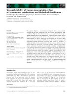
Fig. 2. Model of the ECM and the cell surface events of HPV infection. (1) Most virions bind to primary attachment receptors, HSPG1, pres-
ent in the ECM (basement membrane in vivo) or on the cell surface. HPV11 capsids have also been shown to bind to ECM-resident laminin
5. Viral particles are transported towards the cell body along actin-rich protrusions. (2) Capsids engage with secondary HSPG binding sites
present on the cell surface (HSPG2). Whether transfer from primary ECM binding sites to primary cell surface binding sites occurs has not
been investigated directly. Interaction with the HSPG2 cell surface receptor induces conformational changes in L1 and L2, resulting in the
exposure of the L2 amino terminus and subsequent furin cleavage at a conserved cleavage site. Host cell CyPB facilitates the L2 conforma-
tion changes. (3) These events may induce an additional conformational change that either reduces the affinity of capsids to HSPG or results
in the exposure of sites required for handover to a putative non-HSPG uptake receptor, which then triggers endocytosis.
HPV entry M. Sapp and M. Bienkowska-Haba
7208 FEBS Journal 276 (2009) 7206–7216 ª 2009 The Authors Journal compilation ª 2009 FEBS
was recently confirmed by the use of heparan sulfate
neutralizing drugs applied post-attachment. These
drugs efficiently blocked infection of prebound virions
without inducing their release from the cell surface
[39]. HPV16 virus-like particle binding and HPV11
infection do not appear to require a specific HSPG
protein core for infection in vitro [40]. Because syndec-
an-1 is the predominant HSPG in epithelial tissue, it
was suggested to serve as the primary attachment
receptor in vivo. This is further supported by its high
level of expression in the appropriate target cell and
up-regulation during wound healing [36,41,42]. How-
ever, the in vivo model suggests primary attachment to
the basement membrane rather than the cell surface,
indicating that a secreted HSPG must be involved [11].
HPV31 was reported to not require HSPG interaction
for infection of keratinocytes in vitro, but did interact
with COS-7 in an heparan sulfate-dependent manner
[43]. The in vivo murine cervicovaginal challenge model
yielded results contradicting these observations, where
HPV31 infection was blocked by heparin and heparin-
ase III treatment similar to HPV16 [44]. Neither hepa-
rin nor carrageenan, another sulfated polysaccharide,
was found to inhibit HPV5 infection in vitro despite
having detectable interaction [45]. By contrast, the
in vivo model again suggested a role for HSPG in
HPV5 attachment and infection, albeit with apparently
different requirements regarding sulfation, because
N-desulfated and N-acetylated variants of heparin
rather than the highly sulfated form were found to
preferentially inhibit infection [44].
In vitro studies have shown that PV can also bind to
components of the ECM secreted by keratinocytes and
can be transferred from the ECM to cells in an infec-
tious manner. One ECM component, laminin 5, has
high affinity to HPV11 virions and, in addition to hep-
aran sulfate, may mediate binding to ECM [39,46,47].
However, HPV16 and HPV18 preferentially utilize hep-
aran sulfate moieties for binding to ECM and subse-
quent infectious transfer to cells [39]. Studies using the
murine cervicovaginal challenge model have suggested
that virions bind initially to the basement membrane
prior to transfer to the basal keratinocyte cell surface
[11]. Thus, the ECM might function as the in vitro
equivalent of the epithelial basement membrane.
The minimal length requirement for heparan sulfate
binding to HPV16 virus-like particles is eight monosac-
charide units [48]. For HPV16, positionally conserved
lysine residues K278, K356 and K361, located at the
rim of capsomeres, are involved in primary attach-
ment. Residues from two or more L1 monomers
within a capsomere may form a single receptor binding
site, five of which are present per capsomere [48].
Lysine residue 443 located at the vertex of capsomeres
does not appear to be involved in primary cell attach-
ment. Nevertheless, its exchange for alanine severely
impaired infection, suggesting that secondary binding
events may involve residues found in the cleft between
capsomeres. Another study found that the neutralizing
monoclonal antibody H16.U4 prevented cell surface
but not ECM association of HPV16 and, consequently,
reduced infection [49]. This antibody is specific to a
conformational epitope in the intercapsomeric cleft to
which the invading C-terminal arm contributes [15],
suggesting that elements located within the cleft con-
tribute to cell binding. It is hoped that the determina-
tion of the structure of HPV particles in complex with
its attachment receptor heparan sulfate in combination
with a mutational approach will provide a solution to
these apparent discrepancies.
In recent years, it has become clear that a secondary
non-HSPG receptor is involved in the infectious inter-
nalization of PV particles [28,39]. A study reporting
HSPG-independent infection of HPV16 pseudovirions
pre-cleaved with furin, which processes L2 protein
within capsids, has especially provided evidence for
this notion [10]. Obviously, the treatment of immature
virions with furin induces a conformational change
sufficient to bypass the heparan sulfate-dependent
steps. This indirectly suggests that the engagement of
heparan sulfate is primarily required to induce struc-
tural changes (see below). The identity of the second
non-HSPG binding moiety is still unknown, although
the availability of activated virions with a reduced
affinity to heparan sulfate will potentially allow its
identification. Initial cell surface interactions are pre-
dominantly L1-dependent. However, the L2 protein
may contribute to secondary interactions. Two regions
of L2 that have been described to mediate this engage-
ment encompass residues 13–31 and 108–120 of
HPV16 L2 [29,50].
Attachment-induced conformational
changes
It is well established that engagement with cellular
receptors, most likely HSPG, induces conformational
changes that affect both capsid proteins. The changes
in L1 are not well documented but appear to affect the
BC loop. Improved recognition of a neutralizing L1
epitope in this loop has been observed after virion
attachment to the cell surface [18,38]. Our own unpub-
lished evidence suggests that at least some structural
shifts in L1 precede those in L2 (M. Bienkowska-
Haba, H. D. Patel, K. F. Richards & M. Sapp, unpub-
lished data). On the basis of the relocation of viral
M. Sapp and M. Bienkowska-Haba HPV entry
FEBS Journal 276 (2009) 7206–7216 ª 2009 The Authors Journal compilation ª 2009 FEBS 7209
capsids from cells to ECM under conditions that block
transfer to the secondary receptor, it was proposed
that L1 conformational changes result in a reduced
affinity of the capsid with heparan sulfate, thus aiding
the handover to the secondary receptor [28]. This was
suggested to occur subsequent to L2 conformational
changes [28]. However, no direct evidence for this
notion has yet been provided.
Capsid interaction with HSPG also induces a con-
formational change that results in the exposure of the
L2 amino terminus [28]. Consistent with this idea, the
N-terminal portion of L2 can induce cross-type neu-
tralizing antibodies as a free protein immunogen, but
not when it is assembled into a mature PV capsid [51].
Exposure of the L2 N-terminus allows access to a
highly conserved consensus furin convertase recogni-
tion site and subsequent cleavage by furin on the cell
surface, rendering the cross-neutralizing epitopes acces-
sible to antibody binding [28,52]. Therefore, L2-depen-
dent neutralization must occur subsequent to these
events and not in solution. Proteolytic cleavage is
essential for successful infection. Incorporation of an
N-terminally truncated form of L2 into virions cannot
bypass the furin dependence. This suggests that the
N-terminus is essential for the L2 protein to adopt a
correct conformation within the assembled capsid.
Correct folding may also require the formation of a
disulfide bond between HPV16 L2 residues Cys22 and
Cys28, which was recently identified [30]. Mutation of
the contributing cysteine residues rendered mutant viri-
ons non-infectious [30]. However, it is unclear whether
this is a result of defects in assembly, which only indi-
rectly affect infection processes similar to the N-termi-
nally truncated forms of L2, or whether it has a direct
effect on cell surface and ⁄ or subsequent events.
The cellular peptidyl-prolyl cis ⁄ trans isomerase
cyclophilin B (CyPB) facilitates the exposure of the
HPV16 L2 N-terminus [53]. CyPB has been found on
the cell surface in association with HSPG [54]. Inhi-
bitors of CyPB and its small interfering (si)RNA-
mediated down-regulation prevent exposure of the L2
N-terminus. These treatments induce non-infectious
virus internalization with characteristics similar to
post-attachment treatment with heparan sulfate-block-
ing drugs. Therefore, it was suggested that CyPB acts
prior to or mediates the capsid protein rearrangements,
which are required for transfer to the non-HSPG
receptor [53]. A sequence with homology to a known
CyP binding site is present at surface-exposed L2 resi-
dues 90–110 in many but not all HPV types. Exchang-
ing the central Gly99 and Pro100 of this motif for
alanine made exposure of the HPV16 L2 N-terminus
CyPB-independent [53]. This indicated that the muta-
tions increase flexibility in this loop. The data also sug-
gest that the L2 protein is the substrate for CyPB.
However, exposure of L2 was not achieved in solution
or attached to ECM after addition of bacterially
expressed CyPB [53], indicating that the L2 conforma-
tional change requires engagement with the cell surface
receptor and possibly L1 conformational change(s).
Taken together, these recent advances suggest a
dynamic model of virion-cell surface interactions in
which subsequent engagement with cell surface recep-
tors induce conformational changes in capsid proteins.
It is tempting to speculate that this complex process
evolved to ensure the inaccessibility of critical regions,
thus preventing a host antibody response to conserved
virion epitopes that are essential for infection. The
remarkable conservation of the requirement for L2
furin cleavage suggests that this elaborate process
evolved early in the speciation of papillomaviruses.
Endocytosis
Internalization of HPV16 is highly asynchronous with
an unusually protracted residence on the cell surface.
Similar observations have been made with other PV
types [34,55–57]. In addition to the aforementioned
conformational changes, the reported transport along
filopodia towards the cell body prior to internalization
may contribute to the delayed kinetics [58]. Filopodia-
assisted transport was demonstrated by live cell imag-
ing using HeLa cells. It was suggested that internaliza-
tion can only occur at the cell body. Open questions
regarding this transport include which receptor is link-
ing viral particles to F-actin for retrograde transport
and whether these interactions are sufficient to induce
the observed structural rearrangements. Consistent
with the important role of actin-rich protrusions in
HPV16 infection, it was recently demonstrated that
transport along filopodia also facilitated HPV31 infec-
tion. This study also suggested that particle binding
induced the formation of filopodia [59]. Given the
preferential binding of HPV to the basement mem-
brane, this mechanism might have evolved to allow for
efficient transfer of virions from ECM to the cell body.
A recent study suggested clathrin- and caveolae-
independent internalization of HPV16 pseudovirions in
HeLa and HEK 293TT cells [60]. Entry and infection
was resistant to combined siRNA-mediated down-reg-
ulation of caveolin-1 and clathrin heavy chain and to
over-expression of dominant-negative mutants of dyn-
amin-2, caveolin-1 and eps-15 (EGF receptor pathway
substrate clone no. 15, which plays a role in clathrin-
coated vesicle formation) [60] (Fig. 3). These findings
have now been extended to HaCaT cells (C. Lambert
HPV entry M. Sapp and M. Bienkowska-Haba
7210 FEBS Journal 276 (2009) 7206–7216 ª 2009 The Authors Journal compilation ª 2009 FEBS
& L. Florin, personal communication). Similar
observations were recently presented at the 25th Inter-
national Papillomavirus Workshop by Helenius and
colleagues, who used a large library of siRNA and
inhibitors to interfere with known factors of endocyto-
sis. Furthermore, they found that uptake of HPV16
does not occur via micropinocytosis (M. Schelhaas,
personal communication). As yet, this entry pathway
has not been characterized further but may utilize tet-
raspanin-enriched microdomains as entry platforms
[60]. Earlier studies using biochemical inhibitors such
as chlorpromazine suggested an internalization via
clathrin-mediated endocytosis [55,61]; however, these
findings were mainly based on the use of small drug
inhibitors, which might have unwanted side effects on
cell function. In addition, a recent study also suggested
partial sensitivity of HPV16 pseudovirus infection of
293TT to dynasore, an inhibitor of dynamin GTPase
activity, which is required for clathrin-mediated endo-
cytosis [62]. BPV1 was reported to utilize a clathrin-
dependent endocytic pathway for infectious uptake
based on a combination of microscopic analyses and
biochemical inhibition of known pathways [61]. This
was confirmed using pseudovirions by demonstrating
sensitivity to chlorpromazine and the initial colocaliza-
tion of virions with the early endosomal antigen
(EEA-1) [63], as well as partial sensitivity to dynasore
[62]. For HPV33, internalization was suggested to be
dependent on the actin cytoskeleton [64]. However,
none of these studies was able to demonstrate an effect
of caveolae disruption, via nystatin, methyl-b-cyclodex-
trin or filipin treatment, on HPV16, HPV33 or BPV1
infection. By contrast, HPV31 was reported to depend
on intact caveolae for internalization [55,65]. However,
one study found that treatment with chlorpromazine,
but not with inhibitors of caveolar uptake, prevented
HPV31 pseudovirus infection [66]. As previously
mentioned, HPV31 appears to interact with HSPG
similarly to HPV16 during in vivo infection. Possibly
HPV31 interacts differently with HSPG or has a
unique co-receptor that shunts it into a different inter-
nalization pathway.
Fig. 3. Proposed endocytosis pathways.
Schematic diagrams of the entry pathways
proposed for various PV types. HPV16 is
endocytosed via a clathrin- and caveolin-
independent pathway, whereas BPV1 and
HPV31 were shown to enter via clathrin-
coated pits and caveolae, respectively.
Additional details are provided in the text.
M. Sapp and M. Bienkowska-Haba HPV entry
FEBS Journal 276 (2009) 7206–7216 ª 2009 The Authors Journal compilation ª 2009 FEBS 7211
Vesicular trafficking
A comprehensive study of intracellular trafficking
of different PV types in normal keratinocytes using
siRNA-mediated gene knockdown and dominant-
negative constructs targeting multiple endocytic medi-
ators is still lacking. Given the divergent reports
regarding the endocytic mechanisms, it is not surprising
that the subject of intracellular trafficking of PV-con-
taining vesicles and the cellular compartments involved
is also highly controversial (Fig. 3). The studies are
complicated by the fact that different laboratories uti-
lize different virus sources and cell lines. However,
there is near consensus that successful infection requires
the acidification of endocytic vesicles, suggesting that
PV particles must pass through the endosomal com-
partment [60,61,64,67]. Colocalization with early endo-
some marker EEA-1 has not been observed for HPV16
[60], suggesting they traffic to acidified compartments
via a different route. HPV31 was found to traffic via
caveosomes to early endosomes in a Rab5 GTPase-
dependent manner [67]. Because the infection did not
require functional Rab7, it was suggested that infec-
tious genomes exit the endocytic pathway prior to tran-
sit into late endosomes. However, successful infection
required the acidification of endosomes. By contrast, it
was reported that BPV1 entry via a clathrin-dependent
pathway, which led to colocalization with EEA-1, was
followed by transport to the caveosome and subsequent
entry into the endoplasmic reticulum (ER) in 293TT
cells [63,68,69]. Over-expression of dominant-negative
caveolin-1 and short hairpin RNA-mediated knock-
down of caveolin-1 significantly inhibited infection
without affecting the initial internalization [63]. In
addition, over-expression of a dominant-negative cave-
olin-1 mutant, which is defective for translocation to
the plasma membrane, did not block BPV1 infection,
thus indicating a role for caveolin-1 subsequent to
internalization. However, another study has shown
that the BPV1 genome accumulates in late endosomes
or lysosomes if egress from the endocytic compartment
to the cytosol is blocked [70] and that this requires the
acidification of endosomes [61]. Vesicular transport of
PV particles may also be influenced by capsid protein
interactions with vesicle-resident receptors. It is
intriguing that a binding site for syntaxin-18 was
mapped to a peptide immediately downstream of the
furin cleavage site. Syntaxin-18 is an ER-resident pro-
tein and was found to bind to L2 residues 40–44 of
BPV1. In addition, over-expression of a dominant neg-
ative form of syntaxin-18 impaired BPV1 infection
[68,69]. However, it is unclear whether syntaxin-18 is
present in endocytic vesicles and the mechanism or
consequence of the interaction with L2 has not yet
been fully elucidated. Furthermore, to date, no con-
vincing data demonstrating ER localization of PV
during infectious entry have been made available.
Viral uncoating and egress from
endosomes
Subsequent to the internalization of HPV16, most con-
formational L1 epitopes are lost or are no longer
accessible to antibody binding [39]. L1-specific anti-
bodies to measure uncoating are rare. One such anti-
body, 33L1-7, which has been used for the detection of
internalized particles [60], recognizes an epitope that is
neither accessible in capsids nor in capsomeres [71]. It
remains unclear whether this antibody recognizes a
specific step in uncoating or reacts with protein in the
lysosomal compartment in the process of being com-
pletely degraded. Detection of hidden L2 epitopes and
encapsidated DNA for examination of the uncoating
of papillomaviral pseudoviruses has proven to be more
successful. An HA tag at the L2 C-terminus and
bromodeoxyuridine-labeled viral pseudogenome,
respectively, were used for such a study [72]. The
examination of when these determinants became acces-
sible to antibody staining suggested that uncoating
occurs in endocytic vesicles prior to transfer to the
cytosol. L1 protein appears to be shed from the viral
genome during these events. It could not be detected
in the nucleus of infected cells even when fluorescently-
labeled particles were used. In accordance with this
finding, linear L1 epitopes are continuously detected in
Lamp-3 positive compartments late in infection [60].
Previous studies showing that intact HPV capsids
exceed the size capacity for transit across the central
nuclear pore complex channel had already suggested
that disassembly of the viral particle must occur before
nuclear import [73,74]. L2 protein is not essential for
viral uncoating, as measured by the detection of
bromodeoxyuridine-labeled genome after infection with
L1-only particles [70]. However, L2 protein mediates
the escape of viral DNA from endosomes. An L2
C-terminal peptide harboring a stretch of hydrophobic
residues adjacent to positively-charged amino acids
was shown to contain membrane-disrupting activity
and to mediate the tight association with membranes
in the absence of cellular chaperones. Deletion and
point mutations within this region yielded non-infec-
tious pseudovirus despite unaffected DNA encapsida-
tion and cell surface interactions. A similar deletion in
BPV-1 L2 rendered mutant virus particles non-infec-
tious. Mutant L2 proteins were retained together with
the viral genome within the endosomal compartment
HPV entry M. Sapp and M. Bienkowska-Haba
7212 FEBS Journal 276 (2009) 7206–7216 ª 2009 The Authors Journal compilation ª 2009 FEBS
late after infection [70]. Furin cleavage of L2 is also
essential for endosomal escape despite occurring on
the cell surface [28,52]. However, it remains unclear
how the proteolytic processing contributes to egress
from endosomes. One possibility is that furin cleavage
enables the release of the L2-genome complex from
L1. Alternatively, L2 may promote binding to a spe-
cific receptor that directs virions to vesicles facilitating
uncoating and endosomal membrane passage.
Transport to the nucleus
The issue of how the papillomaviral genome transits
from the endosome to the nucleus has not been sys-
tematically addressed. It is well established that vesicle
trafficking occurs along microtubules. Indeed, the
microtubule disrupting drug nocodazole inhibits PV
infection at a late step [61,64]. However, microtubule-
dependent transport may also be required for the post-
endosomal step involving the delivery of the viral
genome into the nucleus. Cytoplasmic transport along
microtubules is mediated by motor protein complexes
that use cellular energy to move cargo. The L2 protein
of HPV16 and HPV33 was found to interact with the
microtubule network via the motor protein dynein dur-
ing infectious entry [75]. The C-terminal 40 amino
acids of L2 were found to be essential for interaction
with the dynein complex. Other data support the
co-delivery of L2 and genome to the nucleus for
HPV16 and BPV1 L2, possibly in conjunction with a
cell-encoded chaperone [75]. The mechanism by which
the viral genome enters the nucleus is not well under-
stood. L2 protein harbors two terminal peptides that
function as nuclear localization signals when fused
with green fluorescent protein [76–79], raising the
possibility that L2 protein provides the nuclear import
signals. However, these signals overlap with the furin
consensus site and the membrane-destabilizing peptide,
making it difficult to investigate their role in nuclear
entry during infection. A recent study suggested that
nuclear envelope breakdown is required for establish-
ment of HPV16 infection, indicating that active nuclear
import via nuclear pore complexes may not be required
[80]. It is undisputed that L2 protein accompanies the
viral genome to the nucleus. L2 and the viral genome
colocalize in the nucleus at ND10 domains (promyelo-
cytic leukemia nuclear bodies) after infection [72],
suggesting that they are translocated to the nucleus as
a complex. The localization of the genome and L2 at
ND10 is critical for the establishment of infection. Effi-
cient early PV transcription as well as transcription of
the pseudoviral genome under the control of the cyto-
megalovirus immediate early promoter require either
intact ND10 or expression of the promyelocytic leuke-
mia protein [72]. However, the mechanistic explana-
tions for these observations remain unknown.
In summary, our knowledge of PV entry has
increased considerably in recent years. This is espe-
cially a result of the development of systems allowing
the large-scale production of viral particles by bypass-
ing the need for stratified epithelia. However, many
controversies remain, especially regarding the mode of
endocytosis and intracellular trafficking, as well as the
vesicular compartments involved in uncoating. The dis-
crepencies may partially be a result of PV types having
evolved different entry strategies. It is hoped that
future studies will compare several PV types, aiming
to minimize the effect of the different experimental
systems on the findings obtained. In addition, the
recent development of an in vivo model should allow
the significance of the in vitro findings to be tested.
Acknowledgements
We are grateful to members of our laboratory for criti-
cally reading the manuscript. This work was supported
in part by the LSUHSC Foundation (grant:
149741105A) and by the National Center for Research
Resources, a component of the National Institutes of
Health (grant P20-RR018724, entitled ‘Center for
Molecular Tumor Virology’).
References
1 Meyers C, Frattini MG, Hudson JB & Laimins LA
(1992) Biosynthesis of human papillomavirus from a
continuous cell line upon epithelial differentiation.
Science 257, 971–973.
2 Roden RB, Greenstone HL, Kirnbauer R, Booy FP,
Jessie J, Lowy DR & Schiller JT (1996) In vitro
generation and type-specific neutralization of a human
papillomavirus type 16 virion pseudotype. J Virol 70,
5875–5883.
3 Rossi JL, Gissmann L, Jansen K & Muller M (2000)
Assembly of human papillomavirus type 16 pseudoviri-
ons in Saccharomyces cerevisiae. Hum Gene Ther 11,
1165–1176.
4 Unckell F, Streeck RE & Sapp M (1997) Generation
and neutralization of pseudovirions of human papillo-
mavirus type 33. J Virol 71, 2934–2939.
5 Leder C, Kleinschmidt JA, Wiethe C & Mu
¨
ller M (2001)
Enhancement of capsid gene expression: preparing the
human papillomavirus type 16 major structural gene L1
for DNA vaccination purposes. J Virol 75 , 9201–9209.
6 Zhou J, Liu WJ, Peng SW, Sun XY & Frazer I (1999)
Papillomavirus capsid protein expression level depends
M. Sapp and M. Bienkowska-Haba HPV entry
FEBS Journal 276 (2009) 7206–7216 ª 2009 The Authors Journal compilation ª 2009 FEBS 7213
on the match between codon usage and tRNA avail-
ability. J Virol 73, 4972–4982.
7 Buck CB, Pastrana DV, Lowy DR & Schiller JT (2004)
Efficient intracellular assembly of papillomaviral
vectors. J Virol 78, 751–757.
8 Pyeon D, Lambert PF & Ahlquist P (2005) Production
of infectious human papillomavirus independently of
viral replication and epithelial cell differentiation. Proc
Natl Acad Sci USA 102, 9311–9316.
9 Wang HK, Duffy AA, Broker TR & Chow LT (2009)
Robust production and passaging of infectious HPV in
squamous epithelium of primary human keratinocytes.
Genes Dev 23 , 181–194.
10 Day PM, Lowy DR & Schiller JT (2008) Heparan
sulfate-independent cell binding and infection with furin
pre-cleaved papillomavirus capsids. J Virol 82, 12565–
12568.
11 Roberts JN, Buck CB, Thompson CD, Kines R,
Bernardo M, Choyke PL, Lowy DR & Schiller JT
(2007) Genital transmission of HPV in a mouse model
is potentiated by nonoxynol-9 and inhibited by
carrageenan. Nat Med 13, 857–861.
12 Baker TS, Newcomb WW, Olson NH, Cowsert LM,
Olson C & Brown JC (1991) Structures of bovine and
human papillomaviruses. Analysis by cryoelectron
microscopy and three-dimensional image reconstruction.
Biophys J 60, 1445–1456.
13 Chen XS, Garcea RL, Goldberg I, Casini G & Harrison
SC (2000) Structure of small virus-like particles assem-
bled from the L1 protein of human papillomavirus 16.
Mol Cell 5, 557–567.
14 Modis Y, Trus BL & Harrison SC (2002) Atomic model
of the papillomavirus capsid. EMBO J 21, 4754–4762.
15 Carter JJ, Wipf GC, Benki SF, Christensen ND & Gallo-
way DA (2003) Identification of a Human Papillomavirus
Type 16-Specific Epitope on the C-Terminal Arm of the
Major Capsid Protein L1. J Virol 77, 11625–11632.
16 Ludmerer SW, Benincasa D, Mark GE & Christensen
ND (1997) A neutralizing epitope of human papilloma-
virus type 11 is principally described by a continuous
set of residues which overlap a distinct linear, surface-
exposed epitope. J Virol 71, 3834–3839.
17 Ludmerer SW, Benincasa D & Mark GE III (1996)
Two amino acid residues confer type specificity to a
neutralizing, conformationally dependent epitope on
human papillomavirus type 11. J Virol 70, 4791–4794.
18 Roth SD, Sapp M, Streeck RE & Selinka HC (2006)
Characterization of neutralizing epitopes within the
major capsid protein of human papillomavirus type 33.
Virol J 3, 83.
19 White WI, Wilson SD, Palmer-Hill FJ, Woods RM,
Ghim SJ, Hewitt LA, Goldman DM, Burke SJ, Jenson
AB, Koenig S et al. (1999) Characterization of a major
neutralizing epitope on human papillomavirus type
16 L1. J Virol 73, 4882–4886.
20 Bishop B, Dasgupta J, Klein M, Garcea RL, Christen-
sen ND, Zhao R & Chen XS (2007) Crystal structures
of four types of human papillomavirus L1 capsid
proteins: understanding the specificity of neutralizing
monoclonal antibodies. J Biol Chem 282, 31803–31808.
21 Li M, Beard P, Estes PA, Lyon MK & Garcea RL
(1998) Intercapsomeric disulfide bonds in papilloma-
virus assembly and disassembly. J Virol 72, 2160–2167.
22 Sapp M, Fligge C, Petzak I, Harris JR & Streeck RE
(1998) Papillomavirus assembly requires trimerization
of the major capsid protein by disulfides between two
highly conserved cysteines. J Virol 72 , 6186–6189.
23 Buck CB, Thompson CD, Pang YYS, Lowy DR &
Schiller JT (2005) Maturation of papillomavirus
capsids. J Virol 79, 2839–2846.
24 Fligge C, Scha
¨
fer F, Selinka HC, Sapp C & Sapp M
(2001) DNA-induced structural changes in the papillo-
mavirus capsid. J Virol
75, 7727–7731.
25 Buck CB, Cheng N, Thompson CD, Lowy DR, Steven
AC, Schiller JT & Trus BL (2008) Arrangement of L2
within the papillomavirus capsid. J Virol 82, 5190–5197.
26 Liu WJ, Gissmann L, Sun XY, Kanjanahaluethai A,
Muller M, Doorbar J & Zhou J (1997) Sequence close
to the N-terminus of L2 protein is displayed on the
surface of bovine papillomavirus type 1 virions. Virol
227, 474–483.
27 Kondo K, Ishii Y, Ochi H, Matsumoto T, Yoshikawa
H & Kanda T (2007) Neutralization of HPV16, 18, 31,
and 58 pseudovirions with antisera induced by
immunizing rabbits with synthetic peptides representing
segments of the HPV16 minor capsid protein L2
surface region. Virology 358, 266–272.
28 Day PM, Gambhira R, Roden RB, Lowy DR &
Schiller JT (2008) Mechanisms of human papilloma-
virus type 16 neutralization by L2 cross-neutralizing
and L1 type-specific antibodies. J Virol 82, 4638–4646.
29 Yang R, Day PM, Yutzy WH, Lin KY, Hung CF &
Roden RB (2003) Cell surface-binding motifs of L2 that
facilitate papillomavirus infection. J Virol 77, 3531–
3541.
30 Campos SK & Ozbun MA (2009) Two highly conserved
cysteine residues in HPV16 L2 form an intramolecular
disulfide bond and are critical for infectivity in human
keratinocytes. PLoS ONE 4, e4463.
31 Finnen RL, Erickson KD, Chen XS & Garcea RL
(2003) Interactions between papillomavirus L1 and L2
capsid proteins. J Virol 77, 4818–4826.
32 Okun MM, Day PM, Greenstone HL, Booy FP, Lowy
DR, Schiller JT & Roden RB (2001) L1 interaction
domains of papillomavirus L2 necessary for viral
genome encapsidation. J Virol 75, 4332–4342.
33 Sapp M, Volpers C, Mu
¨
ller M & Streeck RE (1995)
Organization of the major and minor capsid proteins
in human papillomavirus type 33 virus-like particles.
J Gen Virol 76, 2407–2412.
HPV entry M. Sapp and M. Bienkowska-Haba
7214 FEBS Journal 276 (2009) 7206–7216 ª 2009 The Authors Journal compilation ª 2009 FEBS
34 Giroglou T, Florin L, Scha
¨
fer F, Streeck RE & Sapp M
(2001) Human papillomavirus infection requires cell
surface heparan sulfate. J Virol 75, 1565–1570.
35 Joyce JG, Tung J-S, Przysiecki CT, Cook JC, Lehman
ED, Sands JA, Jansen KU & Keller PM (1999) The L1
major capsid protein of human papillomavirus type 11
recombinant virus-like particles interacts with heparin
and cell-surface glycosaminoglycans on human kerati-
nocytes. J Biol Chem 274, 5810–5822.
36 Bernfield M, Kokenyesi R, Kato M, Hinkes MT,
Spring J, Gallo RL & Lose EJ (1992) Biology of the
syndecans: a family of transmembrane heparan sulfate
proteoglycans. Annu Rev Cell Biol 8 , 365–393.
37 Fransson LA (2003) Glypicans. Int J Biochem Cell Biol
35, 125–129.
38 Selinka HC, Giroglou T, Nowak T, Christensen ND &
Sapp M (2003) Further evidence that papillomavirus
particles exist in two distinct conformations. J Virol 77,
12961–12967.
39 Selinka HC, Florin L, Patel HD, Freitag K, Schmidtke
M, Makarov VA & Sapp M (2007) Inhibition of trans-
fer to secondary receptors by heparan sulfate-binding
drug or antibody induces non-infectious uptake of
human papillomavirus. J Virol 81, 10970–10980.
40 Shafti-Keramat S, Handisurya A, Kriehuber E, Men-
eguzzi G, Slupetzky K & Kirnbauer R (2003) Different
heparan sulfate proteoglycans serve as cellular receptors
for human papillomaviruses. J Virol 77, 13125–13135.
41 Elenius K, Vainio S, Laato M, Salmivirta M, Thesleff I
& Jalkanen M (1991) Induced expression of syndecan in
healing wounds. J Cell Biol 114 , 585–595.
42 Gallo RL, Ono M, Povsic T, Page C, Eriksson E,
Klagsbrun M & Bernfield M (1994) Syndecans, cell
surface heparan sulfate proteoglycans, are induced by a
proline-rich antimicrobial peptide from wounds.
Proc Natl Acad Sci USA 91, 11035–11039.
43 Patterson NA, Smith JL & Ozbun MA (2005) Human
papillomavirus type 31b infection of human keratino-
cytes does not require heparan sulfate. J Virol 79, 6838–
6847.
44 Johnson KM, Kines RC, Roberts JN, Lowy DR,
Schiller JT & Day PM (2009) Role of heparan sulfate
in attachment to and infection of the murine female
genital tract by human papillomavirus. J Virol 83,
2067–2074.
45 Buck CB, Thompson CD, Roberts JN, Muller M, Lowy
DR & Schiller JT (2006) Carrageenan is a potent inhibi-
tor of papillomavirus infection. PLoS Pathog 2, e69.
46 Culp TD, Budgeon LR & Christensen ND (2006)
Human papillomaviruses bind a basal extracellular
matrix component secreted by keratinocytes which is
distinct from a membrane-associated receptor. Virology
347, 147–159.
47 Culp TD, Budgeon LR, Marinkovich MP, Meneguzzi
G & Christensen ND (2006) Keratinocyte-secreted
laminin 5 can function as a transient receptor for
human papillomaviruses by binding virions and trans-
ferring them to adjacent cells. J Virol 80, 8940–8950.
48 Knappe M, Bodevin S, Selinka HC, Spillmann D,
Streeck RE, Chen XS, Lindahl U & Sapp M (2007)
Surface-exposed amino acid residues of HPV16 L1
protein mediating interaction with cell surface heparan
sulfate. J Biol Chem 282, 27913–27922.
49 Day PM, Thompson CD, Buck CB, Pang YY, Lowy
DR & Schiller JT (2007) Neutralization of human papil-
lomavirus with monoclonal antibodies reveals different
mechanisms of inhibition. J Virol 81, 8784–8792.
50 Kawana Y, Kawana K, Yoshikawa H, Taketani Y,
Yoshiike K & Kanda T (2001) Human papillomavirus
type 16 minor capsid protein L2 N-terminal region
containing a common neutralization epitope binds to
the cell surface and enters the cytoplasm. J Virol 75,
2331–2336.
51 Roden RB, Yutzy WH, Fallon R, Inglis S, Lowy DR &
Schiller JT (2000) Minor capsid protein of human
genital papillomaviruses contains subdominant, cross-
neutralizing epitopes. Virology 270, 254–257.
52 Richards RM, Lowy DR, Schiller JT & Day PM (2006)
Cleavage of the papillomavirus minor capsid protein,
L2, at a furin consensus site is necessary for infection.
Proc Natl Acad Sci USA 103, 1522–1527.
53 Bienkowska-Haba M, Patel HD & Sapp M (2009)
Target cell cyclophilins facilitate human papillomavirus
type 16 infection. PLoS Pathog 5, e1000524.
54 Vanpouille C, Deligny A, Delehedde M, Denys A,
Melchior A, Lienard X, Lyon M, Mazurier J, Fernig
DG & Allain F (2007) The heparin ⁄ heparan sulfate
sequence that interacts with cyclophilin B contains a
3-O-sulfated N-unsubstituted glucosamine residue.
J Biol Chem 282, 24416–24429.
55 Smith JL, Campos SK & Ozbun MA (2007) Human
papillomavirus type 31 uses a caveolin 1- and dynamin
2-mediated entry pathway for infection of human
keratinocytes. J Virol 81, 9922–9931.
56 Christensen ND, Cladel NM & Reed CA (1995) Postat-
tachment neutralization of papillomaviruses by mono-
clonal and polyclonal antibodies. Virology 207, 136–142.
57 Culp TD & Christensen ND (2004) Kinetics of in vitro
adsorption and entry of papillomavirus virions.
Virology 319, 152–161.
58 Schelhaas M, Ewers H, Rajamaki ML, Day PM, Schil-
ler JT & Helenius A (2008) Human papillomavirus type
16 entry: retrograde cell surface transport along actin-
rich protrusions. PLoS Pathog 4, e1000148.
59 Smith JL, Lidke DS & Ozbun MA (2008) Virus activated
filopodia promote human papillomavirus type 31 uptake
from the extracellular matrix. Virology 381, 16–21.
60 Spoden G, Freitag K, Husmann M, Boller K, Sapp
M, Lambert C & Florin L (2008) Clathrin- and
caveolin-independent entry of human papillomavirus
M. Sapp and M. Bienkowska-Haba HPV entry
FEBS Journal 276 (2009) 7206–7216 ª 2009 The Authors Journal compilation ª 2009 FEBS 7215
type 16 – involvement of tetraspanin-enriched microd-
omains (TEMs). PLoS ONE 3, e3313.
61 Day PM, Lowy DR & Schiller JT (2003) Papillomavi-
ruses infect cells via a clathrin-dependent pathway.
Virology 307, 1–11.
62 Abban CY, Bradbury NA & Meneses PI (2008) HPV16
and BPV1 infection can be blocked by the dynamin
inhibitor dynasore. Am J Ther 15, 304–311.
63 Laniosz V, Holthusen KA & Meneses PI (2008) Bovine
papillomavirus type 1: from clathrin to caveolin. J Virol
82, 6288–6298.
64 Selinka HC, Giroglou T & Sapp M (2002) Analysis of
the infectious entry pathway of human papillomavirus
type 33 pseudovirions. Virology 299, 279–287.
65 Bousarghin L, Touze A, Sizaret PY & Coursaget P
(2003) Human papillomavirus types 16, 31, and 58 use
different endocytosis pathways to enter cells. J Virol 77,
3846–3850.
66 Hindmarsh PL & Laimins LA (2007) Mechanisms regu-
lating expression of the HPV 31 L1 and L2 capsid pro-
teins and pseudovirion entry. Virol J 4, 19.
67 Smith JL, Campos SK, Wandinger-Ness A & Ozbun
MA (2008) Caveolin-1 dependent infectious entry of
human papillomavirus type 31 in human keratinocytes
proceeds to the endosomal pathway for pH-dependent
uncoating. J Virol 82, 9505–9512.
68 Bossis I, Roden RB, Gambhira R, Yang R, Tagaya M,
Howley PM & Meneses PI (2005) Interaction of
tSNARE syntaxin 18 with the papillomavirus minor
capsid protein mediates infection. J Virol 79, 6723–6731.
69 Laniosz V, Nguyen KC & Meneses PI (2007) Bovine
papillomavirus type 1 infection is mediated by SNARE
syntaxin 18. J Virol 81, 7435–7448.
70 Ka
¨
mper N, Day PM, Nowak T, Selinka HC, Florin L,
Bolscher J, Hilbig L, Schiller JT & Sapp M (2006) A
membrane-destabilizing peptide in capsid protein L2 is
required for egress of papillomavirus genomes from
endosomes. J Virol 80, 759–768.
71 Rommel O, Dillner J, Fligge C, Bergsdorf C, Wang X,
Selinka HC & Sapp M (2005) Heparan sulfate proteo-
glycans interact exclusively with conformationally intact
HPV L1 assemblies: basis for a virus-like particle
ELISA. J Med Virol 75, 114–121.
72 Day PM, Baker CC, Lowy DR & Schiller JT (2004)
Establishment of papillomavirus infection is enhanced
by promyelocytic leukemia protein (PML) expression.
Proc Natl Acad Sci USA 101, 14252–14257.
73 Merle E, Rose RC, LeRoux L & Moroianu J (1999)
Nuclear import of HPV11 L1 capsid protein is mediated
by karyopherin alpha2beta1 heterodimers. J Cell
Biochem 74, 628–637.
74 Nelson LM, Rose RC, LeRoux L, Lane C, Bruya K &
Moroianu J (2000) Nuclear import and DNA binding
of human papillomavirus type 45 L1 capsid protein.
J Cell Biochem 79, 225–238.
75 Florin L, Becker KA, Lambert C, Nowak T, Sapp C,
Strand D, Streeck RE & Sapp M (2006) Identification
of a dynein interacting domain in the papillomavirus
minor capsid protein L2. J Virol 80, 6691–6696.
76 Becker KA, Florin L, Sapp C & Sapp M (2003) Dissec-
tion of human papillomavirus type 33 L2 domains
involved in nuclear domain (ND) 10 homing and
reorganization. Virology 314, 161–167.
77 Darshan MS, Lucchi J, Harding E & Moroianu J
(2004) The L2 minor capsid protein of human papillo-
mavirus type 16 interacts with a network of nuclear
import receptors. J Virol 78, 12179–12188.
78 Fay A, Yutzy WH, Roden RB & Moroianu J (2004)
The positively charged termini of L2 minor capsid
protein required for bovine papillomavirus infection
function separately in nuclear import and DNA
binding. J Virol 78, 13447–13454.
79 Sun XY, Frazer I, Muller M, Gissmann L & Zhou J
(1995) Sequences required for the nuclear targeting and
accumulation of human papillomavirus type 6B L2
protein. Virology 213, 321–327.
80 Pyeon D, Pearce SM, Lank SM, Ahlquist P & Lambert
PF (2009) Establishment of human papillomavirus
infection requires cell cycle progression. PLoS Pathog 5,
e1000318.
HPV entry M. Sapp and M. Bienkowska-Haba
7216 FEBS Journal 276 (2009) 7206–7216 ª 2009 The Authors Journal compilation ª 2009 FEBS