Báo cáo khoa học: A large complex mediated by Moc1, Moc2 and Cpc2 regulates sexual differentiation in fission yeast ppt
Bạn đang xem bản rút gọn của tài liệu. Xem và tải ngay bản đầy đủ của tài liệu tại đây (532.87 KB, 18 trang )
A large complex mediated by Moc1, Moc2 and Cpc2
regulates sexual differentiation in fission yeast
Swapan Kumar Paul, Yasuo Oowatari and Makoto Kawamukai
Department of Applied Bioscience and Biotechnology, Shimane University, Matsue, Japan
Introduction
The fission yeast Schizosaccharomyces pombe under-
goes sexual differentiation when starved of environ-
mental nutrients. Sexual differentiation in S. pombe is
regulated by at least four signaling pathways: the
cAMP pathway, the stress-responsive Sty1/Spc1 path-
way, the pheromone signaling pathway and the Tor
pathway [1–4]. The cAMP pathway in S. pombe is the
nutrient-sensing pathway that initiates sexual differen-
tiation when opposite mating-type cells coexist [5].
When glucose (or nitrogen) is abundant, the hetero-
trimeric-type guanine nucleotide-binding protein
(Gpa2) becomes activated via the Git3 receptor [6].
The Gpa2 protein subsequently activates adenylyl
cyclase (Cyr1) to generate cAMP from ATP [5]. Cyr1
Keywords
fission yeast; Moc protein;
Schizosaccharomyces pombe; sexual
differentiation; translation
Correspondence
M. Kawamukai, Department of Applied
Bioscience and Biotechnology, Faculty of
Life and Environmental Science, Shimane
University, 1060 Nishikawatsu, Matsue 690-
8504, Japan
Fax: +81 852 32 6092
Tel: +81 852 32 6587
E-mail:
(Received 10 June 2009, revised 2 July
2009, accepted 7 July 2009)
doi:10.1111/j.1742-4658.2009.07204.x
Sexual differentiation in Schizosaccharomyces pombe is triggered by nutri-
ent starvation and is downregulated by cAMP. Screening programs have
identified the moc1/sds23, moc2/ded1, moc3 and moc4/zfs1 genes as inducers
of sexual differentiation, even in the presence of elevated levels of cAMP.
To investigate possible interactions among Moc1, Moc2, Moc3 and Moc4
proteins, we first screened for individual Moc-interacting proteins using the
yeast two-hybrid system and verified the interactions with other Moc pro-
teins. Using this screening process, Cpc2 and Rpl32-2 were highlighted as
factors involved in interactions with multiple Moc proteins. Cpc2 inter-
acted with Moc1, Moc2 and Moc3, whereas the ribosomal protein Rpl32-2
interacted with all Moc proteins in the two-hybrid system. Physical interac-
tions of Cpc2 with Moc1, Moc2 and Rpl32-2, and of Rpl32-2 with Moc2
were confirmed by coimmunoprecipitation. In addition, using Blue Native/
PAGE, we revealed that each Moc protein exists as a large complex. Over-
expression of Moc1, Moc2, Moc3, Moc4 and Rpl32-2 resulted in the effi-
cient induction of a key transcription factor Ste11, suggesting that all
proteins tested are positive regulators of Ste11. Considering that Moc2/
Ded1 is a general translation factor and that Cpc2 associates with many
ribosomal proteins, including Rpl32-2, it is possible that a large Moc-medi-
ated complex, detected in this study, may act as a translational regulator
involved in the control of sexual differentiation in S. pombe through the
induction of Ste11.
Structured digital abstract
l
A list of the large number of protein-protein interactions described in this article is available
via the MINT article ID
MINT-7216191
Abbreviations
EF1a-A, elongation factor 1a-A; GAPDH, glyceraldehyde 3-phosphate dehydrogenase; Gal4-BD, GAL4 DNA-binding domain; X-Gal, 5-bromo-
4-chloro-3-indolyl-
D-galactopyranoside; GFP, green fluorescent protein; moc, multicopy suppressor of over expressed cyr1; P-bodies,
processing bodies; PP2A, protein phosphatase 2A.
5076 FEBS Journal 276 (2009) 5076–5093 ª 2009 The Authors Journal compilation ª 2009 FEBS
interacts with its associated protein Cap1, which plays
a partly regulatory role with respect to adenylyl cyclase
and also interacts with actin [7,8]. When cAMP is
abundant, it associates with the regulatory subunit
Cgs1, and the catalytic protein kinase Pka1 is released
[9]. Pka1 phosphorylates the zinc-finger protein Rst2,
which induces the expression of ste11 , a gene encoding
a key transcription factor for many meiosis-specific
genes [10]. Thus, expression of ste11 is induced in
response to a decrease in the level of cAMP and results
in the initiation of meiosis. The localization shift of
Ste11 in the nucleus and the cytoplasm is controlled by
Rad24 [11] and the pheromone-signaling pathway [12],
which is also negatively controlled by Rad24 [3,13].
The S. pombe ‘multicopy suppressor of overexpres-
sed cyr1’(moc)1 to moc4 genes have been identified as
overcoming a partially sterile S. pombe phenotype
caused by an elevation in cAMP [14,15]. Among the
four moc genes, moc1 is the strongest inducer of sexual
differentiation [15], and the Moc1/Sds23 protein in
S. pombe is known to play important roles in stress
resistance [16,17], the cell cycle [16], chronological life
span [17], survival for Go cells [18] and sexual differen-
tiation [17]. Moc1/Sds23 has also been identified as a
suppressor of dis2 [16] and as a phosphorylated protein
[19]. The Moc1 protein is localized to the cytosol dur-
ing mitotic growth, but accumulates in the nucleus in
mating cells, and this localization shift is inhibited by
cAMP [17]. Moc1 and its orthologous proteins contain
a common domain known as the cystathionine beta
synthase domain, which is predicted to have a multiple
trafficking function for protein–protein interactions
and metabolic regulation, and is found in proteins
such as AMP-activated protein kinase [20]. Moc1 and
its Saccharomyces cerevisiae orthologous proteins
(Sds23/Sds24) are functionally interchangeable [20].
Moc2/Ded1 is an essential RNA helicase, which is
involved in both sexual differentiation [14] and the
mitotic cell cycle [21,22], and is now known to be
a general translational regulator [14,22,23]. Moc3,
a Zn-finger-type protein is localized to the nucleus and
is involved in stress resistance and sexual differentia-
tion [15]. Moc4/Zfs1 contains two Zn-finger motifs, is
localized to the nucleus, and is involved in sexual dif-
ferentiation and septum formation [24,25]. Moc4/Zfs1
has also been identified as an mRNA binding and
destabilizing protein in S. pombe [26]. Whereas the
moc1, moc3 and moc4 genes are dispensable [15,17,24],
moc2 is essential for growth [14]. However, it is not yet
clear how the Moc proteins function in sexual differen-
tiation through interactions with other unidentified
proteins [15].
The possibility that these four Moc proteins might
work together as part of the same complex has never
been considered. Therefore, we decided to search
for Moc-interacting proteins and here we report the
isolation of Moc-interacting proteins in S. pombe using
the yeast two-hybrid system. We then verified the rela-
tionships between the various proteins and proposed
the existence of a Moc-mediated protein complex capa-
ble of regulating sexual differentiation via interactions
with translational components in fission yeast.
Table 1. Interaction of Moc1 interacting proteins with other Moc proteins. A positive signal is indicated by ‘+’ and a negative signal by ‘)’.
The strength of blue color on the X-gal filter is shown by the number of plus marks. Gal4-BD, GAL4 DNA-binding domain.
Moc1 interacting proteins Systematic name Gal4-BD Moc1 Moc2 Moc3 Moc4
Pyruvate decarboxylase SPAC1F8.07c ) + ) ++
Elongation factor 1 a-A SPCC794.09c ) ++ ) ++ +
Glyceraldehyde-3 phosphate dehydrogenase SPBC32F12.11 ) + ) ++ +
Thioredoxin peroxidase SPCC576.03c ) ++ ) ++ +
Mannosyl transferase complex subunit Alg9 SPAC1834.05 ) + ) + )
Srp54 type protein SPCC188.06c ) ++ ) ++
Ribosomal protein L29 SPBC776.01 ) + ) + )
Ribosomal protein L32-2 SPAC3H5.10 ) ++ ++ ++ ++
Ribosomal protein L38 SPBC577.02 ) + ) + )
Ribosomal protein S3a SPAC22H12.04c ) + ) + )
Ribosomal protein S14 SPAC3H5.05c ) + ) + )
Ribosomal protein S16 SPAC664.04c ) ++ ) ++ )
Ribosomal protein S20 SPCC576.09 ) + ) + )
RNA polymerase Rpb3 SPCC1442.10c ) + ) ++ )
Obr1 SPAC3C7.14c ) + ) ++ )
Sfh1 SPCC16A11.14 ) + ) ++ )
Ufd2 SPAC20H4.10 ) ++ ) ++ ++
S. K. Paul et al. Moc proteins in fission yeast
FEBS Journal 276 (2009) 5076–5093 ª 2009 The Authors Journal compilation ª 2009 FEBS 5077
Results
Two-hybrid screening of Moc proteins
To ascertain the relationship between the Moc pro-
teins, we attempted to identify proteins that interact
with Moc1, Moc2, Moc3 and Moc4 using the yeast
two-hybrid system. By cloning each moc gene into the
pGBKT7 vector as bait, we conducted a large-scale
two-hybrid screen using an S. pombe cDNA library,
cloned into the pGAD prey vector in Saccharomy-
ces cerevisiae AH109, as described in Experimental
Procedures. The screened genes were verified by re-
introducing them into the test strain AH109 and the
genes cloned in the pGAD vector were identified by
sequencing. The results of this screening process led to
identification of the following Moc1-interacting pro-
teins: pyruvate decarboxylase, elongation factor 1a-A
(EF1a-A), glyceraldehyde 3-phosphate dehydrogenase
(GAPDH), thioredoxin peroxidase, Alg9, Srp54, Rpb3,
Obr1, Sfh1 and Ufd2; and the ribosomal proteins L29,
L32-2, L38, S3a, S14, S16 and S20 (Table 1). We next
tested whether these proteins also interacted with
Moc2, Moc3 and Moc4 proteins, and we found that
all Moc1-interacting proteins interacted with Moc3,
whereas only the ribosomal protein Rpl32-2 interacted
strongly with Moc1, Moc2, Moc3 and Moc4 proteins.
Pyruvate decarboxylase, EF1a-A, GAPDH, thioredox-
in peroxidase, Srp54 and Ufd2 interacted with Moc1,
Moc3 and Moc4, whereas RNA polymerase subunit
Rpb3, Alg9, Obr1 and Sfh1 interacted with Moc1 and
Moc3 (Table 1). None of the proteins interacted with
the GAL4 DNA-binding domain (Gal4-BD) alone,
indicating that the interactions with the different Moc
proteins were specific.
In a similar-two hybrid screen using Moc2 as bait,
Moc2-interacting proteins were identified as Lys3 (sac-
charopine dehydrogenase) and the ribosomal proteins
L8, L18, L20, L27, L29 and S13 (Table 2). All of the
Moc2-interacting proteins interacted with Moc3,
whereas Lys3 and ribosomal proteins L8, L18, L29
and S13 interacted with Moc1, Moc2 and Moc3. The
ribosomal protein S13 interacted strongly with
Moc1, Moc2 and Moc3, and Lys3 interacted
strongly with Moc2 and Moc3, but loosely with Moc1.
None of the Moc2-interacting proteins interacted with
Moc4, or with the Gal4-BD alone (Table 2), indicating
that the interactions with different Moc proteins were
specific.
Similarly, screening for Moc3-interacting proteins
using the two-hybrid system identified pyruvate decar-
boxylase, enolase, 20S proteasome component alpha 5,
EF1a-A, GAPDH, the ribosomal protein L32-2, super-
oxide dismutase, GluRS [27] and Cpc2 (Table 3). All
Table 2. Interaction of Moc2 interacting proteins with other Moc proteins. A positive signal is indicated by ‘+’ and a negative signal by ‘)’.
The strength of blue color on the X-gal filter is shown by the number of plus marks. Gal4-BD, GAL4 DNA-binding domain.
Moc2 interacting proteins Systematic name Gal4-BD Moc1 Moc2 Moc3 Moc4
Ribosomal protein L8 SPBC29A3.04 ) +++)
Ribosomal protein L18 SPBC11C11.07 ) +++)
Ribosomal protein L20 SPAC3A12.10 ))++)
Ribosomal protein L27 SPCC74.05 ))++)
Ribosomal protein L29 SPBC776.01 ) +++)
Ribosomal protein S13 SPAC6F6.07c ) ++ ++ ++ )
Lys3 (Saccharopine dehydrogenase) SPAC227.18 ) +++++)
Table 3. Interaction of Moc3 interacting proteins with other Moc proteins. A positive signal is indicated by ‘+’ and a negative signal by ‘)’.
The strength of blue color on the X-gal filter is shown by the number of plus marks. Gal4-BD, GAL4 DNA-binding domain.
Moc3 interacting proteins Systematic name Gal4-BD Moc1 Moc2 Moc3 Moc4
Pyruvate decarboxylase SPAC1F8.07c ) + ) ++
Enolase SPBC1815.01 ) + ) ++
20S proteasome component alpha 5 SPAC323.02c ) + ) ++
Elongation factor 1 a-A SPCC794.09c ) + ) ++
Glyceraldehyde-3phosphate dehydrogenase SPBC32F12.11 ) ++ ) ++
Ribosomal protein L32-2 SPAC3H5.10 ) ++ ++ ++ ++
Superoxide dismutase SPAC821.10c ) + ) + )
Glutamyl tRNA synthetase SPAPB1A10.11c ) ++ ) ++ ++
Cpc2 SPAC6B12.15 ) ++ ++ ++ )
Moc proteins in fission yeast S. K. Paul et al.
5078 FEBS Journal 276 (2009) 5076–5093 ª 2009 The Authors Journal compilation ª 2009 FEBS
Moc3-interacting proteins interacted with Moc1, which
is consistent with the results mentioned above in that all
Moc1-interacting proteins interacted with Moc3. This
finding suggests that Moc1 and Moc3 might form indi-
vidual subunits of a putative complex. The ribosomal
protein Rpl32-2 strongly interacted with all four Moc
proteins, and GluRS strongly interacted with Moc1,
Moc3 and Moc4, whereas Cpc2 interacted strongly with
Moc1, Moc2 and Moc3. Pyruvate decarboxylase, eno-
lase, 20S proteasome component alpha 5, EF1a-A and
Table 4. Interaction of Moc4 interacting proteins with other Moc proteins. A positive signal is indicated by ‘+’ and a negative signal by ‘)’.
The strength of blue color on the X-gal filter is shown by the number of plus marks. Gal4-BD, GAL4 DNA-binding domain.
Moc4 interacting proteins Systematic name Gal4-BD Moc1 Moc2 Moc3 Moc4
Glyceraldehyde-3 phosphate dehydrogenase SPBC32F12.11 ) + ) ++
Pyruvate decarboxylase SPAC1F8.07c ) + ) ++
Enolase SPBC1815.01 ) + ) ++
Ribosomal protein L5 SPAC3H5.12c ) )))+
Ribosomal protein L12 SPCC16C4.13c ) ++ ) ++ +
Ribosomal protein L32-2 SPAC3H5.10 ) ++ ++ ++ ++
Ribosomal protein P2B SPBC23G7.15c ) ++ ))+
Elongation factor 2 SPAC513.01c ) )))+
Ebp2 SPAC17H9.05 )))++
Psu1 SPAC1002.13c ) + ) ++ ++
Fba1 (fructose-bisphosphate aldolase) SPBC19C2.07 ) + ) ++ ++
Crb3 SPAC13G7.08c ) + ) ++ ++
mRNA cleavage and polyadenylation
specificity factor complex-associated protein
SPCC74.02c ) + ) ++
Table 5. Schizosaccharomyces pombe strains used in the study.
Strain Genotype Source
SP870 h
90
ade6.210 leu1.32 ura4-D18 [49]
MYM2 h
90
ade6.210 leu1.32 ura4-D18 moc1-3HA<kanMX6 [17]
MYM3 h
90
ade6.210 leu1.32 ura4-D18 moc1-GFP<kanMX6 [17]
HT201 h
90
ade6.210 leu1.32 ura4-D18 cpc2::ura4 [31]
SPB371 h
90
ade6.216 leu1.32 ura4-D18 ste11::ste11-GFP<ura4 [50]
YO7 h
90
ade6.210 leu1.32 ura4-D18 cpc2-13Myc<kanMX6 Lab stock
YO8 h
90
ade6.216 leu1.32 ura4-D18 cpc2-3HA<kanMX6 Lab stock
YM1 h
-
leu1.32 ura4-D18 asf1-13Myc<kanMX6 Lab stock
SKP1 h
90
ade6.210 leu1.32 ura4-D18 moc2-13Myc<kanMX6 This study
SKP2 h
90
ade6.216 leu1.32 ura4-D18 cpc2-3HA<kanMX6 moc2-13Myc<hphMX6 This study
SKP5 h
90
ade6.210 leu1.32 ura4-D18 moc1-13Myc<kanMX6 This study
SKP6 h
90
ade6.216 leu1.32 ura4-D18 cpc2-3HA<kanMX6 moc1-13Myc<hphMX6 This study
SKP7 h
90
ade6.210 leu1.32 ura4-D18 moc3-13Myc<kanMX6 This study
SKP8 h
90
ade6.216 leu1.32 ura4-D18 cpc2-3HA<kanMX6 moc3-13Myc<hphMX6 This study
SKP9 h
90
ade6.210 leu1.32 ura4-D18 moc4-13Myc<kanMX6 This study
SKP10 h
90
ade6.216 leu1.32 ura4-D18 cpc2-3HA<kanMX6 moc4-13Myc<hphMX6 This study
SKP11 h
90
ade6.210 leu1.32 ura4-D18 cpc2::ura4 moc1-13Myc<kanMX6 This study
SKP12 h
90
ade6.210 leu1.32 ura4-D18 cpc2::ura4 moc2-13Myc<kanMX6 This study
SKP13 h
90
ade6.210 leu1.32 ura4-D18 cpc2::ura4 moc3-13Myc<kanMX6 This study
SKP14 h
90
ade6.210 leu1.32 ura4-D18 cpc2::ura4 moc4-13Myc<kanMX6 This study
SKP20 h
90
ade6.210 leu1.32 ura4-D18 rpl32-2-13Myc<kanMX6 This study
SKP21 h
90
ade6.216 leu1.32 ura4-D18 cpc2-3HA<kanMX6 rpl32-2-13Myc<hphMX6 This study
SKP22 h
90
ade6.210 leu1.32 ura4-D18 moc1-3HA<kanMX6 rpl32-2-13Myc<hphMX6 This study
SKP24 h
90
ade6.210 leu1.32 ura4-D18 rpl32-2-3HA<kanMX6 This study
SKP25 h
90
ade6.210 leu1.32 ura4-D18 rpl32-2-3HA<kanMX6 moc2-13Myc<hphMX6 This study
SKP26 h
90
ade6.210 leu1.32 ura4-D18 rpl32-2-3HA<kanMX6 moc3-13Myc<hphMX6 This study
SKP27 h
90
ade6.210 leu1.32 ura4-D18 rpl32-2-3HA<kanMX6 moc4-13Myc<hphMX6 This study
SKP29 h
90
ade6.210 leu1.32 ura4-D18 moc1-GFP<kanMX6 moc2-13Myc<hphMX6 This study
SKP30 h
90
ade6.210 leu1.32 ura4-D18 cpc2::ura4 rpl32-2-13Myc<hphMX6 This study
S. K. Paul et al. Moc proteins in fission yeast
FEBS Journal 276 (2009) 5076–5093 ª 2009 The Authors Journal compilation ª 2009 FEBS 5079
GAPDH interacted with Moc1, Moc3 and Moc4. None
of the Moc3-interacting proteins interacted with the
Gal4-BD (Table 3), again suggesting that the inter-
actions with the different Moc proteins were specific.
Finally, Moc4-interacting proteins identified using
the two-hybrid system were: GAPDH, pyruvate decar-
boxylase, enolase, eEF2, Ebp2, Psu1, Fba1, Crb3,
SPCC74.02c (mRNA cleavage and polyadenylation
specificity factor complex associated protein) and the
ribosomal proteins L5, L12, L32-2 and P2B (Tab le 4).
Among the Moc4-interacting proteins, GAPDH, pyru-
vate decarboxylase, the ribosomal protein L12, Psu1,
Fba1, Crb3 and SPCC74.02c interacted with Moc1,
Moc3 and Moc4, whereas Ebp2 interacted with Moc3
and Moc4. Only the ribosomal protein Rpl32-2 inter-
acted strongly with all the Moc proteins and, except
for Rpl32-2, none of the Moc4-interacting proteins
interacted with Moc2 in a yeast two-hybrid system. In
addition, none of the Moc4-interacting proteins inter-
acted with the Gal4-BD (Table 4).
Interactions of Moc proteins with Cpc2
in fission yeast
The two-hybrid screen revealed that some proteins,
such as Cpc2 and Rpl32-2, interacted strongly with
multiple Moc proteins. We also found that Rpl32-2
interacted with Cpc2 in a two-hybrid system (data not
shown). Cpc2 interacted strongly with Moc1, Moc2
and Moc3, and Rpl32-2 interacted strongly with all
Moc proteins in a yeast two-hybrid system (Table 3);
therefore, we next tested the physical interactions of
Cpc2 with the Moc proteins and with Rpl32-2 by
coimmunoprecipitation, where the protein of interest
was immunoprecipitated with a tagged antibody. Wes-
tern blotting was used to identify proteins that were
pulled down by interaction with the Cpc2 protein. To
determine the physical interactions between Cpc2 and
Moc1, Moc2 and Rpl32-2, cell extracts were prepared
from the double-tagged strains: SKP6 (cpc2–3HA,
moc1–13Myc), SKP2 (cpc2–3HA, moc2–13Myc) and
SKP21 (cpc2–3HA, rpl32-2–13Myc) (Table 5). The HA
mAb was used to immunoprecipitate Cpc2–3HA, and
the precipitate was then analyzed by western blotting,
first using the HA antibody and then the Myc anti-
body (Fig. 1). As shown in Fig. 1, Moc1–13Myc,
Moc2–13Myc and Rpl32-2–13Myc were detected by
immunoprecipitation. Equally, when Moc1–13Myc,
Moc2–13Myc and Rpl32-2–13Myc were first precipi-
tated by a Myc antibody and the precipitated proteins
were analyzed by western blotting using a Myc mAb
followed by the HA antibody (Fig. 1), the result
showed that Cpc2–3HA was present in the anti-Myc
immunoprecipitates of Moc1–13Myc, Moc2–13Myc
and Rpl32-2–13Myc (Fig. 1). These results indicated
that Cpc2 interacted with Moc1, Moc2 and Rpl32-2
in vivo. All the experiments were conducted recipro-
cally and the results of the interactions were consistent
in all cases. However, when we tested the coimmuno-
precipitation of Moc3 and Moc4 with Cpc2, there was
no coimmunoprecipitation in either case (data not
shown). We did not detect any physical interaction
between Moc3 and Cpc2, although they did appear to
interact in the two-hybrid system.
Interactions of Moc proteins and Rpl32-2 in
fission yeast
The interactions between Rpl32-2, fused to the GAL4
activation domain, and each of the Moc1, Moc2,
Moc3 and Moc4 proteins, fused to a Gal4-BD, were
tested in the two-hybrid system (Tables 1, 2 and 3).
We then performed the reciprocal experiment, fusing
Rpl32-2 to the Gal4-BD and fusing Moc1 to Moc4 to
a GAL4 activation domain, and again tested the inter-
actions using the yeast two-hybrid system. The results
showed that Moc1, Moc2, Moc3 and Moc4 interacted
strongly with Rpl32-2 in the GAL4-based two-hybrid
system (data not shown).
Next, we tested the in vivo interactions of Rpl32-2
with Moc1, Moc2, Moc3 and Moc4 by coimmunopre-
cipitation. To determine the physical interactions
between Rpl32-2 and the four Moc proteins, cell
extracts were prepared from the following double-
tagged integrated strains: SKP22 (moc1–3HA, rpl32-2–
13Myc), SKP25 (moc2–13Myc, rpl32-2–3HA), SKP26
(moc3–13Myc, rpl32-2–3HA) and SKP27 (moc4–
13Myc, rpl32-2–3HA) (Table 5). As shown in the
results, only Moc2 was coimmunoprecipitated with
Rpl32-2 (Fig. 2A). A Myc antibody was used to pre-
cipitate the Moc2–13Myc protein and the precipitates
were analyzed by western blotting using the HA mAb.
Conversely, the HA mAb was used to immunoprecipi-
tate Rpl32-2–3HA, and Moc2–13Myc was detected by
a Myc antibody. Our results showed that Rpl32-2–
3HA was present in the Myc immunoprecipitated sam-
ple and, reciprocally, that Moc2–13Myc was present in
the HA immunoprecipitated sample (Fig. 2A), indicat-
ing that Moc2 interacts with Rpl32-2 in vivo. However,
when we tested Moc1, Moc3 and Moc4 with Rpl32-2,
no coimmunoprecipitation was observed (data not
shown), in contrast to the results of the two-hybrid
system.
We then tested the possible interaction of Moc1 and
Moc2 by coimmunoprecipitation using the strain
SKP29 (Moc1–GFP, Moc2–13Myc). A green fluores-
Moc proteins in fission yeast S. K. Paul et al.
5080 FEBS Journal 276 (2009) 5076–5093 ª 2009 The Authors Journal compilation ª 2009 FEBS
cent protein (GFP) mAb was used to precipitate the
Moc1–GFP protein and the precipitates were analyzed
by western blotting using a Myc antibody. As shown
in Fig. 2B, Moc1 was coimmunoprecipitated with
Moc2.
Identification of the Moc complex by
Blue Native/PAGE
The results described above suggested the possibility of
complex formation mediated by some of the Moc pro-
teins, together with Cpc2 and Rpl32-2. To determine
the nature of the putative Moc-mediated complex in
fission yeast, we used Blue Native/PAGE [28]. In these
experiments, cell extracts were prepared from the
S. pombe strains SKP1, SKP5, SKP7 and SKP9 that
expressed Moc2, Moc1, Moc3 and Moc4 proteins,
respectively. The Moc proteins were linked to a 13Myc
tag at the C-terminus (Table 5). When Blue Native/
PAGE was used to separate the proteins from SKP1, a
large Moc2-mediated protein complex of 1000 kDa
was detected by western blotting using the Myc anti-
body (Fig. 3A). The proteins, separated by Blue
Native/PAGE in the first dimension, were further sepa-
rated by SDS/PAGE in the second dimension and sub-
sequently detected by a Myc antibody (Fig. 3B,C).
During electrophoresis in the second dimension, the
complex was separated according to the molecular
masses of the individual subunits and the proteins were
detected by western blotting (Fig. 3B–D), which
revealed a broad signal pattern ranging in size from
large to small. The separation of Cpc2–3HA by 2D
SDS/PAGE following Blue Native/PAGE produced a
similar pattern, indicating that both proteins separate
in a similar manner on a 2D gel. This result also sug-
gested that both proteins exist as complexes that range
in size from high to low molecular masses. The mole-
cular mass ( 1000 kDa) of the complex detected by
Blue Native/PAGE was much greater than its mole-
cular mass ( 100 kDa) detected by SDS/PAGE
(Fig. 3E). A mass of 100 kDa for the Moc2–13Myc
protein detected by SDS/PAGE is reasonable because
the Moc2 protein has a mass of 70 kDa and 13-Myc
is 20 kDa. These results indicated that the Moc2
protein exists as a large complex and associates with
other proteins such as Cpc2.
A broad pattern of molecules ranging in size from
large to small was also detected when proteins from
the strains SKP5 (Moc1–13Myc), SKP7 (Moc3–
13Myc) and SKP9 (Moc4–13Myc) were separated by
Blue Native/PAGE in the first dimension and by SDS/
PAGE in the second dimension, with subsequent
Cpc2-3HA
––
–+
++
+–
Rpl32-2-13Myc
IP:HA
Blot:HA
IP:HA
Blot:Myc
IP:Myc
Blot:Myc
IP:Myc
Blot:HA
Imput
Blot:HA
Imput
Blot:Myc
Cpc2-3HA
––
–
+
++
+–
Moc2-13Myc
IP:HA
Blot:HA
IP:HA
Blot:Myc
IP:Myc
Blot:Myc
IP:Myc
Blot:HA
Imput
Blot:HA
Imput
Blot:Myc
Cpc2-3HA
AB C
Moc1-13Myc
––
–+
++
+–
IP:HA
Blot:HA
IP:HA
Blot:Myc
IP:Myc
Blot:Myc
IP:Myc
Blot:HA
Imput
Blot:HA
Imput
Blot:Myc
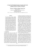
Fig. 1. Interaction between Moc1, Moc2 or Rpl32-2 and Cpc2 in vivo. (A) Cell extract was prepared from fission yeast cells carrying Moc1–
13Myc, Cpc2–3HA, Cpc2–3HA and Moc1–13Myc, or the un-tagged strain (wild-type). (B) Cell extract was prepared from fission yeast cells
carrying Moc2–13Myc, Cpc2–3HA, Cpc2–3HA and Moc2–13Myc, or the un-tagged strain (wild-type). (C) Cell extract was prepared from fis-
sion yeast cells carrying Rpl32-2–13Myc, Cpc2–3HA, Cpc2–3HA and Rpl32-2–13Myc, or the un-tagged strain (wild-type). The individual cell
extract was incubated with an HA antibody and a Myc antibody. Protein A Sepharose beads were added to the mixtures to coimmunoprecip-
itate Cpc2, and protein G Sepharose beads were added to coimmunoprecipitate Moc1, Moc2 or Rpl32-2. The coimmunoprecipitates were
analyzed by western blotting using HA and Myc antibodies.
S. K. Paul et al. Moc proteins in fission yeast
FEBS Journal 276 (2009) 5076–5093 ª 2009 The Authors Journal compilation ª 2009 FEBS 5081
detection using a Myc antibody (Figs 4A, 5A and 6A).
The double-tagged strains SKP2 (cpc2–3HA, moc2–
13Myc), SKP6 (cpc2–3HA, moc1–13Myc), SKP8
(cpc2–3HA, moc3–13Myc) and SKP10 (cpc2–3HA,
moc4–13Myc) showed similar results to the single-
tagged strains (SKP1, SKP5, SKP7 and SKP9) when
analyzed by 2D electrophoresis and western blotting
(Figs 3B, 4B, 5B and 6A). The patterns for Cpc2–3HA
in each strain, detected by the HA antibody, were also
similar to those of the double-tagged strains (Figs 3D,
4C and 5C). The pattern, ranging in size from large to
small, indicated the existence of a large molecule con-
taining the Moc1, Moc2, Moc3, Moc4 and Cpc2 pro-
teins. The pattern of 2D analysis was quite different
upon examination of a protein such as Asf1, which
works as a histone chaperon and exists as a monomer
of 30 kDa (Fig. 6C). 2D analysis of Asf1 13Myc
revealed only a small-sized protein. This control exper-
iment confirmed that the separation of proteins by
Blue Native/PAGE functioned efficiently.
We then performed further tests to determine
whether Cpc2 plays an important role in the
Moc-mediated complex. To this end, we constructed
various cpc2::ura4 strains hosting the different c-myc-
tagged moc genes: SKP11 (cpc2::ura4 moc1–13Myc),
SKP13 (cpc2::ura4 moc3–13Myc) and SKP14
(cpc2::ura4 moc4–13Myc). Cell extracts were prepared
from these strains and the samples were loaded onto
gels for first-dimension separation using Blue Native/
PAGE. Gel strips were then excised and used for elec-
trophoresis in the second dimension. Western blotting
revealed that, because of the cpc2 deletion, the Moc1-
and Moc3-mediated protein complexes produced a
weaker signal and were shifted towards a lower mole-
cular mass (Figs 4D and 5D). The results indicated
that, in the absence of Cpc2, a Moc1- or Moc3-
mediated large protein complex was either not formed,
or was unstable in S. pombe cells. We constructed
the strain cpc2::ura4 moc2–13Myc, but western blot-
ting failed to detect the Moc2 protein against a
cpc2-deleted background. This result indicated that
Cpc2 is important for the existence of the Moc2 pro-
tein in S. pombe cells. To determine whether the sta-
bility of Moc2 is dependent on the presence of Cpc2,
SKP12 (cpc2::ura4 moc2–13Myc) was transformed
with the plasmid pSLF273–cpc2, and the proteins were
analyzed by western blotting. We were able to detect
the Moc2 protein in this transformant (data not
shown), which clearly indicated that, in the absence of
Cpc2, Moc2 is unstable in S. pombe cells. It was previ-
ously reported that loss of Cpc2 did not dramatically
alter the rate of cellular protein synthesis, but caused a
decrease in the steady-state level of variable proteins
Rpl32-2-3HA
A
B
––
–
+
++
+
–
Moc2-13Myc
IP:HA
Blot:HA
IP:HA
Blot:Myc
IP:Myc
Blot:Myc
IP:Myc
Blot:HA
Imput
Blot:HA
Imput
Blot:Myc
Moc1-GFP
Moc2-13Myc
IP:GFP
Blot:GFP
IP:GFP
Blot:Myc
IP:Myc
Blot:Myc
Imput
Blot:GFP
Imput
Blot:Myc
––
–
+
++
+
–
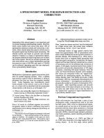
Fig. 2. Interaction between Rpl32-2 or Moc1 and Moc2 in vivo. (A)
Cell extract was prepared from fission yeast cells carrying Moc2–
13Myc, Rpl32-2–3HA, Rpl32-2–3HA and Moc2–13Myc tag, or the un-
tagged strain (wild-type). Individual cell extract was incubated with
an HA antibody and a Myc antibody. Protein A Sepharose beads were
added to the mixtures to coimmunoprecipitate Rpl32-2 and protein G
Sepharose beads were added to coimmunoprecipitate Moc2. The co-
immunoprecipitates were analyzed by western blotting using HA and
Myc antibodies. (B) Cell extract was prepared from fission yeast cell
carrying Moc1–GFP, Moc2–13Myc, Moc1–GFP and Moc2–13Myc
tag or the un-tagged strain (wild-type). Individual cell extract was
incubated with a GFP antibody and a Myc antibody. Protein G Sepha-
rose beads were added to the mixtures to coimmunoprecipitate
Moc1 and Moc2. The coimmunoprecipitates were analyzed by wes-
tern blotting using GFP and Myc antibodies.
Moc proteins in fission yeast S. K. Paul et al.
5082 FEBS Journal 276 (2009) 5076–5093 ª 2009 The Authors Journal compilation ª 2009 FEBS
[29]. We also tested whether Cpc2 affects Rpl32-2 by
2D analysis of the strain SKP30 (cpc2::ura4 rpl32-2-
13Myc). The results revealed that deletion of Cpc2
lowered the total amount of protein present, but did
not alter its molecular size (Fig. 7).
Influence of Moc1 to Moc4 and Rpl32-2 proteins
on the expression of Ste11
Finally, we tested whether overexpression of the Moc1
to Moc4 proteins and of Rpl32-2 induced expression
of the transcription factor Ste11. Following nitrogen
starvation, samples were taken from strains that over-
expressed each protein at regular time intervals
(Fig. 8), and western blotting was used to monitor the
level of Ste11–GFP expressed on the chromosome.
Our results revealed that expression of Ste11 was
clearly induced in response to overexpression of the
individual proteins Moc1, Moc2, Moc3, Moc4 and
Rpl32-2 (Fig. 8). A sharp peak in Ste11 at 3 h after
nitrogen starvation was observed in the wild-type
strain, as observed previously [21]. But, induction of
Ste11–GFP by Moc1 gave the clearest result, consis-
tent with the observation that, of the four Moc pro-
teins, Moc1 is the strongest inducer of sexual
development [15]. Induction of Ste11–GFP by Moc2
was observed after the 9 h time point, which may indi-
cate upregulation of translation. It is interesting to
note that Rpl32-2 also had a positive effect on the
induction of Ste11.
A
kDa
B
Complex
1048
720
480
242
Moc2-13Myc
kDa
95
130
242
146
66
72
55
43
34
26
C
20
Moc2-13Myc
95
130
72
55
43
DE
kDa
43
34
26
95
130
Moc2-
13Myc
72
95
130
Cpc2-3HA
95
72
55
43
34
26
26
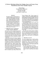
Fig. 3. Western blot analysis of Moc2 following Blue Native/PAGE and 2D SDS/PAGE. (A) Cells were extracted from S. pombe SKP1
(Moc2–13Myc) and proteins were separated on a 4% to 16% Blue Native/PAGE gel. Western blotting was performed using a Myc antibody
(1/3000) followed by anti-mouse IgG (1/3000). The arrow indicates the complex containing the Moc2 protein. (B) One lane was excised from
the first dimension gel and the gel strip was incubated with dissociation buffers and placed horizontally on top of the second dimension gel.
A 10% SDS/PAGE was then performed in the second dimension. When the gel strip was treated with dissociation buffer, the protein com-
plexes dissociated into their constituent polypeptides and the subunits of the protein complexes separated during 2D electrophoresis. Wes-
tern blotting was performed following the 2D SDS/PAGE using a Myc antibody (1/3000), and subsequent anti-mouse IgG (1/3000). 2D
electrophoresis was performed using the S. pombe double-tagged strain SKP2 (Moc2–13Myc, Cpc2–3HA). Western blotting was performed
using a Myc antibody (1/3000) and subsequent anti-mouse IgG (1/3000) (C), or an HA antibody (1/3000) and subsequent anti-mouse IgG (1/
3000) (D), respectively. (E) Western blotting with a Myc antibody (1/3000) and subsequent anti-mouse IgG (1/3000) to detect Moc2 tagged
with Myc on SDS/PAGE alone.
S. K. Paul et al. Moc proteins in fission yeast
FEBS Journal 276 (2009) 5076–5093 ª 2009 The Authors Journal compilation ª 2009 FEBS 5083
Discussion
In this study, we have shown that Moc1, Moc2, Moc3
and Moc4 proteins, which have been identified as posi-
tive regulators of sexual differentiation [14], exist as
A
kDa
95
130
E
Moc1-
13M
72
kDa
95
130
Moc1-13Myc
95
72
55
43
34
26
B
13Myc
Moc1-13Myc
17
95
130
72
55
43
34
26
17
C
Cpc2-3HA
95
130
72
55
43
D
Moc1-13Myc
34
26
17
95
130
72
72
55
43
34
26
17
17
Fig. 4. Western blotting of Moc1 following Blue Native/PAGE and
2D SDS/PAGE. Proteins were extracted from cells of S. pombe
strains SKP5, SKP6 and SKP11, and were separated on a 4–16%
Blue Native/PAGE gel. Individual lanes were excised from the first
dimension gel and treated with dissociation buffers, then slid into
place horizontally on top of the second dimension gel for SDS/
PAGE. Then, western blotting was performed as described in
Fig. 3. (A) S. pombe Moc1–13Myc tagged strain SKP5 was used
for 2D analysis. (B,C) S. pombe double-tagged strain SKP6 was
used for 2D analysis. (D) 2D electrophoresis was performed using
the cpc2 deleted and Moc1–13Myc-tagged strain SKP11. (E) Wes-
tern blotting was performed using a Myc antibody (1/3000) and
subsequent anti-mouse IgG (1/3000) to detect Moc1 protein tagged
with Myc in SKP5 cells on SDS/PAGE alone.
A
E
kDa
Moc3-13Myc
kDa
95
130
72
55
43
B
72
Moc3-
13myc
95
130
Moc3-13Myc
34
26
95
130
72
55
43
34
26
C
Cpc2-3HA
95
130
72
55
43
34
D
M 3 13M
34
26
95
130
Moc3-13Myc
95
72
55
43
34
26
Fig. 5. Western blot analysis of Moc3 following Blue Native/PAGE
and 2D SDS/PAGE. S. pombe (SKP7, SKP8 and SKP13) cells were
extracted and proteins were separated by 4–16% Blue Native/
PAGE and SDS/PAGE. Western blotting was performed as in
Fig. 3. (A) The S. pombe Moc3–13Myc-tagged strain SKP7 was
used for 2D analysis. (B,C) The S. pombe double-tagged strain
SKP8 was used for analysis. (D) 2D electrophoresis was performed
using the cpc2 deleted S. pombe Moc3–13Myc-tagged strain
SKP13. (E) Western blotting was performed using a Myc antibody
(1/3000) and anti-mouse IgG (1/3000) to detect Moc3 protein
tagged with Myc in SKP7 cells on SDS/PAGE alone.
Moc proteins in fission yeast S. K. Paul et al.
5084 FEBS Journal 276 (2009) 5076–5093 ª 2009 The Authors Journal compilation ª 2009 FEBS
high molecular mass complexes, and that Cpc2 plays
an important role in the formation of each complex.
Figure 9 summarizes the interactions revealed in this
study, combined with previously reported results [17].
The interactions revealed by the two-hybrid system
(shown by dashed arrows in Fig. 9) were not always
detected by coimmunoprecipitation in this study. In
general, coimmunoprecipitation detects stable interac-
tions in vivo, whereas the two-hybrid system detects
A
kDa
E
Moc4-13Myc
95
130
72
55
43
34
B
Cpc2-13Myc
kDa
95
130
Moc4-
13Myc
72
95
26
95
130
72
55
43
34
26
C
Asf1-13Myc
95
130
72
55
43
34
26
D
Wild type (SP870)
26
17
95
130
72
55
43
34
26
17
17
Fig. 6. Western blot analysis of Moc4 following Blue Native/PAGE
and 2D SDS/PAGE. S. pombe cells with different tags (Moc4–
13Myc, Cpc2–13Myc, Asf1–13Myc) and wild-type cells (SP870)
were used for this analysis. Proteins were first separated by
4–16% Blue Native/PAGE. (A) The S. pombe Moc4–13Myc-tagged
strain SKP9 was used for 2D analysis. (B) The S. pombe
Cpc2–13Myc tagged strain YO7 was used for 2-D analysis (C) The
S. pombe Asf1–13Myc tagged strain YM1 was used for this analy-
sis. (D) The wild-type strain was treated similarly as a negative con-
trol. (E) Western blotting was performed using a Myc antibody
(1/3000) and anti-mouse IgG (1/3000) to detect Moc4 protein
tagged with Myc in SKP9 cells on SDS/PAGE alone.
72
kDa
55
43
Rpl32-2
-13myc
A
B
C
D
E
Cpc2-3HA
Rpl32-2-13Myc
Rpl32-2-13Myc
Rpl32-2-13Myc
kDa
95
130
72
55
43
34
26
17
95
130
72
55
43
34
26
17
95
130
72
55
43
34
26
17
95
130
72
55
43
34
26
17
Fig. 7. Western blotting of Rpl32-2 following Blue Native/PAGE
and 2D SDS/PAGE. Proteins were extracted from S. pombe
(SKP20, SKP30 and SKP21) cells and were separated by 4–16%
Blue Native/PAGE and subsequent SDS/PAGE. Western blotting
was performed as in Fig. 3. (A) The S. pombe Rpl32-2–13
Myc-tagged strain SKP20 was used for this analysis. (B) 2D
electrophoresis was performed using the cpc2 deleted S. pombe
Rpl32-2–13Myc-tagged strain SKP30. (C,D) 2D electrophoresis was
performed using the S. pombe double-tagged strain SKP21. (E)
Western blotting was performed using a Myc antibody (1/3000)
and anti-mouse IgG (1/3000) to detect Rpl32-2 protein tagged with
Myc in SKP20 cells on SDS/PAGE alone.
S. K. Paul et al. Moc proteins in fission yeast
FEBS Journal 276 (2009) 5076–5093 ª 2009 The Authors Journal compilation ª 2009 FEBS 5085
both stable and transient interactions. Special care is
needed to interpret protein interactions detected in the
two-hybrid system, because the proteins are overex-
pressed in an artificial system, which does not always
reflect the in vivo situation.
Two-dimensional (Blue Native/PAGE and SDS/
PAGE) analysis showed that the gel patterns for each
Moc-mediated complex ranged in size from high to
lower molecular masses, suggesting that a high molecu-
lar mass complex does exist, in some form, in each
case. It is unclear whether all Moc proteins coexist in
the same complex, although it is unlikely, because the
four Moc proteins do not coexist in the same cellular
compartments. Moc1 and Moc2 are mainly localized
to the cytosol [14], whereas Moc3 and Moc4 are found
mainly in the nucleus [15,24]. Moc1 may have a chance
to interact with the nuclear proteins as it migrates
from the cytosol to the nucleus under starvation condi-
tions, although this localization shift is inhibited by
cAMP [17]. All proteins are translated in the cytosol,
thus there will always be the chance of transient inter-
actions between proteins that do not normally coexist,
and although Moc3 and Moc4 have been localized to
the nucleus by GFP fusion analysis, this does not rule
out the transient localization, or the presence of small
amounts, of these proteins in the cytosol.
All Moc1-interacting proteins identified in the two-
hybrid system also interacted with Moc3 (Table 1).
Equally, all Moc3-interacting proteins identified in the
two-hybrid system interacted with Moc1 (Table 3).
These results may imply that Moc1 and Moc3 have
characteristics similar to the target protein, but in fact,
these two proteins have very different primary struc-
tures. Moc1 contains the cystathione beta synthase
domain that is typically found in the gamma subunit of
AMP kinase [17], and Moc3 contains the Zn(2)–Cys(6)
binuclear cluster, which is typically found in transcrip-
tion factors. No direct interaction between Moc1 and
Moc3 has been reported [15] and an explanation for the
formation of a putative complex, mediated by both
Moc1 and Moc3, awaits further analysis. It was shown
very recently that Moc1/Sds23 interacted with protein
phosphatase 2A (PP2A)-related phosphatase Ppe1 [30],
but we did not screen out the related protein in our
two-hybrid screening.
The two-hybrid screening, reported in this study,
using moc1, moc2, moc3 or moc4 genes as bait high-
lighted the involvement of two proteins, namely Cpc2
and Rpl32-2. Cpc2 (a RACKI ortholog) protein is a
highly conserved member of the family of WD-repeat
proteins, exclusively localized in the cytosol [29,31]. It
is involved in sexual differentiation and plays a role in
translation through its interaction with ribosomal pro-
teins [29]. Cpc2 has been shown to interact with Msa2,
an RNA-binding protein that negatively regulates sex-
ual differentiation [31–33], Pck2, a protein kinase C
ortholog [34] and Pat1 kinase, a negative regulator of
meiosis [35]. Our coimmunoprecipitation study addi-
tionally revealed that Cpc2 physically interacted with
Moc1, Moc2 and Rpl32-2 (Fig. 1). Co-immunoprecipi-
tation experiments generally detect tight interactions
between proteins, so we were unable to detect any
interaction between Cpc2 and Moc3 using this
method, although the interaction was detected in the
two-hybrid system. By contrast, Moc1 was coimmuno-
precipitated with Moc2 (Fig. 2), although no interac-
tion between them was observed in the two-hybrid
system. This result suggested the existence of a large
molecule complex and the interaction is explainable by
the presence of a bridging protein such as Cpc2
between Moc1 and Moc2.
0 3 6 9 12 0 3 6 9 12 - Nitrogen starvation (hours)
Vector
Over expression (OE)
cdc2
cdc2
cdc2
cdc2
cdc2
Ste11-GFP (Moc1 OE)
Ste11-GFP (Moc2 OE)
Ste11-GFP (Moc3 OE)
Ste11-GFP (Moc4 OE)
Ste11-GFP (Rpl32-2 OE)
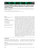
Fig. 8. Ste11 was induced by overexpression of Moc1, Moc2,
Moc3, Moc4 or Rpl32-2. The Ste11–GFP-tagged strain SPB371,
harboring pSLF173 (L), pSLF173(L)–moc1, pSLF173(L)–moc2,
pSLF173(L)–moc3, pSLF173(L)–moc4 and pREP1 or pREP1–rpl32-2
was cultured in PMA liquid medium until mid-log phase (5 · 10
6
cellsÆmL
)1
). The cells were harvested, washed twice with PMA-
N + 0.5% G medium, and shifted to the same medium to induce
mating and sporulation. Approximately 1 · 10
8
cells were harvested
at 0, 3, 6, 9 and 12 h after nitrogen starvation. Western blotting
was performed using the boiling SDS-glass bead method [47]. For
this analysis, a GFP mAb (1/1000) and anti-mouse IgG (1/1000)
were used to detect Ste11–GFP. Cdc2 was detected as an internal
control using a PSTAIRE antibody (1/1000) and anti-rabbit IgG (1/
3000).
Moc proteins in fission yeast S. K. Paul et al.
5086 FEBS Journal 276 (2009) 5076–5093 ª 2009 The Authors Journal compilation ª 2009 FEBS
Most interestingly, the larger molecule of the
Moc1- or Moc3-mediated complex shifted to the
smaller molecule when Cpc2 was absent (Figs. 4 and
5), and the Moc2-mediated complex became undetect-
able (data not shown). Moc2/ded1 is an RNA heli-
case that functions as a general translational factor,
and Cpc2 is a ribosomal-associated protein; therefore,
it is reasonable to think that the complex detected in
this study is also associated with ribosomes. A Moc2-
mediated complex, under native conditions, was
clearly detected at around 1000 kDa by Blue Native/
PAGE (Fig. 3A). This size is close to that of the
small subunit (40S) of the ribosomal complex, which
is estimated to be around 1400 kDa. However,
because 1000 kDa is almost the upper size limit that
can be clearly separated by Blue Native/PAGE, the
estimation of the size may not be accurate, and may
reflect the size of the small subunit (40S) of the ribo-
somal complex. It is possible that each Moc protein
is associated with the ribosomal complex through
Cpc2, although more analysis is needed to prove this.
Alternatively, the large Moc1- Moc2- Moc3- or
Moc4-mediated complex may link to the processing
bodies, which are now implicated in translation
repression, mRNA decay, nonsense-mediated decay
and mRNA storage [36]. In fact, very recently, an
involvement of Ded1 RNA helicase in the formations
of processing bodies was reported in Saccharomyces
cerevisiae [37].
In this study, we focused on the ribosomal protein
Rpl32-2 because it interacts strongly with all Moc pro-
teins in the two-hybrid system. Our analyses revealed
the physical interaction of Rpl32-2 with Moc2 or the
ribosome-associated protein Cpc2, which indicates that
the Moc2 and Cpc2 proteins are associated with the
ribosomal complex. This observation agrees well with
previous results [29,38]. Interestingly, Rpl32-2 of
S. pombe was reported to exhibit a novel extra-ribo-
somal function by acting as a DNA-binding protein and
potential transcriptional regulator [39]. We investigated
whether Rpl32-2 has a positive effect on the sexual dif-
ferentiation of S. pombe , and our results revealed that
when Rpl32-2 was overexpressed in S. pombe cells under
conditions of nitrogen starvation, expression of the key
transcription factor Ste11 was increased (Fig. 8). This
implied a role for Rpl32-2 in sexual differentiation simi-
lar to that for the Moc proteins, which were confirmed
as positive regulators of sexual differentiation and were
found to induce expression of ste11 when overexpressed
(Fig. 8). Lower levels of ste11 expression were shown in
a moc4/zfs1 deletion mutant [21]. Our preliminary data
showed that ste11 expression levels in a moc1 mutant
and a moc3 mutant [12], which are sterile and partly
sterile strains, respectively, were also lower than in wild-
type. Thus, all the moc genes, cpc2 and rpl32-2 investi-
gated in this study appear to have roles as inducers of
sexual differentiation.
These studies collectively suggest that each Moc
protein exists as a large complex in fission yeast and
that these proteins are involved in a regulatory net-
work that functions through interactions with the
ribosome-associated protein Cpc2 and the ribosomal
protein Rpl32-2. Although all Moc proteins may not
coexist in a single complex, it is possible that a large
Cpc2
cAMP
Moc1/Sds23
(CBS)
Cpc2
(Ribosomal
associated)
cAMP
Moc2/Ded1
(
Translation control)
Moc3
(Zn-finger protein)
Rpl32-2
(Ribosomal
protein)
Nucleus
Moc4/Zfs1
(mRNA decay)
p)
Ste11 (Transcription factor)
Translational regulation ?
Nucleus
Ste11
Sexual differentiation
Fig. 9. A summary of the interactions
between Moc proteins, Cpc2 and Rpl32-2.
The interactions detected by coimmunopre-
cipitation experiments are shown by solid
arrows. The interactions detected by the
yeast two-hybrid system are show by
dashed arrows. Moc3 and Moc4/Zfs1 local-
ize to the nucleus, whereas the other pro-
teins are found in the cytosol. Moc1/Sds23
ordinarily localizes to the cytosol, but moves
to the nucleus during meiosis, and this shift
is inhibited by cAMP [17]. Msa2 binds with
Cpc2 [31]. The involvement of the ribo-
some-associated protein Cpc2, and the gen-
eral translation factor Moc2/Ded1, implies
that a Moc-mediated complex may act as a
translational regulator and may be involved
in controlling sexual differentiation in fission
yeast through Ste11.
S. K. Paul et al. Moc proteins in fission yeast
FEBS Journal 276 (2009) 5076–5093 ª 2009 The Authors Journal compilation ª 2009 FEBS 5087
complex mediated by Moc1, Moc2 and Cpc2 might
operate as a translational regulator involved in con-
trolling the sexual differentiation of S. pombe
through activation of the key transcription factor
Ste11.
Experimental procedures
Strains, media and genetic manipulation
The Saccharomyces cerevisiae strain AH109 (MATa, trp1-
901, leu2-3,112 ura3-52, his3-200, gal4D, gal80D, LYS2::
GAL1
UAS
-GAL1
TATA
-HIS3, GAL2
UAS
-GAL2
TATA
-ADE2,
URA3::MEL1
UAS
-MEL1
TATA
-LacZ) was maintained on
YPD media composed of 1% yeast extract, 2% bactopep-
tone, 2% dextrose and 2% agar. Synthetic dropout media
SC–Trp, SC–Trp–Leu and SC–Trp–Leu–His were used for
nutrient auxotrophy in the two-hybrid analyses. The
S. pombe strains used in the study are listed in Table 5.
Standard yeast culture media and genetic manipulations
were used as described previously [40]. The S. pombe strains
were grown in complete YEA medium (0.5% yeast extract,
2% glucose and 0.0075% adenine) or in the synthetic mini-
mal medium, PM (0.3% potassium hydrogen phthalate,
0.22% sodium phosphate, 0.5% ammonium chloride, 2%
glucose, vitamins, minerals and salts), with the addition of
the appropriate auxotrophic supplements (0.0075% ade-
nine, leucine or uracil) when required. Either LiOAc or the
electroporation method, was used to transform yeast cells
[41]. Escherichia coli DH5a grown in LB medium (1%
polypeptone, 0.5% yeast extract, 1% sodium chloride)
hosted all plasmid manipulations using the standard
methods described [42].
Plasmid construction
The bait plasmids pGBKT7–moc1 to pGBKT7–moc4 car-
ried the moc genes fused to the Gal4-BD. The moc1, moc2,
moc3 and moc4 genes were amplified by PCR using
pMCS24, pMCS264, pMCS33 and pMCS65, respectively
(Table 6). The PCR products were digested with SmaI and
SalI. The digested fragments were cloned into the SmaI–
SalI sites of pGBKT7 carrying Gal4-BD, to create
pGBKT7–moc1 to pGBKT7–moc4. The accuracy of the
moc1, moc2, moc3 and moc4 gene sequences were verified
from the resulting constructs.
To create pGAD424–moc1 to pGAD424–moc4, the
moc1-, moc2-, moc3- and moc4-digested fragments were
cloned into the SmaI and SalI sites of pGAD424 carrying
the Gal4 activation domain. The resulting constructs were
confirmed by restriction digestion and PCR amplification
of the respective genes.
The pGBKT7–rpl32-2 construct carried the rpl32-2 gene
fused to Gal4-BD. The rpl32-2 gene was amplified by PCR
from genomic DNA using the relevant primers (Table 6).
The amplified product was digested with the restriction
enzymes EcoRI and SalI, and the digested fragment was
inserted into the EcoRI and SalI sites of pGBKT7 to create
pGBKT7–rpl32-2. To create pREP1–rpl32-2, the rpl32-2
gene was amplified by PCR from genomic DNA using the
relevant primers (Table 6). The PCR product was digested
Table 6. List of oligonucleotide primers used in this study. Restric-
tion enzyme sites are underlined (5¢- and 3¢).
pGBKT7/pGAD424 (Moc1–Moc4)
moc1–F–SmaI ACT
CCCGGGAATGCCTTTGTCAACTCAATC
moc2–F–SmaI CAAA
CCCGGGTATGAGCGACAATGTACAGC
moc3–F–SmaI CCT
CCCGGGTATGAACCCGTATGTTTCTTATC
moc4–F–SmaI TCT
CCCGGGCATGGTTTATTCTCCTATGTC
moc1–R–SalI TAT
GTCGACTCACCGACGTTGTGTATCTAC
moc2–R–SalI TTTA
GTCGACTTACCACCAGGATTGAGCAC
moc3–R–SalI CCA
GTCGACTGACTGTCGTACCGTAATTCG
moc4–R–SalI GAT
GTCGACTCAAGGAGATTGCTTAATAG
pGBKT7/pREP1 (Rpl32-2)
rpl32-2–F–EcoRI CACA
GAATTCATGGCTGCTGCTGTCAATATC
rpl32-2–R–Sal1 GAT
GTCGACTTACTCCTGAGAGCG
rpl32-2–F–SalI CAC
GTCGACAATGGCTGCTGTCAATATC
rpl32-2–R–BamHI CGTGAT
GGATCCTTACTCCTGAGAGC
Moc1 tagging primers
Moc1-W CTTGCTGTTGTCGATGCTCA
Moc1-X GGGGATCCGTCGACCTGCAGCGTACGACCGACG
TTGTGTATCTACAC
Moc1-Y GTTTAAACGAGCTCGAATTCATCGATTGCTAAA
TATTTGATGATT
Moc1-Z CGATTACGCCTCTGTGATTC
Moc2 tagging primers
Moc2-W CGTGGTTTAGATATTCCC
Moc2-X GGGGATCCGTCGACCTGCAGCGTACGA CCACCA
GGATTGAGCAC
Moc2-Y GTTTAAACGAGCTCGAATTCATCGATGGGTTAC
GTGCATCTGTG
Moc2-Z CATGAGCTCAAAGCCTG
Moc3 tagging primers
Moc3-W CTCGAAGTCATGCTCC
Moc3-X GGGGATCCGTCGACCTGCAGCGTACGAAAGTACT
GGTCGATTTAAGAC
Moc3-Y GTTTAAACGAGCTCGAATTCATCGATGCTAGAC
AAAATCACGC
Moc3-Z GCCGTGGTCGGTTCCG
Moc4 tagging primers
Moc4-W CCTAAGCTGTGCGTTCAATC
Moc4-X GGGGATCCGTCGACCTGCAGCGTACGAAGGAGA
TTGCTTAATAGTTGCAC
Moc4-Y GTTTAAACGAGCTCGAATTCATCGATGTTGTTAT
GCAATCTGGGTGAG
Moc4-Z GATTCATGCGTATCGCATTGC
Rpl32-2 tagging primers
Rpl32-2-W CAGTCTGACCGCTTCAAG
Rpl32-2-X GGGGATCCGTCGACCTGCAGCGTACGACTCCTGA
GCGAACCTTAG
Rpl32-2-Y GTTTAAACGAGCTCGAATTCATCGATGGTTAAAC
GTGACGCAGTCG
Rpl32-2-Z CGTCCTCCAGCTCAGATC
Moc proteins in fission yeast S. K. Paul et al.
5088 FEBS Journal 276 (2009) 5076–5093 ª 2009 The Authors Journal compilation ª 2009 FEBS
by the restriction enzymes SalI and BamHI, and the
digested fragment was inserted into the SalI and BamHI
sites of pREP1. The plasmid construct was verified by
restriction digestion and sequence analysis. Plasmid manipu-
lation and bacterial transformation were performed using
standard techniques [42].
Yeast two-hybrid screening
The yeast two-hybrid assay was performed as described
previously [43]. The constructed bait and prey plasmids
were introduced into Saccharomyces cerevisiae AH109 sin-
gly, or in combination with either the pGBKT7 or
pGADGH constructs, using the Li acetate–polyethylene
glycol one-step transformation protocol [44]. Expression of
the bait proteins (Moc1 to Moc4), fused to Gal4-BD, was
verified by western blotting with a c-Myc antibody (data
not shown). Cells transformed with the bait plasmids
pGBKT7-moc1 to pGBKT7-moc4 were incubated in the
synthetic dropout (SC)-Trp medium for 4 days at 30 °C.
Cells harboring bait plasmids were re-transformed with
pGADGH–cDNA and transformants were selected on
SC–Leu–Trp–His + 3-AT plates. The competitive inhibi-
tor 3-AT was used to inhibit low-level expression of the
yeast protein His3, and thus, to suppress background
growth on medium lacking His. Similarly, cells trans-
formed with the plasmids pGBKT7–rpl32-2 and
pGAD424–moc1 to pGAD424–moc4 were incubated on
synthetic dropout SC–Trp–Leu medium for 4 days at
30 °C, followed by culturing the cells in SC–Trp–Leu–His
medium for 4 days at 30 °C. The resulting transformants
were initially screened for b-galactosidase activity by filter
lift assay employing liquid N
2
-lysed cells floated on
5-bromo-4-chloro-3-indolyl-d-galactopyranoside (X-Gal)-
containing phosphate buffer.
b-Galactosidase activity was determined by filter lift
assay, as previously described [43]. Briefly, a sterile What-
man # 5 filter was placed over the surface of the plate.
Fresh colonies that had been grown on plates at 30 ° C for
4 days were carefully picked, individually streaked onto fil-
ter paper in the presence of positive and negative controls,
and cultured for 2 days at 30 °C. The filter was then care-
fully lifted off the agar plate with forceps and transferred
(colonies facing up) to a pool of liquid nitrogen. After the
filter had frozen completely ( 30 s) it was removed and
allowed to thaw at room temperature. The filter was then
carefully placed, colony side up, on another filter that had
been pre-soaked in a clean Petri dish containing Z-buffer
X-Gal solution. The filters were incubated at 30 °C and
checked periodically for the appearance of blue color.
Strain construction
Tag-integrated strains were constructed using a PCR-based
method [45,46]. Fragments of 500 bp from the 5¢ region
of the moc1, moc2, moc3 , moc4 and rpl32-2 genes from the
S. pombe strain SP870 were amplified using the primer
pairs: Moc1-W/Moc1-X, Moc2-W/Moc2-X, Moc3-W/
Moc3-X, Moc4-W/Moc4-X and Rpl32-2-W/Rpl32-2-X.
Similarly, the 3¢ regions of the moc1, moc2, moc3, moc4
and rpl32-2 genes were amplified using the primer pairs:
Moc1-Z/Moc1-Y, Moc2-Z/Moc2-Y, Moc3-Z/Moc3-Y,
Moc4-Z/Moc4-Y and Rpl32-2-Z/Rpl32-2-Y (Table 6). The
amplified fragments were attached to the end of the kan-
MX6 module by PCR using pFA6a–13Myc–kanMX6. The
wild-type strain SP870 was transformed with the tagged
DNA fragments from each of the second PCR products
and G418-resistant transformants were selected. Proper
integration was verified by PCR and western blot analysis.
The resulting strains were named SKP1 (Moc2–13Myc),
SKP5 (Moc1–13Myc), SKP7 (Moc3–13Myc), SKP9
(Moc4–13Myc) and SKP20 (Rpl32-2–13Myc). Both the cor-
responding amplified fragments of Rpl32-2 were attached
to the end of the kanMX6 module by PCR using pFA6a–
3HA–kanMX6. The wild-type strain SP870 was trans-
formed with the tagged DNA fragments from the second
PCR product, G418-resistant transformants were selected
and proper integration was verified by PCR and by western
blot analysis. The resulting strain was named SKP24
(Rpl32-2–3HA).
The double-tagged integrated strains SKP2, SKP6,
SKP8, SKP10 and SKP21 were constructed based on the
YO8 (Cpc2–3HA–kanMX6) strain. The first step involved
the amplification of 500 bp fragments from the 5¢ and
3¢ regions of the moc1, moc2, moc3, moc4 and rpl32-2
genes from S. pombe as previously described. The PCR
products were then attached to the hphMX6 module by
PCR using pFA6a–13Myc–hphMX6. The resulting
tagged fragments were introduced into the S. pombe
strain YO8. Hygromycin B-resistant transformants were
selected and protein expression was analyzed by western
blotting.
The tag-integrated strains SKP11, SKP12, SKP13 and
SKP14 were constructed using the S. pombe strain HT201
(cpc2::ura4). DNA fragments of 500 bp corresponding to
the 5¢ and 3¢ regions of the moc1, moc2, moc3 or moc4
genes were amplified by PCR oligonucleotides. The second
PCR amplified fragments were attached to the end of the
kanMX6 module by PCR using pFA6a–13Myc–kanMX6.
In the case of the tagged strain SKP30, DNA fragments of
500 bp corresponding to the 5¢ and 3¢ regions of the
rpl32-2 gene were amplified by PCR oligonucleotides. The
second PCR amplified fragments were attached to the end
of hphMX6 module by PCR using pFA6a–13Myc–
hphMX6. The fragments were introduced to the S. pombe
strain HT201 (cpc2::ura4) and transformants were selected
by G418 and Hygromycin B, respectively, and also by wes-
tern blotting. The resulting strains were named SKP11
(cpc2::ura4 moc1–13Myc–kanMX6), SKP12 (cpc2::ura4
moc2–13Myc–kanMX6), SKP13 (cpc2::ura4 moc3–13Myc–
S. K. Paul et al. Moc proteins in fission yeast
FEBS Journal 276 (2009) 5076–5093 ª 2009 The Authors Journal compilation ª 2009 FEBS 5089
kanMX6), SKP14 (cpc2::ura4 moc4–13Myc–kanMX6) and
SKP30 (cpc2::ura4 rpl32-2–13Myc–hphMX6).
The tag-integrated strain SKP22 was constructed based
on the S. pombe strain MYM2 (Moc1–3HA). A DNA frag-
ment of 500 bp corresponding to the 5¢ and 3¢ regions of
the rpl32-2 gene was amplified by PCR oligonucleotides.
The second PCR amplified fragments were attached to the
end of the hphMX6 module by PCR using pFA6a–13Myc–
hphMX6. Fragments were introduced into the S. pombe
strain MYM2 and transformants were selected using hygro-
mycin B and western blotting. The tag-integrated strains
SKP25, SKP26 and SKP27 were constructed using the
S. pombe strain SKP24 (Rpl32-2–3HA). DNA fragments of
500 bp corresponding to the 5¢ and 3¢ regions of the
moc2, moc3 or moc4 genes were amplified by PCR oligonu-
cleotides. The second PCR amplified fragments were
attached to the end of kanMX6 module by PCR using
pFA6a–13Myc–hphMX6. The fragments were introduced
into S. pombe SKP24 (Rpl32-2–3HA). Hygromycin B-resis-
tant transformants were selected and protein expression
was analyzed by western blotting. The strain SKP29
(Moc2–13Myc, Moc1–GFP) was constructed based on the
strain MYM3 (Moc1–GFP) in a similar manner. The
tagged protein did not interfere with the normal function of
each protein as judged by phenotypic observation.
Western blotting
Western blotting was performed by the simple alkali-SDS
method [47] and the boiling SDS-glass bead method [48].
Cells were harvested when they reached a density of
1 · 10
8
cells in the appropriate medium. The harvested
cells were washed twice with dH
2
O and dissolved in 100 lL
dH
2
O, and the samples were boiled at 95 °C for 5 min.
Subsequently, 120 lLof2· Laemmli buffer (4% SDS,
20% glycerol, 0.6 m b-mercaptoethanol, 8 m urea and
0.12 m Tris/HCl, pH 6.8) was added and the samples were
vigorously vortexed with an equal volume of acid-washed
glass beads using a bead homogenizer at 2500 rpm for
3 min. Samples were boiled at 95 °C for 5 min and centri-
fuged at 10 000 g for 15 min at 4 °C to remove the glass
beads and large debris. An equal volume of cell extract was
loaded onto SDS/PAGE using a 10% polyacrylamide gel
and then transferred to Immobilon transfer membranes
(Millipore, Bedford, MA, USA) using a wet-type transfer
system. To block unspecific binding, membranes were incu-
bated in a blocking buffer (NaCl/P
i
containing 5% non-fat
dry milk) supplemented with 0.1% Tween 20 at room tem-
perature for 1 h. To detect c-Myc fusion proteins, the mem-
brane was incubated with a Myc mAb (Santa Cruz
Biotechnology, Santa Cruz, CA, USA) diluted 1 : 3000 in
PBS-T (137 mm NaCl, 8 mm Na
2
HPO
4
.12H
2
O, 2.7 mm
KCl, 1.5 mm KHPO
4
and 0.1% Tween 20). The membrane
was washed with PBS-T for 15 and 5 min twice per wash,
and then incubated with horseradish peroxidase-conjugated
anti-mouse secondary IgG (Santa Cruz Biotechnology)
diluted 1 : 3000 in 5% dry milk in PBS-T.
To detect HA fusion proteins, the membrane was incu-
bated with an HA mAb (Santa Cruz Biotechnology) diluted
1 : 3000 in PBS-T (137 mm NaCl, 8 mm Na
2
HPO
4
12H
2
O,
2.7 mm KCl, 1.5 mm KHPO
4
and 0.1% Tween 20). The
membrane was washed three times with PBS-T every 5 min,
and then incubated with an anti-mouse secondary IgG
(Santa Cruz Biotechnology), diluted 1 : 3000 in 5% dry
milk in PBS-T. The membrane was washed three times with
PBS-T every 5 min, and then secondary antibodies were
detected by the chemiluminescence (ECL) system as
described by the manufacturer (Amersham, Little Chalfont,
UK).
Co-immunoprecipitation
S. pombe cells were grown in YES medium to the mid-log-
arithmic phase, then harvested (2 · 10
8
cellsÆmL
)1
) by cen-
trifugation, and washed once with ice-cold stop buffer
(150 mm NaCl, 50 mm NaF, 10 mm EDTA and 1 mm
NaN
3
pH 8). The cells were then lysed in 100 lL ice-cold
lysis buffer [50 mm Tris, 150 mm NaCl, 0.8% Nonid-
et-P40, 5 mm EDTA, 10% glycerol, 1 mm phenyl-
methanesulfonyl fluoride (PMSF) and protease inhibitor].
The samples were vortexed vigorously with 0.5 mm diameter
zirconia/silica beads using a bead homogenizer at
2500 rpm for 3 min. After centrifugation (10 000 g for 15
min at 4 °C), the protein concentration in the supernatant
was estimated.
An HA mAb and a Myc antibody were used in the
immunoprecipitation of HA and Myc fusion proteins,
respectively, in which 1 mg of each cell extract was incu-
bated with 1 lg of HA antibody and 1 lg of Myc antibody
for 4 h at 4 °C. Then 40 lL of protein A Sepharose beads
and the same volume of protein G Sepharose beads were
washed twice with 0.5 mL lysis buffer. The cleaned protein
A Sepharose beads were added to the HA antibody mixture
and the protein G Sepharose beads were added to the Myc
antibody mixture, followed by incubation with rotation for
4 h at 4 °C.
Sepharose beads were collected by centrifugation at
10 000 g for 10 min at 4 °C. The supernatant was dis-
carded by aspiration and the beads were washed six to
eight times using 0.5 mL lysis buffer, including protease
inhibitor and PMSF. The bead pellet was suspended in
30 lL lysis buffer (including protease inhibitor and PMSF)
and 60 lLof2· Laemmli buffer (4% SDS, 20% glycerol,
0.6 m b-mercaptoethanol, 8 m urea and 0.12 m Tris/HCl
pH 6.8) was added and vortexed. The suspended beads
were boiled at 95 °C for 5 min to dissociate the immuno-
complexes from the beads. After centrifugation (10 000 g
for 10 min at 4 °C), the supernatant was collected in a
new Eppendorf tube and then used for SDS/PAGE and
western blotting.
Moc proteins in fission yeast S. K. Paul et al.
5090 FEBS Journal 276 (2009) 5076–5093 ª 2009 The Authors Journal compilation ª 2009 FEBS
Blue Native/PAGE
Blue Native/PAGE is a method for the isolation of intact
protein complexes. The method followed the manufacturer’s
instructions (Invitrogen Corp., Tokyo, Japan). Protein com-
plexes were separated by their apparent molecular mass using
this standard PAGE system. In the first dimension, separa-
tion of the complexes under native conditions occurs accord-
ing to their molecular mass, and in the second dimension,
where electrophoresis is performed under denaturing condi-
tions, the individual subunits of the complexes are resolved,
again on the basis of their molecular mass [28].
Cells were grown in YES medium to the mid-logarithmic
phase, then harvested (1–2 · 10
8
cellsÆmL
)1
) by centrifuga-
tion. Cells were washed once with dH
2
O and stored at )80 °C.
The cell pellets were dissolved with 4 · Native PAGE sample
buffer (25 lL) + dH
2
O (72 lL) + 1 mm PMSF (1 lL) +
protease inhibitor (2 lL). Samples were vortexed vigorously
with an equal volume of acid-washed glass beads using a bead
homogenizer at 2500 rpm for 3 min. After centrifugation
(10 000 g for 15 min at 4 °C), the protein concentration in
the supernatant was estimated. Approximately 50 lg protein
was loaded per lane for electrophoresis.
Gel strips were cut from the Blue Native/PAGE gel, each
strip was transferred individually to a 15 mL conical tube,
and 5 mL of reducing solution [0.5 mL sample reducing
agent (10·), 1 mL LDS sample buffer (4·) and 3.5 mL H
2
O]
was added to each tube. Samples were incubated for 15 min
with shaking at room temperature and the reducing solution
was then decanted. Then 5 mL of alkylating solution [1 mL
LDS sample buffer (4·), 3.72 mL H
2
O and 28 lL DMA] was
added to each tube, incubated for 30 min at room tempera-
ture with shaking and then decanted. This was followed by
5 mL of quenching solution [0.50 mL sample reducing agent
(10·), 3 mL LDS sample buffer (4·), 1 mL EtOH and
3.5 mL H
2
O] being added to each tube and incubated at
room temperature. The quenching solution was decanted and
the gel strips were used for 2D SDS/PAGE.
Acknowledgements
We thank H. Kato and Y. Matsuo for technical
advices and R. Ogawa for her technical assistance.
This research was supported by grant-aid from the
Ministry of Education, Culture, Sports, Science and
Technology of Japan.
References
1 Yamamoto M, Imai Y & Watanabe Y (1997) Mating
and sporulation in Schizosaccharomyces pombe .InThe
Molecular and Cellular Biology of the Yeast Saccharo-
myces (Pringle JT, Broach JR & Jones EW eds), pp.
1037–1106. Cold Spring Harbor Laboratory Press, Cold
Spring Harbor, NY.
2 Matsuo T, Otsubo Y, Urano J, Tamanoi F & Yamam-
oto M (2007) Loss of the TOR kinase Tor2 mimics
nitrogen starvation and activates the sexual develop-
ment pathway in fission yeast. Mol Cell Biol 27, 3154–
3164.
3 Ozoe F, Kurokawa R, Kobayashi Y, Jeong HT,
Tanaka K, Sen K, Nakagawa T, Matsuda H &
Kawamukai M (2002) The 14-3-3 proteins Rad24 and
Rad25 negatively regulate Byr2 by affecting its localiza-
tion in Schizosaccharomyces pombe. Mol Cell Biol 22,
7105–7119.
4 Yamamoto TG, Chikashige Y, Ozoe F, Kawamukai M
& Hiraoka Y (2004) Activation of the pheromone-
responsive MAP kinase drives haploid cells to undergo
ectopic meiosis with normal telomere clustering and
sister chromatid segregation in fission yeast. J Cell Sci
117, 3875–3886.
5 Kawamukai M, Ferguson K, Wigler M & Young D
(1991) Genetic and biochemical analysis of the adenylyl
cyclase of Schizosaccharomyces pombe. Cell Regul 2 ,
155–164.
6 Hoffman CS (2005) Glucose sensing via the protein
kinase A pathway in Schizosaccharomyces pombe.
Biochem Soc Trans 33, 257–260.
7 Kawamukai M, Gerst J, Field J, Riggs M, Rodgers L,
Wigler M & Young D (1992) Genetic and biochemical
analysis of the adenylyl cyclase-associated protein, cap,
in Schizosaccharomyces pombe. Mol Biol Cell 3, 167–
180.
8 Zhou GL, Yamamoto T, Ozoe F, Yano D, Tanaka K,
Matsuda H & Kawamukai M (2000) Identification of a
14-3-3 protein from Lentinus edodes that interacts with
CAP (adenylyl cyclase-associated protein), and conser-
vation of this interaction in fission yeast. Biosci Biotech-
nol Biochem 64, 149–159.
9 Maeda T, Watanabe Y, Kunitomo H & Yamamoto M
(1994) Cloning of the pka1 gene encoding the catalytic
subunit of the cAMP-dependent protein kinase in
Schizosaccharomyces pombe. J Biol Chem 269, 9632–
9637.
10 Higuchi T, Watanabe Y & Yamamoto M (2002) Pro-
tein kinase A regulates sexual development and gluco-
neogenesis through phosphorylation of the Zn finger
transcriptional activator Rst2p in fission yeast. Mol Cell
Biol 22, 1–11.
11 Kitamura K, Katayama S, Dhut S, Sato M, Watanabe
Y, Yamamoto M & Toda T (2001) Phosphorylation of
Mei2 and Ste11 by Pat1 kinase inhibits sexual differen-
tiation via ubiquitin proteolysis and 14-3-3 protein in
fission yeast. Dev Cell 1, 389–399.
12 Qin J, Kang W, Leung B & McLeod M (2003) Ste11p,
a high-mobility-group box DNA-binding protein,
undergoes pheromone- and nutrient-regulated
nuclear–cytoplasmic shuttling. Mol Cell Biol 23, 3253–
3264.
S. K. Paul et al. Moc proteins in fission yeast
FEBS Journal 276 (2009) 5076–5093 ª 2009 The Authors Journal compilation ª 2009 FEBS 5091
13 Oowatari Y, Toma K, Ozoe F & Kawamukai M (2009)
Identification of sam4 as a rad24 allele in Schizosacchar-
omyces pombe. Biosci Biotechnol Biochem 73, 1591–
1598.
14 Kawamukai M (1999) Isolation of a novel gene, moc2,
encoding a putative RNA helicase as a suppressor of
sterile strains in Schizosaccharomyces pombe. Biochim
Biophys Acta 1446, 93–101.
15 Goldar MM, Jeong HT, Tanaka K, Matsuda H &
Kawamukai M (2005) Moc3, a novel Zn finger type
protein involved in sexual development, ascus forma-
tion, and stress response of Schizosaccharomyces pombe.
Curr Genet 48, 345–355.
16 Ishii K, Kumada K, Toda T & Yanagida M (1996)
Requirement for PP1 phosphatase and 20S cyclosome/
APC for the onset of anaphase is lessened by the dosage
increase of a novel gene sds23
+
. EMBO J 15, 6629–6640.
17 Yakura M, Ishikura Y, Adachi Y & Kawamukai M
(2006) Involvement of Moc1 in sexual development and
survival of Schizosaccharomyces pombe. Biosci Biotech-
nol Biochem 70, 1740–1749.
18 Shimanuki M, Chung SY, Chikashige Y, Kawasaki Y,
Uehara L, Tsutsumi C, Hatanaka M, Hiraoka Y,
Nagao K & Yanagida M (2007) Two-step, extensive
alterations in the transcriptome from G0 arrest to cell
division in Schizosaccharomyces pombe. Gene Cell 12,
677–692.
19 Jang YJ, Won M, Chung KS, Kim DU, Hoe KL, Park
C & Yoo HS (1997) A novel protein, Psp1, essential for
cell cycle progression of Schizosaccharomyces pombe is
phosphorylated by Cdc2–Cdc13 upon entry into G0-like
stationary phase of cell growth. J Biol Chem 272,
19993–20002.
20 Goldar MM, Nishie T, Ishikura Y, Fukuda T, Takega-
wa K & Kawamukai M (2005) Functional conservation
between fission yeast moc1/sds23 and its two orthologs,
budding yeast SDS23 and SDS24, and phenotypic dif-
ferences in their disruptants. Biosci Biotechnol Biochem
69, 1422–1426.
21 Forbes KC, Humphrey T & Enoch T (1998) Suppres-
sors of cdc25p overexpression identify two pathways
that influence the G2/M checkpoint in fission yeast.
Genetics 150, 1361–1375.
22 Grallert B, Kearsey SE, Lenhard M, Carlson CR,
Nurse P, Boye E & Labib K (2000) A fission yeast
general translation factor reveals links between protein
synthesis and cell cycle controls. J Cell Sci 113,
1447–1458.
23 Liu HY, Nefsky BS & Walworth NC (2002) The Ded1
DEAD box helicase interacts with Chk1 and Cdc2.
J Biol Chem 277, 2637–2643.
24 Kanoh J, Sugimoto A & Yamamoto M (1995) Schizo-
saccharomyces pombe zfs1
+
encoding a zinc-finger pro-
tein functions in the mating pheromone recognition
pathway. Mol Biol Cell 6, 1185–1195.
25 Beltraminelli N, Murone M & Simanis V (1999) The
S. pombe zfs1 gene is required to prevent septation
if mitotic progression is inhibited. J Cell Sci 112,
3103–3114.
26 Cuthbertson BJ, Liao Y, Birnbaumer L & Blackshear
PJ (2008) Characterization of zfs1 as an mRNA-binding
and -destabilizing protein in Schizosaccharomyces pom-
be. J Biol Chem 283, 2586–2594.
27 Paul SK, Goldar MM, Yakura M, Oowatari Y &
Kawamukai M (2009) Glutamyl tRNA synthetases and
glutamic acid induce sexual differentiation of
Schizosaccharomyces pombe. Biosci Biotechnol Biochem
73, 1339–1347.
28 Nijtmans LG, Henderson NS & Holt IJ (2002) Blue
Native electrophoresis to study mitochondrial and other
protein complexes. Methods 26, 327–334.
29 Shor B, Calaycay J, Rushbrook J & McLeod M (2003)
Cpc2/RACK1 is a ribosome-associated protein that
promotes efficient translation in Schizosaccharo-
myces pombe. J Biol Chem 278, 49119–49128.
30 Hanyu Y, Imai KK, Kawasaki Y, Nakamura T,
Nakaseko Y, Nagao K, Kokubu A, Ebe M, Fujisawa
A, Hayashi T et al. (2009) Schizosaccharomyces pombe
cell division cycle under limited glucose requires Ssp1
kinase, the putative CaMKK, and Sds23, a PP2A-
related phosphatase inhibitor. Gene Cell 14, 539–554.
31 Jeong HT, Oowatari Y, Abe M, Tanaka K, Matsuda H
& Kawamukai M (2004) Interaction between a negative
regulator (Msa2/Nrd1) and a positive regulator (Cpc2)
of sexual differentiation in Schizosaccharomyces pombe.
Biosci Biotechnol Biochem 68, 1621–1626.
32 Jeong HT, Ozoe F, Tanaka K, Nakagawa T, Matsuda
H & Kawamukai M (2004) A novel gene, msa1, inhibits
sexual differentiation in Schizosaccharomyces pombe.
Genetics 167, 77–91.
33 Katayama S, Ozoe F, Kurokawa R, Tanaka K, Naka-
gawa T, Matsuda H & Kawamukai M (1996) Genetic
analysis of the sam mutations, which induce sexual
development with no requirement for nutritional starva-
tion in fission yeast. Biosci Biotechnol Biochem 60, 994–
999.
34 Won M, Park SK, Hoe KL, Jang YJ, Chung KS,
Kim DU, Kim HB & Yoo HS (2001) Rkp1/Cpc2,
a fission yeast RACK1 homolog, is involved in actin
cytoskeleton organization through protein kinase C,
Pck2, signaling. Biochem Biophys Res Commun 282,
10–15.
35 McLeod M, Shor B, Caporaso A, Wang W, Chen H &
Hu L (2000) Cpc2, a fission yeast homologue of mam-
malian RACK1 protein, interacts with Ran1 (Pat1)
kinase to regulate cell cycle progression and meiotic
development. Mol Cell Biol 20, 4016–4027.
36 Sheth U & Parker R (2006) Targeting of aberrant
mRNAs to cytoplasmic processing bodies. Cell 125,
1095–1109.
Moc proteins in fission yeast S. K. Paul et al.
5092 FEBS Journal 276 (2009) 5076–5093 ª 2009 The Authors Journal compilation ª 2009 FEBS
37 Beckham C, Hilliker A, Cziko AM, Noueiry A,
Ramaswami M & Parker R (2008) The DEAD-box
RNA helicase Ded1p affects and accumulates in
Saccharomyces cerevisiae P-bodies. Mol Biol Cell 19,
984–993.
38 Linder P (2003) Yeast RNA helicases of the DEAD-
box family involved in translation initiation. Biol Cell
95, 157–167.
39 Wang J, Yuan S & Jiang S (2006) The ribosomal
protein L32-2 (RPL32-2) of S. pombe exhibits a novel
extraribosomal function by acting as a potential tran-
scriptional regulator. FEBS Lett 580, 1827–1832.
40 Alfa C, Fantes P, Hyams J, McLeod M & Warbrick E
(1993) Experiments with Fission Yeast . Cold Spring
Harbor Laboratory Press, Cold Spring Harbor, NY.
41 Prentice HL (1992) High efficiency transformation of
Schizosaccharomyces pombe by electroporation. Nucleic
Acids Res 20, 621.
42 Sambrook J, Fritsch EF & Maniatis T (1989) Molecular
Cloning: A Laboratory Manual, 2nd edn. Cold Spring
Harbor Laboratory Press, Cold Spring Harbor, NY.
43 Chien CT, Bartel PL, Sternglanz R & Fields S (1991)
The two-hybrid system: a method to identify and clone
genes for proteins that interact with a protein of inter-
est. Proc Natl Acad Sci USA 88, 9578–9582.
44 Ito H, Fukuda Y, Murata K & Kimura A (1983)
Transformation of intact yeast cells treated with alkali
cations. J Bacteriol 153, 163–168.
45 Krawchuk MD & Wahls WP (1999) High-effi-
ciency gene targeting in Schizosaccharomyces pombe
using a modular, PCR-based approach with
long tracts of flanking homology. Yeast 15,
1419–1427.
46 Matsuo Y, Matsuura Y, Tanaka K, Matsuda H &
Kawamukai M (2004) Chr4, a Schizosaccharo-
myces pombe homologue of the Saccharomyces
cerevisiae Chs4p/Skt5p protein, is related to septum
formation and is required for the proper localization of
Chs2. Yeast 21, 1005–1019.
47 Matsuo Y, Asakawa K, Toda T & Katayama S
(2006) A rapid method for protein extraction
from fission yeast. Biosci Biotechnol Biochem 70,
1992–1994.
48 Yakura M, Ozoe F, Ishida H, Nakagawa T, Tanaka K,
Matsuda H & Kawamukai M (2006) zds1, a novel gene
encoding an ortholog of Zds1 and Zds2, controls sexual
differentiation, cell wall integrity and cell morphology
in fission yeast. Genetics 172, 811–825.
49 Tanabe K, Ito N, Wakuri T, Ozoe F, Umeda M,
Katayama S, Tanaka K, Matsuda H & Kawamukai
M (2003) Sla1, a Schizosaccharomyces pombe homolog
of the human La protein, induces ectopic meiosis
when its C terminus is truncated. Eukaryot Cell 2,
1274–1287.
50 Tanabe K, Tanaka K, Matsuda H & Kawamukai M
(2004) Truncated Sla1 induces haploid meiosis through
the Pat1–Mei2 system in fission yeast. Biosci Biotechnol
Biochem 68, 266–270.
S. K. Paul et al. Moc proteins in fission yeast
FEBS Journal 276 (2009) 5076–5093 ª 2009 The Authors Journal compilation ª 2009 FEBS 5093