Báo cáo khoa học: The b-N-acetylglucosaminidases NAG1 and NAG2 are essential for growth of Trichoderma atroviride on chitin doc
Bạn đang xem bản rút gọn của tài liệu. Xem và tải ngay bản đầy đủ của tài liệu tại đây (452.55 KB, 12 trang )
The b-N-acetylglucosaminidases NAG1 and NAG2 are
essential for growth of Trichoderma atroviride on chitin
Rube
´
nLo
´
pez-Monde
´
jar*, Valentina Catalano, Christian P. Kubicek and Verena Seidl
Research Area Gene Technology and Applied Biochemistry, Institute of Chemical Engineering, Vienna University of Technology, Austria
Introduction
Chitin is a natural polysaccharide consisting of b-1,4-
linked N-acetylglucosamine (GlcNAc) units, and,
although it is the second most abundant biopolymer,
relatively little is known about its turnover in marine
and soil ecosystems. In the sea, chitin is found as the
main compound of the exoskeleton of crustaceans, and
on land it is an essential structural component of
insects and the cell walls of filamentous fungi, where it
is covalently linked to other carbohydrates and pro-
teins [1,2]. Degradation of chitin biomass is achieved
Keywords
chitin degradation; chitinases;
mycoparasitism; N-acetylglucosaminidases;
Trichoderma atroviride
Correspondence
V. Seidl, Research Area Gene Technology
and Applied Biochemistry, Institute of
Chemical Engineering, Vienna University of
Technology, Getreidemarkt 9 ⁄ 166-5, 1060
Vienna, Austria
Fax: +43 1 58801 17299
Tel: +43 1 58801 17227
E-mail:
Website: />Seidl
Present addresses
*Department of Soil Water Conservation
and Organic Waste Management, Centro de
Edafologı
´
a y Biologı
´
a Aplicada del Segura
(CEBAS-CSIC), PO Box 164, 30100 Espi-
nardo, Murcia, Spain
Department of Tree Science, Entomology
and Plant Pathology ‘G. Scaramuzzi’, Plant
Pathology Section, Faculty of Agriculture,
University of Pisa, Via del Borghetto 80,
I-56124 Pisa, Italy
(Received 10 June 2009, revised 26 June
2009, accepted 13 July 2009)
doi:10.1111/j.1742-4658.2009.07211.x
The chitinolytic enzyme machinery of fungi consists of chitinases and b-N-
acetylglucosaminidases. These enzymes are important during the fungal life
cycle for degradation of exogenous chitin, which is the second most abun-
dant biopolymer, as well as fungal cell-wall remodelling. In addition,
involvement of chitinolytic enzymes in the lysis of the host cell wall in
mycoparasitic Trichoderma spp. has been reported. In view of the fact that
fungi have on average 15–20 chitinases, but only two b-N-acetylglucosami-
nidases, the question arises how important the latter enzymes actually are
for various aspects of chitin degradation. In this study, the role of two
b-N-acetylglucosaminidases, NAG1 and NAG2, was analysed in the myco-
parasitic fungus Trichoderma atroviride.Nob-N-acetylglucosaminidase
activity was detected in T. atroviride Dnag1Dnag2 strains, suggesting that
NAG1 and NAG2 are the only enzymes in T. atroviride that possess this
activity. Dnag1Dnag2 strains were not able to grow on chitin and chitobi-
ose, but the presence of either NAG1 or NAG2 was sufficient to restore
growth on chitinous carbon sources in solid media. Our results demon-
strated that T. atroviride cannot metabolize chitobiose but only the mono-
mer N-acetylglucosamine, and that N-acetylglucosaminidases are therefore
essential for the use of chitin as a nutrient source. NAG1 is predominantly
secreted into the medium, whereas NAG2 mainly remains attached to the
cell wall. No physiological changes or reduction of the mycoparasitic
potential of T. atroviride was detected in the double knockout strains, sug-
gesting that the use of chitin as carbon source is only of minor importance
for these processes.
Abbreviations
GH, glycoside hydrolase; GlcNAc, N-acetylglucosamine; NAGase, b-N-acetylglucosaminidase; PDA, potato dextrose agar.
FEBS Journal 276 (2009) 5137–5148 ª 2009 The Authors Journal compilation ª 2009 FEBS 5137
by the concerted action of chitinases and b- N-acetyl-
glucosaminidases (NAGases; EC 3.2.1.52). NAGases
belong to glycoside hydrolase (GH) family 20 in the
CAZy classification (), and, by def-
inition, catalyse the hydrolytic release of terminal,
non-reducing GlcNAc residues, but their highest sub-
strate affinity is for the dimer chitobiose (GlcNAc)
2
,
which they convert into two GlcNAc monomers [3].
The genomes of ascomycetous filamentous fungi
contain on average 15–20 genes encoding chitinases,
but only two or three genes encoding GH family 20
proteins. Potential functions of chitin-degrading
enzymes in fungi include use of exogenous chitin as a
nutrient source and cell-wall remodelling during the
fungal life cycle [4].
Some species of the fungal genus Hypocrea ⁄ Tricho-
derma, such as T. atroviride (teleomorph Hypo-
crea atroviridis), T. harzianum, T. virens (H. virens)
and T. asperellum, are mycoparasites, i.e. they invade
and destroy fungal cells and feed on the contents of
dead cells. Chitinases and NAGases have been repeat-
edly implicated in cell-wall hydrolysis during myco-
parasitic attack (for reviews, see [5,6]). Two NAGases
have been cloned from several Trichoderma spp., and
it was shown that they are active as dimers and that
their gene expression can be induced by chitinous
carbon sources such as GlcNAc, chito-oligosaccha-
rides, colloidal chitin and fungal cell walls [7–13].
Further, enhanced NAGase activities were detected
on non-chitinous carbon sources such as a-glucans
and oligosaccharides containing galactose, and tran-
scriptional upregulation of nag1 and nag2 under the
respective growth conditions was shown [11]. In the
same study, basal transcript levels of nag1 and nag2
and corresponding NAGase activities were detected
under non-inducing growth conditions, possibly sug-
gesting a role during fungal cell-wall remodelling.
However, while transcriptional regulation of genes
encoding NAGases has been studied in detail, rela-
tively little is known about their functions and impor-
tance in Trichoderma. Aspergillus nidulans has only
one NAGase, nagA, which was shown to be strongly
induced during autolysis [14,15]. In contrast, in a
T. atroviride Dnag1 strain, residual NAGase activity
was found to be as high as 80%, depending on the
substrate [11], which is most likely due to the fact
that T. atroviride has two NAGases. The mycopara-
sitic abilities of the Dnag1 strain were similar to those
of the wild-type in plate confrontation assays with
plant pathogenic fungi [16], and no phenotypic
changes or alteration of mycoparasitim were detected
in a T. asperellum exc2y knockout strain (where
exc2y is equivalent to nag2) [10].
It is obvious that there is still a severe lack of
understanding of the physiological relevance of
NAGases in fungi. NAGase gene expression was
found to be upregulated under a variety of growth
conditions, but few effects were observed in single
knockout strains in Trichoderma. Therefore, the
question arises as to how important NAGases actu-
ally are for various chitinolyitc processes. Are they
involved in cell-wall remodelling, attack and defence
mechanisms (e.g. mycoparasitism) and ⁄ or are they
solely important for chitin sequestration, independent
of the chitinous carbon source? Chitin is the second
most abundant biopolymer on earth, but how fungi
handle its degradation is still not understood, espe-
cially in view of the fact that they have up to 35
chitinases, but only two extracellular NAGases. Are
NAGases of particular importance for chitin catabo-
lism in fungi or are they fully dispensable for this
process?
We addressed these questions by creating Dnag1D
nag2 strains in T. atroviride . Here we present data
showing that NAG1 and NAG2 are the only extracel-
lular NAGases in T. atroviride,asDnag1Dnag2
strains
exhibit no residual extracellular NAGase activity, and
that their function in cleaving the dimer chitobiose in
GlcNAc monomers is essential for the use of chitin
as a nutrient source in T. atroviride. This is the first
time that the ability of a fungus to catabolize chitin
has been abolished. However, the mycoparasitic
potential was not altered in Dnag1Dnag2 strains, sug-
gesting that use of chitin as a carbon or nitrogen
source is not of major importance for the mycopara-
sitic process.
Results
Construction of T. atroviride Dnag2 and Dnag1D
nag2 strains
Two NAGases have been cloned and characterized
from several Trichoderma species so far [7–10]. In
T. atroviride,aDnag1 strain has been reported previ-
ously [16], whereas NAG2 has only been analysed
indirectly by comparison of the growth of T. atrovi-
ride wild-type (WT) and Dnag1 strains on various car-
bon sources [11]. To elucidate the role of nag2 and
the combined roles of nag1 and nag2 in T. atroviride,
nag2 knockout and nag1 nag2 double knockout
strains were generated. The Dnag1 strain carries the
amdS selection marker, and an hph cassette, confer-
ring resistance to hygromycin B, was therefore used
for generation of both Dnag2 and Dnag1Dnag2 knock-
out strains.
Chitin degradation in Trichoderma atroviride R. Lo
´
pez-Monde
´
jar et al.
5138 FEBS Journal 276 (2009) 5137–5148 ª 2009 The Authors Journal compilation ª 2009 FEBS
The deletion cassette (Fig. S1A) was amplified by
PCR from the generated plasmid pVCNAG2 and
transformed into T. atroviride WT and Dnag1 strains
(see Experimental procedures). Purified transformants
were screened for deletion of nag2 by PCR (data not
shown). Five Dnag2 strains and four Dnag1Dnag2
strains of the positively identified transformants were
subjected to Southern analysis (Fig. S1B), which
confirmed that nag2 had been replaced by the hph
cassette, and showed that only a single copy of the
deletion cassette had been integrated into the genome.
Carbon source utilization profiles of T. atroviride
Dnag2 and Dnag1Dnag2 strains
The carbon source utilization profiles of all positively
identified Dnag2 and Dnag1Dnag2 knockout strains were
assessed using the Biolog Phenotype MicroArray sys-
tem, which has previously been established for Tricho-
derma spp. [11,17] and allows fast and reliable screening
of growth rates on 95 carbon sources. Carbon source
profiling enabled us to analyse the phenotypical vari-
ability of growth among the knockout strains in order
to check for any possible defects unrelated to the nag2
gene knockout due to the transformation procedure,
and also to compare the growth profiles of the knock-
out strains with those of the WT strain. Specific growth
rates of the strains were calculated from the increase in
the absorbance at 750 nm between 24 and 42 h – the
time at which active growth occurs on most carbon
sources – and are shown in Fig. S2. The inter-strain var-
iability among the five Dnag2 strains and four Dnag1
Dnag2 strains that were studied was extremely low, as
can be seen from the error bars in Fig. S2, representing
the standard deviation of the growth rate for the respec-
tive groups of strains.
The average carbon source utilization profiles of
the Dnag2 and Dnag1Dnag2 strains were highly simi-
lar to those of the WT (Fig. S2), showing that assimi-
lation of the 95 carbon sources assayed was not
altered in the Dnag2 or Dnag1Dnag2 knockout strains.
The Dnag1 strain, which has already been character-
ized in detail using the Biolog system [11], also
displayed similar growth rates (data not shown).
These data indicate that NAGases are not essential
for normal growth on non-chitinous carbon sources.
Dnag2 strains C2332 and A523 and Dnag1Dnag2
strains 713 and 1921 were randomly chosen and used
together with the WT and Dnag1 strain in subsequent
experiments for thorough characterization of their
phenotypes. D
nag2-I and Dnag2-II are Dnag2 strains
C2332 and A523, and nag1Dnag2-I and nag1Dnag2-II
are Dnag1Dnag2 strains 713 and 1921, respectively.
NAG1 and NAG2 are essential for growth on
chitin and chitobiose
Having shown that T. atroviride Dnag2 and Dnag1
Dnag2 strains grew normally on non-chitinous carbon
sources, we next investigated the role of these enzymes
in growth on chitin. Although T. atroviride has more
than 25 chitinases, chitin is not a good carbon source
for T. atroviride, even in its pre-treated colloidal form.
The fungus does not readily use chitin but first forms
a thin mycelium on the surface of the whole agar plate
before actually starting to form biomass, which is then
strongly linked to sporulation. The WT, Dnag1 and
Dnag2 strains formed a firm layer of biomass and
spores on chitin plates, whereas the Dnag1Dnag2
strains only produced very few spots of sporulating
biomass (Fig. 1A). On control plates containing potato
dextrose agar (PDA) all strains grew and sporulated
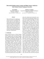
normally (Fig. 1B). These results suggest that the pres-
ence of at least one of the two enzymes NAG1 and
NAG2 is essential for growth on chitin by hydrolysing
the dimer chitobiose (GlcNAc)
2
, and imply that extra-
cellular conversion of the dimer into monomers is nec-
essary for assimilation of this carbon source, and that
only the monomer can be taken up by the fungus.
The small amount of biomass that Dnag1Dnag2
strains formed on chitin plates could theoretically
result either from the presence of an as yet unidentified
third NAGase, or be due to release of GlcNAc mono-
mers resulting from the random cleavage of chito-olig-
omers by chitinases. To test this, we grew the strains
on chitobiose (Fig. 1C). Under these conditions,
growth of the WT and Dnag1 and Dnag2 strains
occurred and was identical, whereas the Dnag1Dnag2
strains did not grow at all, except for a very few extre-
mely thin aerial hyphae, which were also found on
control medium containing no carbon source; these
hyphae therefore most likely result from internal car-
bohydrate reserves of the spores. These findings prove
that NAG1 and NAG2 are together responsible for
chitobiose degradation by T. atroviride, and that there
are no further enzymes in T. atroviride that account
for this ability.
As growth on plates can be misleading, e.g. due to
varying hyphal thickness, we also quantified the
biomass formed on chitin plates. The results from
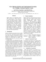
biomass quantifications (Fig. 2A) reflected the macro-
scopic observations from Fig. 1B, showing a statisti-
cally significant reduction of biomass formation in the
Dnag1Dnag2 strains to less than 25% of the WT strain
(one-way ANOVA, F(5,6) = 138.44, P < 0.01). Bio-
mass formation in the Dnag1 and Dnag2 strains was
similar to that in the WT (P > 0.05).
R. Lo
´
pez-Monde
´
jar et al. Chitin degradation in Trichoderma atroviride
FEBS Journal 276 (2009) 5137–5148 ª 2009 The Authors Journal compilation ª 2009 FEBS 5139
To further prove that NAG1 and NAG2 are the
only enzymes responsible for NAGase activity in T. at-
roviride, we assayed their activity in the various dele-
tion strains. Fig. 2B shows that the activity was indeed
completely absent in the Dnag1Dnag2 strains. In the
single knockout strains, a significant reduction of
NAGase levels was detected (one-way ANOVA,
F(5,6) = 71.72, P < 0.01), but the residual activity
was still approximately 60% of that of the WT strain.
In addition, the finding that the sum of the NAGase
activities in the Dnag1 and Dnag2 strains totalled more
than 100% of that in the WT suggested that expres-
sion of NAG2 and NAG1, respectively, may be
enhanced in each other’s absence to compensate for
the absence of the other enzyme.
NAGase activity is not essential for induction of
chitinases
Brunner et al. [16] reported a reduction of chitinase
activities in the Dnag1 strain during growth on colloi-
dal chitin in shake flask cultures, and concluded that
NAG1 may be involved in formation of the inducer
for chitinase gene expression. We were therefore
expecting an even more drastic reduction in the double
mutant. Consequently, we measured chitinase activities
in the single and double mutants on chitin plates
(Fig. 2C). The data confirmed the significant reduction
(one-way ANOVA, F(5,6) = 8.20, P < 0.01) of chitin-
ase formation in the Dnag1 strain. However, this
reduction did not occur in the Dnag2 strains, and,
most importantly, not in the Dnag1Dnag2 strains
either. While the reason for the unique behaviour of
the Dnag1 strain remains to be elucidated, we neverthe-
less conclude that this observation is not connected to
the reduction of NAGase activity in the Dnag1 strain
because even the complete loss of NAGase activity in
the double mutants did not affect chitinase formation
on chitin in these strains (see Discussion for details).
Dnag2 and Dnag1Dnag2 strains have no
morphological defects
A hypothesis that was raised previously in several
reviews, e.g. [18,19], postulated that chitin-degrading
enzymes, including NAGases, are involved in cell-wall
remodelling during hyphal growth. To study the poten-
tial involvement of NAG1 and NAG2 in these pro-
cesses, a detailed morphological characterization of the
T. atroviride Dnag1, Dnag2 and Dnag1D nag2 strains
was carried out. It should be noted that no NAGase
activity was detected under these growth conditions,
using glucose as the carbon source (data not shown).
Germination of the strains was followed in liquid
A
B
C
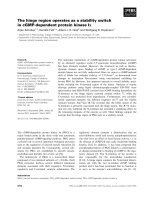
Fig. 1. Growth on chitin and chitobiose. T. atroviride strains (WT, Dnag1, Dnag2 and Dnag1Dnag2) were grown on solid medium (1.5% w ⁄ v
agar) containing (A) minimal medium with colloidal chitin, (B) PDA as a control to show normal growth and sporulation of the knockout
strains, and (C) minimal medium with chitobiose. Dnag2-I and Dnag2-II are Dnag2 strains C2332 and A523, and nag1Dnag2-I and nag1Dnag2-
II are Dnag1Dnag2 strains 713 and 1921, respectively.
Chitin degradation in Trichoderma atroviride R. Lo
´
pez-Monde
´
jar et al.
5140 FEBS Journal 276 (2009) 5137–5148 ª 2009 The Authors Journal compilation ª 2009 FEBS
cultures, but no differences between the knockout
strains and the WT could be detected with respect to
the timing and frequency of spore swelling and germi-
nation, and the morphology of the germ tubes was
also completely normal, indicating that the NAGases
NAG1 and NAG2 are not essential for germination in
T. atroviride (Fig. S3A).
Hyphal morphology was investigated macroscopi-
cally and microscopically on agar plates using a num-
ber of carbon sources including glucose, glycerol,
maltotriose, glycogen, glucosamine, GlcNAc and PDA.
Hyphal extension and colony diameter were measured,
but no differences between the knockout strains and
the WT were observed on any carbon source, confirm-
ing the data from the Biolog analysis (see above). A
microscopical analysis of hyphal growth and branching
patterns did also not reveal any differences among the
analysed strains (Fig. S3B), indicating that the NAG-
ases NAG1 and NAG2 are not essential for hyphal
growth in T. atroviride.
Further, sporulation rates were measured on various
carbon sources by quantification of the numbers of
spores formed on agar plates, but again no significant
influence of the loss of nag1 and nag2 could be
detected. The only exception was the carbon source
GlcNAc, on which the WT and Dnag1Dnag2 strains
produced a similar number of spores, while the Dnag2
strains only produced 11 ± 1% of the number of
spores produced by the WT and the number of spores
in the Dnag1 strain was 466 ± 105%. The results on
all other carbon sources showed no differences
between the WT and the single knockout strains, and,
most importantly, on none of the investigated carbon
sources could any changes in sporulation rates be
detected in the Dnag1Dnag2 strains.
Comparison of growth and chitinolytic activities
on chitin in liquid and solid media
Having determined that NAG1 and NAG2 are essential
for growth on chitin in plates and that no residual
NAGase activity remained in the double knockout
A
B
C
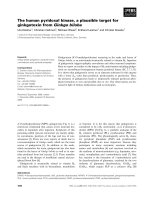
Fig. 2. Biomass and chitinolytic enzyme activities on chitin agar
plates. (A) Biomass, measured as total protein concentration, (B)
NAGase activities and (C) chitinase activities of T. atroviride strains
(WT, Dnag1, Dnag2 and Dnag1Dnag2). Values for biomass are given
per mL of protein extracts under normalized extraction conditions,
and enzyme activities were normalized to the biomass and are
shown per mg of biomass (total protein). Error bars show SEM val-
ues of the measurements. D2-I and D2-II are Dnag2 strains C2332
and A523, and DD-I and DD-II are Dnag1Dnag2 strains 713 and
1921, respectively.
R. Lo
´
pez-Monde
´
jar et al. Chitin degradation in Trichoderma atroviride
FEBS Journal 276 (2009) 5137–5148 ª 2009 The Authors Journal compilation ª 2009 FEBS 5141
strains, we were interested to assess how these results
compare to those of previous studies using submerged
shake flask cultures [12,16], which, however, do not
resemble natural growth conditions for T. atroviride.
On agar plates, growth on chitin was linked to sporula-
tion (see Fig. 1A), and, similarly, visual inspection of
the shake flask cultures showed that the biomass
formed by the WT strain was already green due to spor-
ulation after 48 h, whereas the biomass of the Dnag2
strains was only light green and no sporulation was
observed in the Dnag1 and Dnag1Dnag2 strains. To
quantify these observations, samples were taken after
30, 48 and 72 h, and the total protein concentration,
corresponding to biomass formation, was measured for
mycelial and conidial biomass after extraction with
NaOH (Fig. 3A). Our results show that growth on chi-
tin in shake flask cultures was similar to that of the WT
in the Dnag2 strains and slightly reduced in the Dnag1
strain, and that almost no growth of the Dnag1Dnag2
strains occurred at all. Extracellular NAGase activities
of the Dnag2 strains, normalized to the amount of bio-
mass, were similar to WT levels, whereas those of the
Dnag1 strain were reduced to below 2% and in the
Dnag1Dnag2 strains no NAGase activity could be
detected at all (Fig. 3B). This showed that NAG1 and
NAG2 are also the only two enzymes responsible for
extracellular NAGase activity in T. atroviride in shake
flask cultivations. Similar results were obtained when
cell-wall-bound NAGase activities were also taken into
account, except that activities in the Dnag1 strain were
approximately 28% and 35% of the WT levels at 48
and 72 h, respectively (Fig. 3C). This suggests that
NAG2 remains attached to the fungal cell wall although
the protein sequence does not contain any membrane-
anchoring signals. Chitinase activities, normalized to
the biomass, paralleled NAGase activities, with a strong
reduction of chitinase activities in the Dnag1 and
Dnag1Dnag2 strains (Fig. 3D). In summary, these
results revealed differences in the kinetics of NAG1 and
NAG2 formation between submerged and solid-surface
cultivations, which also seemed to affect chitinase for-
mation in the Dnag1 strain, but confirmed our finding
that the presence of either NAG1 or NAG2 is essential
for growth on chitin.
A
B
CD
Fig. 3. Biomass and chitinolytic enzyme activities upon growth on chitin in shake flask cultures. Values for biomass are given per mL of trea-
ted culture extract. Enzyme activities were normalized to the biomass and are shown per mg of biomass (total protein). (A) Biomass, mea-
sured as total protein concentration, (B) extracellular NAGase activities, (C) total (extracellular and cell-wall-bound) NAGase activities, and (D)
extracellular chitinase activities. Mean values from one representative experiment are shown. Filled diamonds, WT strain; filled triangles,
Dnag1 strain; grey circles, Dnag1 strain A523 (D2-II); grey diamonds, Dnag2 strain C2332 (D2-I); open circles, Dnag1Dnag2 strain 713 (DD-I);
crosses, Dnag1Dnag2 strain 1921(DD-II).
Chitin degradation in Trichoderma atroviride R. Lo
´
pez-Monde
´
jar et al.
5142 FEBS Journal 276 (2009) 5137–5148 ª 2009 The Authors Journal compilation ª 2009 FEBS
Autolysis is not altered in Dnag1Dnag2 strains
As T. atroviride Dnag1Dnag2 strains could not grow on
chitin, we reasoned that T. atroviride would also no
longer be able to recycle GlcNAc from its own cell
wall, and its ability to autolyse would be altered.
Autolysis was studied by growing T. atroviride strains
in submerged cultivations with glucose as the carbon
source and measuring the decrease of biomass after
entering the stationary phase (i.e. when glucose had
been depleted). This phase was observed after 35 h of
cultivation for all strains. However, up to 90 h of culti-
vation, no differences in the decline of biomass due to
autolysis and the corresponding concentration of
extracellular proteins were detected between the strains
(data not shown). NAGase activities under these con-
ditions were consistent with the results for the respec-
tive strains shown during submerged growth on chitin,
i.e. no NAGase activity was observed in the double
knockout strains and NAG1 activitiy seemed to be
predominantly extracellular, whereas the majority of
NAG2 was apparently attached to the cell walls (data
not shown). These results indicate that recycling of
GlcNAc via NAG1 and NAG2 during autolysis is not
of major importance for T. atroviride.
Mycoparasitism is not affected by the lack of
NAGase activity
To analyse whether the inability to use chitin would
affect the mycoparasitic activity of T. atroviride, plate
confrontation assays with two plant pathogenic fungi,
the basidiomycete Rhizoctonia solani and the ascomy-
cete Botrytis cinerea, were performed. The experiment
was carried out on both PDA plates and on plates
with minimal medium and nutrient limitations (various
nitrogen and glucose concentrations, see Experimental
procedures for details). However, no effect was
observed under any of these conditions (data not
shown); all tested knockout strains were as efficient as
the WT in parasitizing both host fungi. This result
shows that the inability to use chitin as a carbon
source during mycoparasitism does not affect the
antagonistic potential of T. atroviride . This finding
does not eliminate the possibility that chitin in the cell
walls of the host fungi is hydrolysed by chitinases, but
indicates that it does not need to be metabolized.
Discussion
In this study, we assessed the function of fungal GH
family 20 NAGases in fungal chitin catabolism. Rela-
tively little is known about chitin turnover by fungi, and
it is especially difficult to determine the importance of
chitin degradation for the mycoparastic process. The
number of chitinolytic enzymes in mycoparasitic
Trichoderma spp. is much higher than in other fungal
genera [20], but the number (two) and amino acid
sequences of NAGases are much more conserved when
compared to other fungal genomes. Until now, apart
from their transcriptional regulation, nothing was
known about the roles and physiological relevance of
NAGases in fungi. We therefore generated Dnag1Dnag2
double knockout strains in T. atroviride in order to
study the role of NAGases in chitin degradation.
No extracellular NAGase activity was detected in
Dnag1Dnag2 strains under any of the tested growth con-
ditions, which indicates that NAG1 and NAG2 are
indeed the only extracellular NAGases in T. atroviride
under the tested conditions. Our data show that the
presence of either of these enzymes is essential and
sufficient for degradation of chitobiose and growth on
chitin. Analysis of the T. atroviride genome database
( re-
vealed that, in addition to NAG1 (protein ID 136120)
and NAG2 (protein ID 41039), the genome contains a
third ORF encoding a GH family 20 protein (ID 33962);
however, this is highly dissimilar to NAGases [NAG1
and NAG2 have a sequence similarity of 70% positives
(e 0.0), but compared to the third GH 20 protein the
similarity is only 37% positives (e-04 to e-08)], strongly
suggesting a different substrate spectrum for this
enzyme. Interestingly, while most fungi possess three
NAGases, the A. nidulans genome also contains only
two GH family 20 proteins, one of which is highly simi-
lar to NAGases and the other to T. atroviride protein
33962. The complete absence of NAGase activity under
all tested growth conditions reported in this study –
including enzyme assays of mycelial extracts from agar
plates that would also reveal any intracellular NAGase
activities – suggests that NAG1 and NAG2 are the only
two enzymes that possess this activity in T. atroviride.In
accordance with these findings, it is interesting to note
that previous studies showed that nag1 gene expression
is induced by a variety of carbon sources and other
stimuli, and is in our experience one of the strongest
inducible genes in T. atroviride. Therefore, NAGases
constitute a ‘genomic bottleneck’ for chitin catabolism
in fungi with respect to chitin degradation, as chitin can-
not be used as a nutrient source if the essential NAGase
activity, dependent on only two enzymes, is absent,
despite the presence of approximately 30 chitinases.
However, the large variability of chitinases, but limited
arsenal of NAGases, also suggests that chitinases might
have many additional functions (defence mechanisms,
enabling accessibility to other substrates, e.g. during
R. Lo
´
pez-Monde
´
jar et al. Chitin degradation in Trichoderma atroviride
FEBS Journal 276 (2009) 5137–5148 ª 2009 The Authors Journal compilation ª 2009 FEBS 5143
mycoparasitism, loosening up of the cell wall during the
fungal life cycle, etc.), for which complete degradation
of chitin into GlcNAc monomers is not important.
Of course, we cannot rule out the possibility that
NAGase activities will be detected in subgroups of other
GH families, which could be membrane-bound proteins
and hence be involved in fungal cell-wall remodelling,
but it should be noted that the third GH family 20 pro-
tein in T. atroviride does not contain a membrane-
anchoring signal. GlcNAc recycling has been implicated
in cell-wall formation during the fungal life cycle,
including germination, hyphal growth, fusion and spor-
ulation [18,19,21], but we found no alteration of the
phenotype in Dnag1Dnag2 strains, and these two
enzymes are missing to form GlcNAc from chitobiose
during chitin degradation. This shows that neither
NAG1 or NAG2 are involved in these processes in
T. atroviride. As mentioned above, it is possible that
chitinases, but not NAGases, are sufficient for loosening
of the chitin structure in the cell wall during formation
of hyphal branches and fusions. With respect to sporula-
tion, an effect could only be detected on GlcNAc as car-
bon source. Although all tested strains showed similar
growth rates on GlcNAc plates, the number of spores
produced was altered in the single knockout strains, but
not in the double knockout strains. This suggests the
existence of regulatory mechanisms between GlcNAc
metabolism, NAGases and sporulation during growth
on this carbon source. Nevertheless, we conclude from
the finding that there was no difference between the WT
and Dnag1Dnag2 strains on any of the tested carbon
sources with regard to spore formation that NAG1 and
NAG2 are not directly important for this process.
We also found no differences during autolysis
between the WT and the Dnag1Dnag2 strains. This can
be explained by the fact that the cell walls of hyphal
fragments that undergo autolysis are permeabilized by
chitinases and other hydrolytic enzymes, e.g. glucanas-
es. The intracellular components, such as mono-
saccharides and proteins, are of higher nutritional value
for the hyphal fragments than the chitinous cell wall and
therefore the recycling of GIcNAc is apparently not of
major nutritional importance during this process. It
should be noted that we assayed autolysis with glucose
as carbon source, on which all strains showed the same
growth rate, whereas the previous finding that the Dnag1
strain showed delayed autolysis [16] was based on an
experiment with colloidal chitin as carbon source. How-
ever, we do not consider this to be a suitable carbon
source for this experiment, because, as can be seen in
Fig. 3A, the Dnag1 strain exhibits slower growth in sub-
merged cultures with chitin than the WT strain does,
and therefore the suspected delay of autolysis reported
by Brunner et al. may have been due to the slower
growth rate on this carbon source.
Despite our findings that extracellular NAGases are
not important for any of the studied morphogenetic
aspects in T. atroviride, our results clearly showed that
they are essential for growth on chitin. Dnag1Dnag2
strains showed significantly reduced biomass formation
upon growth on colloidal chitin, and did not grow at all
on chitobiose. This demonstrates that T. atroviride
Dnag1Dnag2 strains cannot hydrolyse chitobiose, and
shows that, even though this fungus has a large array of
chitinases, the last step of cleaving the dimer into two
monomers is performed by only two enzymes: NAG1
and NAG2. Further, it can be concluded from our
results that T. atroviride cannot take up the dimer and
use it as a carbon source, as has been reported for bacte-
ria [22], but depends on extracellular cleavage of chitobi-
ose into the monomer GlcNAc, which can then be taken
up by the hyphae and catabolized. The small amount of
residual biomass that was formed upon growth of the
double knockout strain on chitin was probably due to
small amounts of GlcNAc that were released by those
chitinases, which can randomly cleave chito-oligomers,
occasionally leading to the formation of monomers.
A comparison between extracellular and total (extra-
cellular and cell wall-bound) NAGase activities in
liquid medium showed that NAG2 – the only NAGase
in the Dnag1 strain – was predominantly cell-wall
bound, whereas large amounts of NAG1 were found
to be released from the cell wall in Dnag2 strains. The
fact that the remaining NAGase in Dnag1 strains was
cell-wall-bound in liquid media possibly limited its
access to the substrate, and this limitation of GlcNAc
availability in turn resulted in less biomass formation.
In Dnag2 strains, on the other hand, the large amounts
of extracellular NAG1 were sufficient to enable a
growth rate in submerged cultures similar to that of
WT. It is important to consider, however, that sub-
merged cultures are not a natural growth medium for
T. atroviride, and whether the NAGases were cell-wall-
bound or extracellular did not influence growth under
more natural conditions on agar plates, and therefore
all single knockout strains reached WT growth levels.
Another interesting finding was that the sum of the
NAGase activities measured in single knockout strains
exceeded that of the WT. This indicates that NAG1 and
NAG2 can compensate for each other. Such findings are
reminiscent of similar data for knockouts of the cello-
biohydrolases CBHI and CBHII in Trichoderma reesei,
for which a Dcbh1 strain showed increased cbh2 tran-
script levels in comparison to the parental strain [23].
Chitinase formation on chitin plates was reduced in
the Dnag1 strain but not in the Dnag2 and Dnag1Dnag2
Chitin degradation in Trichoderma atroviride R. Lo
´
pez-Monde
´
jar et al.
5144 FEBS Journal 276 (2009) 5137–5148 ª 2009 The Authors Journal compilation ª 2009 FEBS
strains. The decrease in chitinase activities in the Dnag1
strain therefore cannot be directly related to the absence
of NAG1, because it was not observed in the double
knockout strains, of which the Dnag1 strain is the pro-
genitor. A more likely explanation is that this effect is
caused by NAG2, possibly also due to its increased
expression in the Dnag1 strain in comparison to the WT.
Elevated NAG2 levels could affect the concentration of
the chitinase inducer formed from chitin, e.g. by hydro-
lysis or by transglycosylation. This will be an interesting
topic for further studies. In shake flask cultures, growth
on chitin is generally slow and inefficient. As can be seen
by the delayed onset of chitinase formation, measured
activities at 30 h were extremely low for all strains.
However, although the WT and single knockout strains
showed an increase in biomass at later time points, bio-
mass in the double knockout strains stayed constant or
even decreased slightly. This suggests that due the lim-
ited contact time of chitinases with the substrate in
shake flask cultivations, and possibly also the altered
expression profile of various chitinases, chitinases do
not release enough GlcNAc from the random cleavage
of chito-oligomers to enable residual biomass forma-
tion, as hypothesized for growth on chitin agar plates.
Therefore, we conclude that the small amount of myce-
lial biomass that is formed in the first 30 h of the cultiva-
tion – probably from the 0.05% w ⁄ v of peptone that is
added to liquid cultures to ensure efficient and homoge-
nous germination – is most likely dead at later time
points, which explains why no chitinase activities were
found in the double knockout strains in the shake flask
experiment. The sensitivity of the enzymatic measure-
ment was not the limiting factor, because biomass in the
other strains was also relatively low (Fig. 3A), the
attenuance of the enzymatic assays were in a good sensi-
tivity range of the method. Chitin is an insoluble carbon
source, and to avoid effects due to limited substrate
accessibility, we conclude from the comparison of solid
and liquid cultures that growth of T. atroviride on chitin
should be preferably carried out in solid substrate or
stationary cultivations.
The role of chitinolytic enzymes in the mycoparasitic
process has received a lot of attention and has been
the subject of several studies in Trichoderma spp. [5].
Our findings imply that NAGases are fully dispensable
for this process. These results do not rule out the pos-
sibility that chitinases are important for attack of the
host, but clearly show that the use of chitinous cell
walls from the host as a carbon source is not relevant
for the antagonistic potential of T. atroviride. Our find-
ings suggest that in soil or on decaying wood – the
two natural habitats of T. atroviride – the mycoparasit-
ic lifestyle probably involves successful competition for
nutrients and living space with other fungi rather than
sequestration of chitin as a nutrient source.
Analysis of chitin metabolism in fungi is compli-
cated due to the large number of enzymes that are
involved. In this study, we elucidated the final extra-
cellular steps of this process, and found that, in
T. atroviride, NAG1 and NAG2 are the only enzymes
responsible for the final step in chitin degradation. The
availability of these mutants will enable us to perform
further studies on the use of chitinous carbon sources
and chitinase expression in T. atroviride, which will be
the next steps towards understanding this versatile
aspect of fungal metabolism.
Experimental procedures
Strains
T. atroviride P1 (ATCC 74058), referred to as wild-type
(WT), and the amdS
+
nag1 disruption strain T. atroviride
P1ND1 [16] were maintained on potato dextrose agar (Lab
M Limited, Bury, UK), and stock cultures were kept at
)80 °C. Escherichia coli strain JM109 (Promega, Madison,
WI, USA) was used for plasmid propagation.
Fungal cultivation conditions
The growth of fungal transformants on 95 carbon sources
was assessed using Biolog phenotype microarrays (Biolog,
Hayward, CA, USA) according to the protocol recently
developed for Trichoderma spp. [17]. For growth assays on
agar plates, minimal medium [24] with 1.5% w ⁄ v agar and
supplemented with 1% w ⁄ v of the various carbon sources
was used. The carbon sources included mono- and disaccha-
rides, which were purchased from Sigma (St Louis, MO,
USA), and colloidal chitin, which was prepared as described
previously [25]. Agar plates were incubated at 25 °C under a
12 h light ⁄ dark diurnal cycle. Chitobiose growth assays were
performed in six- well plates containing 650 lL minimal
medium + 1.5% agar and 0.5% carbon source per well due
to the high costs of the substrate. Plate confrontation assays
with Rhizoctonia solani and Botrytis cinerea were performed
as described previously [26] on PDA and also on agar plates
with minimal medium salt composition and (a) glucose
limitation (0.2% w ⁄ v), (b) nitrogen limitation (0.14 gÆL
)1
ammonium sulfate), or (c) glucose and nitrogen limitation.
Shake flask cultivations were prepared in minimal medium
containing 0.05% peptone to ensure efficient germination
and 1% w ⁄ v of either glucose or colloidal chitin. Cultures
were grown in rotary incubators (Multitron 2 shaking incu-
bator, Infors, Bottmingen, Switzerland) at 28 °C and
220 rpm, and kept in constant light to enable sporulation,
which is linked to growth on this carbon source on agar
plates (this study) and was also observed previously to occur
R. Lo
´
pez-Monde
´
jar et al. Chitin degradation in Trichoderma atroviride
FEBS Journal 276 (2009) 5137–5148 ª 2009 The Authors Journal compilation ª 2009 FEBS 5145
in shake flask cultures (V. Seidl, unpublished results). All
experiments were performed at least in duplicate.
Determination of fungal growth, biomass
production and sporulation
The increase in colony diameter on agar plates was measured
daily. To measure biomass from agar plates containing col-
loidal chitin as carbon source, agar pieces of equal size were
cut out, ground to a fine powder in liquid nitrogen and
suspended in 1 mL of buffer (100 mm Tris, pH 8.0, 1 mm
EDTA). The suspension was kept on ice and sonicated five
times for 10 s each, and centrifuged for 10 min at 13 000 g,
4 °C. The supernatant was subsequently used to measure
total protein concentration, corresponding to the biomass,
and also NAGase and chitinase enzyme activities (see below).
The protein content was determined using the Bradford pro-
tein assay (Bio-Rad, Hercules, CA, USA) with BSA as the
standard. Sporulation rates on agar plates were determined
by quantitatively harvesting spores from an agar plate using
a 0.9% NaCl + 0.05% Tween solution and counting the
spores using a haemocytometer.
For submerged cultures containing soluble carbon
sources, mycelial dry weight was recorded by withdrawing
40 mL aliquots from the cultures, suction filtration through
a glass wool filter, followed by extensive washing with tap
water, and drying at 80 °C to constant weight. For sub-
merged cultures containing colloidal chitin, the biomass
was determined by taking 1 mL samples and lysing them
by addition of 0.2 mL 0.1 m NaOH for 3 h at 30 °C. The
samples were then centrifuged for 10 min at 13 000 g and
the supernatant was used to measure the total protein con-
centration, corresponding to the biomass, by the Bradford
protein assay using BSA as the standard. All extractions
and measurements were performed at least in duplicate.
Enzyme assays
Samples from shake flask cultures were centrifuged for
10 min at 13 000 g and 4 °C, and the supernatants were used
for extracellular enzyme activity measurements. Total (extra-
cellular and cell-wall-bound) enzyme activities from shake
flask cultures were measured using samples containing myc-
elia and from agar plates using protein extracts as described
above. NAGase activities were measured using a modifica-
tion of the method described by Yagi et al. [27], which is
based on the release of p-nitrophenol from the respective aryl
chitosides. Samples of between 5 and 100 lL were added to
0.5 mL of a solution containing 50 mm potassium phosphate
buffer, pH 6.7, and 300 lgÆmL
)1
4-nitrophenyl N-acetyl-b-d-
glucosaminide, and the volume made up to 600 lL with
buffer. Enzyme assays were incubated at 30 °C with gentle
agitation, reactions were terminated after 15 min by addition
of 0.4 mL 0.4 m Na
2
CO
3
, and absorbance was measured at
405 nm. Control measurements of enzyme activities were
performed by omitting the substrate from the phosphate buf-
fer. Chitinase activities were determined using the same
method but with 4-nitrophenyl b-d-N,N¢,N¢¢-triacetylchitotri-
ose as substrate. Enzymatic activities were calculated based
on the release of 4-nitrophenol using a molar extinction
coefficient of 18.5 mmol
)1
Æcm
)1
.
Statistical evaluation
Statistical analysis of the results, as specified in the various
sections, was performed using graphpad instat software
version 8.0 (San Diego, CA, USA).
Microscopic analysis
For microscopic analysis, an inverted T300 microscope (Ni-
kon, Tokyo, Japan), equipped with differential interference
contrast optics, was used, and images were captured using
a DXM1200F digital camera (Nikon) and digitally pro-
cessed using photoshop CS3 (Adobe, San Jose, CA, USA).
Germination was observed by placing 50 lL samples on
large cover slips, and hyphae were imaged directly on agar
pieces that were cut out from plates using the inverted agar
method described previously [28].
Plasmid construction
The UniProt accession number of T. atroviride NAG1 is
P87258. The T. atroviride nag2 gene was identified in the
T. atroviride genome database ( />Triat1/Triat1.home.html) using a previously cloned frag-
ment of nag2 [11], GenBank ⁄ EMBL ⁄ DDBJ accession num-
ber DQ364461 (UniProt Q0ZLH7), for a BLAST search.
The query yielded a single specific hit (protein ID 41039).
For the nag2 deletion vector, 1.5 kb of the up- and down-
stream non-coding regions of T. atroviride nag2 were ampli-
fied from T. atroviride P1 genomic DNA using primer pairs
A ⁄ B and C ⁄ D, respectively (Table 1), using the GoTaq
Ò
system (Promega), with 200 nm of each primer in the PCR
reactions and reaction conditions according to the manu-
facturer’s instructions. The hph cassette from pRLMEX30
[29] was cut out using XhoI ⁄ HindIII and ligated into an
XhoI ⁄ HindIII-digested pBluescript SK(+) vector (Strata-
gene, La Jolla, CA), resulting in vector pBS31. The PCR
fragment of the nag2 upstream region was ligated into the
pGEM-T Easy vector (Promega), cut out again using the
NotI restriction sites, and ligated into NotI-digested pBS31.
The resulting plasmid was digested with ApaI, and the
amplified nag2 downstream region was digested correspond-
ingly and ligated, resulting in the nag2 knockout vector
pVCNAG2. The correct orientation of the fragments was
checked using several control restriction digests. The 5.8 kb
nag2 deletion cassette was amplified using primers E and F
(Table 1) using the Long Template Expand PCR System
Chitin degradation in Trichoderma atroviride R. Lo
´
pez-Monde
´
jar et al.
5146 FEBS Journal 276 (2009) 5137–5148 ª 2009 The Authors Journal compilation ª 2009 FEBS
(Roche, Indianapolis, IN, USA) and PCR conditions
according to the manufacturer’s instructions.
Transformation of T. atroviride
Protoplast preparation and DNA-mediated transformation
were performed essentially as described previously [30], with
the minor modifications that 7.5 mgÆmL
)1
lysing enzymes
(Sigma) were used, and, for protoplast generation, that myc-
elia immersed in the lysing solution were incubated at 30 °C
for 2 h under gentle agitation and mycelial clumps were
gently separated with sterile tweezers every 30 min. After
transformation, protoplasts were stabilized and regenerated
on PDA containing d-sorbitol (1 m) and 50 lgÆmL
)1
hygro-
mycin B, and inoculated at 28 °C. Colonies emerging from
the transformation plates were sub-cultivated on PDA ⁄
hygromycin plates and subsequently purified by single spore
isolation on plates that also contained 0.1% Triton X-100.
Characterization of the transformants
Analysis of all transformants was performed by diagnostic
PCR using primer pairs G ⁄ I and H ⁄ I (Table 1) to amplify
the hph cassette and the native nag2 locus, respectively,
with the GoTaq system (Promega). In addition, the integra-
tion type of selected strains was verified by Southern analy-
sis. DNA isolation was performed as described by Hartl
and Seiboth [31]. Southern analysis of the deletion strains
was performed by digesting the genomic DNA with ApaI.
Standard methods [32] were used for DNA electrophoresis
and blotting. Hybridization and labelling of the probe by
PCR were performed using the DIG non-radioactive system
(Roche). The 1.5 kb nag2 probe was amplified using
primers A and B (Table 1). A 4.6 kb hybridizing fragment
indicated an endogenous nag2 locus, whereas homologous
integration of the deletion cassette resulted in a 2.0 kb
fragment (for details, see Fig. S1).
Acknowledgements
We thank Monika Schmoll (Research Area Gene
Technology and Applied Biochemistry, Institute of
Chemical Engineering, Vienna University of Technol-
ogy) for kindly providing pBS31. This work was sup-
ported by grant P20559-B03 from the Austrian Science
Fund. R.L.M’s stay in Vienna was funded by the I3P
Program from the Consejo Superior de Investigaciones
Cientı
´
ficas (CSIC), Spain. V.C.’s stay in Vienna was
funded by the Italian Ministero dell’Istruzione
dell’Universita
`
e della Ricerca for her PhD program,
and the work of V.S. is supported by a Hertha-Firn-
berg Program (T390-B03) from the Austrian Science
Fund.
References
1 Latge
´
JP (2007) The cell wall: a carbohydrate armour
for the fungal cell. Mol Microbiol 66, 279–290.
2 Tharanathan RN & Kittur FS (2003) Chitin – the
undisputed biomolecule of great potential. Crit Rev
Food Sci Nutr 43, 61–87.
3 Horsch M, Mayer C, Sennhauser U & Rast DM (1997)
b-N-acetylhexosaminidase: a target for the design of
antifungal agents. Pharmacol Ther 76, 187–218.
4 Seidl V (2008) Chitinases of filamentous fungi: a large
group of diverse proteins with multiple physiological
functions. Fungal Biol Rev 22, 36–42.
5 Benitez T, Rincon AM, Limon MC & Codon AC
(2004) Biocontrol mechanisms of Trichoderma strains.
Int Microbiol 7, 249–260.
6 Hjeljord L & Tronsmo A (1998) Trichoderma and Glioc-
ladium in biological control: an overview. In Trichoder-
ma and Gliocladium. Vol. 2: Enzymes, Biological Control
and Commercial Applications (Harman GE & Kubicek
CP eds), pp. 131–152. Taylor and Francis Ltd, London.
7 Draborg H, Kauppinen S, Dalboge H & Christgau S
(1995) Molecular cloning and expression in S. cerevisiae
of two exochitinases from Trichoderma harzianum. Bio-
chem Mol Biol Int 36, 781–791.
8 Kim DJ, Baek JM, Uribe P, Kenerley CM & Cook DR
(2002) Cloning and characterization of multiple glycosyl
hydrolase genes from Trichoderma virens. Curr Genet
40, 374–384.
9 Peterbauer CK, Lorito M, Hayes CK, Harman GE &
Kubicek CP (1996) Molecular cloning and expression of
the nag1 gene (N-acetyl-b-d-glucosaminidase-encoding
gene) from Trichoderma harzianum P1. Curr Genet 30,
325–331.
10 Ramot O, Viterbo A, Friesem D, Oppenheim A & Chet
I (2004) Regulation of two homodimer hexosaminidases
in the mycoparasitic fungus Trichoderma asperellum by
glucosamine. Curr Genet 45, 205–213.
11 Seidl V, Druzhinina IS & Kubicek CP (2006) A screen-
ing system for carbon sources enhancing b-N-acetylglu-
cosaminidase formation in Hypocrea atroviridis
(Trichoderma atroviride). Microbiology 152, 2003–2012.
Table 1. Primers used in this study.
Primer Sequence (5¢-to3¢)
A nag2-prom-fw ATCAGATGGCGATGTGAAGAG
B nag2-prom-rv ACCAAGAGTTGAGCCCGTC
C Nag2_term_fw_ApaI TCTTGGGCCCTGATACAGACAC
D nag2_term-rv_ApaI AGTTTGGGCCCTGCGAGTTTG
E nag2-prom-fwnest TGGCATACAGACTGGGCG
F nag2-term-rvnest AGAACTCGGCTCCATAGGC
G Nag2-cds-fw TTGAAGAAGAGCTGCGAG
H hph-fw TGAATGAGGATACACGGG
I nag2-prom-test-rv TGGATGTTTGAGTGAGCGG
R. Lo
´
pez-Monde
´
jar et al. Chitin degradation in Trichoderma atroviride
FEBS Journal 276 (2009) 5137–5148 ª 2009 The Authors Journal compilation ª 2009 FEBS 5147
12 Mach RL, Peterbauer CK, Payer K, Jaksits S, Woo SL,
Zeilinger S, Kullnig CM, Lorito M & Kubicek CP
(1999) Expression of two major chitinase genes of
Trichoderma atroviride (T. harzianum P1) is triggered by
different regulatory signals. Appl Environ Microbiol 65,
1858–1863.
13 Peterbauer CK, Brunner K, Mach RL & Kubicek CP
(2002) Identification of the N-acetyl-d -glucosamine-
inducible element in the promoter of the Trichoderma
atroviride nag1 gene encoding N-acetyl-glucosaminidase.
Mol Genet Genomics 267, 162–170.
14 Pusztahelyi T, Molnar Z, Emri T, Klement E, Miskei M,
Kerekgyarto J, Balla J & Pocsi I (2006) Comparative
studies of differential expression of chitinolytic enzymes
encoded by chiA, chiB, chiC and nagA genes in Aspergil-
lus nidulans. Folia Microbiol (Praha) 51, 547–554.
15 Shin KS, Kwon NJ, Kim YH, Park HS, Kwon GS &
Yu JH (2009) Differential roles of the ChiB chitinase
in autolysis and cell death of Aspergillus nidulans.
Eukaryot Cell 8, 738–746.
16 Brunner K, Peterbauer CK, Mach RL, Lorito M,
Zeilinger S & Kubicek CP (2003) The Nag1 N-acetyl-
glucosaminidase of Trichoderma atroviride is essential
for chitinase induction by chitin and of major relevance
to biocontrol. Curr Genet 14, 289–295.
17 Druzhinina IS, Schmoll M, Seiboth B & Kubicek CP
(2006) Global carbon utilization profiles of wild-type,
mutant, and transformant strains of Hypocrea jecorina.
Appl Environ Microbiol 72, 2126–2133.
18 Gooday GW (1990) Physiology of microbial degrada-
tion of chitin and chitosan. Biodegradation 1, 177–190.
19 Merz RA, Horsch M, Nyhlen LE & Rast DM (1999)
Biochemistry of chitin synthase. EXS 87, 9–37.
20 Karlsson M & Stenlid J (2009) Evolution of family 18
glycoside hydrolases: diversity, domain structures and
phylogenetic relationships. J Mol Microbiol Biotechnol
16, 208–223.
21 Latge
´
JP & Calderone R (2006) The fungal cell wall.
In The Mycota – Growth, Differentiation and Sexuality
(Ku
¨
es U & Fischer R eds), pp. 73–104. Springer,
Heidelberg.
22 Uchiyama T, Kaneko R, Yamaguchi J, Inoue A,
Yanagida T, Nikaidou N, Regue M & Watanabe T
(2003) Uptake of N,N¢-diacetylchitobiose [(GlcNAc)
2
]
via the phosphotransferase system is essential for chitin-
ase production by Serratia marcescens 2170. J Bacteriol
185, 1776–1782.
23 Seiboth B, Hakola S, Mach RL, Suominen PL &
Kubicek CP (1997) Role of four major cellulases in
triggering of cellulase gene expression by cellulose in
Trichoderma reesei. J Bacteriol 179, 5318–5320.
24 Seidl V, Seiboth B, Karaffa L & Kubicek CP (2004)
The fungal STRE-element-binding protein Seb1 is
involved but not essential for glycerol dehydrogenase
(gld1) gene expression and glycerol accumulation in
Trichoderma atroviride during osmotic stress. Fungal
Genet Biol 41, 1132–1140.
25 Seidl V, Huemer B, Seiboth B & Kubicek CP (2005) A
complete survey of Trichoderma chitinases reveals three
distinct subgroups of family 18 chitinases. FEBS J 272,
5923–5939.
26 Seidl V, Schmoll M, Scherm B, Balmas V, Seiboth B,
Migheli Q & Kubicek CP (2006) Antagonism of Pythi-
um blight of zucchini by Hypocrea jecorina does not
require cellulase gene expression but is improved by
carbon catabolite derepression. FEMS Microbiol Lett
257, 145–151.
27 Yagi T, Hisada R & Shibata H (1989) 3,4-Dinitrophe-
nyl N-acetyl-b-d-glucosaminide, a synthetic substrate
for direct spectrophotometric assay of N-acetyl-b-d-
glucosaminidase or N-acetyl-b-d-hexosaminidase. Anal
Biochem 183, 245–249.
28 Hickey PC, Swift SR, Roca MG & Read ND (2005)
Live-cell imaging of filamentous fungi using vital fluo-
rescent dyes and confocal microscopy. Methods Micro-
biol 34, 63–87.
29 Mach RL, Schindler M & Kubicek CP (1994) Transfor-
mation of Trichoderma reesei based on hygromycin B
resistance using homologous expression signals. Curr
Genet 25, 567–750.
30 Gruber F, Visser J, Kubicek CP & de Graaff LH (1990)
The development of a heterologous transformation sys-
tem for the cellulolytic fungus Trichoderma reesei based
on a pyrG-negative mutant strain. Curr Genet 18, 71–76.
31 Hartl L & Seiboth B (2005) Sequential gene deletions in
Hypocrea jecorina using a single blaster cassette. Curr
Genet 48, 204–211.
32 Sambrook J & Russell DW (2001) Molecular Cloning:
A Laboratory Manual, 2nd edn. Cold Spring Harbor
Laboratory Press, Cold Spring Harbor, NY.
Supporting information
The following supplementary material is available:
Fig. S1. Deletion of nag2 in T. atroviride.
Fig. S2. Carbon source profiling of T. atroviride.
Fig. S3. Microscopical characterization of the mor-
phology of nag2 knockout strains.
This supplementary material can be found in the
online article.
Please note: As a service to our authors and readers,
this journal provides supporting information supplied
by the authors. Such materials are peer-reviewed and
may be re-organized for online delivery, but are not
copy-edited or typeset. Technical support issues arising
from supporting information (other than missing files)
should be addressed to the authors.
Chitin degradation in Trichoderma atroviride R. Lo
´
pez-Monde
´
jar et al.
5148 FEBS Journal 276 (2009) 5137–5148 ª 2009 The Authors Journal compilation ª 2009 FEBS