Báo cáo khoa học: Genomic structure and expression analysis of the RNase j family ortholog gene in the insect Ceratitis capitata pptx
Bạn đang xem bản rút gọn của tài liệu. Xem và tải ngay bản đầy đủ của tài liệu tại đây (1.56 MB, 11 trang )
Genomic structure and expression analysis of the
RNase j family ortholog gene in the insect
Ceratitis capitata
Theodoros N. Rampias*, Emmanuel G. Fragoulis and Diamantis C. Sideris
Department of Biochemistry and Molecular Biology, Faculty of Biology, University of Athens, Greece
Ribonucleases have been extensively studied in terms
of structure, function and enzymatic properties in
terms of their specific RNA degradation in a variety of
organisms. In addition to the well known ribonucleases
of pancreatic type [1,2], there are numerous intracellu-
lar ribonucleases whose biological roles have not yet
been established. Additionally, the burst of determina-
tion of genome sequences in recent years has led to
identification of homologs of many ribonucleases in a
variety of diverse organisms. Many close homologs of
bacterial or yeast ribonucleases have also been detected
in insect genome sequences [3,4].
In Drosophila melanogaster, the JhI-1 gene encodes
both forms of the tRNA 3¢ endonuclease RNase Z,
which participates in endonucleolytic tRNA 3¢ end
processing [5]. Additionally, JhI-1 is a juvenile hor-
mone-regulated gene with several important functions
in insects, such as moulting regulation during pre-adult
Keywords
alternative polyadenylation; AUUUA motifs;
Cc RNase; RNase j; specific ribonuclease
Correspondence
D. C. Sideris, Department of Biochemistry
and Molecular Biology, Faculty of Biology,
University of Athens, Panepistimioupolis,
15701 Athens, Greece
Fax: +30 210 7274158
Tel: +30 210 7274515
E-mail:
*Present address
Department of Molecular Biophysics and
Biochemistry, Yale University, New Haven,
CT, USA
Database
The nucleotide sequences of Ceratitis capi-
tata RNase j have been submitted to the
DDBJ ⁄ EMBL ⁄ GenBank databases under the
accession numbers AJ874689 (cDNA) and
AJ874690 (genomic DNA)
(Received 28 July 2008, revised 9
September 2008, accepted 16 October
2008)
doi:10.1111/j.1742-4658.2008.06746.x
Cc RNase is the founding member of the recently identified RNase j
family, which is represented by a single ortholog in a wide range of animal
taxonomic groups. Although the precise biological role of this protein is
still unknown, it has been shown that the recombinant proteins isolated so
far from the insect Ceratitis capitata and from human exhibit ribonucleo-
lytic activity. In this work, we report the genomic organization and molec-
ular evolution of the RNase j gene from various animal species, as well as
expression analysis of the ortholog gene in C. capitata. The high degree of
amino acid sequence similarity, in combination with the fact that exon sizes
and intronic positions are extremely conserved among RNase j orthologs
in 15 diverse genomes from sea anemone to human, imply a very
significant biological function for this enzyme. In C. capitata, two forms of
RNase j mRNA (0.9 and 1.5 kb) with various lengths of 3¢ UTR were
identified as alternative products of a single gene, resulting from the use of
different polyadenylation signals. Both transcripts are expressed in all
insect tissues and developmental stages. Sequence analysis of the extended
region of the longer transcript revealed the existence of three mRNA
instability motifs (AUUUA) and five poly(U) tracts, whose functional
importance in RNase j mRNA decay remains to be explored.
Abbreviations:
ARE, AU-rich element; miRNA, micro RNA.
FEBS Journal 275 (2008) 6217–6227 ª 2008 The Authors Journal compilation ª 2008 FEBS 6217
development, and control of reproductive maturation
and sexual behaviour in adults [6]. Drosophila RNase
H
1
is one of the best-studied insect ribonucleases [7],
and its expression is essential for development but not
for proliferation [8]. The two RNase III proteins, Dro-
sha and Dicer, collaborate in the stepwise processing
of micro RNAs (miRNAs), and play key roles in
miRNA-mediated gene regulation of processes such as
development and differentiation [9,10].
In Bombyx mori, a homolog of the RNase L inhibi-
tor has been detected, cloned and characterized [11].
This finding implies the existence of RNase L, which
plays an important role in the response of cells to
dsRNA during events such as virus infection in insects.
In Ceratitis capitata, three RNA-degrading enzymes
have been detected. Two of these have been purified
and characterized as alkaline [12] and acidic RNases
[13], and a third poly(U)- and poly(C)-specific ribo-
nuclease has also been isolated and characterized
[14,15]. In order to further characterize this poly(U)-
and poly(C)-specific ribonuclease, designated as Cc
RNase, the full-length cDNA encoding this protein was
cloned and characterized [16,17]. Cc RNase belongs to
the recently identified RNase j family represented by a
single ortholog in a wide spectrum of metazoans from
anthozoans to humans. All family members are highly
conserved, indicating a significant biological role
for these molecules. Although the members of the
RNase j family do not show any significant similarities
to other known ribonucleases, the recombinant proteins
isolated from two representatives (human and the
insect C. capitata) exhibited ribonucleolytic activity on
different RNA substrates. Additionally, the EST
sequences deposited in NCBI databank for the human
protein show that human RNase j is widely expressed
in a large number of normal fetal, juvenile and adult
tissues, a fact that strongly indicates a basic housekeep-
ing cellular role for this ribonuclease [18].
This paper reports the identification, isolation and
characterization of a novel Cc RNase transcript in the
insect C. capitata. Moreover, we describe the expression
profile of the RNase j gene at various developmental
stages and in several tissues of the insect C. capitata. We
have also determined the general genomic organization
of this gene and analyzed its structural evolution.
Results
Expression analysis of Cc RNase
In our previous work, we reported the cloning and
overexpression of a cDNA sequence of 864 bp
(Cc RNase cDNA) that encodes a specific RNase of
the insect C. capitata [17]. In order to investigate the
expression pattern of this novel RNase, a northern
blot analysis was performed using RNA isolated from
various developmental insect stages, as well as from
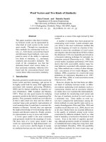
male and female adults (Fig. 1). Our results show that
the Cc RNase gene is expressed at all stages of the
medfly’s life cycle, with higher levels of expression evi-
dent in 6-day-old larvae. In addition to a Cc RNase
mRNA band at 0.9 kb, a second unexpected band at
1.5 kb was also detected. Densitometric analysis of
northern blot data revealed that the 1.5 kb transcript
was more abundant than the 0.9 kb transcript at all
the developmental stages studied, with ratios ranging
from 1.1 (6-day-old larvae) to 2.2 (female adults). In
order to investigate the tissue-specific distribution of
the Cc RNase transcripts, total RNA isolated from the
0
2
4
6
8
10
12
14
16
18
20
EL3L6WPP3P4M F
Stages
Intensity of signal (arbitrary
densitometric units)
1.5 kb
0.9 kb
E L
3
L WP P
3
P
4
M F
1.5 Kb
0.9 Kb
L
6
M F
1.5 Kb
0.9 Kb
C
28S rRNA
A
B
Fig. 1. Developmental northern blot analysis of Cc RNase.
(A) Approximately 15 lg of total RNA isolated from various develop-
mental stages of the insect C. capitata (E, eggs; L, larvae; WP,
white pupae; P, pupae; M, adult male; F, adult female; the num-
bers indicate the age in days) were loaded per lane, separated by
electrophoresis in a 1.2% agarose–formaldehyde gel and trans-
ferred to GeneScreen membranes. The membrane was hybridized
with a
32
P-labeled probe corresponding to nucleotides 69–767 of
the previously isolated Cc RNase cDNA (accession number
AJ441124). Sizes were estimated by comparison with RNA size
markers. (B) 28S rRNA was used as a control for the amounts of
RNA loaded. (C) Densitometric analysis of the northern blot data,
normalized to 28S rRNA, was performed using
NIH IMAGEJ software.
The vertical black and grey bars represent the 0.9 and 1.5 kb tran-
scripts, respectively. Error bars denote the standard error of the
mean for three independent measurements.
Genomic structure and expression analysis of the Cc RNase T. N. Rampias et al.
6218 FEBS Journal 275 (2008) 6217–6227 ª 2008 The Authors Journal compilation ª 2008 FEBS
epidermis and intestine of 6-day-old larvae and adult
heads and ovaries, was blotted and hybridized using
the Cc RNase cDNA as a probe. As shown in Fig. 2,
both transcripts were expressed in all tissues examined.
The signal for the longer transcript was found to be
2.5 times more intense than the signal for the shorter
transcript in adult heads.
Isolation and characterization of the Cc RNase
02 cDNA
Given that the shorter transcript corresponds to the
previously reported Cc RNase cDNA, a modified
version of the hybrid selection technique was used in
order to isolate and clone the longer transcript.
Poly(A)
+
RNA from 6-day-old larvae was reverse-
transcribed and the single-stranded cDNA molecules
were hybridized with a 5¢-biotinylated DNA probe cor-
responding to the main part of the Cc RNase cDNA.
The selected hybrids were amplified by PCR and cloned
into the pCR 2.1 vector. Twenty-four cDNA clones
were selected and verified by restriction endonuclease
mapping and DNA sequence analysis. The sequencing
results revealed that the majority of the clones matched
the Cc RNase cDNA, but seven clones corresponded to
a longer cDNA molecule of 1.5 kb, referred to as Cc
RNase cDNA 02 (EMBL database accession number
AJ874689). This novel cDNA clone has a precise length
of 1466 bp and contains a 5¢ UTR of 138 bp, an ORF
of 288 nucleotides and a long 3¢ UTR of 1040 bp. The
Cc RNase 02 cDNA encodes a 95 amino acid polypep-
tide whose amino acid sequence is identical to that of
the protein encoded by the previously isolated and
characterized Cc RNase cDNA clone. Nucleotide
sequence alignment between these cDNAs (Fig. 3)
showed that Cc RNase cDNA 02 contains a 626 bp
DNA sequence extension in the 3¢ UTR. The extended
region is extremely AU-rich (71% A and U residues)
and contains three mRNA instability motifs (AUUUA)
and five poly(U) tracts (one U
16
stretch, one U
10
stretch, one U
9
stretch, one U
7
stretch and one U
9
G
stretch). Three potential poly(A) signals were also
found in the 3¢ UTR, one of them just 46 bp upstream
of the poly(A) tail. The remaining two poly(A) signals
are upstream of the region that contains the AUUUA
motifs and the poly(U) tracts, so that if one of them
were used as the poly(A) signal, a transcript encoding
the same protein but without any instability motif in
the 3¢ UTR would be produced.
To confirm that the cloned Cc RNase cDNA 02 cor-
responds to the 1.5 kb transcript seen in the northern
blot experiments, the membranes were re-probed using
a specific cDNA fragment (nucleotides 825–1314) from
the extended 3¢ UTR sequence. In this case, only the
1.5 kb mRNA hybridized (data not shown), which
strongly suggests that the cloned Cc RNase cDNA 02
corresponds to the longer Cc RNase transcript. Addi-
tionally, the recombinant protein expressed in Escheri-
chia coli by Cc RNase 02 ORF exhibits ribonucleolytic
activity, as verified by direct visualization of the puri-
fied recombinant protein on an SDS–PAGE gel into
which poly(U) had been incorporated (Fig. 4). The fact
that the recombinant proteins expressed by both Cc
RNase cDNAs exhibits similar mobility and enzymatic
activity confirms experimentally that the translation
product of the two different cDNAs is the same.
Epidermis
Gut
Head
Ovary
1.5 kb
0.9 kb
A
B
28S rRNA
0
2
4
6
8
10
12
Epidermis
Gut
Head Ovary
Tissues
Intensity of signal (arbitrary
densitometric units)
1.5 kb
0.9 kb
C
Fig. 2. Tissue-specific northern blot analysis of Cc RNase.
(A) Approximately 15 lg of total RNA isolated from various tissues
of the insect C. capitata (epidermis, guts, heads and ovaries) were
loaded per lane, separated by electrophoresis in a 1.2% agarose–
formaldehyde gel and transferred to GeneScreen membrane. The
membrane was hybridized with a
32
P-labeled probe corresponding
to nucleotides 69–767 of the previously isolated Cc RNase cDNA
(accession number AJ441124). Sizes were estimated by compari-
son with RNA size markers (Pharmacia). (B) 28S rRNA was used as
a control for the amounts of RNA loaded. (C) Densitometric analy-
sis of the northern blot data, normalized to 28S rRNA, was per-
formed using
NIH IMAGEJ software. The vertical black and grey bars
represent the 0.9 and 1.5 kb transcripts, respectively. Error bars
denote the standard error of the mean for three independent mea-
surements.
T. N. Rampias et al. Genomic structure and expression analysis of the Cc RNase
FEBS Journal 275 (2008) 6217–6227 ª 2008 The Authors Journal compilation ª 2008 FEBS 6219
Cc RNase is present as a single-copy gene in the
C. capitata genome
Southern blot analysis was performed on genomic
DNA of C. capitata digested with HindIII, SalI and
EcoRI and hybridized with probe A (nucleotides 58–
784), which corresponds to the common sequence of
the two cDNA clones, at high stringency (Fig. 5A).
The autoradiograph shows a single band in lanes con-
taining genomic DNA digested with HindIII or SalI,
Fig. 3. Nucleotide and deduced amino acid sequences of the Cc RNase cDNAs. The cDNA 01 (top line, accession number AJ441124),
cDNA 02 (middle line, accession number AJ874689) and amino acid sequences (bottom line) are numbered on the right. Nucleotides of the
cDNA 02 that are identical those of the cDNA 01 are replaced by asterisks, and alignment gaps are indicated by dashes. The three potential
poly(A) signals are boxed with a white background, and the poly(T) tracts and ATTTA motifs in the 3¢ UTR are boxed with a gray background.
Genomic structure and expression analysis of the Cc RNase T. N. Rampias et al.
6220 FEBS Journal 275 (2008) 6217–6227 ª 2008 The Authors Journal compilation ª 2008 FEBS
and two bands (approximately 0.9 and 1.5 kb) when
the genomic DNA was digested with EcoRI. These
data show that the Cc RNase gene occurs as single
copy in the C. capitata diploid genome. Additionally,
the fact that only the 0.9 kb EcoRI fragment disap-
pears from the hybridization pattern when probe B
(nucleotides 825–1314) specific for the 3¢ UTR of Cc
RNase 02 cDNA was used (Fig. 5B) indicates that
both Cc RNase mRNAs derive from the same gene.
The Cc RNase gene contains two introns and
three exons
To determine the structure of the Cc RNase gene, a
PCR reaction was performed using a primer pair
designed to anneal to the 5¢ and 3¢ UTR terminal
sequences of Cc RNase 02 cDNA, with genomic DNA
of C. capitata as a template. The amplified product was
found to be 1642 bp long upon cloning and sequencing
in both directions (EMBL database accession number
AJ874690). Multiple alignment of cDNAs and genomic
nucleotide sequences revealed that the Cc RNase gene
consists of three exons (218, 120 and 1150) interrupted
by two introns (180 and 214 bp). All intron ⁄ exon
boundaries follow the GT ⁄ AG rule, with GT being the
splice donor and AG the splice acceptor [19].
Alternative polyadenylation of the Cc RNase gene
generates two mRNA isoforms
The finding that no additional intronic sequence is
present in the 3¢ UTR of the Cc RNase gene indicates
that the derived mRNAs 01 and 02 cannot derive from
a primary transcript by alternative splicing. An exten-
sive analysis of the Cc RNase cDNA 02 sequence
revealed that two putative polyadenylation signals are
found in the 3¢ UTR, located at nucleotide positions
788–793 (AAUAUA) and 1386–1391 (AAUAAA). The
existence of these polyadenylation signals in the same
exon of the gene encoding Cc RNase, in combination
with the fact that the first signal (AAUAUA) is
located just 19 bp upstream of the poly(A) tail of
cDNA 01 and the second one is located 46 bp
upstream of the poly(A) tail of cDNA 02, suggest that
the two Cc RNase mRNA isoforms probably derive
from a primary transcript by alternative polyadenyla-
tion (Fig. 6).
Intron/exon structure comparison of RNase j
family genes from various taxa
In addition to the characterized Cc RNase gene
reported above, a search against all the available
-94
-67
-43
-30
-20.1
-14.4
C
1 2
1 2
-94
-67
-43
-30
-20.1
-14.4
AB
Fig. 4. SDS–PAGE and activity staining of the purified Cc RNase.
One microgram of the purified recombinant protein expressed in
E. coli from the ORF of the previously isolated cDNA (lane 1) and
from the Cc RNase cDNA 02 described here (lane 2) were analysed
on an 11–15% polyacrylamide gel (A) or on the same gel containing
0.25 mgÆmL
)1
poly(U) (B). After electrophoresis, the gel in (A) was
stained with Coomassie blue and the gel in (B) was stained for
RNase activity as described in Experimental procedures. A set of
marker proteins of known molecular weights (94, 67, 43, 30, 20.1
and 14.4 kDa, top to bottom) (C) was run in parallel and the pro-
teins were stained with Coomassie blue.
Hind III
Sal I
EcoR I
Hind III
Sal I
EcoR I
10 kb
8 kb
5 kb
2.5 kb
1.5 kb
1 kb
A
B
Fig. 5. Southern blot analysis of Cc RNase. Approximately 50 lgof
chromosomal DNA were loaded per lane after complete digestion
with HindIII, SalIorEcoRI, separated by electrophoresis in a 0.8%
agarose gel and transferred to GeneScreen membrane. The mem-
brane was then hybridized with (A) probe A (nucleotides 58–784) or
(B) probe B (nucleotides 825–1314) of the alternative Cc RNase
cDNA 02 sequence. Positions of the DNA markers are given on
the right.
T. N. Rampias et al. Genomic structure and expression analysis of the Cc RNase
FEBS Journal 275 (2008) 6217–6227 ª 2008 The Authors Journal compilation ª 2008 FEBS 6221
genome nucleotide databases was performed in order
to identify other RNase j homolog genes. This search
resulted in the retrieval of 14 genomic sequences from
various animal species, including mammals, fish,
insects, echinoderms and anthozoans. Alignment of the
genomic sequences with the corresponding ESTs led to
identification of their intron ⁄ exon structure. In all
cases, only one functional gene was detected in each
genome. The intron ⁄ exon organization of the ORF
region of the RNase j family genes is shown in
Fig. 7A. In all organisms examined, the region coding
for RNase j is interrupted by two introns, with the
exception of the sea urchin Strongulocentrotus purpura-
tus and the hymenopteran insects Nasonia vitripennis
and Apis mellifera, in which only one intron is present.
The intronic regions among these 15 species demon-
strate an expectedly higher degree of variability than
the exons, although remarkable similarities exist
among several species. For example, the higher-verte-
brate lineages represented by dog, cow, rat, mouse and
human exhibit relatively few differences in the size of
their respective introns. Furthermore, there is a strong
conservation of exon organization. The exon size and
the intronic positions are identical within mammals
and only slightly different compared to species from
other taxa. The intronic architecture of the selected
RNase j genes was mapped onto a multiple protein
sequence alignment (Fig. 7B). In addition to the high
degree of amino acid sequence similarity, there is a
considerable preservation of both the position and size
of exons among RNase j genes in several taxa. For
example, there is an absolute conservation of the loca-
tion, size and phase of exon 1 from primitive animals
to humans. However, the amino acid sequence from
the C-terminus of exon 2 to the N-terminus of exon 3
is more divergent between the vertebrates and the
other taxa analyzed.
Discussion
In the insect C. capitata, an RNase j mRNA of
864 bp has previously been cloned and characterized
[17]. Northern blot analysis of expression of the
RNase j revealed that, in addition to the band at
0.9 kb, a second band at 1.5 kb was also detected at
all developmental stages and in all tissues examined.
Using a modified version of the hybrid selection tech-
nique, we cloned the full-length longer transcript.
Nucleotide sequence alignment between the two alter-
native transcripts revealed that the longer transcript is
identical to the previously isolated Cc RNase mRNA
in coding sequence, and differs only in the length of
the 3¢ UTR, which contains a 626 bp extension. Addi-
tionally, the recombinant protein expressed by the
longer transcript exhibits the same molecular weight
and the same enzymatic activity as the recombinant
protein expressed by the shorter transcript. This result
was expected due to the fact that the ORF is totally
conserved between the two mRNA isoforms.
The first question is whether the longer transcript is
produced from the same RNase j gene by alternative
RNA processing. The data obtained by Southern blot
analysis revealed that there is a single copy of RNase j
in the C. capitata genome. The genomic DNA frag-
ATG
TAA
Poly A
Poly A
AAUAAA
AAUAUA
Gene
mRNA 01
PA S1 PA S2
mRNA 02
100 bp
Fig. 6. Schematic organization of the Cc RNase gene and its derived
mRNA isoforms. The rectangles represent the exons and the horizon-
tal bold lines the introns. The black parts of the rectangles show the
protein-coding region, the white parts represent the non-coding
regions and the gray parts show the poly(A) tail. The positions of
the ATG, TAA and two putative polyadenylation signals PA S1 and PA
S2 are indicated by vertical lines. The non-horizontal broken lines
represent the alternative polyadenylation signal selection.
Fig. 7. Gene structure and deduced amino acid alignment of RNase j among various species. (A) A schematic representation of the exon (rect-
angles) and intron (horizontal lines) structure of the ORF region of the RNase j genes from Homo sapiens (NC_000017:6856522–6858575),
Mus musculus (NC_000077:c70053348–70051628), Rattus norvegicus (NC_0051109), Bos taurus (NC_007317:c57078994–57077154),
Canis familiaris (NC_006587:35037037–35038710), Danio rerio (NC_007118:c20053898-20052649), Nasonia vitripennis (NW_001820416
c1450413–1449631), Apis mellifera (AmeLGUn_WGA1021_4:8811–9751), Drosophila melanogaster (NT_033778:c75661–74991), Ceratitis cap-
itata (reported here), Anopheles gambiae (NT_078265:c31050091–31048592), Culex pipiens (NW_001886701:1836614–1837565), Triboli-
um castaneus (TcaLG7_WGA100_1: c3103336–3102941), Strongulocentrotus purpuratus (SpuUn_WGA56223_2:75333–76952) and
Nematostella vectensis (NW_001834409:c1450413–1449631). The numbers above the boxes indicate the size of the exons, and those below
the lines indicate the size of the introns. (B) Multiple amino acid sequence alignment of the RNase j family orthologs. Amino acid sequences
are numbered on the right. Exon 1 is shown in dark gray and exon 2 in light gray. Where an intron does not fall at phase 0, the corresponding
amino acid residue is highlighted. Dashes indicate alignment gaps.
Genomic structure and expression analysis of the Cc RNase T. N. Rampias et al.
6222 FEBS Journal 275 (2008) 6217–6227 ª 2008 The Authors Journal compilation ª 2008 FEBS
A
B
T. N. Rampias et al. Genomic structure and expression analysis of the Cc RNase
FEBS Journal 275 (2008) 6217–6227 ª 2008 The Authors Journal compilation ª 2008 FEBS 6223
ment with the terminal sequences of the longer tran-
script was amplified by PCR and the RNase j gene
structure was determined. The C. capitata RNase j
gene consists of three exons interrupted by two
introns. Multiple alignment between the cDNA and
genomic nucleotide sequences did not reveal any
intronic sequences that could be involved in any alter-
native splicing processes. We have therefore excluded
this possibility for generation of the two alternative
transcripts, and present evidence that C. capitata
RNase j pre-mRNA undergoes processing at two
polyadenylation sites.
Sequence analysis of the cDNA corresponding to
the longer transcript revealed the presence of two puta-
tive polyadenylation signals in the 3¢ UTR, located at
nucleotide positions 788–793 (AAUAUA) and 1386–
1391 (AAUAAA). The first signal (AAUAUA) is
located just 19 bp upstream of the poly(A) tail of the
shorter transcript and the second one is located 46 bp
upstream of the poly(A) tail of the alternative
transcript. The hexanucleotide AAUAAA is the pre-
dominant cleavage-directing and polyadenylation
sequence among eukaryotic pre-mRNAs; however, the
AAUAUA sequence is also present at a lower
frequency in Diptera transcripts [20,21]. These two
polyadenylation signals were found in the C. capitata
RNase j gene sequence. Alternative use of these
polyadenylation signals in RNase j pre-mRNA is
likely to generate the two mRNA isoforms. As the
putative polyadenylation signal for the shorter tran-
script (AAUAUA) is slightly different from the con-
sensus signal that is used for formation of the longer
transcript, it is possible that these transcripts are
expressed at different levels due to a lower efficiency of
polyadenylation driven by the AAUAUA hexamer.
Northern blot analysis data demonstrated that the
levels of the longer transcript are higher than the levels
of the shorter transcript. A tblastn search of available
EST sequences from Drosophila melanogaster revealed
the existence of a single RNase j transcript containing
the AAUAAA polyadenylation signal 15 nucleotides
upstream of the polyA tail. This difference in the poly-
adenylation process of the RNase j transcript between
D. melanogaster and C. capitata could be explained by
accelerated gene evolution in the group of dipterans
[22,23].
As the first functional polyadenylation signal used
to produce the smaller transcript (0.9 kb) is also pres-
ent in the longer transcript (1.5 kb), a question con-
cerning the functional significance of the 3¢ UTR
extension was raised. It is well known that the presence
of a long 3¢ UTR is frequently inductive of post-tran-
scriptional regulation of gene expression via modula-
tion of mRNA stability [24,25]. For this reason, we
further investigated the existence of regulatory ele-
ments in the 3¢ extended UTR of the longer transcript.
An extensive analysis revealed that this particular
region is extremely AU-rich, containing three copies of
the AUUUA motif and five poly(U) tracts. Repeats of
the pentamer AUUUA in the 3¢ UTRs of mRNAs
promote de-adenylation and subsequent degradation in
a wide variety of mRNAs [26,27]. Additionally, in sev-
eral AU-rich elements (AREs), U stretches combined
with AUUUA motifs have a destabilizing effect, lead-
ing to rapid mRNA decay [28]. The complexity of
ARE-mediated pathways is manifested by the presence
of multiple ARE-binding proteins, including stabilizing
proteins [29,30] or proteins that promote degradation
of mRNAs that contain AREs [31]. Recent findings
reveal that the RNase L expression is regulated by
binding of the stabilizing HuR protein to ARE
sequences located in the 3¢ UTR [32].
Alternative polyadenylation is also associated with
miRNA targeting, considering that individual miRNA
target sites may be included in or excluded from vari-
ous mRNA isoforms [33,34]. Using the regulatory
RNA motifs and elements finder [35], a search for
putative miRNA binding sites in the extended 3¢ UTR
sequence of the longer RNase j mRNA was per-
formed. This search resulted in retrieval of a putative
site for hsa-miR-206 miRNA (located at position 818–
840), whose homolog in Drosophila (dme-miR-1) was
found to regulate the expression of a large number of
genes [36]. These results generate an attractive hypoth-
esis that alternative polyadenylation is one of the
mechanisms that regulates RNase j expression in
C. capitata.
Another focus of this work was the study of the
exon ⁄ intron structural evolution of the RNase
j gene,
by means of integration of blast-based analysis data
obtained from all available EST and genome
sequences from various taxa. No RNase j gene
sequences were detected in the genomes of fungi,
plants, bacteria or protists, a finding that clearly indi-
cates that the RNase j family is limited to metazoans.
It should be noted that, in all metazoan genomes ana-
lyzed, only one copy of RNase j was identified, con-
firming the conclusion that RNase j is an orthologous
protein family. In early animal evolution, there appear
to have been two intronic insertion events, leading to
the extremely conserved three-exon structure of the
RNase j gene that is observed from the old eumetazo-
an phylum, the Cnidaria (Nematostella vectensis), to
humans. As a result of this conservation, there is
considerable preservation of the position and size of
exons.
Genomic structure and expression analysis of the Cc RNase T. N. Rampias et al.
6224 FEBS Journal 275 (2008) 6217–6227 ª 2008 The Authors Journal compilation ª 2008 FEBS
An intronic deletion event appears to have occurred
within the phylum Echinodermata, leading to the
two-exon configuration identified in S. purpuratus. Prior
to divergence of hymenoptera from diptera (> 290 mil-
lion years ago) [37], an additional independent intronic
deletion may have taken place and led to the two-exon
structure seen in the insects N. vitripennis and A. mellif-
era. As a result of these events, the sum of the sizes
exons 2 and 3 detected in all other analyzed species is
very similar to the size of exon 2 in S. purpuratus, N. vit-
ripennis and A. mellifera. Additionally, there is absolute
conservation of the location, size and phase of exon 1
among all RNase j genes in the available species.
The above results imply that there has been a highly
conservative selective pressure imposed on the RNase
j gene structure during evolution, a fact that further
supports the conclusion that the encoded protein plays
a central role in RNA metabolism.
Experimental procedures
Animals
The insect C. capitata was grown under controlled condi-
tions of temperature and humidity as described previously
[14]. Six-day-old larvae were collected and stored in liquid
nitrogen prior to use.
Materials
Oligonucleotides were custom-synthesized by MWG Bio-
tech (Ebersberg, Germany). Restriction enzymes and T
4
DNA ligase were purchased from New England Biolabs
(Hitchin, UK). DyNAZymeÔ II DNA polymerase was
obtained from Finnzymes Oy (Espoo, Finland). Plasmid
preparation kits were obtained from Qiagen Inc. (Hilden,
Germany). All expression vectors, bacterial strains and the
His-Bind resin were from Novagen (Darmastadt, Ger-
many). Hepes and poly(U) were purchased from Serva
(Heidelberg, Germany). Marker proteins for SDS–PAGE
molecular weight estimations and RNA markers were
obtained from Pharmacia (Uppsala, Sweden). All other
reagents used were of analytical grade and obtained from
Merck (Darmstadt, Germany).
Isolation of the Cc RNase cDNA 02 clone
Total RNA was prepared from 6-day-old larvae of the
insect C. capitata (Bouhin et al. [38]) and poly (A)
+
RNA
was isolated using DynabeadsÒ oligo(dT)
25
(Dynal, Oslo,
Norway). Poly(A)
+
RNA (1 lg) was reverse-transcribed
using the SMART PCR cDNA synthesis kit (Clontech
Laboratories, Palo Alto, CA, USA) as described by the
manufacturer. In order to isolate the alternative Cc RNase
cDNA, a modified version of the hybrid selection technique
[39] was employed. A 5¢-biotinylated DNA probe was syn-
thesized by PCR using the complete previously character-
ized cDNA clone as template [17]. The biotinylated strand
of the PCR product was attached to DynabeadsÒ M-280
streptavidin (Dynal), and hybridized overnight to the
single-stranded cDNAs at 63 ° Cin3· SSC, 0.1% SDS,
1.25· Denhardt’s. Following hybridization, the beads were
selected and washed three times in 2· SSC, 0.1% SDS at
60 °C for 10 min each, twice in TE buffer (10mm Tris-HCl,
pH 7.4, 1 mm EDTA) at room temperature for 5 min each,
and finally resuspended in distilled H
2
O. The selected
hybrids were then amplified by PCR using the LD primer
(5¢-AAGCAGTGGTAACAACGCAGAGT-3¢), which anneals
at the 5¢ and 3¢ ends of the cDNA strands. The PCR prod-
ucts were cloned into the pCR 2.1 cloning vector (Invitro-
gen, Paisley, UK), and the isolated clones were sequenced
in both directions.
Amplification and cloning of the Cc RNase gene
The Cc RNase gene was amplified by PCR using genomic
DNA isolated from the insect C. capitata as described
previously [40] as template and the DS74 (5¢-TTG
TGGAAAATCATACGAGA-3¢) and R1286 (5¢-CAAAC
ACACATCGAGGAGC-3¢) oligonucleotides corresponding
to the 5¢ and 3¢ terminal regions of Cc RNase cDNA 02,
respectively, as primers. PCR amplification was performed
using a Perkin-Elmer (Waltham, MA, USA) 9600 thermal
cycler, and the cycle conditions were as follows: one cycle
at 95 °C for 2 min, followed by 35 cycles at 95 °C for
1 min, 56 °C for 1 min and 72 °C for 2 min, with a 10 min
final extension at 72 °C. The PCR product was analyzed by
agarose gel electrophoresis, and the extracted DNA was
cloned into the pCR 2.1 cloning vector. Three clones were
selected, and their authenticity was verified initially by
restriction endonuclease mapping and then by DNA
sequencing in both directions.
Northern and Southern blot analysis
Total RNA (15 lg) extracted from various developmental
stages and tissues of the insect C. capitata were resolved by
electrophoresis in 1% agarose containing 1.25 m formalde-
hyde and transferred to a GeneScreen (DuPont-NEN, Bos-
ton, MA, USA) membrane. For Southern blotting, 50 lg
of genomic DNA were digested overnight using 30 units of
the restriction endonucleases. Hybridization was performed
at 68 °C as described previously [41], using two
32
P-labeled
probes corresponding to nucleotides 58–784 (probe A) and
825–1314 (probe B) of the alternative Cc RNase cDNA 02
sequence. After hybridization, the membranes were washed
and exposed to X-ray film at )80 °C for 2 days. Densito-
metric analysis of Northern blot data was performed using
nih imagej software ( />T. N. Rampias et al. Genomic structure and expression analysis of the Cc RNase
FEBS Journal 275 (2008) 6217–6227 ª 2008 The Authors Journal compilation ª 2008 FEBS 6225
Expression and purification of Cc RNase
recombinant protein
The ORF of the Cc RNase cDNA 02 sequence was ampli-
fied by PCR and then sub-cloned into the pSCREEN-
1b(+) expression vector (Novagen) producing the PF2 con-
struct. This construct was initially transformed into the
Nova blue E. coli host strain for characterization, and then
into BL21(DE3) pLysS E. coli cells for protein expression.
Purification of the recombinant protein was performed as
previously described [17], and the ribonucleolytic activity
towards poly(U) was tested by analyzing the purified
enzyme in an activity staining gel [42].
Bioinformatics analysis of RNase j genes
The dbEST database [43] and genome sequences deposited
in the NCBI web server were searched using tblastn soft-
ware [44] using the deduced amino acid sequence of
Cc RNase (accession number AJ441124) as the query. Mul-
tiple sequence alignment was performed using the clustalx
program [45] with default parameters.
Acknowledgements
This work was supported by grants from the Greek
Secretariat of Research and Technology and from the
Research Committee of the National Kapodistrian
University of Athens.
References
1 Beintema JJ & Kleineidam RG (1998) The ribonucle-
ase A superfamily: general discussion. Cell Mol Life Sci
54, 825–832.
2 Benner B & Allemann RK (1989) The return of pancre-
atic ribonucleases. Trends Biochem Sci 14, 396–397.
3 Filippov V, Solovyev V, Filippova M & Gill SS (2000)
A novel type of RNase III family proteins in eukary-
otes. Gene 245, 213–221.
4 Schiffer S, Ro
¨
sch S & Marchfelder A (2002) Assigning
a function to a conserved group of proteins: the tRNA
3¢-processing enzymes. EMBO J 21, 2769–2777.
5 Dubrovsky EB, Dubrovskaya VA, Levinger L, Schiffer
S & Marchfelder A (2004) Drosophila RNase Z pro-
cesses mitochondrial and nuclear pre-tRNA 3¢ ends
in vivo. Nucleic Acids Res 32, 255–262.
6 Dubrovsky EB, Dubrovskaya VA, Bilderback AL &
Berger EM (2000) The isolation of two juvenile hor-
mone-inducible genes in Drosophila melanogaster. Dev
Biol 224, 486–495.
7 Filippov V, Filippova M & Gill SS (1997) Functional
characterization of RNase H1 from Drosophila
melanogaster. Biochem Biophys Res Commun 240, 844–
849.
8 Filippov V, Filippova M & Gill SS (2001) Drosophila
RNase H1 is essential for development but not for pro-
liferation. Mol Genet Genomics 265, 771–777.
9 Lee Y, Ahn C, Han J, Choi H, Kim J, Yim J, Lee J,
Provost P, Radmark O, Kim S et al. (2003) The nuclear
RNase III Drosha initiates microRNA processing.
Nature 425, 415–419.
10 Zeng Y, Yi R & Cullen BR (2005) Recognition and
cleavage of primary microRNA precursors by the nuclear
processing enzyme Drosha. EMBO J 24, 138–148.
11 Maeda T, Lee JM, Miyagawa Y, Koga K, Kawaguchi
Y & Kusakabe T (2005) Cloning and characterization
of a ribonuclease L inhibitor from the silkworm,
Bombyx mori. DNA Seq 16, 21–27.
12 Garcı
´
a-Segura JM, Fominaya JM, Orozco MM &
Gavilanes JG (1985) Alkaline ribonuclease from the
insect Ceratitis capitata. Biochim Biophys Acta 826,
129–136.
13 Garcı
´
a-Segura JM, Orozco MM, Fominaya JM &
Gavilanes JG (1986) Purification, molecular and enzy-
mic characterization of an acid RNase from the insect
Ceratitis capitata. Eur J Biochem 158, 367–372.
14 Sideris DC & Fragoulis EG (1987) Purification and
characterization of a ribonuclease specific for poly(U)
and poly(C) from the larvae of Ceratitis capitata. Eur J
Biochem 164
, 309–315.
15 Lalioti VS, Sideris DC & Fragoulis EG (1992) Further
characterization of a poly(U), poly(C) ribonuclease
from the insect Ceratitis capitata. Insect Biochem Mol
Biol 22, 125–135.
16 Sideris DC, Rampias TN & Fragoulis EG (2000) Isola-
tion and sequencing of a cDNA encoding for a ribonu-
clease from the insect Ceratitis capitata. Insect Biochem
Mol Biol 30, 153–161.
17 Rampias TN, Sideris DC & Fragoulis EG (2003)
Cc RNase: the Ceratitis capitata ortholog of a novel
highly conserved protein family in metazoans. Nucleic
Acids Res 31, 3092–3100.
18 Economopoulou MA, Fragoulis EG & Sideris DC
(2007) Molecular cloning and characterization of the
human RNase j, an ortholog of Cc RNase. Nucleic
Acids Res 35, 6389–6398.
19 Mount SM (1982) A catalogue of splice junction
sequences. Nucleic Acids Res 10, 459–472.
20 Graber JH, Cantor CR, Mohr SC & Smith TF (1999)
In silico detection of control signals: mRNA 3¢-end-pro-
cessing sequences in diverse species. Proc Natl Acad Sci
USA 96, 14055–14060.
21 Retelska D, Iseli C, Bucher P, Jongeneel CV & Naef F
(2006) Similarities and differences of polyadenylation
signals in human and fly. BMC Genomics 7, 176.
22 Friedrich M & Tautz D (1997) An episodic change of
rDNA nucleotide substitution rate has occurred during
the emergence of the insect order Diptera. Mol Biol
Evol 14, 644–653.
Genomic structure and expression analysis of the Cc RNase T. N. Rampias et al.
6226 FEBS Journal 275 (2008) 6217–6227 ª 2008 The Authors Journal compilation ª 2008 FEBS
23 Savard J, Tautz D & Lercher MJ (2006) Genome-wide
acceleration of protein evolution in flies (Diptera).
BMC Evol Biol 25,7.
24 Schiavone N, Rosini P, Quattrone A, Donnini M,
Lapucci A, Citti L, Bevilacqua A, Nicolin A &
Capaccioli S (2000) A conserved AU-rich element in the
3¢ untranslated region of bcl-2 mRNA is endowed with
a destabilizing function that is involved in bcl-2 down-
regulation during apoptosis. FASEB J 14, 174–184.
25 Nishimori T, Inoue H & Hirata Y (2004) Involvement
of the 3¢-untranslated region of cyclooxygenase-2 gene
in its post-transcriptional regulation through the gluco-
corticoid receptor. Life Sci 74, 2505–2513.
26 Voeltz GK & Steitz JA (1998) AUUUA sequences
direct mRNA deadenylation uncoupled from decay dur-
ing Xenopus early development. Mol Cell Biol 18, 7537–
7545.
27 Laroia G, Sarkar B & Schneider RJ (2002) Ubiquitin-
dependent mechanism regulates rapid turnover of
AU-rich cytokine mRNAs. Proc Natl Acad Sci USA 99,
1842–1846.
28 Chen CY & Shyu AB (1994) Selective degradation of
early-response-gene mRNAs: functional analyses of
sequence features of the AU-rich elements. Mol Cell
Biol 14, 8471–8482.
29 Ruth JH, Esnault S, Jarzembowski JA & Malter JS
(1999) Calcium ionophore upregulation of AUUUA-
specific binding protein activity is contemporaneous
with granulocyte macrophage colony-stimulating factor
messenger RNA stabilization in AML14.3D10 cells.
Am J Respir Cell Mol Biol 21, 621–628.
30 Levy N, Chung S, Furneaux H & Levy A (1998) Hyp-
oxic stabilization of vascular endothelial growth factor
mRNA by the RNA-binding protein HuR. J Biol Chem
273, 6417–6423.
31 Stoecklin G, Colombi M, Raineri I, Leuenberger S,
Mallauan M, Schmidlin M, Gross B, Lu M, Kitamura
T & Moroni C (2002) Functional cloning of BRF1, a
regulator of ARE-dependent mRNA turnover. EMBO
J 21, 4709–4718.
32 Li XL, Andersen JB, Ejelle HJ, Wilson GM & Bassel
BA (2007) Post-transcriptional regulation of RNase-L
expression is mediated by the 3¢ untranslated region of
its mRNA. J Biol Chem 282, 7950–7960.
33 Legendre M, Ritchie W, Lopez F & Gautheret D
(2006) Differential repression of alternative tran-
scripts: a screen for miRNA targets. PLoS Comput Biol
2, e43.
34 Majoros WH & Ohler U (2007) Spatial preferences
of microRNA targets in 3¢ untranslated regions.
BMC Genomics 8, 152.
35 Huang HY, Chien CH, Jen KH & Huang HD (2006)
RegRNA: an integrated web server for identifying regu-
latory RNA motifs and elements. Nucleic Acids Res 34,
W429–W434.
36 Grun D, Wang YL, Langenberger D, Gunsalus KC &
Rajewsky N (2005) microRNA target predictions across
seven Drosophila species and comparison to mammalian
targets. PLoS Comput Biol 1, e13.
37 Gaunt MW & Miles MA (2002) An insect molecular
clock dates the origin of the insects and accords with
palaeontological and biogeographic landmarks. Mol
Biol Evol 19, 748–761.
38 Bouhin H, Charles JP, Quennedey B & Delachambre J
(1992) Developmental profiles of epidermal mRNAs
during the pupal-adult molt of Tenebrio molitor and
isolation of a cDNA clone encoding an adult cuticular
protein: effects of a juvenile hormone analogue. Dev
Biol 149, 112–122.
39 Tagle DA, Swaroop M, Lovett M & Collins FS (1993)
Magnetic bead capture of expressed sequences encoded
within large genomic segments. Nature 361, 751–753.
40 Holmes DS & Bonner J (1973) Preparation, molecular
weight, base composition, and secondary structure of
giant nuclear ribonucleic acid. Biochemistry 12, 2330–
2338.
41 Sambrook J, Fritsch EF & Maniatis T (1989) Molecular
Cloning: A Laboratory Manual, 2nd edn. Cold Spring
Harbor Laboratory Press, Cold Spring Harbor, NY.
42 Blank A, Sugiyama RH & Dekker CA (1982) Activity
staining of nucleolytic enzymes after sodium dodecyl
sulfate–polyacrylamide gel electrophoresis: use of aque-
ous isopropanol to remove detergent from gels. Anal
Biochem 120, 267–275.
43 Boguski MS, Lowe TM & Tolstoshev CM (1993)
dbESTD database for ‘expressed sequence tags’. Nat
Genet 4, 332–333.
44 Altschul SF, Madden TL, Schaffer AA, Zhang J, Zhahg
Z, Miller W & Lipman DJ (1997) Gapped BLAST and
PSI-BLAST: a new generation of protein database
search programs. Nucleic Acids Res 25, 3389–3402.
45 Thompson JD, Gibson TJ, Plewniak F, Jeanmougin F
& Higgins DG (1997) The Clustal X Windows interface:
flexible strategies for multiple sequence alignment aided
by quality analysis tools. Nucleic Acids Res 25, 4876–
4882.
T. N. Rampias et al. Genomic structure and expression analysis of the Cc RNase
FEBS Journal 275 (2008) 6217–6227 ª 2008 The Authors Journal compilation ª 2008 FEBS 6227