Báo cáo khoa học: Recruitment of coregulator complexes to the b-globin gene locus by TFII-I and upstream stimulatory factor doc
Bạn đang xem bản rút gọn của tài liệu. Xem và tải ngay bản đầy đủ của tài liệu tại đây (616.04 KB, 9 trang )
Recruitment of coregulator complexes to the b-globin gene
locus by TFII-I and upstream stimulatory factor
Valerie J. Crusselle-Davis, Zhuo Zhou, Archana Anantharaman, Babak Moghimi, Tihomir Dodev,
Suming Huang and Jo
¨
rg Bungert
Department of Biochemistry and Molecular Biology, Center for Mammalian Genetics, Shands Cancer Center, Powell Gene Therapy Center,
Genetics Institute, University of Florida, College of Medicine, Gainesville, FL, USA
Gene expression is regulated at multiple steps involv-
ing the relocation of genes in the nucleus, the modifi-
cation of chromatin structure and, ultimately, the
recruitment of transcription complexes [1–3]. Each step
is regulated by transcription factors that interact in a
sequence-specific manner with DNA control elements
located in promoters or enhancers [4]. These sequence-
specific binding proteins recruit coregulators that
modify histones, mobilize nucleosomes or recruit
components of the basal transcription machinery [5].
Tissue-specific genes are often regulated by tissue-
specific activators and repressors that act in concert
with ubiquitously expressed transcription factors.
The sequential stage-specific expression of the five
b-like globin genes is regulated by gene proximal
regulatory elements that recruit transcription factors
either activating or repressing gene expression [6].
The regulation of globin gene transcription involves
the recruitment of chromatin modifying activities
that regulate accessibility to subregions of the globin
gene locus in a developmental stage-specific manner.
High-level expression of the globin genes requires a
locus control region (LCR) located far upstream of the
embryonic e-globin gene and composed of five DNaseI
hypersensitive (HS) sites that are 200–400 bp in size
and separated form each other by 2–4 kbp [7–9]. The
LCR HS sites function together in a synergistic or
additive manner to stimulate globin gene expression
[10–12]. There is increasing evidence that transcription
of at least some, perhaps highly expressed, genes takes
Keywords
coregulator; globin genes; locus control
region; transcription
Correspondence
J. Bungert, Department of Biochemistry and
Molecular Biology, College of Medicine,
University of Florida, 1600 SW Archer Road,
PO Box 100245, Gainesville, FL 32610, USA
Fax: +1 352 392 2853
Tel: +1 352 273 8098
E-mail: jbungert@ufl.edu
(Received 16 May 2007, revised 3 October
2007, accepted 5 October 2007)
doi:10.1111/j.1742-4658.2007.06128.x
Upstream stimulatory factor and TFII-I are ubiquitously expressed helix-
loop-helix transcription factors that interact with E-box sequences and or
initiator elements. We previously demonstrated that upstream stimulatory
factor is an activator of b-globin gene expression whereas TFII-I is a
repressor. In the present study, we demonstrate that upstream stimulatory
factor interacts with the coactivator p300 and that this interaction is
restricted to erythroid cells expressing the adult b-globin gene. Further-
more, we demonstrate that Suz12, a component of the polycomb repressor
complex 2, is recruited to the b-globin gene. Reducing expression of Suz12
significantly activates b-globin gene expression in an erythroid cell line with
an embryonic phenotype. Suz12 also interacts with the adult b-globin gene
during early stages of erythroid differentiation of mouse embryonic stem
cells. Our data suggest that TFII-I contributes to the recruitment of the
polycomb repressor complex 2 complex to the b-globin gene. Together,
these data demonstrate that the antagonistic activities of upstream stimula-
tory factor and TFII-I on b-globin gene expression are mediated at least in
part by protein complexes that render the promoter associated chromatin
accessible or inaccessible for the transcription complex.
Abbreviations
ChIP, chromatin immunoprecipitation; EPO, erythropoietin; ES, embryonic stem; GAPDH, glyceraldehyde 3-phosphate dehydrogenase;
HDAC, histone deacetylase; HS, hypersensitive; LCR, locus control region; MEL, murine erythroleukemia; PRC, polycomb repressor
complex; siRNA, short interfering RNA; USF, upstream stimulatory factor.
FEBS Journal 274 (2007) 6065–6073 ª 2007 The Authors Journal compilation ª 2007 FEBS 6065
place in so-called transcription factories, specific
domains in the nucleus enriched for RNA polymer-
ase II and splicing factors [13,14]. A recent study by
Ragoczy et al. [15] demonstrates that the LCR medi-
ates the association of globin genes with transcription
factories during the differentiation of erythroid cells.
Observations that the LCR recruits transcription com-
plexes before they become detectable at globin gene
promoters support the hypothesis that the LCR is the
primary attachment site for the recruitment of macro-
molecular complexes involved in chromatin structure
alterations and transcription [16,17].
Regulation of the globin genes involves the action of
many transcription factors, some of which have been
characterized in detail. GATA-1, EKLF and NF-E2
are hematopoietic transcription factors that have all
been shown to participate in LCR function and b-glo-
bin gene expression [6]. In addition to these tissue
restricted transcription factors, ubiquitously expressed
transcription regulatory proteins such as Sp1, upstream
stimulatory factor (USF) and TFII-I have also been
demonstrated to regulate globin gene expression
[18,19]. Previous studies have shown that the helix-
loop-helix protein USF activates b-globin gene expres-
sion and interacts with E-box elements located in LCR
element HS2 and in the b-globin downstream pro-
moter region [20]. TFII-I, another helix-loop-helix pro-
tein, interacts with the b-globin initiator sequence and
represses b-globin gene expression. TFII-I exerts part
of its function by recruiting histone deacetylase
(HDAC) to the b-globin gene promoter and rendering
the chromatin inaccessible for transcription complexes
[20].
The polycomb repressor complex (PRC) has origi-
nally been identified in Drosophila, in which it plays an
important role in regulating the expression of segment
polarity genes, including the Hox gene cluster [21].
Homologous proteins have also been identified in
mammalian cells. There are two main PRC complexes,
PRC1 and PRC2. PRC2 contains the histone methyl-
transferase Ezh2, which methylates lysine 27 on the
histone H3 N-terminal tail [21]. This modification is
absent or reduced in promoters of transcribed genes.
PRC1 interacts with PRC2 and contains subunits that
recruit DNA methyltransferases. Current models pro-
pose that the PRC2 complex initially represses gene
activity by H3K27 methylation. Subsequent interac-
tions with the PRC1 complex appear to stabilize the
repressed chromatin structure by recruitment of DNA
methyl-transferases [22].
In the present study, we demonstrate that USF
interacts with coactivator p300 in mouse erythroleuke-
mia cells that express the b-globin gene. No inter-
actions are detectable between USF and p300 in K562
cells, a human erythroleukemia cell line that does not
express significant levels of the b-globin gene.
CBP ⁄ p300 interacts with LCR element HS2 and the
b-globin promoter in murine erythroleukemia (MEL)
cells, but only with LCR HS2 in K562 cells. Further-
more, we demonstrate that the polycomb group pro-
tein Suz12 associates with the b-globin gene promoter
in K562 cells, but not in MEL cells. Inactivation of
Suz12 led to a three- to five-fold increase in b-globin
gene expression in K562 cells. Our data suggest that
TFII-I recruits HDAC3 and the PRC2 complex to the
b-globin gene promoter to establish an inaccessible
chromatin configuration.
Results
CBP
⁄
p300 interacts with USF and the b-globin
promoter in MEL but not in K562 cells
We have shown previously that USF is required for
high-level b-globin gene expression in MEL cells [20].
USF functions as a classical transcription factor that is
able to stimulate transcription in in vitro transcription
systems [23]. Recent data from the Felsenfeld labora-
tory have shown that USF is also a critical part of a
chromatin boundary in the chicken b-globin gene locus
[24]. It was shown that USF interacts with CBP ⁄ p300,
suggesting that it can function at least in part by
recruiting chromatin modifying activities. MEL cells
expressing a dominant negative mutant of USF exhib-
ited a reduction in Pol II loading to LCR element HS2
and to the b-globin gene promoter [20]. At the same
time, we observed a reduction in the recruitment of
CBP and p300 to these sites.
To examine in more detail whether USF recruits co-
activators to the b-globin gene locus, we first examined
the recruitment of different coactivators to regions in
the globin gene locus (Fig. 1A). The data show that
CBP and p300 are efficiently crosslinked at the tran-
scribed b-globin gene promoter and at LCR element
HS2 in MEL cells (Fig. 1B). No interactions were
detected at the repressed embryonic ec-globin gene
promoter (Fig. 1B). In K562 cells, CBP and p300
interact efficiently with HS2 but not with the repressed
b-globin gene promoter (Fig. 1C). There is some inter-
action between CBP and the expressed e- and c-globin
genes in K562 cells but the interaction appears to be
less efficient compared to LCR HS2.
We next analyzed interactions between USF and p300
in MEL and K562 cells by co-immunoprecipitation
(Fig. 1D). The results demonstrate that USF interacts
with p300 in MEL cells but not in K562 cells. This
Coregulators of USF and TFII-I in erythroid cells V. J. Crusselle-Davis et al.
6066 FEBS Journal 274 (2007) 6065–6073 ª 2007 The Authors Journal compilation ª 2007 FEBS
interaction is specific because no interactions between
HDAC3 and p300 are observed. Taken together, the
data demonstrate that USF interacts with the coactiva-
tor p300 in erythroid cells and suggest that it recruits
p300 to specific regions in the b-globin gene locus.
Interaction of Suz12 with the b-globin gene
promoter in K562 but not in MEL cells
TFII-I interacts with the b-globin initiator and
represses b-globin gene expression in embryonic
erythroid cells [20]. We have shown previously that
TFII-I interacts with HDAC3 in K562 cells. Polycomb
group proteins were originally identified as repressors
of gene expression during development in Drosophila.
Recently, it was shown that the PRC2 is located at
and represses developmentally regulated genes in
undifferentiated, embryonic stem (ES) cells [22]. To
examine whether PRCs are located at the b-globin
gene promoter in embryonic cells, we carried out chro-
matin immunoprecipitation (ChIP) experiments using
antibodies against Suz12, a component of PRC2, in
K562 and MEL cells (Fig. 2). The data demonstrate
that Suz12 can be crosslinked to the repressed b-globin
gene promoter in K562 cells (Fig. 2A) but not to the
transcribed b
maj
-globin gene promoter in MEL cells
(Fig. 2B). We did not detect any interactions of Suz12
with the embryonic e-globin gene promoter. The Suz12
antibody used in these experiments specifically recog-
nizes both mouse and human proteins and Suz12 is
expressed in both K562 and MEL cells as determined
in western blotting experiments (data not shown).
Interaction of TFII-I with HDAC3 and Suz12
We previously demonstrated that TFII-I interacts with
HDAC3 and recruits this protein to the b-globin gene
promoter in K562 cells. In the present study, we ana-
lyzed the interaction between TFII-I and HDAC3 in
both MEL and K562 cells and show that this inter-
action is restricted to K562 cells. We next wished to
examine whether the PRC2 complex, which interacts
with the b-globin gene in K562 cells, could be recruited
to the gene by TFII-I. Co-immunoprecipitation experi-
ments demonstrate that TFII-I interacts with HDAC3
in K562 cells, consistent with our previous data, but
AB
CD
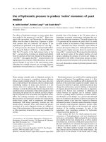
Fig. 1. CBP ⁄ p300 interact with the b-globin gene locus and with USF1 in MEL cells but not in K562 cells. (A) Schematic of the organization
of the human and mouse b-globin gene loci. The human b -globin locus depicted on top consists of five genes which are expressed in a
developmental stage-specific manner in erythroid cells as outlined. The expression of the genes is regulated by a LCR composed of five
DNaseI HS and located approximately 15–27 kbp upstream of the embryonic e-globin gene. The murine b-globin gene locus, which is
depicted on the bottom, consists of four genes which are expressed either in erythroid cells of the embryonic yolk sac (EY or bH1) or in
definitive erythroid cells derived from fetal liver or bone marrow hematopoiesis (b
maj
and b
min
). The murine LCR also contains multiple HS
required for high-level globin gene expression. K562 (B) and MEL cells (C) were subjected to ChIP analysis with antibodies against p300,
CBP or with a nonspecific antibody (IgG). Purified DNA was analyzed by real-time quantitative PCR with primers specific for LCR HS2 or the
e-, c-, b-, b-major or ec-globin gene promoters. (D) K562 or MEL cell extracts were precleared with anti-(rabbit IgG) beads and precipitated
with a-USF1, a-p300, a-CBP or a-HDAC3 (as negative control), and complexes were captured by incubation with anti-(rabbit IgG) beads.
Complexes were eluted off the beads with Laemmli buffer and incubation at 95 °C for 10 min and loaded onto a 5% Ready gel (Bio-Rad).
The membrane was probed with a-p300.
V. J. Crusselle-Davis et al. Coregulators of USF and TFII-I in erythroid cells
FEBS Journal 274 (2007) 6065–6073 ª 2007 The Authors Journal compilation ª 2007 FEBS 6067
not in MEL cells, in which the b-globin gene is tran-
scribed (Fig. 3A). We also detected interactions
between TFII-I and Suz12 in K562 cells (Fig. 3B). The
interaction between TFII-I and Suz12 is not as efficient
as that involving HDAC3 and it is not restricted to
K562 cells, because interactions are also detectable in
MEL cells (data not shown).
Reduction of Suz12 expression in K562 cells
increases b-globin gene expression
We previously used SMART-pool short interfering
RNA (siRNA) reagent from Dharmacon and effi-
ciently reduced expression of TFII-I and HDAC3 [20].
Reductions in both TFII-I and HDAC3 expression by
more than 80% led to an approximately three-fold
increase in b-globin gene expression. To examine
whether Suz12 and the PRC2 complex participate in
the repression of the adult b-globin gene in K562 cells,
we reduced expression of Suz12 by RNA interference.
The western blot results demonstrate that siRNA
transfected cells reveal a drastic reduction in Suz12
protein levels compared to mock transfected cells or
cells transfected with nonspecific siRNA (Fig. 4A).
Expression of the adult b-globin gene was increased by
three- to five-fold in cells transfected with Suz12
siRNA compared to mock or negative control siRNA
transfected cells (Fig. 4B). These results demonstrate
that the PRC2 complex, or components thereof, partic-
ipate in the repression of the adult b-globin gene in
embryonic erythroid cells. We also detected an increase
in the expression of the embryonic e-globin gene.
However, this increase was not as pronounced as the
one seen for b-globin gene expression.
The PRC2 complex contains the histone H3K27
specific histone methyltransferase Ezh2 [21]. Ezh2
catalyzes the di- and tri-methylation of H3K27 [25].
Using ChIP, we did not detect high levels of tri-
methylated H3K27 at the globin gene locus in K562
cells, consistent with studies from the Blobel labora-
AB
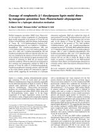
Fig. 2. Suz12 interacts with the adult b-globin gene in K562 but not in MEL cells. Antibodies against Suz12 and nonspecific IgG were used
in ChIP assays using K562 (A) and MEL (B) cells. Quantitative PCR was performed with primers that amplified the promoters of the genes
as indicated.
A
B
Fig. 3. Interaction of Suz12 and HDAC3 with TFII-I. (A) Interaction
of HDAC3 with TFII-I in K562 but not MEL cells. K562 or MEL cell
extract was precleared with anti-(a)-(rabbit IgG) beads and precipi-
tated with a-HDAC3 or IgG, and complexes were captured by incu-
bation with anti-(rabbit IgG) beads. Complexes were eluted off the
beads with Laemmli buffer and incubation at 95 °C for 10 min and
loaded onto a 10% Ready gel (Bio-Rad). The membrane was
probed with a-TFII-I and then stripped and probed with a-HDAC3
as a positive control. (B) Interaction of Suz12 with TFII-I in K562
cells. K562 extracts were precleared with anti-(rabbit IgG) beads
and precipitated with 2.5 lgofa-Suz12 or 2.5 lgofa-IgG (as nega-
tive control) antibodies. The complexes were captured by incuba-
tion with anti-(rabbit IgG) beads. Complexes were eluted off the
beads with Laemmli buffer and incubation at 95 °C for 10 min and
loaded onto a 10% Ready gel (Bio-Rad). The membrane was
probed with a-TFII-I. The lane labeled control represents a regular
western blot for TFII-I with protein extract from K562 cells.
Coregulators of USF and TFII-I in erythroid cells V. J. Crusselle-Davis et al.
6068 FEBS Journal 274 (2007) 6065–6073 ª 2007 The Authors Journal compilation ª 2007 FEBS
tory [26]. In addition, the association of trimethylated
H3K27 was not significantly altered in cells that
express, or do not express, the adult b-globin gene
(data not shown).
Interaction of Suz12 with the b
maj
-globin gene
promoter decreases during activation of the
b
maj
-globin gene in differentiating murine ES cells
We next examined the association of Suz12 with the
b
maj
-globin gene promoter during differentiation of
mouse ES cells. We previously demonstrated that the
adult b-globin gene is expressed at low levels in ES
cell cultures incubated for 5 days with erythropoietin
(EPO), which mediates the differentiation and prolif-
eration of erythroid cells [17]. High-level expression
of the adult b-globin gene was observed at day 12 in
the ES cell differentiation system. Figure 5A,B dem-
onstrates that b
maj
-globin gene expression is up-regu-
lated by more than 30-fold between days 5 and 12.
We observed that the association of Suz12, TFII-I
and trimethylated H3K27 (H3K27me3) with the
b
maj
-globin gene promoter is high at day 5 but
undetectable at day 12 (Fig. 5C). Quantitation of the
Suz12 levels at the b
maj
-globin gene promoter dem-
onstrate that the changes between days 5 and 12 are
significant. The control experiment demonstrates that
interaction of LCR HS2 associated dimethylated
H3K4 (H3K4me2) does not change during the
course of differentiation. Neither Suz12, nor TFII-I
were found to associate with a control region located
between LCR elements HS2 and HS3 (data not
shown).
Discussion
We provide evidence that USF and TFII-I regulate
b-globin gene expression through the recruitment of
coactivator complexes that render the b-globin pro-
moter accessible or inaccessible to the transcription
complex. USF recruits the histone acetyltransferase
p300 to the b-globin promoter and this activity
increases the accessibility for transcription factors.
TFII-I recruits HDAC3 and the PRC2 complex, which
render the chromatin structure inaccessible to the tran-
scription complex.
Previous data from the Felsenfeld laboratory have
shown that USF interacts with the coregulators p300,
CBP, SET7 ⁄ 9 and PCAF and perhaps recruits these
activities to a chromosomal boundary element [24]. In
the present study, we show that p300 and CBP are
located at the promoter of the active adult b-globin
gene and also at LCR element HS2. USF1 is observed
to interact with p300 exclusively in erythroid cells with
an adult phenotype. These data suggest that USF1
recruits p300 to the promoter of the active b-globin
gene to aid in transcriptional activation.
CBP ⁄ p300 is located at LCR HS2 in K562 cells, in
which we did not detect interactions between USF and
p300. This suggests that the recruitment of CBP ⁄ p300
to the LCR, at least in K562 cells, is mediated by pro-
teins other than USF, and potential candidates are
GATA-1 and NF-E2, which have been shown to inter-
act with CBP [27,28]. Both of these proteins were
shown to be required for histone acetylation of spe-
cific regions in the globin gene locus. However, the
recruitment of CBP ⁄ p300 to the adult b-globin gene
AB
Fig. 4. Suz12 represses b-globin gene expression in K562 cells. K562 cells were nucleofected with Suz12 siRNA, nontargeting siRNA (neg),
or mock transfected. (A) Protein was collected after 2 days and electrophoresed on gels for western blotting. Blots were probed with
a-Suz12 in the upper panel and then stripped and reprobed with a-GAPDH for a loading control. (B) Relative b-, c- and e-globin expression in
Suz12 knockdown cells. RNA was collected, reverse transcribed, and analyzed by quantitative PCR. Expression is set relative to either non-
targeting siRNA (neg) samples or mock transfected cells, with GAPDH as the internal reference.
V. J. Crusselle-Davis et al. Coregulators of USF and TFII-I in erythroid cells
FEBS Journal 274 (2007) 6065–6073 ª 2007 The Authors Journal compilation ª 2007 FEBS 6069
promoter is mediated by USF. We previously demon-
strated that the expression of a dominant negative
mutant of USF in MEL cells resulted in decreased
interactions between CBP ⁄ p300 with both b-globin
gene promoter and LCR HS2, suggesting that, in adult
erythroid cells, USF is required for the recruitment of
CBP ⁄ p300 to both the b-globin gene and the LCR.
Expression of the dominant negative mutant also
reduced interactions of RNA polymerase II with LCR
HS2 and the b-globin gene promoter [20]. E-box
motifs are present downstream of the transcription
start sites of both the b-globin promoter and HS2, sug-
gesting that USF is important for transcription in both
of these regions in adult erythroid cells [29].
Components of PRC2 are expressed at high levels
in embryonic tissues and are essential for the earliest
stages of vertebrate development. They also have
been found to occupy a special set of developmental
genes in ES cells that must be repressed to maintain
pluripotency and that are poised for activation during
ES cell differentiation [30,31]. As the adult b-globin
gene is repressed during the early stages of develop-
ment and is poised for activation, we reasoned that
PCR2 could be involved in repressing b-globin
expression at the embryonic and fetal stage of devel-
opment.
Reducing the activity of Suz12 in K562 cells led to
an increase in b-globin gene expression and also a
modest increase in e-globin gene expression (Fig. 4). It
should be noted that the K562 cells were not induced
by hemin, which has been shown to increase the
expression of e- and c-globin gene expression in these
cells. However, the effect of hemin on globin gene
expression in these cells is relatively low [32]. Neverthe-
less, it is possible that the PRC complex represses
globin gene expression in a differentiation and devel-
opmental stage-specific manner. This is consistent with
previous findings demonstrating that the PRC2
complex localizes to developmentally regulated genes
in undifferentiated ES cells [30], and with our own
data demonstrating that Suz12 and TFII-I interact
with the b
maj
-globin promoter during early but not
late stages of EPO-induced ES cell differentiation [20]
(Fig. 5).
AC
B
D
Fig. 5. The interaction of Suz12 with the b-globin gene promoter decreases with increased b-globin gene expression during erythroid differ-
entiation of mouse ES cells. (A) RT-PCR analysis of e- and b-globin gene expression during erythroid differentiation of mouse ES cells. RNA
was isolated from ES cells incubated in the presence of EPO for 5 or 12 days, as indicated. The RNA was reverse transcribed and subjected
to RT-PCR using primers specific for the control genes Rex1 and b-actin as well as the embryonic e- and adult b
maj
-globin genes. (B) Quanti-
tative RT-PCR analysis of b
maj
-globin gene expression at days 5 and 12 of erythroid differentiation of mouse ES cells. RNA was isolated from
the cells at the indicated time points after addition of EPO and subjected to quantitative RT-PCR analysis using primers specific for the b
maj
-
globin gene. (C) Analysis of modified histones, Suz12 and TFII-I interactions with the b
maj
-globin gene promoter at days 5 and 12 of erythroid
differentiation. Cells were collected at the indicated time points and subjected to ChIP analysis using antibodies specific for histone H3 dime-
thylated at K4 (H3K4me2), histone H3 trimethylated at K27 (H3K27me3), Suz12, TFII-I and the control IgG. (D) Quantitative analysis of Suz12
interactions with the b
maj
-globin gene promoter at days 5 and 12 of erythroid differentiation. Cells were taken at the indicated time points
and subjected to ChIP with the indicated antibodies. The precipitated DNA was subjected to quantitative PCR using primers specific for the
b
maj
-globin gene. In the left panel, chromatin was precipitated with antibodies specific for histone H3 dimethylated at K4 (H3K4me2) and
analysed by quantitative PCR using primers specific for LCR element HS2.
Coregulators of USF and TFII-I in erythroid cells V. J. Crusselle-Davis et al.
6070 FEBS Journal 274 (2007) 6065–6073 ª 2007 The Authors Journal compilation ª 2007 FEBS
Suz12 has previously been shown to be required for
the di- and tri-methylation of H3K27, which are marks
for inactive chromatin [33]. We did not observe high
levels of trimethylated H3K27 associated with the
b-globin gene promoter in K562 cells. However, trime-
thylated H3K27 was present at the b
maj
-globin gene
promoter in early stage EPO-induced ES cell cultures
(day 5) when the b
maj
-globin gene is expressed at low
levels. This modification was absent in day 12 cultures
that express high levels of the b
maj
-globin gene
(Fig. 5).
The interactions between TFII-I and Suz12 in K562
cells are weak. This interaction may be unstable or
transient in nature. In this context, it is interesting to
note that another initiator binding protein, YY1, has
also been shown to directly interact with both HDAC3
and the PRC complex [34,35]. Furthermore, YY1 has
been implicated in the silencing of e-globin gene
expression in adult erythroid cells [36].
Suz12 could also be recruited to the b-globin gene
promoter through the interaction with other DNA
binding proteins involved in repressing the adult glo-
bin gene. One possible candidate is BP1, a homeo-
domain-containing protein, which binds to a negative
regulatory element in the upstream b-globin gene
region and reduces expression [37,38]. It is not
known how this protein represses b-globin gene
expression.
Another corepressor recruited by TFII-I to the glo-
bin gene locus is HDAC3. TFII-I and HDAC3 interact
with each other exclusively in K562 cells and not in
MEL cells. It has been observed that TFII-I and
HDAC3 have very similar expression patterns in the
developing mouse embryo [39]. Therefore, the results
presented in the present study, along with the previous
studies, suggest that the transcription activity of TFII-
I may be controlled by HDAC3 during early develop-
ment. Our data demonstrate that TFII-I functions as a
repressor of b-globin gene expression by recruiting
HDAC activity to the promoter. This is consistent
with observations of the acetylation of the human
b-globin promoter region being reduced in erythroid
cells with an embryonic phenotype [16,40].
EKLF is present and active in both primitive and
adult erythroid cells but somehow is unable to activate
b-globin gene expression in embryonic erythroid cells
[41]. Perhaps EKLF functions in conjunction with
other factors to activate b-globin gene expression and
these factors are not present or active in embryonic
cells. Alternatively, EKLF requires an accessible chro-
matin structure to recruit nucleosome mobilizing activ-
ities to the promoter. Initial accessibility may be
provided by proteins that recruit histone acetyl or
methyl transferases to the adult b -globin gene pro-
moter, thereby counteracting the repressive activity of
TFII-I.
Experimental procedures
Cell culture
Human erythroleukemia (K562) cells were grown in
RPMI 1640 medium supplemented with 10% fetal bovine
serum and 5% penicillin-streptomycin. Murine erythroleu-
kemia (MEL) cells were grown in RPMI containing 10%
fetal bovine serum and 5% antibiotic-antimycotic. Cells
were grown at 37 °C and maintained at a cell density of
approximately 1–5 · 10
5
cellsÆmL
)1
. Mouse ES cells were
cultured and induced to differentiate with EPO as described
by Levings et al. [17].
ChIP, co-immunoprecipitation
Chromatin immunoprecipitation was carried out as
described by Leach et al. [19]. Briefly, cells were washed in
NaCl ⁄ P
i
, crosslinked using 1% formaldehyde, and
quenched with glycine. After isolation of nuclei and lysis,
the crosslinked chromatin was fragmented by sonication to
average size fragments of 500 bp. Chromatin fragments
were precipitated with specific or nonspecific control (IgG)
antibodies. Antibody ⁄ chromatin fragments were subjected
to several rounds of stringent washing. The DNA was puri-
fied from the chromatin fragments and analyzed by real-
time quantitative PCR using pairs of primers specific for
the murine or human b-globin gene locus as described
by Crusselle-Davis et al. [20] in addition to mouse ec-glo-
bin promoter (upstream: 5¢-ATGACCTGGCTCCACC
CAT-3¢, downstream: 5¢-TCTTTGAGCCATTGGTCAGC-
3¢); human HS2 (upstream: 5¢-CGCCTTCTGGTTCTGTG
TAA-3¢, downstream: 5¢-GAGAACATCTGGGCACAC
AC-3¢); human c-globin promoter (upstream: 5¢-CCTTCA
GCAGTTCCACACAC-3¢, downstream: 5¢-CTCCTCTGT
GAAATGACCCA-3¢); human HS3 ⁄ 2 (upstream: 5¢-GTG
ACCTCAGTGCCTCAGAA-3¢, downstream: 5¢-ACCTAT
CACAGCACCACACC-3¢); and human glyceralde-
hyde 3-phosphate dehydrogenase (GAPDH) promoter
(upstream: 5¢-ACGTAGCTCAGGCCTCAAGACCTTG-3¢,
downstream: 5¢-GACTGTCGAACAGGAGGAGCAGA
GA-3¢).
Immunoprecipitation was carried out essentially as
described by Crusselle-Davis et al. [20]. The antibodies used
in the experiments comprised: monomethyl-histone H3
Lys27 07-448, trimethyl-H3 Lys27, Suz12 07-379 (Upstate,
Charlotteville, VA, USA); USF1 (H-86) sc-8983, p300 (N-15)
sc-584, CBP (A-22) sc-369, GAPDH (FL-335) sc-25778
(Santa Cruz Biotechnologies, Santa Cruz, CA, USA);
Suz12 ab12073, TFII-I ab10464 (Abcam, Cambridge, MA,
V. J. Crusselle-Davis et al. Coregulators of USF and TFII-I in erythroid cells
FEBS Journal 274 (2007) 6065–6073 ª 2007 The Authors Journal compilation ª 2007 FEBS 6071
USA); and IgG C6409 anti-(chicken IgG); (Sigma,
St Louis, MO, USA).
RNA interference
siGENOME SMART-pool reagents for Suz12
(NM 015355) and siCONTROL nontargeting pool (D-
001810-10-05) were obtained from Dharmacon (Lafayette,
CO, USA) and used for transfection as described by Crus-
selle-Davis et al. [20]. A total of 10
6
K562 cells were nucleo-
fected with siRNA or mock transfected using an
optimized protocol for K562 cells (Amaxa Biosystems,
Cologne, Germany). Protein and RNA were collected from
transfected K562 cells after 48, 72 and 96 h, as described
previously [20]. Relative expression was determined using
real-time RT-PCR and primers as described by Crusselle-
Davis et al. [20]. In addition, human c-globin primers
(upstream: 5¢-TGAATGTCCAAGATGCTGGA-3¢, down-
stream: 5¢-CATGATGGCAGAGGCAGAG-3¢) were used.
Protein isolation and western blotting
Protein isolation and western blotting was performed as
described by Leach et al. [19]. A total of 20 lg of protein
was loaded onto 7.5% or 5% Ready gels (Bio-Rad, Hercu-
les, CA, USA), electrophoresed, and transferred to nylon
membranes. The detection of proteins on the membranes
was performed using the ECL Plus system as described in
the manufacturer’s instructions (Amersham Pharmacia Bio-
tech, Piscataway, NJ, USA). The primary antibodies used
were the same as those used in the ChIP and immunopre-
cipitation assays in addition to HDAC3 (H-99) sc-11417
(Santa Cruz). The secondary antibodies used were as
described by Crusselle-Davis et al. [20]. The antibodies were
used at concentrations recommended in the manufacturer’s
guidelines.
Acknowledgements
We are grateful to our colleagues I-Ju Lin, Shermi
Liang, Kunjal Gandhi and JoAnne Andersen for
assistance and stimulating discussions. This work was
supported by grants from the NIH (DK 52356) and
American Heart Association.
References
1 Misteli T (2007) Beyond the sequence: cellular organiza-
tion of genome function. Cell 128, 787–800.
2 Li B, Carey M & Workman JL (2007) The role of chro-
matin during transcription. Cell 128, 707–719.
3 Thomas MC & Chiang CM (2006) The general tran-
scription machinery and general cofactors. Crit Rev
Biochem Mol Biol 41, 105–178.
4 Orphanides G & Reinberg D (2002) A unified theory of
gene expression. Cell 108, 439–451.
5 Spiegelman BM & Heinrich R (2004) Biological control
through regulated transcriptional coactivators. Cell 119,
157–167.
6 Stamatoyannopoulos G (2005) Control of globin gene
expression during development and erythroid differenti-
ation. Exp Hematol 33, 259–271.
7 Grosveld F, van Assendelft GB, Greaves DR & Kollias
G (1987) Position-independent, high-level expression of
the human b-globin gene in transgenic mice. Cell 51,
975–985.
8 Forrester WC, Thompson C, Elder JT & Groudine M
(1986) A developmentally stable chromatin structure in
the human b-globin gene cluster. Proc Natl Acad Sci
USA 83, 1359–1363.
9 Tuan D, Solomon W, Li Q & London IM (1985) The
‘b-like globin’ gene domain in human erythroid cells.
Proc Natl Acad Sci USA 81, 2718–2722.
10 Bungert J, Dave U, Lim KC, Lieuw KH, Shavit JA,
Liu Q & Engel JD (1995) Synergistic regulation of
human b-globin gene switching by locus control region
elements HS3 and HS4. Genes Dev 9, 3083–3096.
11 Bungert J, Tanimoto K, Patel S, Liu Q, Fear M &
Engel JD (1999) Hypersensitive site 2 specifies a unique
function within the human b-globin locus control region
to stimulate globin gene transcription. Mol Cell Biol 19,
3062–3072.
12 Bender MA, Roach JN, Halow J, Close J, Alami R,
Bouhassira EE, Groudine M & Fiering SN (2001) Tar-
geted deletion of 5¢HS1 and 5¢HS4 of the beta-globin
locus control region reveals additive activity of the
DNaseI hypersensitive sites. Blood 98, 2022–2027.
13 Marenduzzo D, Faro-Trindade I & Cook PR (2007)
What are the molecular ties that maintain genomic
loops? Trends Genet 23, 126–133.
14 Chakalova L, Debrand E, Mitchell JA, Osborne CS &
Fraser P (2005) Replication and transcription: shaping
the landscape of the genome. Nat Rev Genet 6, 669–677.
15 Ragoczy T, Bender MA, Telling A, Byron R & Grou-
dine M (2006) The locus control region is required for
association of the murine beta-globin locus with
engaged transcription factories during erythroid matura-
tion. Genes Dev 20, 1447–1457.
16 Vieira KF, Levings PP, Hill MA, Crusselle VJ, Kang
SHL, Engel JD & Bungert J (2004) Recruitment of tran-
scription complexes to the beta-globin gene locus in vivo
and
in vitro. J Biol Chem 279, 50350–50357.
17 Levings PP, Zhou Z, Vieira KF, Crusselle-Davis VJ &
Bungert J (2006) Recruitment of transcription com-
plexes to the b-globin locus control region and tran-
scription of hypersensitive site 3 prior to erythroid
differentiation of murine embryonic stem cells. FEBS J
273, 746–755.
Coregulators of USF and TFII-I in erythroid cells V. J. Crusselle-Davis et al.
6072 FEBS Journal 274 (2007) 6065–6073 ª 2007 The Authors Journal compilation ª 2007 FEBS
18 Feng D & Kan YW (2005) The binding of the ubiqui-
tous transcription factor Sp1 at the locus control region
represses the expression of beta-like globin genes. Proc
Natl Acad Sci USA 102, 9896–9900.
19 Leach KM, Vieira KF, Kang SH, Aslanian A, Teich-
mann M, Roeder RG & Bungert J (2003) Characteriza-
tion of the human beta-globin downstream promoter
region. Nucleic Acids Res 31, 1292–1301.
20 Crusselle-Davis VJ, Vieira KF, Zhou Z, Anantharaman
A & Bungert J (2006) Antagonistic regulation of b-glo-
bin gene expression by helix-loop-helix proteins USF
and TFII-I. Mol Cell Biol 26, 6832–6843.
21 Schwartz YB & Pirrotta V (2007) Polycomb silencing
mechanisms and the managements of genomic pro-
grammes. Nat Rev Genet 8 , 9–22.
22 Bantignies F & Cavalli G (2006) Cellular memory and
dynamic regulation of polycomb group proteins. Curr
Opin Cell Biol 18, 275–283.
23 Bungert J, Kober I, Du
¨
ring F & Seifart KH (1992)
Transcription factor eUSF is an essential component of
isolated transcription complexes on the duck histone H5
gene and it mediates the interaction of TFII-D with a
TATA-deficient promoter. J Mol Biol 223, 885–898.
24 West AG, Huang S, Gaszner M, Litt MD & Felsenfeld
G (2004) Recruitment of histone modifications by USF
proteins at a vertebrate barrier element. Mol Cell 16,
453–463.
25 Montgomery ND, Yee D, Chen A, Kalantry S, Cham-
berlain SJ, Otte AP & Magnuson T (2005) The murine
polycomb group protein Eed is required for global his-
tone H3 lysine-27 methylation. Curr Biol 15, 942–947.
26 Vakoc CR, Sachdeva MM, Wang H & Blobel GA
(2006) Profile of histone lysine methylation across
transcribed mammalian chromatin. Mol Cell Biol 26,
9185–9195.
27 Blobel GA, Nakajima T, Eckner R, Montminy M &
Orkin SH (1998) CREB binding protein cooperates with
transcription factor GATA-1 and is required for ery-
throid differentiation. Proc Natl Acad Sci USA 95,
2061–2066.
28 Johnson KD, Christensen H, Zhao B & Bresnick EH
(2001) Distinct mechanisms control RNA polymerase II
recruitment to a tissue-specific locus control region and
a downstream promoter. Mol Cell 8, 465–471.
29 Leach KM, Nightingale K, Igarashi K, Levings PP,
Engel JD, Becker PB & Bungert J (2001) Reconstitution
of human b-globin locus control region hypersensitive
sites in the absence of chromatin assembly. Mol Cell
Biol 21, 2629–2640.
30 Boyer LA, Plath K, Zeitlingert J, Brambrink T, Medei-
ros LA, Lee TI, Levine SS, Wernig M, Tajonar A, Ray
MK et al. (2006) Polycomb complexes repress develop-
mental regulators in murine embryonic stem cells.
Nature 441, 349–353.
31 Boyer LA, Mathur D & Jaenisch R (2006) Molecular
control of pluripotency. Curr Opin Genet Dev 16, 455–
462.
32 Kim A & Dean A (2004) Developmental stage differ-
ences in chromatin subdomains of the beta globin locus.
Proc Natl Acad Sci USA 101, 7028–7033.
33 Barski A, Cuddapah S, Cui K, Roh TY, Schones DE,
Wang Z, Wei G, Chepelev I & Zhao K (2007) High-res-
olution profiling of histone methylations in the human
genome. Cell 129, 823–837.
34 Yao YL, Yang WM & Seto E (2001) Regulation of
transcription factor YY1 by acetylation and deacetyla-
tion. Mol Cell Biol 21, 5979–5991.
35 Wilkinson FH, Park K & Atchison ML (2006)
Polycomb recruitment to DNA by the YY1 REPO
domain. Proc Natl Acad Sci USA 103, 19296–19301.
36 Raich N, Clegg CH, Grofti J, Romeo PH & Stamato-
yannopoulos G (1995) Demonstration of a human
epsilon-globin gene silencer with studies in transgenic
mice. EMBO J 14, 801–809.
37 Chase MB, Fu S, Haga SB, Davenport G, Stevenson H,
Do K, Morgan D, Mah AL & Berg PE (2002) BP1, a
homeodomain-containing isoform of DLX4, represses
the beta-globin gene. Mol Cell Biol 22, 2505–2514.
38 Mpollo MS, Beaudoin M, Berg PE, Beauchemin H,
D’Agati V & Trudel M (2006) BP1 is a negative modu-
lator of definitive erythropoiesis. Nucleic Acids Res 34,
5232–5237.
39 Tussie-Luna MI, Bayarsihan D, Seto E, Ruddle FH &
Roy AL (2002) Physical and functional interactions
of histone deacetylase 3 with TFII-I family proteins
and PIASxb. Proc Natl Acad Sci USA 99, 12807–12812.
40 Forsberg EC, Downs KM, Christensen HM, Im H,
Nuzzi EH & Bresnick EH (2000) Developmentally
dynamic histone acetylation pattern of a tissue-specific
chromatin domain. Proc Natl Acad Sci USA 97, 14494–
14499.
41 Bieker JJ (2005) Probing the onset and regulation of
erythroid-cell specific gene expression. Mt Sinai J Med
72, 333–338.
V. J. Crusselle-Davis et al. Coregulators of USF and TFII-I in erythroid cells
FEBS Journal 274 (2007) 6065–6073 ª 2007 The Authors Journal compilation ª 2007 FEBS 6073