Báo cáo khoa học: The Drosophila jumonji gene encodes a JmjC-containing nuclear protein that is required for metamorphosis pot
Bạn đang xem bản rút gọn của tài liệu. Xem và tải ngay bản đầy đủ của tài liệu tại đây (639.6 KB, 13 trang )
The Drosophila jumonji gene encodes a JmjC-containing
nuclear protein that is required for metamorphosis
Nobuhiro Sasai
1,2,3,
*, Yasuko Kato
2,3
, Gaku Kimura
2,3
, Takashi Takeuchi
4
and
Masamitsu Yamaguchi
2,3
1 Venture Laboratory, Kyoto Institute of Technology, Japan
2 Department of Applied Biology, Kyoto Institute of Technology, Japan
3 Insect Biomedical Research Center, Kyoto Institute of Technology, Japan
4 Mitsubishi Kagaku Institute of Life Sciences (MITILS), Machida, Japan
The basic unit of chromatin in eukaryotes is the
nucleosome, which consists of 146 bp of DNA
wrapped around an octamer of histones H2A, H2B,
H3 and H4 [1]. Covalent modifications of histone
tails, such as acetylation, methylation, phosphoryla-
tion and ubiquitination, modulate interaction affinities
for chromatin-associated proteins, leading to the
formation of either transcriptionally active or silent
chromatin structures [2]. For example, methylation
at Lys9 of histone H3 (H3-K9) by the su(var)3-9,
enhancer of zeste, trithorax (SET) domain-containing
protein SUV39H1 creates binding sites for the chromo-
domain-containing protein HP1, resulting in the
establishment of heterochromatin [3]. In addition,
methylation of H3-K27 and H4-K20 and hypoacetyla-
tion of histones are associated with transcriptionally
silenced chromatin, whereas methylation of H3-K4
and hyperacetylation of histones are connected with
active transcription [4].
The JmjC domain was initially characterized as
a conserved domain among jumonji (Jmj) family
proteins, including Jmj, RBP2 and SMCX, and has
Keywords
euchromatin; JmjC domain; metamorphosis;
suppressor of PEV; transcriptional silencing
Correspondence
M. Yamaguchi, Department of Applied
Biology, Kyoto Institute of Technology,
Matsugasaki, Sakyo-ku, Kyoto 606-8585
Japan
Fax: +81 75 724 7760
Tel: +81 75 724 7781
E-mail:
*Present address
CNRS ⁄ UMR218, Institute Curie, Paris,
France
(Received 25 July 2007, revised 4 October
2007, accepted 10 October 2007)
doi:10.1111/j.1742-4658.2007.06135.x
Jumonji (Jmj) is a transcriptional repressor that plays important roles in
the suppression of cell proliferation and development of various tissues in
the mouse. To further clarify the roles of Jmj during development and gain
insight into mechanisms of Jmj-mediated transcriptional regulation, we
have taken advantage of Drosophila as a model organism. Drosophila Jmj
(dJmj) shares high homology with mammalian Jmj in the JmjN, JmjC and
AT-rich interaction domains, as well as in the N-terminal repression
domain. dJmj localizes to hundreds of euchromatic sites but not to chro-
mocenter heterochromatin on salivary gland polytene chromosomes. In
addition, dJmj is excluded from regions stained with an antibody against
Ser5-phosphorylated RNA polymerase II, suggesting a function of dJmj in
transcriptionally inactive chromatin. Loss of djmj results in larval and
pupal lethality with phenotypes similar to those observed in mutants of
ecdysone-regulated genes, implying the involvement of dJmj in the repres-
sion of gene expression in the ecdysone pathway. Transgenic mouse Jmj
mostly colocalizes with dJmj and partially rescues the phenotypes of djmj
mutants, indicating that dJmj is a functional homolog of mammalian Jmj.
Furthermore, mutation in djmj suppresses position effect variegation of the
T(2;3)Sb
V
rearrangement. These findings suggest that dJmj controls
expression of developmentally important genes through modification of
chromatin into a transcriptionally silenced state.
Abbreviations
ARID, AT-rich interaction domain; DAPI, 4¢,6-diamidino-2-phenylindole; dJmj, Drosophila Jmj; GST, glutathione S-transferase; Jmj, jumonji;
Lid, little imaginal disks; mJmj, mouse jumonji; PolII, RNA polymerase II.
FEBS Journal 274 (2007) 6139–6151 ª 2007 The Authors Journal compilation ª 2007 FEBS 6139
subsequently been identified in more than 100 proteins
in prokaryotic and eukaryotic organisms [5–7]. JmjC-
containing proteins have been shown to play important
roles in various biological processes, including cellular
differentiation, DNA repair and regulation of hetero-
chromatin [8–10]. These JmjC-containing proteins are
considered to regulate chromatin or transcription, as
they are generally associated with chromatin- or
DNA-binding domains, such as the plant homeo-
domain (PHD) finger, the TUDOR domain, the AT-
rich interaction domain (ARID) and the zinc finger
motif [11–13]. Recent studies revealed that the JmjC-
containing proteins are histone demethylases and that
the JmjC domain is responsible for their enzymatic
activity [14–19]. However, as several JmjC-containing
proteins are predicted to be enzymatically inactive
[11,20], additional mechanisms might be involved in
JmjC-mediated regulation of chromatin or transcrip-
tion.
The jmj gene was originally identified by a gene trap
strategy in the mouse and shown to be required for
the appropriate development of various tissues, includ-
ing brain, liver, thymus and heart [7,21,22]. jmj
encodes a transcriptional repressor containing the
JmjC domain, JmjN domain and ARID. The latter
two mediate the interaction of Jmj with A ⁄ T-rich
DNA sequences [23]. Although the N-terminal region
of Jmj itself is known to be responsible for its repres-
sor activity [23,24], the mechanisms remain unknown.
The JmjC domain of Jmj is predicted to be enzymati-
cally inactive as a histone demethylase [11,12] and its
function remains to be clarified.
Jmj appears to have an important role in suppres-
sion of cellular proliferation. In the developing heart,
Jmj binds to the promoter and represses the expression
of cyclinD1, which is essential for G
1
⁄ S phase transi-
tion, thereby suppressing cell proliferation and regulat-
ing morphogenesis of cardiac cells [24]. Jmj also
represses E2F activity and reduces cell cycle progres-
sion by associating with the Rb protein [25]. Further-
more, it represses expression of ANF, which encodes a
hormonal mediator that is required for heart develop-
ment, by counteracting the function of ANF activators
Nkx2.5 and GATA4 [26]. As jmj is widely expressed
and is required for the correct development of various
tissues, involvement in the regulation of a diverse
range of developmental programs, not limited to car-
diac cells, is likely.
To further clarify the roles of Jmj during develop-
ment and gain insight into mechanisms of Jmj-medi-
ated chromatin regulation, we have taken advantage of
Drosophila melanogaster as a model organism. We
show here that loss of the Drosophila jumonji (djmj)
gene results in larval and pupal lethality with pheno-
types similar to those with ecdysone-regulated genes.
On salivary gland polytene chromosomes, Drosophila
Jmj (dJmj) localizes to euchromatic sites excluded from
highly transcribed regions that are stained with an
antibody against RNA polymerase II (PolII), suggest-
ing a function of dJmj in transcriptionally inactive
chromatin. Moreover, a djmj mutant suppresses the
position effect variegation (PEV) of the T(2;3)Sb
V
rearrangement. These observations suggest that dJmj
controls expression of developmentally important
genes through modification of chromatin into a trans-
criptionally silenced state.
Results
The CG3654 gene encodes a Drosophila ortholog
of mammalian Jmj
JmjC-containing proteins are classified into subgroups
on the basis of their protein structures [11,17]. Jmj
belongs to the JARID family, which is characterized
by possession of the conserved domains, JmjN, JmjC
and ARID [13]. Drosophila contains two JARID fam-
ily proteins, little imaginal disks (Lid) and a novel pro-
tein CG3654 (Fig. 1A). Lid has been identified as a
gene that enhances the phenotype of ash1 mutants,
and is classified as a trithorax group gene [27]. Lid is
considered to be a sole ortholog of mammalian
JARID1 proteins, including RBP2, PLU-1, SMCX
and SMCY, as all of them contain additional PHD
fingers [12,13].
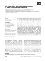
Mouse Jmj (mJmj) and Drosophila CG3654 share
40%, 45% and 37% identities in the JmjN domain,
JmjC domain and ARID, respectively (Fig. 1A). In
addition to these conserved domains, mJmj contains a
zinc finger motif at its C-terminus, whereas CG3654
possesses two AT-hook motifs (Fig. 1A). The N-termi-
nal repression domain of Jmj is also conserved in
CG3654 (Fig. 1B), but not in Lid. Therefore, we con-
cluded that CG3654 is a Drosophila counterpart of
mammalian Jmj and designated it as Drosophila jum-
onji (dJmj). Jmj proteins are also found in various spe-
cies, from insects to mammals, but not in worms and
yeasts. Importantly, all the Jmj proteins share high
homology in the N-terminal region (data not shown),
suggesting that this is important for Jmj function,
probably acting as a repression domain.
djmj
e03131
is a loss of function allele of djmj
The djmj gene localizes in the 67B9-10 cytological
region and is composed of four exons, including
Characterization of Drosophila jumonji N. Sasai et al.
6140 FEBS Journal 274 (2007) 6139–6151 ª 2007 The Authors Journal compilation ª 2007 FEBS
7053 bp of an ORF (Fig. 2A). To confirm the expres-
sion of dJmj protein, we generated a polyclonal anti-
body to dJmj by immunizing rabbits with the
C-terminal region of dJmj (amino acids 1635–2351) as
an antigen. Western blot analysis with affinity purified
antibody to dJmj recognized a protein corresponding
to the calculated molecular mass of dJmj (252 kDa)
from embryo to adult stages, indicating continuous
expression of dJmj throughout development (Fig. 2B,
lanes 1–7). The lower band (120 kDa) detected by
antibody to dJmj is evident in extracts of embryos
(Fig. 2B, lanes 1 and 2) and embryo-derived Kc cells
(Fig. 2B, lane 8). dsRNA-mediated knockdown of
dJmj in Kc cells reduced the amount of the 250 kDa
dJmj protein to an undetectable level at 4 days after
dsRNA treatment, whereas that of the 120 kDa band
was unchanged throughout dsRNA treatment (Fig. 2B,
lane 9). Therefore, we concluded that the 120 kDa
A
B
Fig. 1. Identification of the Drosophila Jmj
protein. (A) Schematic structures of mouse
Jmj, Drosophila Jmj and Lid. The locations
of the JmjN domain, JmjC domain, ARID,
PHD, AT-hook domain and C5HC2 zinc fin-
ger domain are shown. (B) Amino acid align-
ment of the N-terminal repression domain
of mouse and Drosophila Jmj. Identical and
similar residues are shaded in black and
gray, respectively.
AB
CEF
D
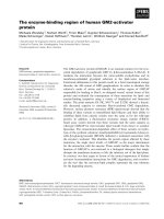
Fig. 2. Characterization of transposon-inserted djmj mutants. (A) The structure of djmj and the location of the transposon insertion in e03131
(piggyBac) is shown. The noncoding and coding regions of the djmj transcript are depicted as open and filled boxes, respectively. (B) Devel-
opmental western blot analysis of dJmj. Protein extracts from various developmental stages were probed with polyclonal antibody to dJmj.
Anti-a-tubulin antibody was used to compare the amount of protein loading. An asterisk shows nonspecific bands. Lane 1: 0–12 h embryo.
Lane 2: 12–24 h embryo. Lane 3: third larva. Lane 4: early pupa. Lane 5: late pupa. Lane 6: adult male. Lane 7: adult female. Lane 8: Kc
cells. Lane 9: Kc cells treated with dsRNA. (C) Protein extracts from third instar larvae were subjected to western blotting with antibody to
dJmj (upper). The same blot was reprobed with antibody to a-tubulin to compare protein loading (lower). Lane 1: wild type. Lane 2:
djmj
e03131
. Lane 3: djmj
e03131
⁄ Df(3L)AC1. (D) RT-PCR analysis of expression of djmj in third instar larvae from wild-type and djmj
e03131
mutants. Rp49 was used as an internal control. (E) Immunostaining for dJmj in whole salivary gland cells in wild-type and djmj
e03131
mutant
larvae. DNA was visualized with DAPI. (F) Semiquantitative RT-PCR analysis of cell cycle regulators in wild-type and djmj
e03131
third instar lar-
vae. Expression of rp49 was used as an internal control.
N. Sasai et al. Characterization of Drosophila jumonji
FEBS Journal 274 (2007) 6139–6151 ª 2007 The Authors Journal compilation ª 2007 FEBS 6141
band is a nonspecific protein that is cross-reactive
with the antibody. It should be noted that this
cross-reactive 120 kDa band is undetectable in extracts
from flies at later developmental stages.
To clarify the in vivo roles of djmj, we analyzed
transposon-inserted djmj mutants. Two fly strains that
contain the P or piggyBac transposons in the djmj gene
locus were identified. The djmj
EY02717
allele is an inser-
tion of the EY element [28] in the 5¢-UTR of djmj.
However, this insertion does not affect djmj expression,
and homozygous djmj
EY02717
flies proved to be viable
and fertile (data not shown). The djmj
e03131
allele car-
ries the insertion of the piggyBac construct RB, which
contains the splice acceptor and an FLP recombination
target (FRT) site [29], in the first intron of the djmj
gene (Fig. 2A), and djmj
e03131
homozygotes, in con-
trast, showed a lethal phenotype. The dJmj protein
was found to be absent in larval extracts of djmj
e03131
homozygotes or heterozygotes with the deficiency chro-
mosome, Df(3L)AC1, which lacks a genomic region
including the entire djmj locus (Fig. 2C). RT-PCR
analysis also indicated a decrease of djmj transcripts in
djmj
e03131
homozygotes (Fig. 2D). Immunostaining of
whole salivary gland cells from third instar larvae
showed predominant localization of dJmj protein in
the nuclei of wild-type but not of djmj
e03131
homozy-
gous cells (Fig. 2E).
As it has been reported that mammalian Jmj
represses cyclinD1 expression via binding to its pro-
moter [24], we investigated whether dJmj also represses
the expression of cyclinD, the sole ortholog of mam-
malian cyclinD genes in Drosophila [30]. Semiquantita-
tive RT-PCR analysis showed that cyclinD is not
misregulated in djmj
e03131
mutant third instar larvae
(Fig. 2F). The expression of other cell cycle regulators,
including cyclinE, cdk4, E2Fs, Rbfs and stg, was also
unaltered by loss of djmj (Fig. 2F and data not
shown). These results suggest that dJmj does not play
a dominant role in the repression of cell cycle regula-
tors in Drosophila.
dJmj localizes to euchromatic regions on
polytene chromosomes
The JmjC-containing proteins are thought to regulate
chromatin or transcription [11,12]. To gain insight into
the roles of dJmj in chromatin regulation, we analyzed
its chromosomal localization by immunostaining of
polytene chromosomes of salivary glands from third
instar larvae (Fig. 3). DNA was visualized with 4¢,6-di-
amidino-2-phenylindole (DAPI), which stains brightly
at condensed DNA regions on euchromatic arms that
are divided into bands and interbands and at chromo-
center heterochromatin (Fig. 3A,D). Immunostaining
of chromosomes with antibody to dJmj showed dJmj
at hundreds of euchromatic sites with 10–20 bright
signals (Fig. 3B,C). In contrast, no dJmj signals were
detected in chromosomes of djmj
e03131
mutants
(Fig. 3E,F). Higher magnification of merged images of
dJmj and DAPI staining showed that dJmj was local-
ized mostly to bands, but it was also observed in inter-
bands and at band–interband boundaries, and no
correlation was observed between dJmj localization
and DNA density (Fig. 3G–I). dJmj was not localized
in chromocenter heterochromatin, as confirmed by co-
immunostaining of chromosomes with antibodies for
dJmj and HP1, a marker of heterochromatin (Fig. 3J–
L). These findings suggest that dJmj is involved in the
regulation of specific target genes at euchromatin.
dJmj is excluded from highly transcribed
chromatin regions
Given that mammalian Jmj functions as a transcrip-
tional repressor [23,24], dJmj is likely to be associated
with transcriptionally inactive chromatin. To investi-
gate the correlation between dJmj localization and
transcriptional activity, we performed coimmunostain-
ing of polytene chromosomes with antibodies for dJmj
(Fig. 4A,D) and PolII (Fig. 4B,E). Immunostaining
with an antibody against Ser5-phosphorylated PolII
detected numerous euchromatic bands in actively tran-
scribed regions of the genome. Merged images of dJmj
and PolII staining revealed no overlap in the distribu-
tions of these two proteins (Fig. 4C,F), suggesting that
dJmj is associated with transcriptionally inactive chro-
matin.
djmj is a suppressor of position effect
variegation
To address whether dJmj regulates the organization of
chromatin structure, we examined the effect of djmj on
position effect variegation (Table 1). In chromosomes
with T(2;3)Sb
V
rearrangement, the dominant Stubble
mutation (Sb
1
), which results in a short bristle pheno-
type, is relocated close to pericentromeric hetero-
chromatin, resulting in heterochromatin-induced
silencing of Sb
1
and a wild-type bristle phenotype [31].
Female flies of wild-type, djmj
e03131
⁄ TM6B and
SUV4-20
BG00814
, a known suppressor of Sb
V
variega-
tion [32], were each crossed with T(2;3)Sb
V
⁄ TM3
males, and the bristles of the progeny were scored for
Sb expression. On the wild-type genetic background,
29.7% of bristles showed the Sb phenotype. As a posi-
tive control, we confirmed that on the background of
Characterization of Drosophila jumonji N. Sasai et al.
6142 FEBS Journal 274 (2007) 6139–6151 ª 2007 The Authors Journal compilation ª 2007 FEBS
SUV4-20
BG00814
, Sb bristles were increased to 54.7%.
In the djmj
e03131
mutant background the Sb bristles
were significantly increased to 52.6%, indicating that
djmj
e03131
acts as a suppressor of PEV. Similar results
were obtained for the Df(3L)AC1 chromosome, which
lacks a djmj locus in the genome. These results suggest
the involvement of dJmj in the establishment and ⁄ or
maintenance of the closed chromatin structure.
djmj is required for metamorphosis
To investigate in more detail the lethal phenotypes
and lethal phases associated with djmj mutants, the
djmj
e03131
allele was balanced with the green fluores-
cent protein-expressing balancer chromosome, and via-
ble larvae were counted in each developmental stage.
Almost all nonfluorescent djmj
e03131
homozygous lar-
vae developed to the end of the third instar larvae,
similarly to control animals. Approximately 95% of
djmj
e03131
homozygous animals initiated pupation, but
this was delayed for 2–3 days as compared to control
animals, whereas the remaining animals continued to
wander and did not undergo pupation. Of pupated
djmj
e03131
homozygotes, 23% died in the early pupal
stage (Fig. 5A,C). Other animals developed to the late
pupal stage or pharate adults, with a few escapers that
A
B
C
D
E
F
G
H
I
J
K
L
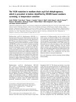
Fig. 3. dJmj localizes to euchromatic regions on polytene chromosomes. (A–I) Polytene chromosomes of third instar larvae from wild-type
(A–C, G–I) and djmj
e03131
mutants (D–F) were immunostained with antibody to dJmj (B, E, H). DNA was counterstained with DAPI (A, D, G).
(C, F, I) Merged images of dJmj and DAPI staining. (G–I) Higher-magnification images of dJmj localization on polytene chromosomes of
another spread. (J–L) Higher magnification of dJmj staining at chromocenter heterochromatin. Polytene chromosomes were coimmuno-
stained with antibodies for HP1 (J) and dJmj (K). (L) Merged image of dJmj and HP1 staining.
N. Sasai et al. Characterization of Drosophila jumonji
FEBS Journal 274 (2007) 6139–6151 ª 2007 The Authors Journal compilation ª 2007 FEBS 6143
died shortly after eclosion (Fig. 5A). Precise excision
of the piggyBac transposon reversed the lethality,
indicating that the transposon insertion was indeed
responsible for the phenotype (data not shown). Hemi-
zygous djmj
e03131
⁄ Df(3L)AC1 animals also exhibited
larval and pupal lethality and displayed similar pheno-
types as homozygous djmj
e03131
mutants (Fig. 5A and
data not shown), confirming that djmj
e03131
is a loss of
function allele of djmj.
Phenotypic characterization of pharate adults
revealed some mutants to have defects in leg elonga-
tion and to show a crooked leg phenotype (Fig. 5D,F).
These phenotypes are similar to those with loss of
function of the genes involved in the ecdysone pathway
[33,34], suggesting the participation of dJmj in ecdy-
sone signaling.
The jmj gene is functionally conserved from flies
to mammals
To investigate whether djmj is a functional homolog of
mammalian jmj, we tested the chromosomal distribu-
tion of mJmj and its ability to rescue the phenotypes
of the djmj mutants. To this end, transgenic flies that
Table 1. The djmj gene is a suppressor of position effect variega-
tion of the T(2;3)Sb
V
rearrangement.
Genotype
Number
of flies
Total
bristles
Number
of Sb Sb (%)
+ ⁄ Sb
V
115 1610 478 29.7
SUV4-20
BG00814
⁄ Sb
V
80 1120 613 54.7
djmj
e03131
⁄ Sb
V
77 1078 567 52.6
Df(3L)AC1 ⁄ Sb
V
92 1288 684 53.1
A
B
C
D
E
F
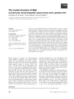
Fig. 4. dJmj is excluded from highly transcribed chromatin regions. (A–F) Polytene chromosomes from wild-type third larvae were stained
with antibodies for dJmj (A, D) and PolII (B, E). Higher-magnification images of dJmj (D) and PolII (E) staining of another spread are also
shown. (C, F) Merged images of dJmj and PolII staining.
A
B
C
D
E
F
Fig. 5. The djmj gene is required for metamorphosis. (A) Lethal phases were determined in animals with the following genotypes: + ⁄ +,
djmj
e03131
and djmj
e03131
⁄ Df(3L)AC1. (B–F) Lethal phenotypes of djmj
e03131
homozygotes. (B) Wild-type control animal 4 days after pupation.
(C, D) djmj
e03131
mutant animals 5 days after pupation. (E, F) djmj
e03131
mutants show a crooked leg phenotype. Third legs dissected from
wild-type (E) and djmj
e03131
pharate adults (F) are shown.
Characterization of Drosophila jumonji N. Sasai et al.
6144 FEBS Journal 274 (2007) 6139–6151 ª 2007 The Authors Journal compilation ª 2007 FEBS
express FLAG-tagged full-length mJmj (FLAG–mJmj)
under the control of the GAL4–UAS system [35] were
established. To minimize the expression of FLAG–
mJmj, the hsp70–GAL4 driver line was used without
heat shock treatment, which results in leaky expression
of FLAG–mJmj that is barely detected by western
blotting with antibody to FLAG (Fig. 6A). Immuno-
staining of polytene chromosomes from FLAG–mJmj-
expressing salivary gland cells detected numerous
euchromatic bands (Fig. 6C,F), whereas no FLAG sig-
nals were detected in chromosomes without hsp70–
GAL4 (Fig. 6H–J). Coimmunostaining of chromo-
somes with antibodies for dJmj (Fig. 6B,E) and FLAG
(Fig. 6C,F) showed that most, but not all, mJmj sites
colocalize with endogenous dJmj (Fig. 6D,G), suggest-
ing that mJmj has similar function as dJmj on chroma-
tin. The number of mJmj-binding sites was much
greater than that for dJmj. This could be due to higher
expression of FLAG–mJmj on transgenic lines as
compared to endogenous dJmj or to stronger affinity
of the antibody for FLAG.
We then expressed mJmj under the background of
djmj
e03131
and investigated the lethal phases of the res-
cued flies (Table 2). As most djmj
e03131
homozygotes
develop to the pupal stage (Fig. 5), third larvae with
the desired genotype were picked up and tested for
their lethal phases and phenotypes during pupal stages.
Of the control flies that contain the either FLAG–mjmj
(line 35) transgene or the hsp70–GAL4 driver under
the background of the djmj mutation, 10.7–14.7% of
pupae showed the abnormal leg phenotype and 0.6–
7.1% of animals eclosed, which is similar to what was
seen with djmj
e03131
homozygous mutants. In contrast,
when mJmj was ubiquitously and modestly expressed
by the hsp70–GAL4 driver, the abnormal leg pheno-
type was restored and 21.2% of rescued animals
eclosed, indicating that mJmj can partially compensate
for loss of djmj. The FLAG–mjmj transgene inserted in
A
B
C
D
E
F
G
H
I
J
Fig. 6. Transgenic mouse Jmj mostly colocalizes with endogenous dJmj. (A) Western blot analysis of FLAG–mJmj expression in larval
extracts with the indicated genotypes using antibody to FLAG (upper). The same blot was reprobed with antibody to tubulin to compare pro-
tein loading (lower). Lane 1: FLAG–mjmj ⁄ +. Lane 2: FLAG–mjmj ⁄ hsp70–GAL4. (B–J) Polytene chromosomes from FLAG–mjmj ⁄ hsp70–GAL4
(B–G) or FLAG–mjmj ⁄ + (H–J) larvae were coimmunostained with antibodies to dJmj (B, E, H) and FLAG (C, F, I). (D, G, J) Merged images of
dJmj and FLAG–mJmj staining. (E–G) Higher-magnification images of each staining.
Table 2. Transgenic mJmj partially rescues the phenotypes of djmj
e03131
mutants.
Genotype
Lethal phase
Early pupa Abnormal leg
a
Late pupa Adult Total
+ ⁄ hsp70–GAL4; djmj
e03131
⁄ djmj
e03131
6 (10.7%) 6 (10.7%) 40 (71.4%) 4 (7.1%) 56
FLAG–mjmj(35) ⁄ + djmj
e03131
⁄ djmj
e03131
13 (8.0%) 24 (14.7%) 127 (77.9%) 1 (0.6%) 163
FLAG–mjmj(19) ⁄ + djmj
e03131
⁄ djmj
e03131
5 (4.9%) 15 (14.7%) 81 (79.4%) 1 (1.0%) 102
FLAG–mjmj(35) ⁄ hsp70–GAL4; djmj
e03131
⁄ djmj
e03131
0 (0.0%) 2 (3.8%) 39 (75.0%) 11 (21.2%) 52
FLAG–mjmj(19) ⁄ hsp70–GAL4; djmj
e03131
⁄ djmj
e03131
0 (0.0%) 2 (4.9%) 37 (90.0%) 2 (4.9%) 41
a
The number of late pupae that show the crooked leg phenotype.
N. Sasai et al. Characterization of Drosophila jumonji
FEBS Journal 274 (2007) 6139–6151 ª 2007 The Authors Journal compilation ª 2007 FEBS 6145
the independent genomic locus (line 19) showed simi-
lar, but less pronounced, effects on the rescue experi-
ment. It is not possible to draw definitive conclusions
regarding the degree to which mJmj can rescue the
djmj mutant phenotype, as we have not yet succeeded
in cloning the full-length cDNA for djmj to make djmj-
expressing flies, due to its large size. However, these
findings strongly suggest the functional conservation of
the jmj gene from flies to mammals.
Discussion
Although the Drosophila genome contains at least 13
genes encoding JmjC domain-containing proteins [11],
little is known about their biological roles and their
contributions to chromatin regulation. In this study,
we showed that a novel JmjC-containing protein, dJmj,
a Drosophila homolog of mammalian Jmj, is associated
with euchromatic sites excluded from highly tran-
scribed regions on polytene chromosomes and is
required for metamorphosis during development.
The mjmj gene appears to be involved in many devel-
opmental pathways, as clarified by analysis of mutant
mice that show various developmental abnormalities
[7,21,22]. In the present study, loss of djmj function
caused lethality during larval and pupal stages (Fig. 5),
indicating that djmj is also important in Drosophila
development. Jmj plays critical roles in suppression of
cellular proliferation via repression of cyclinD1 [24].
However, dJmj is not likely to regulate Drosophila cyc-
linD, as the expression of cyclinD was unchanged in
djmj mutant larvae (Fig. 2F) and in dJmj-depleted Kc
cells (data not shown). It is important to note that,
unlike mammalian D-type cyclin proteins, Drosophila
cyclin D is not required for G
1
⁄ S phase transition but
instead plays a role in cellular growth, whereas cyclin E
plays an essential role in G
1
⁄ S phase progression [36].
However, cyclinE and several other cell cycle-related
genes were not misregulated in djmj mutant larvae
(Fig. 2F and data not shown). Furthermore, dJmj
depletion did not affect cell growth in Kc cells (data not
shown). Therefore, cyclinD repression and subsequent
suppression of cellular proliferation might be a mam-
mal-specific event. However, these data do not rule out
the possibility that dJmj might repress cyclinD expres-
sion in restricted tissues, which would not be detected
by expression analysis of extracts of whole animals. In
addition, although relatively high expression of dJmj
was observed during embryonic stages (Fig. 2B), it
remains unclear whether dJmj is required for the repres-
sion of cell cycle regulators during early development,
as maternally deposited dJmj protein might contribute
to embryogenesis in djmj mutants. Further studies are
required to investigate the involvement of dJmj in cell
cycle regulation during early embryonic development.
The detailed mechanism by which Jmj represses tran-
scription remains to be clarified. Although it has been
shown to counteract the function of DNA-binding tran-
scription factors [25,26], Jmj directly binds to the
cyclinD1 promoter to repress its expression [24]. As
our data do not show direct evidence that dJmj has a
transcriptional repression activity, we cannot conclude
that dJmj is indeed a transcriptional repressor like
mammalian Jmj. However, the observation that dJmj
localizes on specific chromatin domains excluded from
PolII sites on polytene chromosomes suggests that dJmj
mediates transcriptional repression through modifica-
tion of chromatin. In addition, djmj is not likely to
affect global modification of histone tails that are
associated with transcriptional activity (supplementary
Fig. S1). Therefore, our findings suggest that dJmj is
involved in the regulation of specific target genes at spe-
cific chromosomal loci in response to developmental
signals rather than acting as a global regulator of chro-
matin.
The finding that the phenotypes of djmj mutants
resemble those of Drosophila lacking ecdysone-regu-
lated genes [33,34] suggests the involvement of dJmj in
the ecdysone pathway. Expression of early and late
puff genes are regulated in a direct or indirect manner
by a subset of chromatin-modifying proteins, including
NURF, p66, dGcn5, dAda2a, Bonus, Rpd3 and dG9a
[37–43]. In addition, one property of JmjC-containing
proteins is to associate with chromatin modification
enzymes, such as the NCoR corepressor and histone
deacetylase (HDACs) [8,44,45]. Investigation of
whether dJmj links with these proteins to control
metamorphosis is clearly warranted. The possible inter-
action domain of dJmj for these factors is the N-termi-
nal repression domain, which is evolutionarily
conserved among Jmj proteins (Fig. 1). Detailed analy-
sis of the role of N-terminal and the JmjC domains in
dJmj function may provide clues with which to address
these issues.
Several studies have clarified that JmjC-containing
proteins act as histone demethylases [11]. Lid, the clos-
est protein to dJmj, was recently shown to be a histone
demethylase that removes dimethyl and trimethyl K4
of H3 [46–48]. Although our results showed that the
mutation in the djmj gene does not affect global modi-
fication of histone tails, including dimethyl K4 of H3
(supplementary Fig. S1), we cannot rule out the possi-
bility that dJmj might demethylate histones at specific
chromosomal loci or target a nonhistone protein as
a substrate. However, importantly, both mammalian
and Drosophila Jmj proteins are predicted to be
Characterization of Drosophila jumonji N. Sasai et al.
6146 FEBS Journal 274 (2007) 6139–6151 ª 2007 The Authors Journal compilation ª 2007 FEBS
catalytically inactive as histone demethylases because
of the amino acid changes in the catalytic domain
[11,12]. Several other JmjC-containing proteins are
considered to be enzymatically inactive as histone
demethylases [11]. Epe1 has been shown to counteract
heterochromatin formation by interacting with Swi6, a
yeast homolog of HP1. This event requires an enzy-
matically inactive JmjC domain, suggesting a novel
function of the JmjC domain of Epe1 in heterochro-
matin formation [49]. As the JmjC domain is also
found in bacteria, it might have diverse functions, and
its analysis in dJmj should provide novel insights.
Despite the finding of djmj as a suppressor of PEV,
the detailed roles of dJmj in chromatin organization
remain unclear. Several different genes are reported to
similarly act as suppressors, including Su(var)2-5,
Su(var)3-7 and Su(var)3-9, which encode structural
components of heterochromatin localizing to chromo-
center heterochromatin [50,51], and Z4, which encodes
a zinc finger protein that localizes to interbands of
euchromatin and regulates chromatin organization at
band–interband boundaries [52]. In addition, JIL-1 his-
tone kinase functions to maintain euchromatic regions
via antagonizing heterochromatinization by Su(var)3-9
[53,54]. On polytene chromosomes, dJmj signals were
excluded from chromocenter heterochromatin, and het-
erochromatin components, including dimethyl K9-H3
and HP1, were not altered by loss of dJmj (data not
shown). In addition, dJmj does not affect PEV of the
white
m4
rearrangement (data not shown). Taken
together, these findings strongly suggest that dJmj is
not a structural element in heterochromatin and acts
at particular domains rather than functioning as a gen-
eral modifier of chromatin.
In conclusion, our data suggest that dJmj plays
important roles during metamorphosis by regulating
gene expression in response to developmental signals.
As mJmj shows similar distributions to dJmj on poly-
tene chromosomes (Fig. 6) and partially rescues the
phenotypes of djmj mutants (Table 2), the Drosophila
system could be a powerful tool with which to analyze
Jmj functions in chromatin regulation and development.
Experimental procedures
Fly stocks
Fly stocks were raised at 25 °C on standard medium.
Canton-S was used as the wild-type strain. The piggy-
Bac-inserted djmj
e03131
⁄ TM6B fly was obtained from the
Harvard stock center [29], and djmj
EY02717
, Df(3L)AC1
rn
roe-1
p
p
⁄ TM3, SUV4-20
BG00814
and T(2;3)Sb
V
, In(3R)Mo,
Sb
1
,sr
1
⁄ TM3Ser flies were from the Bloomington stock
center. The hsp70–GAL4 ⁄ CyO and white
m4
flies were
obtained from the Drosophila Genetic Resource Center at
Kyoto Institute of Technology.
Lethal phase analysis and phenotypic
characterization
The djmj
e03131
and Df(3L)AC1 alleles were rebalanced with
TM6BGFP and TM3GFP balancer chromosomes, respec-
tively. Lethal phase analysis and phenotypic characteriza-
tion were performed as previously described [34].
Generation of transgenic flies and rescue
experiment
For constructing the pUAST–FLAG–mjmj vector, a cDNA
for FLAG–mjmj in pBluescript was digested with Cla I,
blunt-ended and inserted into the pUAST vector [35], which
was blunt-ended after EcoRI digestion. Transgenic fly lines
were generated as described previously [55,56], and three
independent fly lines carrying the transgene on the second
chromosome were established. The GAL4–UAS system [35]
was used for ubiquitous expression of FLAG–mJmj using
the hsp70–GAL4 driver.
For the rescue experiment, FLAG–mjmj (line 35),
djmj
e03131
⁄ TM6B or FLAG–mjmj (line 19) ⁄ CyOGFP,
djmj
e03131
⁄ TM6B females were crossed with hsp70–
GAL4 ⁄ CyOGFP, djmj
e03131
⁄ TM6B males at 25 °C. As con-
trol crosses, djmj
e03131
⁄ TM6BGFP females and males were
mated with hsp70–GAL4 ⁄ CyOGFP, djmj
e03131
⁄ TM6B males
and FLAG-mjmj ⁄ (CyOGFP), djmj
e03131
⁄ TM6B females,
respectively. Nontubby and nonfluorescent third larvae
were picked up, and their lethal phases and phenotypes
during pupal development were analyzed.
PEV analysis
To examine the effect of djmj on the white
m4
variegation,
w
m4
⁄ w
m4
females were crossed with w ⁄ Y, djmj
e03131
⁄ TM6B
males, and the eyes of w
m4
⁄ Y, djmj
e03131
⁄ +males were scored
and compared with those of w
m4
⁄ Y, TM6B ⁄ +males. The
effect of djmj on the Sb
V
variegation was studied by crossing
SUV4-20
BG00814
, djmj
e03131
⁄ TM6B, Df(3L)AC1 ⁄ TM3Ser-
GFP or Canton S females with T(2;3)Sb
V
⁄ TM3Ser males
[31], and 14 defined bristles were scored as being wild type or
Sb. Male and female scores were combined because no differ-
ences between sexes were observed.
Production of polyclonal antibody to dJmj
To construct an expression vector for the glutathione
S-transferase (GST)-fused C-terminal region of the dJmj
protein (dJmjC, amino acids 1635–2351), the djmj cDNA
fragment was inserted into the SalI and NotI sites of
N. Sasai et al. Characterization of Drosophila jumonji
FEBS Journal 274 (2007) 6139–6151 ª 2007 The Authors Journal compilation ª 2007 FEBS 6147
the pGEX4T-1 vector. GST–dJmjC was expressed in the
bacterial strain BL-21(DE3), affinity purified with a glutathi-
one Sepharose column (GE Healthcare, Little Chalfont,
UK), and injected into rabbits. The antiserum generated
was applied to GST-conjugated sepharose, and this was fol-
lowed by purification with GST–dJmj-conjugated sepharose.
Cell culture and knockdown experiments
Kc cells were cultured at 25 °C in M3 medium (Sigma, St
Louis, MO, USA) supplemented with 2% fetal bovine
serum. For dsRNA production, a 621 bp fragment spanning
from nucleotide 6485 to the 3¢-UTR (40 bp downstream of
the stop codon) of djmj were amplified using 5¢-CAC
GGGCGTATACCTCAAGC-3¢ and 5¢-TGTGCCTGA
ATCTTTCGTGC-3¢ primers and cloned into the pGEM-T
vector. Sense and antisense RNAs were synthesized in vitro
and annealed. For knockdown experiments, 1 · 10
6
cells
were plated on 6 cm dishes and transfected with 10 lgof
dsRNA using cellfectin transfection reagent (Invitrogen,
Carlsbad, CA, USA) according to the manufacturer’s proto-
col. The cells were collected, directly suspended in SDS sam-
ple buffer, and subjected to western blotting.
Western blotting
Protein extracts were prepared by homogenization of ani-
mals in ice-cold SDS sample buffer followed by boiling for
5 min. After centrifugation at 12 000 g for 10 min at 4 °C,
protein samples were separated by SDS ⁄ PAGE and trans-
ferred to poly(vinylidene difluoride) membranes (Millipore,
Billerica, MA, USA). Antibodies used were anti-dJmj
(1 : 2000), anti-a-tubulin (1 : 5000, Sigma), anti-FLAG
(M2, 1 : 2000; Sigma), anti-acetyl H3 (06–599, 1 : 5000),
anti-dimethyl K4-H3 (07-030, 1 : 2000), anti-monometh-
yl K9-H3 (07–450, 1 : 1000), anti-dimethyl K9-H3 (07–212,
1 : 1000), and anti-trimethyl K27-H3 (07–449, 1 : 1000)
from Upstate (Lake Placid, NY, USA), and anti-H3
(1 : 1,000; Cell Signaling, Danvers, MA, USA). Horseradish
peroxidase-conjugated anti-rabbit and anti-mouse IgGs
(GE Healthcare) were used as secondary antibodies, and
proteins were detected with ECL-plus (GE Healthcare).
Immunostaining of polytene chromosomes and
whole salivary glands
For immunostaining of polytene chromosomes, salivary
glands from wandering third instar larvae were dissected in
0.7% NaCl, fixed for 5 min, and squashed in 45% acetic
acid ⁄ 3.7% formaldehyde. The slides were frozen in liquid
nitrogen and were then blocked in blocking buffer (5%
skimmed milk in NaCl ⁄ P
i
⁄ 0.1% Triton X-100) for 1 h at
25 °C. Slides were incubated with primary antibodies for
16 h at 4 °C. The antibodies used were anti-dJmj (1 : 400),
anti-FLAG (M2, 1 : 5,000; Sigma), anti-PolII (H-14, 1 : 100;
Covance, Princeton, NJ, USA) and anti-HP1 (C1A9, 1 : 100;
Developmental Studies Hybridoma Bank at the University
of Iowa). After being washed with NaCl ⁄ P
i
⁄ 0.1% Triton X-
100 twice for 15 min each, the slides were incubated with
Alexa-488-conjugated anti-rabbit IgG, Alexa-488-conjugated
anti-mouse IgM, or Alexa-594-conjugated anti-mouse IgG
or anti-rabbit IgG (1 : 400) from Invitrogen for 2 h at 25 °C.
DNA was visualized with DAPI. Preparations were mounted
in FluoroGuard Antifade Reagent (Bio-Rad, Hercules, CA,
USA), and images were obtained using an Olympus (Tokyo,
Japan) BX-50 microscope equipped with a cooled CCD cam-
era. Each staining experiment was performed at least three
times, and representative spreads are shown.
For immunostaining of whole salivary glands, dissected
glands were fixed in 4% formaldehyde ⁄ 0.15% Triton X-100
for 20 min on ice. After blocking in NaCl ⁄ P
i
containing
2% goat serum and 0.15% Triton X-100 for 30 min at
25 °C, the glands were incubated with antibody to dJmj
(1 : 400) for 16 h at 4 °C, and this was followed by incuba-
tion with Alexa-488-conjugated anti-rabbit IgG (1 : 400)
for 2 h at 25 °C. DNA was stained with DAPI.
Semiquantitative RT-PCR
Total RNA was extracted with Sepasol RNA I (Nacalai,
Kyoto, Japan). First-strand cDNA was synthesized using
oligo(dT)
20
and Superscript III reverse transcriptase (Invi-
trogen). PCR reactions were performed over a range of
cDNA dilutions to ensure exponential amplification. Primer
sequences used were as follows: cycD-F, 5¢-GGGATCCCA
CATTGTATTCG-3¢; cycD-R, 5¢-ACGGAGCTTTGAAG
CCAGTA-3¢; cycE-F, 5¢-AAGGTGCAGAAGACGCA
CTT-3¢; cycE-R, 5¢-AATCACCTGCCAATCCAGAC-3¢;
cdk4-F, 5¢-TACAACAGCACCGTGGACAT-3¢; cdk4-R,
5¢-TGGGCATCGAGACTATAGGG-3¢; rp49-F, 5¢-CGG
ATCGATATGCTAAGCTG-3¢; and rp49-R, 5¢-GAACG
CAGGCGACCGTTGGGG-3¢.
Acknowledgements
We would like to thank Haruki Shirato for providing
the FLAG–mjmj plasmid and members of the Yamagu-
chi laboratory for helpful comments and advice. We
also acknowledge the contribution of Malcolm Moore
in critical reading of the manuscript. This work was
supported in part by grants-in-aid from the Ministry
of Education, Sciences, Sports and Culture of Japan.
References
1 Luger K, Mader AW, Richmond RK, Sargent DF &
Richmond TJ (1997) Crystal structure of the nucleo-
Characterization of Drosophila jumonji N. Sasai et al.
6148 FEBS Journal 274 (2007) 6139–6151 ª 2007 The Authors Journal compilation ª 2007 FEBS
some core particle at 2.8 A resolution. Nature 389 ,
251–260.
2 Jenuwein T & Allis CD (2001) Translating the histone
code. Science 293, 1074–1080.
3 Lachner M, O’Carroll D, Rea S, Mechtler K & Jenuw-
ein T (2001) Methylation of histone H3 lysine 9 creates
a binding site for HP1 proteins. Nature 410, 116–120.
4 Lachner M, O’Sullivan RJ & Jenuwein T (2003) An epi-
genetic road map for histone lysine methylation. J Cell
Sci 116, 2117–2124.
5 Balciunas D & Ronne H (2000) Evidence of domain
swapping within the jumonji family of transcription
factors. Trends Biochem Sci 25, 274–276.
6 Clissold PM & Ponting CP (2001) JmjC: cupin metal-
loenzyme-like domains in jumonji, hairless and phos-
pholipase A2beta. Trends Biochem Sci 26, 7–9.
7 Takeuchi T, Yamazaki Y, Katoh-Fukui Y, Tsuchiya R,
Kondo S, Motoyama J & Higashinakagawa T (1995)
Gene trap capture of a novel mouse gene, jumonji, requi-
red for neural tube formation. Genes Dev 9, 1211–1222.
8 Ahmed S, Palermo C, Wan S & Walworth NC (2004) A
novel protein with similarities to Rb binding protein 2
compensates for loss of Chk1 function and affects his-
tone modification in fission yeast. Mol Cell Biol 24,
3660–3669.
9 Ayoub N, Noma K, Isaac S, Kahan T, Grewal SI &
Cohen A (2003) A novel jmjC domain protein modu-
lates heterochromatization in fission yeast. Mol Cell Biol
23, 4356–4370.
10 Benevolenskaya EV, Murray HL, Branton P, Young
RA & Kaelin WG Jr (2005) Binding of pRB to the
PHD protein RBP2 promotes cellular differentiation.
Mol Cell 18, 623–635.
11 Klose RJ, Kallin EM & Zhang Y (2006) JmjC-domain-
containing proteins and histone demethylation. Nat Rev
Genet 7, 715–727.
12 Takeuchi T, Watanabe Y, Takano-Shimizu T & Kondo
S (2006) Roles of jumonji and jumonji family genes in
chromatin regulation and development. Dev Dyn 235,
2449–2459.
13 Wilsker D, Patsialou A, Dallas PB & Moran E (2002)
ARID proteins: a diverse family of DNA binding pro-
teins implicated in the control of cell growth, differenti-
ation, and development. Cell Growth Differ 13, 95–106.
14 Tsukada Y, Fang J, Erdjument-Bromage H, Warren
ME, Borchers CH, Tempst P & Zhang Y (2006)
Histone demethylation by a family of JmjC domain-
containing proteins. Nature 439, 811–816.
15 Klose RJ, Yamane K, Bae Y, Zhang D, Erdjument-
Bromage H, Tempst P, Wong J & Zhang Y (2006) The
transcriptional repressor JHDM3A demethylates tri-
methyl histone H3 lysine 9 and lysine 36. Nature 442,
312–316.
16 Yamane K, Toumazou C, Tsukada Y, Erdjument-
Bromage H, Tempst P, Wong J & Zhang Y (2006)
JHDM2A, a JmjC-containing H3K9 demethylase, facili-
tates transcription activation by androgen receptor. Cell
125, 483–495.
17 Fodor BD, Kubicek S, Yonezawa M, O’Sullivan RJ,
Sengupta R, Perez-Burgos L, Opravil S, Mechtler K,
Schotta G & Jenuwein T (2006) Jmjd2b antagonizes
H3K9 trimethylation at pericentric heterochromatin in
mammalian cells. Genes Dev 20, 1557–1562.
18 Cloos PA, Christensen J, Agger K, Maiolica A, Rapp-
silber J, Antal T, Hansen KH & Helin K (2006) The
putative oncogene GASC1 demethylates tri- and dime-
thylated lysine 9 on histone H3. Nature 442, 307–311.
19 Whetstine JR, Nottke A, Lan F, Huarte M, Smolikov
S, Chen Z, Spooner E, Li E, Zhang G, Colaiacovo M
et al. (2006) Reversal of histone lysine trimethylation by
the JMJD2 family of histone demethylases. Cell 125,
467–481.
20 Trewick SC, McLaughlin PJ & Allshire RC (2005) Meth-
ylation: lost in hydroxylation? EMBO Rep 6, 315–320.
21 Motoyama J, Kitajima K, Kojima M, Kondo S &
Takeuchi T (1997) Organogenesis of the liver, thymus
and spleen is affected in jumonji mutant mice. Mech
Dev 66, 27–37.
22 Kitajima K, Kojima M, Nakajima K, Kondo S, Hara
T, Miyajima A & Takeuchi T (1999) Definitive but not
primitive hematopoiesis is impaired in jumonji mutant
mice. Blood 93, 87–95.
23 Kim TG, Kraus JC, Chen J & Lee Y (2003) JUMONJI,
a critical factor for cardiac development, functions as a
transcriptional repressor. J Biol Chem 278, 42247–
42255.
24 Toyoda M, Shirato H, Nakajima K, Kojima M, Takah-
ashi M, Kubota M, Suzuki-Migishima R, Motegi Y,
Yokoyama M & Takeuchi T (2003) Jumonji downregu-
lates cardiac cell proliferation by repressing cyclin D1
expression. Dev Cell 5, 85–97.
25 Jung J, Kim TG, Lyons GE, Kim HR & Lee Y (2005)
Jumonji regulates cardiomyocyte proliferation via inter-
action with retinoblastoma protein. J Biol Chem 280,
30916–30923.
26 Kim TG, Chen J, Sadoshima J & Lee Y (2004) Jumonji
represses atrial natriuretic factor gene expression by
inhibiting transcriptional activities of cardiac transcrip-
tion factors. Mol Cell Biol 24, 10151–10160.
27 Gildea JJ, Lopez R & Shearn A (2000) A screen for
new trithorax group genes identified little imaginal discs,
the Drosophila melanogaster homologue of human reti-
noblastoma binding protein 2. Genetics 156, 645–663.
28 Bellen HJ, Levis RW, Liao G, He Y, Carlson JW,
Tsang G, Evans-Holm M, Hiesinger PR, Schulze KL,
Rubin GM et al. (2004) The BDGP gene disruption
project: single transposon insertions associated with
40% of Drosophila genes. Genetics 167, 761–781.
29 Thibault ST, Singer MA, Miyazaki WY, Milash B,
Dompe NA, Singh CM, Buchholz R, Demsky M,
N. Sasai et al. Characterization of Drosophila jumonji
FEBS Journal 274 (2007) 6139–6151 ª 2007 The Authors Journal compilation ª 2007 FEBS 6149
Fawcett R, Francis-Lang HL et al. (2004) A comple-
mentary transposon tool kit for Drosophila melanogas-
ter using P and piggyBac. Nat Genet 36, 283–287.
30 Finley RL Jr, Thomas BJ, Zipursky SL & Brent R
(1996) Isolation of Drosophila cyclin D, a protein
expressed in the morphogenetic furrow before entry into
S phase. Proc Natl Acad Sci USA 93, 3011–3015.
31 Sinclair DAR, Mottus RC & Grigliatti TA (1983)
Genes which suppress position effect variegation in
Drosophila melanogaster are clustered. Mol Gen Genet
191, 326–333.
32 Schotta G, Lachner M, Sarma K, Ebert A, Sengupta R,
Reuter G, Reinberg D & Jenuwein T (2004) A silencing
pathway to induce H3-K9 and H4-K20 trimethylation at
constitutive heterochromatin. Genes Dev 18, 1251–1262.
33 Beckstead RB, Lam G & Thummel CS (2005) The
genomic response to 20-hydroxyecdysone at the onset
of Drosophila metamorphosis. Genome Biol 6,
doi: 10.1186/gb-2005-6-12-r99
34 D’Avino PP & Thummel CS (1998) Crooked legs
encodes a family of zinc finger proteins required for leg
morphogenesis and ecdysone-regulated gene expression
during Drosophila metamorphosis. Development 125,
1733–1745.
35 Brand AH & Perrimon N (1993) Targeted gene
expression as a means of altering cell fates and
generating dominant phenotypes. Development 118,
401–415.
36 Datar SA, Jacobs HW, de la Cruz AF, Lehner CF &
Edgar BA (2000) The Drosophila cyclin D–Cdk4 com-
plex promotes cellular growth. EMBO J 19, 4543–4554.
37 Badenhorst P, Xiao H, Cherbas L, Kwon SY, Voas M,
Rebay I, Cherbas P & Wu C (2005) The Drosophila
nucleosome remodeling factor NURF is required for
Ecdysteroid signaling and metamorphosis. Genes Dev
19, 2540–2545.
38 Beckstead R, Ortiz JA, Sanchez C, Prokopenko SN,
Chambon P, Losson R & Bellen HJ (2001) Bonus, a
Drosophila homolog of TIF1 proteins, interacts with
nuclear receptors and can inhibit betaFTZ-F1-depen-
dent transcription. Mol Cell 7, 753–765.
39 Kon C, Cadigan KM, da Silva SL & Nusse R (2005)
Developmental roles of the Mi-2 ⁄ NURD-associated
protein p66 in Drosophila. Genetics 169, 2087–2100.
40 Carre C, Szymczak D, Pidoux J & Antoniewski C
(2005) The histone H3 acetylase dGcn5 is a key player
in Drosophila melanogaster metamorphosis. Mol Cell
Biol 25, 8228–8238.
41 Ciurciu A, Komonyi O, Pankotai T & Boros IM (2006)
The Drosophila histone acetyltransferase Gcn5 and tran-
scriptional adaptor Ada2a are involved in nucleosomal
histone H4 acetylation. Mol Cell Biol 26, 9413–9423.
42 Stabell M, Eskeland R, Bjorkmo M, Larsson J, Aalen
RB, Imhof A & Lambertsson A (2006) The Drosophila
G9a gene encodes a multi-catalytic histone methyltrans-
ferase required for normal development. Nucleic Acids
Res 34, 4609–4621.
43 Pile LA & Wassarman DA (2000) Chromosomal locali-
zation links the SIN3–RPD3 complex to the regulation
of chromatin condensation, histone acetylation and gene
expression. EMBO J 19, 6131–6140.
44 Gray SG, Iglesias AH, Lizcano F, Villanueva R, Ca-
melo S, Jingu H, Teh BT, Koibuchi N, Chin WW, Kok-
kotou E et al. (2005) Functional characterization of
JMJD2A, a histone deacetylase- and retinoblastoma-
binding protein. J Biol Chem 280
, 28507–28518.
45 Zhang D, Yoon HG & Wong J (2005) JMJD2A is a
novel N-CoR-interacting protein and is involved in
repression of the human transcription factor achaete
scute-like homologue 2 (ASCL2 ⁄ Hash2). Mol Cell Biol
25, 6404–6414.
46 Eissenberg JC, Lee MG, Schneider J, Ilvarsonn A,
Shiekhattar R & Shilatifard A (2007) The trithorax-
group gene in Drosophila little imaginal discs encodes a
trimethylated histone H3 Lys4 demethylase. Nat Struct
Mol Biol 14, 344–346.
47 Lee N, Zhang J, Klose RJ, Erdjument-Bromage H,
Tempst P, Jones RS & Zhang Y (2007) The trithorax-
group protein Lid is a histone H3 trimethyl-Lys4
demethylase. Nat Struct Mol Biol 14, 341–343.
48 Secombe J, Li L, Carlos L & Eisenman RN (2007) The
Trithorax group protein Lid is a trimethyl histone
H3K4 demethylase required for dMyc-induced cell
growth. Genes Dev 21, 537–551.
49 Zofall M & Grewal SI (2006) Swi6 ⁄ HP1 recruits a JmjC
domain protein to facilitate transcription of heterochro-
matic repeats. Mol Cell 22, 681–692.
50 Schotta G, Ebert A, Krauss V, Fischer A, Hoffmann J,
Rea S, Jenuwein T, Dorn R & Reuter G (2002) Central
role of Drosophila SU(VAR)3-9 in histone H3-K9
methylation and heterochromatic gene silencing. EMBO
J 21, 1121–1131.
51 Delattre M, Spierer A, Tonka CH & Spierer P (2000)
The genomic silencing of position-effect variegation in
Drosophila melanogaster: interaction between the het-
erochromatin-associated proteins Su(var)3–7 and HP1.
J Cell Sci 113, 4253–4261.
52 Eggert H, Gortchakov A & Saumweber H (2004) Identi-
fication of the Drosophila interband-specific protein Z4
as a DNA-binding zinc-finger protein determining chro-
mosomal structure. J Cell Sci 117, 4253–4264.
53 Ebert A, Schotta G, Lein S, Kubicek S, Krauss V,
Jenuwein T & Reuter G (2004) Su(var) genes regulate
the balance between euchromatin and heterochromatin
in Drosophila. Genes Dev 18, 2973–2983.
54 Zhang W, Deng H, Bao X, Lerach S, Girton J, Johan-
sen J & Johansen KM (2006) The JIL-1 histone H3S10
kinase regulates dimethyl H3K9 modifications and het-
erochromatic spreading in Drosophila. Development
133, 229–235.
Characterization of Drosophila jumonji N. Sasai et al.
6150 FEBS Journal 274 (2007) 6139–6151 ª 2007 The Authors Journal compilation ª 2007 FEBS
55 Robertson HM, Preston CR, Phillis RW, Johnson-
Schlitz DM, Benz WK & Engels WR (1988) A stable
genomic source of P element transposase in Drosoph-
ila melanogaster. Genetics 118, 461–470.
56 Spradling AC (1986) P element-mediated transforma-
tion. In Drosophila: a Practical Approach (Roberts DB,
ed.), pp. 175–197. IRL Press, Oxford.
Supplementary material
The following supplementary material is available
online:
Fig. S1. dJmj is not required for global modification of
histone tails. Protein extracts from third instar larvae
of wild-type and djmje
03131
mutants were subjected to
western blotting with antibodies for modified histones.
Antibody to H3 was used as a loading control.
This material is available as part of the online article
from
Please note: Blackwell Publishing is not responsible
for the content or functionality of any supplementary
materials supplied by the authors. Any queries (other
than missing material) should be directed to the corre-
sponding author for the article.
N. Sasai et al. Characterization of Drosophila jumonji
FEBS Journal 274 (2007) 6139–6151 ª 2007 The Authors Journal compilation ª 2007 FEBS 6151