Báo cáo khoa học: The complex of the insect LDL receptor homolog, lipophorin receptor, LpR, and its lipoprotein ligand does not dissociate under endosomal conditions pot
Bạn đang xem bản rút gọn của tài liệu. Xem và tải ngay bản đầy đủ của tài liệu tại đây (517.22 KB, 16 trang )
The complex of the insect LDL receptor homolog,
lipophorin receptor, LpR, and its lipoprotein ligand does
not dissociate under endosomal conditions
Sigrid D. Roosendaal
1
, Jana Kerver
1
, Maria Schipper
2
, Kees W. Rodenburg
1
and
Dick J. Van der Horst
1
1 Division of Endocrinology and Metabolism, Department of Biology, Institute of Biomembranes, Utrecht University, the Netherlands
2 Department of Biology, Centre for Biostatistics, Utrecht University, the Netherlands
Lipoproteins are used to transport lipids in the circula-
tion of vertebrates as well as invertebrates. Whereas
mammals employ an array of different lipoproteins,
insects rely on one single multifunctional lipoprotein,
high-density lipophorin (HDLp), which is synthesized
in the fat body and released into the blood (hemo-
lymph). In addition to lipid, the particle harbors
two nonexchangeable apolipoproteins, apolipophorin I
Keywords
acidic pH; apolipophorin; calcium; endocytic
recycling compartment; ligand recycling
Correspondence
K. W. Rodenburg, Division of Endocrinology
and Metabolism, Department of Biology and
Institute of Biomembranes, Utrecht
University, NL-CH Utrecht, the Netherlands
Fax: +31 30 253 2837
Tel: +31 30 253 9331
E-mail:
(Received 26 December 2007, revised 10
February 2008, accepted 12 February 2008)
doi:10.1111/j.1742-4658.2008.06334.x
The insect low-density lipoprotein (LDL) receptor (LDLR) homolog, lipo-
phorin receptor (LpR), mediates endocytic uptake of the single insect lipo-
protein, high-density lipophorin (HDLp), which is structurally related to
LDL. However, in contrast to the fate of LDL, which is endocytosed by
LDLR, we previously demonstrated that after endocytosis, HDLp is sorted
to the endocytic recycling compartment and recycled for resecretion in a
transferrin-like manner. This means that the integrity of the complex
between HDLp and LpR is retained under endosomal conditions. There-
fore, in this study, the ligand-binding and ligand-dissociation capacities of
LpR were investigated by employing a new flow cytometric assay, using
LDLR as a control. At pH 5.4, the LpR–HDLp complex remained stable,
whereas that of LDLR and LDL dissociated. Hybrid HDLp-binding recep-
tors, containing either the b-propeller or both the b-propeller and the hinge
region of LDLR, appeared to be unable to release ligand at endosomal
pH, revealing that the stability of the complex is imparted by the ligand-
binding domain of LpR. The LpR–HDLp complex additionally appeared
to be EDTA-resistant, excluding a low Ca
2+
concentration in the endo-
some as an alternative trigger for complex dissociation. From binding of
HDLp to the above hybrid receptors, it was inferred that the stability
upon EDTA treatment is confined to LDLR type A (LA) ligand-binding
repeats 1–7. Additional (competition) binding experiments indicated that
the binding site of LpR for HDLp most likely involves LA-2–7. It is there-
fore proposed that the remarkable stability of the LpR–HDLp complex is
attributable to this binding site. Together, these data indicate that LpR
and HDLp travel in complex to the endocytic recycling compartment,
which constitutes a key determinant for ligand recycling by LpR.
Abbreviations
apoLp-I, apolipophorin I; apoLp-II, apolipophorin II; CHO, Chinese hamster ovary; EGF, epidermal growth factor; ERC, endocytic recycling
compartment; FACS, fluorescence-activated cell sorter; FITC, fluorescein isothiocyanate; HDLp, high-density lipophorin; LA, low-density
lipoprotein receptor type A; LDL, low-density lipoprotein; LDLR, low-density lipoprotein receptor; LpR, lipophorin receptor; LRP, low-density
lipoprotein receptor-related protein; OG, Oregon green; PCSK9, proprotein convertase subtilisin type 9; R1, region 1; RAP, receptor-
associated protein.
FEBS Journal 275 (2008) 1751–1766 ª 2008 The Authors Journal compilation ª 2008 FEBS 1751
(apoLp-I) and apolipophorin II (apoLp-II), which are
derived from the post-translational cleavage of their
common precursor, apoLp-II ⁄ I [1,2]. ApoLp-II ⁄ I was
demonstrated to be a homolog of apolipoprotein
B-100 [3,4], the nonexchangeable apolipoprotein of
mammalian lipoproteins such as very-low-density lipo-
protein and low-density lipoprotein (LDL) [5,6].
Despite this homology, HDLp appears to function
differentially from these mammalian lipoproteins, as
upon conversion of very-low-density lipoprotein to
LDL, the latter is endocytosed by the LDL receptor
(LDLR) and subsequently lysosomally degraded [7],
whereas the insect lipoprotein iteratively loads and
unloads lipid at various target tissues without being
internalized or degraded, and thus acts as a reusable
shuttle [8–11]. However, in apparent contrast to this
concept of HDLp as a reusable shuttle, receptor-medi-
ated endocytic uptake of HDLp was discovered in fat
body tissue of larval and young adult locusts [12], and
was shown to be mediated by an LDLR homolog [13].
This first lipophorin receptor (LpR) was molecularly
and functionally characterized [13–18], and since then
the LpR sequences of several other insect species have
been reported [19–24]. Sequence analysis showed that
LpR is a classic LDLR family member, encompassing
all the typical domains in an LDLR-like sequential
manner [13]: (a) a ligand-binding domain consisting of
LDLR type A (LA) repeats; (b) an epidermal growth
factor (EGF) precursor homology domain composed
of two EGF repeats (EGF-A and EGF-B), a b-propel-
ler containing YWTD repeats, and a third EGF repeat
(EGF-C); (c) an O-linked glycosylation domain; (d) a
transmembrane domain; and (e) an intracellular C-ter-
minal domain [25]. Three-dimensional models of the
elements representing the ligand-binding domain and
EGF precursor homology domain of locust LpR bear
a striking resemblance to those of mammalian LDLR
[10]. On the other hand, the ligand-binding domain of
LpR contains one additional LA repeat as compared
to the cluster of seven repeats in LDLR [13,14], and
despite their pronounced structural similarity, the spec-
ificity of LpR and LDLR for their ligands (HDLp and
LDL, respectively) is mutually exclusive [14].
Remarkably, however, when the functioning of LpR
was compared directly with that of LDLR in a mam-
malian cell line [Chinese hamster ovary (CHO) cells
transfected with LpR], the insect lipoprotein, in con-
trast to LDL, was shown to remain colocalized with
its receptor and was targeted to the endocytic recycling
compartment (ERC). From the latter compartment,
HDLp is resecreted, following a recycling pathway
similar to that of transferrin [14]. In the insect system,
LpR appeared to function similarly. Although an
insect fat body cell line is not available, HDLp inter-
nalized by fat body tissue from young adult locusts
endogenously expressing LpR is also resecreted, consis-
tent with the above concept of ligand recycling in
LpR-transfected CHO cells [12,18]. Trafficking of
ligand to the ERC constitutes a highly unusual prop-
erty among LDLR family members, and in addition,
LDLR mutants that remain in complex with LDL are
targeted to lysosomes [17,26].
During LDLR-mediated endocytosis of LDL, the
receptor–ligand complex ends up in early endosomes
that have a lumenal pH of 6–6.5 [27]. At this acidic
pH, the ectodomain of LDLR, composed of the
ligand-binding domain, EGF precursor homology
domain and glycosylation domain, is proposed to
undergo a conformational change, resulting in the
release of bound LDL. In this model, the ligand-bind-
ing domain is hypothesized to fold onto the b-propel-
ler after protonation of His residues located at the
interface of LA-4, LA-5 and the b-propeller [28],
whereas other residues at this interface, i.e. Gln540,
Glu581, and Lys582, are important for docking of the
ligand-binding domain onto the b-propeller [29]. In
addition, the linker between LA-7 and EGF-A was
demonstrated to constitute a rigid structure stabilized
by a cluster of hydrophobic residues that includes
Phe261, Val274, and Ile313 [29]. Because of the rigidity
of this linker as well as that between EGF-A and
EGF-B, the three repeats serve as a rigid scaffold, pro-
viding a favorable overall topology that permits the
ligand-binding domain to fold over the b-propeller
[29,30]. As a result of this conformational change, the
b-propeller displaces bound LDL [28,31]. In addition
to the low pH, there is a drop in Ca
2+
concentration
in the endosome [32], which is predicted to destabilize
the Ca
2+
-binding properties of the LA repeats and of
EGF-A and EGF-B, and thus might additionally con-
tribute to the pH-dependent ligand release [25,33–35].
In the LA repeats, which consist of approximately
40 amino acids and are organized in a two-loop con-
formation stabilized by three disulfide bonds, a Ca
2+
is chelated by a conserved stretch of acidic amino acids
(DCxDxSDE) and is essential to stabilize the C-termi-
nal fold of the repeat [34,36–39]. Consequently,
removal of Ca
2+
abolishes ligand binding by LDLR
[40]. Whereas the released LDL is targeted to lyso-
somes for degradation [31], LDLR is directed to the
ERC, and from there is efficiently recycled to the
plasma membrane for another round of endocytosis
[7,41].
As, in contrast to the different fates of LDLR and
LDL, LpR and HDLp are both directed to the ERC,
functional studies with LpR–LDLR hybrid receptors
Ligand binding to the insect LDLR homolog, LpR S. D. Roosendaal et al.
1752 FEBS Journal 275 (2008) 1751–1766 ª 2008 The Authors Journal compilation ª 2008 FEBS
were performed to determine the molecular mechanism
of LpR-mediated ligand sorting and subsequent recy-
cling. The data obtained indicate that the ability of
LpR to deliver HDLp to the ERC is not attributable
to the C-terminal intracellular domain, both the length
and sequence of which are very different from that of
LDLR, but appears to be a function of the ecto-
domain [16]. The mechanism of HDLp recycling by
LpR implies that during its intracellular itinerary, the
LpR–HDLp complex is not dissociated. Therefore, in
this study, a novel binding assay using flow cytometry
was used to demonstrate that, in contrast to what is
found with control experiments involving LDLR and
LDL, LpR and HDLp remain in complex at endo-
somal pH. This remarkable stability of the receptor–
ligand complex appeared to be accounted for by the
ligand-binding domain. In addition, treatment of the
LpR–HDLp complex with an EDTA-containing buffer
to mimic the effect of the low Ca
2+
concentration in
the endosome did not induce complex dissociation
either, once again in contrast to the LDLR–LDL com-
plex. Together, our new findings provide ample evi-
dence that endosomal conditions fail to result in
dissociation of the complex, signifying that HDLp and
LpR travel in complex to the ERC. Experiments using
an LpR–LDLR hybrid receptor containing LA-1–7 of
LpR and the complementary part of LDLR suggest that
the stability of the complex is imparted by LA-1–7,
which were shown to comprise the binding site for
HDLp. The data accumulated imply that the stability
of the complex is engendered by the specific interaction
between LpR and HDLp.
Results
Measurement of ligand binding and subsequent
endocytosis by flow cytometry
To assess the binding of HDLp to LpR, a flow cyto-
metric assay was developed in which living, attached
LDLR-deficient CHO cells [42] transfected with LpR
were incubated with Oregon green (OG)-labeled HDLp
(OG–HDLp). The analysis of bound ligand was per-
formed using flow cytometry, which requires cells in
suspension. Therefore, a three-step procedure was
used. The first step involved the binding of ligand at
4 °C to prevent endocytosis, allowing the binding to
reach equilibrium. Second, the cells were incubated at
37 °C for 5 min in serum-free medium to mediate
endocytosis of bound ligand by LpR [14], protecting
bound ligand from the trypsin treatment applied in the
next step. The third step involved resuspension of the
cells by trypsinization and measurement of the fluores-
cence by flow cytometry. This measurement resulted in
a dotplot displaying two populations of cells with dif-
ferent fluorescence intensities (Fig. 1A): first, a small
population with a relatively high fluorescence intensity
(Fig. 1A, population 1), representing LpR-transfected
cells that bound and subsequently endocytosed HDLp;
and second, a population of LpR-transfected cells with
a lower fluorescence intensity (Fig. 1A, population 2).
The second population is located at the same position
as the negative control cells (Fig. 1B), indicating that
A
B
C
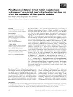
Fig. 1. FACS analysis of HDLp binding by LpR. After binding and
subsequent endocytosis of OG–HDLp, the cells were trypsinized
and analyzed by flow cytometry (A). The amount of fluorescence is
plotted on the y-axis (relative values), and the forward scatter (rela-
tive values) on the x-axis. Cells in the population indicated by 1
(population 1) are transfected cells with a higher fluorescence
intensity than the cells in the population indicated by 2 (popula-
tion 2). (B) A similar experiment using untransfected cells. (C)
Measurements of cells that were incubated at 4 °C with anti-
body 2189 ⁄ 90, and then with an FITC-labeled secondary antibody
at 4 °C, and then at 37 °C for 5 min before trypsinization and
analysis. The data shown in the plots are representative of four
independent experiments.
S. D. Roosendaal et al. Ligand binding to the insect LDLR homolog, LpR
FEBS Journal 275 (2008) 1751–1766 ª 2008 The Authors Journal compilation ª 2008 FEBS 1753
these cells did not bind HDLp. Detection of the recep-
tor on the plasma membrane, enabled by the use of
antibody 2189 ⁄ 90, yielded a similar distribution of the
two populations (Fig. 1C, cf. Fig. 1A). Quantification
of the number of cells in population 1 revealed that
the number of cells that bound ligand was
91.5 ± 6.3% of the number that bound antibody,
indicating that binding of HDLp was proportional to
the amount of receptor on the plasma membrane.
HDLp and LpR remain in complex at endosomal
pH
To investigate whether the LpR–HDLp complex disso-
ciates upon exposure to endosomal pH, OG–HDLp
was bound at neutral pH (7.4) to LpR-transfected
cells, after which the cells were washed at 4 °C with a
buffer of low pH (5.4). After endocytosis of bound
ligand, the fluorescence of the cells appeared to be not
affected when compared to cells that had been washed
at pH 7.4 (Fig. 2A,B). In contrast, similar experiments
performed with cells transfected with LDLR cDNA,
which were incubated with OG–LDL and subsequently
washed at pH 5.4, resulted in a decrease in fluores-
cence in comparison to cells that had been washed at
pH 7.4 (Fig. 2C,D). After washing at pH 5.4, the pop-
ulation with low fluorescence intensity was located at
the same position as after washing at pH 7.4, indicat-
ing that the different pH values of the buffers used in
the incubations did not affect the size or the morphol-
ogy of the cells, and thereby the amount of fluores-
cence. The population with low fluorescence intensity
was excluded from the analysis by defining region 1
(R1) (Fig. 2). To quantify the amount of bound
ligand, the mean fluorescence in R1 (Fig. 2) was deter-
mined and compared to the mean fluorescence in R1
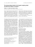
after washing at pH 5.4. In the case of LpR, the fluo-
rescence of cells washed at pH 5.4 was 94.3 ± 7.6%
of the fluorescence of cells washed at pH 7.4, indicat-
ing that OG–HDLp remained bound to LpR upon
exposure to pH 5.4. As expected, in the case of LDLR,
the fluorescence of cells washed at pH 5.4 had
decreased significantly, and amounted to only
51.4 ± 2.2% of the fluorescence of cells washed at
pH 7.4 (Fig. 2E). A longer incubation at pH 5.4 (1 h
instead of 30 min; data not shown) did not result in
further dissociation of either the LDLR–LDL or
LpR–HDLp complexes, suggesting that an incubation
time of 30 min was sufficient to achieve maximum dis-
sociation. Furthermore, the expression level of recep-
tor, which varied between different cell lines and thus
between different experiments, did not influence the
relative amount of dissociation (data not shown).
Exposure of the LpR–HDLp complex to pH values
between 4.0 and 5.0 resulted in a substantial decrease
in fluorescence of the cells (data not shown). However,
at this pH, HDLp appeared to be precipitated (data
not shown). Moreover, because in LpR-transfected
CHO cells HDLp is transported from the early endo-
some to the ERC, and from there is returned to the
plasma membrane [14], it is unlikely that the LpR–
HDLp complex encounters a pH lower than 5.4. Addi-
tionally, it should be noted that the pH of endosomes
in the insect fat body is similar to that of mammalian
AB
C
E
D
Fig. 2. LpR and HDLp remain in complex at endosomal pH.
CHO(LpR) cells were incubated with OG–HDLp and washed at
pH 7.4 (A) or pH 5.4 (B). (C, D) Similar experiment for binding of
OG–LDL to cells transfected with LDLR, and washed at pH 7.4 (C)
or pH 5.4 (D). Plots are representative of at least eight independent
experiments performed on cell lines created by four different trans-
fections. (E) Amount of bound ligand after washing at pH 5.4. The
mean fluorescence (y-mean) in R1 (A–D) was determined for each
sample. The relative y-mean (Rel. y-mean) after washing at pH 5.4
was calculated by the formula y-mean
pH 5.4
⁄ y-mean
pH 7.4
, and is
plotted on the y-axis. Data are the means of at least eight indepen-
dent experiments. Error bars indicate the SEM. See legend to
Fig. 1 for more details.
Ligand binding to the insect LDLR homolog, LpR S. D. Roosendaal et al.
1754 FEBS Journal 275 (2008) 1751–1766 ª 2008 The Authors Journal compilation ª 2008 FEBS
cells [43], indicating that also after LpR-mediated
uptake of HDLp in insect fat body tissue the
LpR–HDLp complex does not encounter a pH lower
than 5.4.
The lack of LpR–HDLp complex dissociation is
caused by the ligand-binding domain
Sequence alignment of the amino acid sequence of
LDLR with that of LpR revealed that several of the
residues crucial for LDL release by LDLR are not con-
served in LpR (Table 1). To investigate whether the
deficiency of these crucial residues in LpR may be
responsible for the lack of dissociation of the LpR–
HDLp complex, the binding and dissociation capacities
of different hybrid receptors (Fig. 3A [16]) were
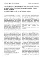
assessed. LDLR(1–292)LpR(343–850) was able to bind
LDL, but unable to release this ligand at endosomal
pH (Fig. 3B), suggesting that the absence of Gln540,
His562, Glu581 and Lys582 in the b-propeller of LpR
causes the lack of HDLp release by LpR. However,
the reciprocal hybrid, LpR(1–342)LDLR(293–839)
(Fig. 3A), appeared to be equally incapable of releasing
its ligand, HDLp (Fig. 3B). The presence of Gln540,
His562, Glu581 and Lys582 in this hybrid suggests that
the b-propeller of this hybrid may able to interact with
the ligand-binding domain. However, although LpR(1–
342)LDLR(293–839) contains Ile313, it does not
contain the complete hinge region of LDLR. The
presence of two Gly residues in LpR at the correspond-
ing positions of His264 and Ser265 of LDLR (Table 1)
might decrease the rigidity of the hinge region of
LpR(1–342)LDLR(293–839), thereby abolishing ligand
release. To investigate whether the complete hinge
region and b-propeller of LDLR were able to induce
HDLp release by LpR, the hybrid receptor LpR(1–
301)LDLR(252–839) (Fig. 3A) was created. This
hybrid receptor was able to bind HDLp, but, like
wild-type LpR, was unable to release it (Fig. 3B). As
these functional LDLR domains failed to evoke HDLp
release, the lack of dissociation of the complex is pro-
posed to result from the specific binding interaction of
the ligand-binding domain of LpR with HDLp.
HDLp binding to LpR is not sensitive to EDTA
Ligand binding by LDLR family members is known to
be dependent on Ca
2+
[33,44], and the removal of
Ca
2+
from LDLR, e.g. by EDTA, prevents ligand
binding [40]. To investigate whether the drop in Ca
2+
level that occurs in the early endosome could result in a
disruption of the interaction between LpR and HDLp,
LpR-transfected cells that had bound OG–HDLp were
exposed to an EDTA-containing buffer (Fig. 4A,B).
After washing and subsequent endocytosis of bound
ligand, the fluorescence of the cells was measured by
flow cytometry. The mean fluorescence of cells that
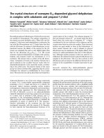
bound OG–HDLp did not change upon EDTA treat-
ment, as 96.6 ± 7.5% of OG–HDLp remained bound
to the receptor (Fig. 4E), demonstrating that the com-
plex was not disrupted. In contrast, when the same
experimental approach was employed using the
LDLR–LDL complex as a control, this resulted, as
expected, in a significant decrease of receptor-bound
LDL fluorescence (Fig. 4C,D). Only 37.8 ± 4.1% of
OG–LDL remained bound to the receptor (Fig. 4E).
This indicates that the low Ca
2+
concentration in the
early endosome is not able to induce dissociation of the
LpR–HDLp complex. To determine whether the stabil-
ity of the complex upon EDTA treatment is caused by
the LA repeats or by the two N-terminal EGF repeats
(EGF-A and EGF-B), which also contain a Ca
2+
, simi-
lar experiments were performed with the hybrid recep-
tors (Fig. 3A). The interaction between HDLp and
Table 1. LDLR amino acid residues that are essential for LDL release and the corresponding amino acid of LpR.
Residue in LDLR
Corresponding residue
in LpR Location Function Ref.
His190 His270 LA-5 Interaction with b-propeller [29,44]
Phe261 Phe344 LA-7 Required for rigidity of the hinge region [29]
His264 Gly346 LA-7 Unknown />Ser265 Gly347 LA-7 Unknown />Val274 Val357 LA-7 Required for rigidity of the hinge region [29]
Ile313 Ala395 EGF-A Anchorage of EGF-A and EGF-B with LA-7 [29,49]
Gln540 Lys621 b-Propeller Interaction with ligand-binding domain [29,49]
His562 Asn643 b-Propeller Induction of conformational change [29]
Glu581 Pro662 b-Propeller Interaction with ligand-binding domain [29,49]
Lys582 Glu663 b-Propeller Interaction with ligand-binding domain [29]
His586 His667 b-Propeller Interaction with ligand-binding domain [29,44]
S. D. Roosendaal et al. Ligand binding to the insect LDLR homolog, LpR
FEBS Journal 275 (2008) 1751–1766 ª 2008 The Authors Journal compilation ª 2008 FEBS 1755
LpR(1–342)LDLR(293–839) was not abrogated by
EDTA treatment (Fig. 4E). As this receptor contains
EGF-A and EGF-B of LDLR, this suggests that the
stability results from the ligand-binding domain of
LpR. In addition, the binding of HDLp to LpR(1–
301)LDLR(252–839) was shown to be EDTA-resistant,
indicating that the resistance resides in the first seven
LA repeats of LpR. In contrast, the binding of LDL to
the reciprocal hybrid receptor LDLR(1–251)LpR(302–
850) (Fig. 4F), the ligand-binding domain of which is
A
B
LDLR(1–251)LpR(302–850) LDLR(1–292)LpR(343–850)
LDLR(1–292)LpR(343–850)
LDLR
LpR
LpR(1–342)LDLR(293–839)
LpR(1–342)LDLR(293–839)
LpR(1–301)LDLR(252–839)
LpR(1–301)LDLR(252–839)
Fig. 3. Hybrid receptors and relative amount of pH-dependent
ligand dissociation. (A) Schematic models of the hybrid receptors.
LDLR domains are depicted in gray and LpR domains in black. Each
receptor contains a ligand-binding domain composed of LA repeats
(squares), an EGF-precursor homology domain composed of two
EGF repeats (diamonds) that are separated from a third by a b-pro-
peller containing YWTD repeats (circle), an O-linked glycosylation
domain (oval), a transmembrane domain (trapezoid), and an intracel-
lular C-terminal domain (long rectangle). The wide and open rectan-
gle represents the plasma membrane. The numbers indicate the
amino acids of the mature proteins. Amino acids that are important
for LDL release and not conserved in LpR are indicated by white
dots. (B) Amount of ligand bound to different hybrid receptors after
incubation at pH 5.4. CHO cells transfected with the different
hybrid receptors were incubated with OG–LDL [LDLR and LDLR(1–
292)LpR(343–850)] or OG–HDLp [LpR, LpR(1–342)LDLR(293–839),
LpR(1–301)LDLR(252–839)] and washed at pH 7.4 or pH 5.4. The
fluorescence was measured by flow cytometry. The y-mean of the
receptor-expressing population was determined for each sample.
The relative y-mean (Rel. y-mean) after a wash at pH 5.4 was calcu-
lated by the formula y-mean
pH 5.4
⁄ y-mean
pH 7.4
, and is plotted on
the y-axis. The values represented are the averages of at least
three independent experiments. Error bars indicate the SEM. See
legend to Fig. 1 for more details.
AB
C
D
EF
LpR
LpR(1–342)LDLR(293–839)
LpR(1–301)LDLR(252–839)
LDLR(1–251)LpR(343–850)
LDLR(1–251)LpR(343–850)
LDLR
Fig. 4. HDLp remains in complex with LA-1–7 of LpR after EDTA
treatment. CHO(LpR) cells were incubated with OG–HDLp and
washed at pH 7.4 without (A) or with (B) EDTA. (C, D) A similar
experiment for binding of OG–LDL to cells transfected with
LDLR, washed in the absence (C) and presence (D) of EDTA.
(E) Amount of OG–HDLp bound to LpR, LpR(1–342)LDLR(293–839)
or LpR(1–301)LDLR(252–839) or of OG–LDL bound to LDLR (con-
trol) and LDLR(1–251)LpR(302–850) after washing with EDTA. The
mean fluorescence (y-mean) in R1 was determined for each sam-
ple. The relative y-mean (Rel. y-mean) after a wash with an EDTA-
containing buffer was calculated by the formula y-mean
EDTA
⁄
y-mean
pH 7.4
, and is plotted on the y-axis. Data are the means of at
least six independent experiments. Error bars indicate the SEM.
See legend to Fig. 1 for more details. (F) Schematic model of
LDLR(1–251)LpR(302–850). LDLR domains are depicted in gray and
LpR domains in black. See legend to Fig. 3 for more details.
Ligand binding to the insect LDLR homolog, LpR S. D. Roosendaal et al.
1756 FEBS Journal 275 (2008) 1751–1766 ª 2008 The Authors Journal compilation ª 2008 FEBS
composed of the six most N-terminal LA repeats of
LDLR and LA-8 of LpR, was not EDTA-resistant
(Fig. 4E). Collectively, these results indicate that the
EDTA resistance of the binding of HDLp to LpR is
imparted by LA-1–7.
HDLp binding by LpR is similar to ligand binding
by other LDLR family members
As neither treatment with EDTA nor endosomal pH
was able to disrupt the complex, the issue was
addressed of whether LpR binds HDLp in a different
manner than other LDLR family members bind their
ligands. Therefore, the ability of receptor-associated
protein (RAP), a general ligand for LDLR family
members [45–47], to compete with HDLp binding to
LpR was assayed. LpR-transfected cells incubated with
OG–HDLp in the presence of an equimolar concentra-
tion of RAP displayed a fluorescence similar to that of
untransfected cells incubated with OG–HDLp and
RAP (Fig. 5), indicating that RAP completely blocked
the binding of OG–HDLp to LpR. Thus, RAP and
HDLp apparently use the same binding site. Therefore,
LpR probably binds HDLp using the general mecha-
nism of binding of ligands by LDLR family members
[48,49].
LA-8 and EGF-A of LpR are not involved in the
binding site of LpR for HDLp
To characterize the binding site for HDLp in LpR, the
binding of ligand by wild-type LpR was compared with
the binding of HDLp by LpR(1–301)LDLR(252–839).
To exclude the possibility that differences in ligand
binding were caused by differences in receptor expres-
sion, binding of antibody to the receptor was used as a
measure for the amount of receptor on the plasma mem-
brane. After binding, cells were allowed to endocytose
bound ligand or antibody, by incubation of the cells at
37 °C. Following endocytosis, the cells were trypsinized,
and the fluorescence was analyzed with flow cytometry.
As a control, the binding of LDL to LDLR was com-
pared to the binding of LDL to LDLR(1–251)LpR
(302–850). For wild-type LDLR, both ligand binding
and antibody binding yielded similar numbers of fluo-
rescent cells in R1 (Table 2), indicating that the amount
of LDL binding was proportional to the amount of
LDLR on the plasma membrane. However, in the case
of the hybrid receptor LDLR(1–251)LpR(302–850)
(Fig. 4F), the ligand-binding domain of which is com-
posed of the six most N-terminal LA repeats of LDLR
and LA-8 of LpR, the number of cells that bound ligand
was only 58.7 ± 4.2% of the number of cells that
bound antibody. This suggests a reduction in affinity of
the hybrid receptor for LDL, which may be expected, as
the binding site of LDLR for LDL encompasses LA-3–7
and EGF-A [50,51]. Despite the presence of LA-8 of
LpR in this receptor, LDLR(1–251)LpR(302–850) was
not able to bind HDLp. Moreover, as was previously
found for ligand binding to LpR and LDLR [14], for
binding to the hybrid receptors the ligands are not inter-
changeable (data not shown) [16]. With respect to the
binding of HDLp to LpR, the number of cells that
bound ligand was 91.5 ± 6.3% of the number of cells
that bound antibody, showing that also for LpR the
AB
C
D
Fig. 5. RAP competes with OG–HDLp for binding. CHO(LpR) cells
were incubated with OG–HDLp (A) or OG–HDLp in the presence of
RAP (B). (C, D) A similar experiment using untransfected cells incu-
bated with OG–HDLp in the absence (C) or presence (D) of RAP.
Plots are representative of three independent experiments. See
legend to Fig. 1 for more details.
Table 2. Binding efficiency of expressed receptors. CHO cells
transfected with the different (hybrid) receptors were incubated
with OG–LDL [LDLR, LDLR(1–251)LpR(302–850)] or with OG–HDLp
[LpR, LpR(1–301)LDLR(252–839), LpR
splice
] or a primary antibody
detected by an FITC-labeled secondary antibody. The percentage of
cells that bound ligand relative to the percentage of cells that
bound antibody was determined. The percentages shown are the
means ± SEM of at least five independent experiments.
Receptor Binding efficiency (%)
LDLR 96.1 ± 7.0
LDLR(1–251)LpR(302–850) 58.7 ± 4.2
LpR 91.5 ± 6.3
LpR(1–301)LDLR(252–839) 84.4 ± 7.5
LpR
splice
57.3 ± 5.9
S. D. Roosendaal et al. Ligand binding to the insect LDLR homolog, LpR
FEBS Journal 275 (2008) 1751–1766 ª 2008 The Authors Journal compilation ª 2008 FEBS 1757
binding of HDLp is proportional to LpR expression. As
for the hybrid receptor LpR(1–301)LDLR(252–839),
which contains LA-1–7 of LpR, followed by LA-7 of
LDLR, the binding of HDLp yielded 84.4 ± 7.5% of
fluorescent cells as compared to the number of cells that
bound antibody (Table 2). As 84.4 ± 7.5% is not sig-
nificantly lower than the 91.5 ± 6.3% measured for
LpR, this suggests that LpR(1–301)LDLR(252–839)
binds HDLp with a similar affinity as wild-type LpR.
These results indicate that LA-7 and EGF-A of LDLR
were able to replace the corresponding region of LpR
(LA-8 and EGF-A) without disrupting the binding site
for HDLp. This suggests that these repeats of LpR are
not involved in the ligand-binding site of LpR, in con-
trast to the same structure (LA-7 and EGF-A) in
LDLR.
LA-3 is involved in the binding site of LpR for
HDLp
Recently, a putative splice variant of LpR, LpR
splice
,
has been identified in ovaries of young animals
(J. Kerver and K. W. Rodenburg, unpublished results),
in which the sequence of LA-3 is altered. Although
sequence alignment revealed a high similarity between
LA-3 of the two variants, the central Trp present in
LA-3 of wild-type LpR is absent in LA-3 of LpR
splice
(Fig. 6). As the central Trp plays an important role in
the interaction between LDLR family members and
their ligands [49], we investigated whether the binding
of OG–HDLp to LpR
splice
deviates from that to wild-
type LpR. The binding of HDLp to LpR
splice
yielded
only 57.3 ± 6.9% of fluorescent cells as compared to
the number of cells that bound antibody (Table 2),
implying that LpR
splice
binds HDLp with a lower affin-
ity than wild-type LpR. This indicates that LA-3 is
involved in the binding of HDLp to wild-type LpR,
suggesting that the Trp in wild-type LpR may be
involved in the interface between HDLp and LpR.
LA-1 is not essential for binding of ligand to LpR
To further investigate which LA repeats are involved
in HDLp binding, we investigated whether anti-
body 2189 ⁄ 90, directed against the first LA repeat of
LpR, was able to compete with HDLp for binding by
LpR. After a preincubation with OG–HDLp at 4 °C,
followed by incubation with antibody 2189 ⁄ 90 at 4 °C,
the fluorescence after uptake of the bound OG–HDLp
appeared to be 73.2 ± 6.3% of the fluorescence of the
cells in such an experiment without incubation with
antibody 2189 ⁄ 90. In a similar experiment in which the
order of the incubations with OG–HDLp and anti-
body 2189 ⁄ 90 was reversed, the fluorescence of the
cells was 78.9 ± 7.0% of the fluorescence of cells incu-
bated with OG–HDLp alone. Although the presence
of antibody resulted in a significant decrease in fluores-
cence of the cells as compared to cells that were incu-
bated with OG–HDLp only, these results indicate that
LpR is still able to efficiently bind a major amount of
OG–HDLp in the presence of the antibody. Moreover,
the amount of competition was similar to that in the
corresponding control experiment using LDLR and
antibody C7, an antibody against the first repeat of
LDLR (data not shown) [52,53]. As LA-1 of LDLR is
not involved in LDL binding [51], the inhibition of
binding that was measured probably results from steric
hindrance due to the size of the antibody and not from
competition for the same binding site. No competition
was observed between RAP and the antibody (data
not shown), suggesting that LA-1 is not involved in
the binding of LpR to RAP or HDLp.
Discussion
Previous studies have demonstrated that in a mam-
malian model (CHO) cell line transfected with insect
LpR, the receptor recycles its ligand, HDLp, in a
transferrin-like manner, in contrast to endogenously
expressed LDLR, the ligand of which (LDL) is
released and undergoes intracellular degradation [14].
Also during insect development, LpR appeared to
function similarly, since HDLp internalized by fat
body tissue from young adult locusts endogenously
expressing LpR appeared to be resecreted, supporting
the concept of LpR-mediated ligand recycling [12,18].
To investigate the mechanism underlaying the highly
unusual behavior of this insect LDLR family member,
Fig. 6. Sequence of LA-3 of a putative splice variant in LpR. Alignment of the sequences of LA-3 of wild-type LpR and LpR
splice
. Identical
residues are boxed in black, and conserved residues are shaded in gray. The arrow indicates the position of the central Trp in the sequence
of wild-type LpR.
Ligand binding to the insect LDLR homolog, LpR S. D. Roosendaal et al.
1758 FEBS Journal 275 (2008) 1751–1766 ª 2008 The Authors Journal compilation ª 2008 FEBS
we examined the stability of the binding of HDLp to
LpR in direct comparison with that of LDL to LDLR,
and additionally explored the subset of structural fea-
tures in LpR that may allow for the occurrence of the
difference in ligand delivery as compared to that in
mammals.
Our present studies provide the new findings that
the complex of LpR and HDLp is stable at endosomal
pH and EDTA-resistant, both in contrast to the com-
plex of LDLR and LDL. This stability of the LpR–
HDLp complex is proposed to be caused by the spe-
cific interaction between HDLp and LA-2–7. Together,
our data indicate that the complex of LpR and HDLp
remains intact during its intracellular itinerary, which
is in complete agreement with the occurrence of ligand
recycling [14,16–18], and may provide a vital determi-
nant of the ligand-recycling capacity of LpR. In sev-
eral studies, flow cytometry has been used to quantify
lipoprotein binding and uptake [29,53–59]. In most
cases, the experiments were performed on blood cells.
As these cells are already in suspension, they can be
easily measured by flow cytometry. In the case of
attached cells, resuspending the cells may destroy the
interaction between receptor and ligand or antibody.
For this reason, the actual binding experiment was
performed at 4 °C to prevent endocytosis, so that equi-
librium binding was achieved. After binding, the cells
were allowed to endocytose bound ligand to protect it
from the subsequent trypsin treatment. Fluorescence
images of the cells after binding at 4 °C and after
endocytosis at 37 °C showed that the bound ligand or
antibodies were efficiently endocytosed, indicating that
the amount of intracellular fluorescence was propor-
tional to the amount of bound ligand at equilibrium
(data not shown). For these experiments, stably trans-
fected polyclonal cell lines were used to provide hetero-
geneous samples of cells that express the receptor. This
resulted in flow cytometry plots containing two popu-
lations, one of which comprised cells whose fluores-
cence did not exceed that of untransfected cells
(Fig. 1). In order to analyze only the cells that
expressed the receptor, R1 was defined to exclude
the population with lower fluorescence intensity from
the analysis (Figs 2 and 4). However, for LDLR, the
decrease in fluorescence after treatment at pH 5.4 or
with EDTA resulted in a decrease of the number of
cells in R1. As this reduction in sample size introduced
a bias into the analysis, the number of cells in the
analysis was restored by using random measurements
from the population with low fluorescence intensity.
After correction of the mean fluorescence, similar val-
ues for LDL release by LDLR were obtained as mea-
sured by Blacklow et al. for monoclonal cell lines [29].
The relative amount of dissociation was not affected
by differences in receptor expression; we therefore con-
clude that the results obtained with the flow cytometric
assay represent physiologically relevant receptor prop-
erties.
Our data indicate that, unlike the complex of LDLR
and LDL, the complex of LpR and HDLp remains
stable at a pH as low as 5.4, which is significantly
lower than that encountered in endosomes (pH 6–6.5)
[27]. This indicates that, despite the substantial
sequence similarity between LpR and LDLR, LpR is
unable to release HDLp in the early endosome. LDLR
is hypothesized to release LDL at endosomal pH by
undergoing a conformational change in which the
b-propeller displaces LDL [31]. Blacklow et al. ele-
gantly identified domains and residues that are impor-
tant for LDL release by LDLR [29,59] (Table 1). In
agreement with these results, the b-propeller of LpR,
lacking the important residues Gln540, His562,
Glu581, and Lys582, was incapable of inducing LDL
release. Similar results were obtained for the swap of
the b-propeller of LDLR with b-propeller 4 of LDLR
related protein (LRP) 6, in which two Lys residues
and one His are not conserved. However, when
b-propeller 2 of LRP6 was introduced, containing
these residues, the receptor was able to release LDL,
indicating that a different b-propeller is able to substi-
tute for the wild-type propeller of LDLR [29]. How-
ever, introducing the b-propeller of LDLR into LpR
did not lead to HDLp release by the hybrid receptor
LpR(1–342)LDLR(293–839), implying that other
domains produce the remarkable stability of the
complex. In LDLR, the interface between LA-7 and
EGF-A, the hinge region, also plays an important role
in LDL release, as this region functions as a rigid
scaffold allowing the b-propeller to fold over the
ligand-binding domain. To investigate the importance
of this hinge region for the lack of HDLp release by
LpR, both the hinge region and b-propeller of LDLR
were introduced into LpR. The resulting hybrid recep-
tor, LpR(1–301)LDLR(252–839), did not release
HDLp, despite the fact that this hybrid contains all
the domains of LDLR that are essential for LDL
release. This suggests that the b-propeller of LDLR is
not able to compete with HDLp for binding to the
ligand-binding domain of LpR, implying that the lack
of HDLp release is mainly caused by the interaction
between HDLp and the ligand-binding domain of
LpR, and suggests that LpR may use a different mech-
anism to release HDLp, in contrast to the mechanism
of LDL release by LDLR, in which the b-propeller is
of vital importance [28,29,60]. Interestingly, our earlier
localization studies of the hybrid receptors revealed
S. D. Roosendaal et al. Ligand binding to the insect LDLR homolog, LpR
FEBS Journal 275 (2008) 1751–1766 ª 2008 The Authors Journal compilation ª 2008 FEBS 1759
that the intracellular fate of the complex is determined
by the extracellular domain as a whole [16]. In view of
the mechanism of ligand recycling by LpR, this implies
that for the stability of the complex, the ligand-binding
domain is sufficient, but for proper targeting of LpR
to the ERC, the combination of the ligand-binding
domain and b-propeller of LpR is essential [16].
Ligand binding to LDLR family members is known
to depend on Ca
2+
, due to the stabilization of the LA
repeats by a central Ca
2+
[33,34,36–39]. Sequence
comparison of the LA repeats of LpR with those of
other LDLR family members, as well as modeling and
molecular dynamics studies of LA-4–6 of LpR, indi-
cate that this also applies for the LA repeats of LpR
(S. D. Roosendaal, S. Cuesta-Lo
´
pez, J. Sancho and
K. W. Rodenburg, unpublished results). In addition to
a decrease in pH, the Ca
2+
concentration in the early
endosome drops within minutes to the low micromolar
range [32], possibly contributing to ligand release by
LDLR [33]. Therefore, the LpR–HDLp complex was
exposed to a Ca
2+
-chelating agent (EDTA) to mimic
the effect of low Ca
2+
in the early endosome. In con-
trast to the binding of LDL to LDLR, the binding of
HDLp to LpR appeared to be resistant to EDTA
treatment. A possible explanation for this phenomenon
might be that LpR binds HDLp by using a different
binding mode than that used by other LDLR family
members for binding of their ligands. For example, a
different, Ca
2+
-independent binding mode is used in
the interaction between the single LA repeat of Tva,
the cellular receptor for subgroup A Rous sarcoma
virus [61] and its ligand. However, as the ligand bound
Tva with an aberrant binding mode, RAP appeared to
be unable to compete with the ligand for binding to
Tva [62]. Our studies show that RAP efficiently com-
petes with HDLp for binding to LpR, indicating that
HDLp binds LpR using the same binding mode as
other ligands for LDLR family members, which again
implies the presence of Ca
2+
in LA repeats of LpR.
Therefore, the resistance of the LpR–HDLp complex
may be caused by a higher affinity of the LA repeats
of LpR for Ca
2+
, or by the ability of HDLp to shield
the calcium ions from EDTA. Although it is unclear
what precisely causes this remarkable stability, our
data emerging from the use of hybrid receptors indi-
cate that the stability of the complex at low pH and
upon EDTA treatment is caused by the interaction
between HDLp and LA-1–7 of LpR. The general bind-
ing mode of LDLR family members and their ligands
consists of an acidic binding pocket present in the LA
repeats that entraps a Lys from the ligand. The bind-
ing is augmented by an essential aromatic residue,
preferentially Trp, of the LA repeat, positioned next to
the binding pocket [45,47,49,63]. To obtain more infor-
mation about the recognition interface between LpR
and HDLp, the LA repeats of LpR were aligned with
those of LDLR. This revealed that only LA-1–6 of
LpR contain a central aromatic residue, in all cases
Trp (data not shown). As LA-1 appeared not to be
involved in the binding site for HDLp, this suggests
that only LA-2–6 are involved in the interface. LA-3
of wild-type LpR contains the central Trp, but impor-
tantly, in addition to other amino acid changes, LA-3
of LpR
splice
lacks this Trp (Fig. 6). As LpR
splice
binds
HDLp with a lower affinity, indicating that LA-3 is
involved in the interaction with HDLp, it may very
well be that the absence of the Trp weakens the inter-
action between HDLp and LpR. In this respect, it is
interesting to note that the binding of HDLp to the
splice variant is also resistant to low pH and EDTA
treatment (data not shown). This suggests that the Trp
and the other residues that are different between LA-3
of wild-type LpR and LpR
splice
are not important for
determining the stability of the interaction under endo-
somal conditions. Additionally, from these results, it is
apparent that the stability of the complex at endoso-
mal pH and upon EDTA treatment is not merely the
result of the affinity of the interaction, but may require
additional contacts or a slightly alternative mode of
binding of HDLp to LpR. An alternative mechanism
for a stable complex at endosomal pH may be pro-
vided by the binding of proprotein convertase subtili-
sin type 9 (PCSK9) to LDLR. PCSK9 has been shown
to be involved in the regulation of cell surface LDLR
levels. After endocytosis, the LDLR–PCSK9 complex
is also not dissociated at endosomal pH. Instead, the
affinity of PCSK9 for LDLR is enhanced by the low
pH [64,65], possibly through protonation of the abun-
dant His residues on the surface of PCSK9 [65,66].
Even though HDLp binds to the LA repeats of the
receptor and PCSK9 to EGF-A of LDLR, similar
effects may play a role in the stability of the complex
of HDLp and LpR. An important difference is, how-
ever, that binding of PCSK9 seems to target LDLR to
lysosomes for degradation [67,68], whereas the com-
plex of HDLp and LpR is transported to the ERC for
recycling.
The acidic residues involved in Ca
2+
binding of spe-
cific LA repeats are proposed to interact with the basic
residues of the ligand, in particular one protruding Lys
[49]. A consensus sequence containing the protruding
Lys was proposed, in which the Lys is surrounded by
basic and hydrophobic residues [69,70]. Such sequences
are numerous in both apolipoproteins of HDLp,
apoLp-I and apoLp-II. Interestingly, the three-dimen-
sional model of their protein precursor apoLp-II ⁄ I
Ligand binding to the insect LDLR homolog, LpR S. D. Roosendaal et al.
1760 FEBS Journal 275 (2008) 1751–1766 ª 2008 The Authors Journal compilation ª 2008 FEBS
reveals that at least one of these motifs is situated at
the end of an a-helix [71], as is the case for the binding
site of RAP and apolipoprotein E for LRP [49]. Fur-
thermore, this helix is probably exposed on the surface
of the HDLp particle, and is thus available for interac-
tion with LpR [71]. Because of the multitude of puta-
tive binding sites in apoLp-I and apoLp-II, it cannot
excluded that one HDLp particle binds several recep-
tors concomitantly, as is the case for apolipoprotein E-
containing lipoproteins [72,73]. In this respect, it is
important to note that apolipoprotein B-100, which is
a homolog of apoLp-II ⁄ I [3,4], also contains several of
these consensus sequences (data not shown). However,
LDL binds to LDLR with a stoichiometry of 1 : 1.
Moreover, RAP is able to efficiently compete at equi-
molar concentrations with the binding of HDLp.
Although several RAP molecules may be able to bind
LpR, as RAP binds to two LA repeats [49,74], compe-
tition binding studies indicated that RAP and anti-
body 2189 ⁄ 90 against LA-1 of LpR do not compete
(data not shown), suggesting that RAP does not bind
LA-1. On the basis of the presence of an important
acidic residue [74], sequence analysis of the LA repeats
of LpR suggests that RAP may bind either LA-4–5 or
LA-5–6, suggesting that the stoichiometry is one RAP
molecule per LpR molecule. Therefore, it seems unli-
kely that LpR binds more than one HDLp molecule.
In conclusion, our results indicate that the inter-
action between HDLp and LA-2–7 of LpR is stable
upon exposure to endosomal pH as well as EDTA
treatment, implying that the integrity of the complex is
maintained during intracellular trafficking of LpR and
HDLp in LpR-transfected CHO cells and most likely
also in insect cells. Similar to transferrin recycling, the
intracellular transfer of lipid or other hydrophobic
compounds from or to the HDLp particle may change
its affinity for LpR, thus allowing HDLp resecretion.
Indeed, binding studies using a partially delipidated
HDLp particle revealed that the affinity of LpR
for HDLp is modulated by the amount of lipids
(S. D. Roosendaal, J. M. Van Doorn, K. M. Valentijn,
D. J. Van der Horst and K. W. Rodenburg, unpub-
lished results), suggesting that changes in lipid content
may trigger HDLp resecretion. The stability of the
complex and the modulation thereof may be deter-
mined by secondary contacts between HDLp and non-
conserved residues of LpR. Although the function of
recycling of endocytosed lipoprotein ligand during
insect development remains to be defined, our study
reveals the molecular mechanism underlying the stabil-
ity of the LpR–HDLp complex; this is likely to pro-
vide a crucial key to the process of ligand recycling,
and might additionally help to explain the ability of
LDLR family members to bind a wide range of struc-
turally unrelated ligands.
Experimental procedures
Proteins and antibodies
Insect HDLp was isolated from locust hemolymph by den-
sity gradient ultracentrifugation as described previously
[14]. Human LDL was isolated from blood plasma (Bloed-
bank Midden Nederland, the Netherlands) as described by
Redgrave et al. [75], with minor adaptations to the original
protocol. The salt solutions of different densities used
in the procedure contained 86.89 gÆmL
)1
KBr (density
1.063 gÆmL
)1
), 18.36 gÆmL
)1
KBr (density 1.019 gÆmL
)1
),
and 8.68 gÆmL
)1
KBr (density 1.006 gÆmL
)1
). Polyclonal
rabbit antibody to LpR (antibody 2189 ⁄ 90) was raised
against a synthetic peptide representing the unique N-termi-
nal 20 amino acids (34–53) of LA-1 of LpR [15]. Mouse
antibody to LDLR (antibody C7) was a generous gift from
I. Braakman (Utrecht University, Utrecht, the Nether-
lands). Human His-tagged RAP (RAP–His) was a generous
gift from M. Etzerodt (IMSB, Aarhus University, A
˚
rhus,
Denmark).
Construction of expression vectors encoding
lipoprotein receptor cDNA
The cloning of the expression vectors was performed
according to standard laboratory procedures and according
to the protocols supplied with enzymes and kits. Site-spe-
cific mutations were generated with a QuickChange site-
directed mutagenesis kit using PfuTurbo DNA polymerase
(Stratagene, Amsterdam, the Netherlands), according to the
manufacturer’s protocol. PCR fragments were generated
using PfuTurbo DNA polymerase and synthetic oligo-
nucleotide primers (Biolegio, Nijmegen, the Netherlands).
Endonucleases were from New England BioLabs (Westburg
B.V., Leusden, the Netherlands) and Fermentas (St Leon-
Rot, Germany). Plasmid pcDNA3–LpR(1–297)LDLR(248–
839) was made as follows. First, by mutagenesis, a unique
AgeI site was introduced in pcDNA3–LpR (piLR-e
[13], causing a silent mutation in the Pro301 codon
(CCA fi CCG; the first amino acid is that of the mature
protein), using the oligonucleotides 5¢-GAGAATTGCAC
ATCACC
GGTGCCAAAGTGTGACCC-3¢ (forward pri-
mer) and 5¢-GGGTCACACTTTGGCACCGGTGATGTG
CAATTCTC-3¢ (reverse primer), yielding the construct
pcDNA3–LpR(AgeI). Subsequently, to replace the sequence
encoding LA-8 of LpR with that encoding LA-7 of human
LDLR, a 1668 bp fragment containing the 5¢-flanking AgeI
and 3¢-flanking AccIII sites was generated by PCR from
pGEM-T–LDLR(1–292)LpR(343–850) [16], using the oligo-
nucleotides 5¢-GGCCGCACCGGTGACACTCTGCGAGG
S. D. Roosendaal et al. Ligand binding to the insect LDLR homolog, LpR
FEBS Journal 275 (2008) 1751–1766 ª 2008 The Authors Journal compilation ª 2008 FEBS 1761
GACCC-3¢ (forward primer) and 5¢-GCGGCCGCTTATA
CATAATCATTTGTCCC-3¢ (reverse primer). The AgeI–
AccIII fragment encoding the 5¢-end LA-7 of LDLR
obtained by PCR was cloned in pcDNA3–LpR(AgeI), using
the enzymes AgeI and AccIII, thereby replacing the sequence
encoding LA-8 of LpR to yield the mosaic receptor construct
pcDNA3–LpR(1–301)LDLR(252–292)LpR(343–850). Sub-
sequently, the 1267 bp EcoRI–KpnI fragment [16] from the
mosaic receptor construct was isolated and cloned into
pGEM-T–LpR(1–342)LDLR(293–839) digested with the
same two endonucleases to replace the sequence encoding
LA-1 through LA-8 of LpR with the sequence encoding
LA-1 through LA-7 of LpR combined with LA-7 of human
LDLR, thereby generating pGEM-T–LpR(1–301)LDLR(252–
839). Finally, the EcoRI–NotI fragment encoding the
LpR(1–301)LDLR(252–839) sequence was cloned in
pcDNA3 digested with the same enzymes to yield
pcDNA3–LpR(1–301)LDLR(252–839).
Plasmid pcDNA3–LDLR(1–251)LpR(302–850) was con-
structed similarly. First, a unique HpaI site was introduced
in pcDNA3–LDLR [16], causing a silent mutation in
the Asn251 codon (AAT fi AAC), using the oligonucleo-
tides 5¢-GGCTGCGTTAACGTGACACTCTGCGAG-3¢
(forward primer) and 5¢-CTCGCATGTCAGGTTAACG
CAGCC-3¢ (reverse primer), yielding the construct pBS–
LDLR(HpaI). Subsequently, to replace the sequence encod-
ing LA-7 of human LDLR with that encoding LA-8 of
LpR, a 1790 bp fragment, containing the unique HpaI and
Bsu361 sites, was generated from pcDNA3–LpR(1–
342)LDLR(293–839) by PCR, using the oligonucleotide
primers 5¢-CCCGGGGTTAACGTGCCAAAGTGTGA
CCCC-3¢ (forward primer) and 5¢-ATTTAAATTCACGCC
AGCTCATCCTCC-3¢ (reverse primer). The 473 bp HpaI–
Bsu361 fragment, encoding LA-8 at the 5¢-end, obtained by
PCR, was then cloned in pBS–LDLR(HpaI), using the
enzymes HpaI and Bsu361, replacing the sequence encoding
LA-7 of LDLR with that en coding LA-8 of LpR, to yield the
mosaic receptor pBS–LDLR(1–2 51)LpR(301–342)LDLR(293–
839). To obtain the construct pcDNA3–LDLR(1–
251)LpR(301–342)LDLR(293–839), the sequence encoding
the mosaic receptor was cloned in pcDNA3using the XbaI
restriction enzyme. Subsequently, the 225 bp EcoRI–KpnI
fragment from the mosaic receptor construct was isolated
and cloned into pGEM-T–LDLR(1–292)LpR(343–850)
digested with the same two endonucleases to replace the
sequence encoding the seven LA repeats of LDLR with
that encoding LA-1–6 of LDLR followed by LA-8 of LpR,
thereby generating pGEM-T–LDLR(1–251)LpR(301–850).
Finally, the HindI–
NotI fragment encoding the LDLR(1–
251)LpR(301–850) sequence was cloned in pcDNA3
digested with the same enzymes to yield pcDNA3–
LDLR(1–251)LpR(301–850). All PCR- and mutagenesis-
generated LpR fragments were sequenced, and their
sequences, apart from the intended mutations, were
confirmed to be identical to that of LpR as indicated in
the EMBL sequence database (accession number
AJ000010).
Cell culture
CHO cells were cultured in 25 cm
2
polystyrene culture
flasks in growth medium [Ham F10 nutrient mixture
(GibcoBRL, Invitrogen, Breda, the Netherlands)] contain-
ing 5% heat-inactivated fetal bovine serum (GibcoBRL)
and 100 UÆmL
)1
penicillin G sodium and 100 lgÆmL
)1
streptomycin sulfate in 85% saline (GibcoBRL). The cells
were maintained at 37 ° C and 5% CO
2
.
Transfections
LDLR-deficient CHO(ldlA) cells [42] were grown up to
40% confluency in 12-well multidishes (Costar, Corning BV
Life Sciences, Schiphol-Rijk, the Netherlands). After wash-
ing of the cells once, the growth medium was replaced with
500 lL of fresh growth medium. Subsequently, 2 lgof
DNA and 4 lg of poly(ethylenimine) (Polysciences,
Eppelheim, Germany) in 50 lL of serum-free medium
(Ham F10 nutrient mixture supplemented with 100 UÆmL
)1
penicillin G sodium and 100 lgÆmL
)1
streptomycin sulfate
in 85% saline) was administered to the cells. After 4 h,
500 lL of growth medium was added and cells were cul-
tured overnight. The next day, cells were detached from
dishes and cultured in 25 cm
2
culture flasks in growth med-
ium supplemented with 400 lgÆmL
)1
geneticin (GibcoBRL)
or 400 lgÆmL
)1
zeocin (Cayla, Toulouse, France). Ten days
after transfection, cells were used for experiments.
Fluorescence labeling of LDL and HDLp
LDL and HDLp were covalently labeled with OG 488 car-
boxylic acid (Molecular Probes, Leiden, the Netherlands) as
described previously [14].
Binding experiments using flow cytometry
The cells were grown up to a confluency of 70%. Sixteen
hours before the experiment, the growth medium was
replaced with serum-free medium. At the start of the exper-
iment, the cells were placed on ice. Subsequently, the cells
were washed with ice-cold binding buffer (50 mm Tris ⁄ HCl,
2mm CaCl
2
, 150 mm NaCl, pH 7.4, 4 °C) and incubated
with OG-labeled LDL (35 lgÆ mL
)1
) or HDLp (25 lgÆmL
)1
)
in binding buffer for 30 min. After binding, the cells were
washed once with either ice-cold binding-buffer, low-pH
buffer (25 mm Tris, 25 mm sodium succinate, 2 mm CaCl
2
,
150 mm NaCl, pH 5.4 or pH 4.0, 4 °C) or EDTA-contain-
ing buffer (50 mm Tris ⁄ HCl, 150 mm NaCl, 5 mm EDTA,
pH 7.4, 4 °C). The cells were then incubated with the
buffer for 30 min. After washing of the cells, the cells were
Ligand binding to the insect LDLR homolog, LpR S. D. Roosendaal et al.
1762 FEBS Journal 275 (2008) 1751–1766 ª 2008 The Authors Journal compilation ª 2008 FEBS
incubated for 5 min at 37 °C in serum-free medium, to
allow the cells to endocytose bound ligand. After endocyto-
sis, the cells were detached using trypsin ⁄ EDTA (Invitro-
gen), according to the manufacturer’s instructions, and
resuspended in growth medium. Resuspended cells were
fixed in 0.5% paraformaldehyde in NaCl ⁄ P
i
at 4 °C for at
least 30 min or overnight.
The receptor on the plasma membrane was detected
using the same protocol as for ligand binding. However,
the cells were incubated with an antibody against the first
LA repeat (antibody C7 [76] for LDLR, and anti-
body 2189 ⁄ 90 [15] for LpR) for 30 min in binding buffer.
After being washed with binding buffer, the cells were
incubated for 30 min with fluorescein isothiocyanate
(FITC)-labeled secondary anti-IgG (Jackson Immuno-
Research Laboratories Inc., Brunschwig, Amsterdam, the
Netherlands). Then, the complex was endocytosed, and cells
were detached and fixed as described above.
Competition binding experiments
Competition experiments were performed similarly to the
binding experiments. However, the cells were first incubated
for 30 min with primary antibody 2189 ⁄ 90, and then for
30 min with OG–HDLp, or vice versa. The degree of bind-
ing was compared to the degree of binding without the
antibody incubation. For competition experiments with
RAP, RAP–His (3.6 lgÆ mL
)1
) was added simultaneously
with (OG)–HDLp or primary antibody 2189 ⁄ 90. Then,
bound OG–HDLp or antibody 2189 ⁄ 90 was detected as
described previously. RAP–His was detected by subsequent
washing and incubation of mouse antibody to His (Amer-
sham Biosciences, Roosendaal, the Netherlands) in binding
buffer, and this was followed by washing and incubation
with FITC-labeled secondary anti-IgG (Jackson Immuno-
Research Laboratories Inc.).
Flow cytometry data analysis
Samples were measured using a fluorescence-activated cell
sorter (FACS; Becton Dickinson FACS Calibur). Flow
cytometry data were collected using cellquest (Becton
Dickinson) and downloaded into the program winmdi
(TSRI FACS Core Facility, La Jolla, CA, USA) for analy-
sis. For each sample (100 000 cells), the fluorescence was
plotted against the forward scatter. On the basis of samples
of untransfected cells, for each series of experiments R1
was defined to exclude cells whose fluorescence did not
exceed that of untransfected cells from the analysis. Then,
the number of cells and the mean fluorescence (y-mean) in
R1 were determined. If the number of cells in R1 decreases
with the different treatments of the cells, the y-mean in R1
is overestimated. Therefore, for each cell line, the number
of cells in R1 after different treatments was compared by a
t-test for paired samples performed on the logarithms of
the number of cells. In cases of a significant (P < 0.05) dif-
ference in sample size due to the different treatments, the
y-mean was corrected by using random values of the
missing number of cells from the population with lower
fluorescence intensity. After correction, for each sample the
relative amount of fluorescence as compared to control
samples was determined. Data presented as means ± SEM
were obtained from at least three independent experiments.
To test whether samples were significantly different from
control samples, a t-test for paired samples was performed
on the logarithms of the y-means.
Acknowledgements
We thank Steve Blacklow, Sander Meijer, Ineke Bra-
akman, Ju
¨
rgen Gent, Manon Wildenberg and Masja
van Oort for stimulating discussions, Ger Arkesteijn
for his help with flow cytometry, Wim Busschers for
help with quantification of the flow cytometry data,
and Santiago Cuesta-Lo
´
pez and Javier Sancho for
molecular dynamics studies on LA-4–6 of LpR.
References
1 Weers PM, Van Marrewijk WJ, Beenakkers AM & Van
der Horst DJ (1993) Biosynthesis of locust lipophorin.
Apolipophorins I and II originate from a common pre-
cursor. J Biol Chem 268 , 4300–4303.
2 Bogerd J, Babin PJ, Kooiman FP, Andre M, Ballagny
C, Van Marrewijk WJ & Van der Horst DJ (2000)
Molecular characterization and gene expression in the
eye of the apolipophorin II ⁄ I precursor from Locusta
migratoria. J Comp Neurol 427, 546–558.
3 Babin PJ, Bogerd J, Kooiman FP, Van Marrewijk WJ
& Van der Horst DJ (1999) Apolipophorin II ⁄ I, apoli-
poprotein B, vitellogenin, and microsomal triglyceride
transfer protein genes are derived from a common
ancestor. J Mol Evol 49, 150–160.
4 Mann CJ, Anderson TA, Read J, Chester SA, Harrison
GB, Kochl S, Ritchie PJ, Bradbury P, Hussain FS,
Amey J et al. (1999) The structure of vitellogenin pro-
vides a molecular model for the assembly and secretion
of atherogenic lipoproteins. J Mol Biol 285, 391–408.
5 Mahley RW & Innerarity TL (1983) Lipoprotein recep-
tors and cholesterol homeostasis. Biochim Biophys Acta
737, 197–222.
6 Davidson NO & Shelness GS (2000) Apolipoprotein B:
mRNA editing, lipoprotein assembly, and presecretory
degradation. Annu Rev Nutr 20, 169–193.
7 Brown MS & Goldstein JL (1986) A receptor-mediated
pathway for cholesterol homeostasis. Science 232, 34–47.
8 Ryan RO & Van der Horst DJ (2000) Lipid transport
biochemistry and its role in energy production. Annu
Rev Entomol 45, 233–260.
S. D. Roosendaal et al. Ligand binding to the insect LDLR homolog, LpR
FEBS Journal 275 (2008) 1751–1766 ª 2008 The Authors Journal compilation ª 2008 FEBS 1763
9 Van der Horst DJ, Van Marrewijk WJ & Diederen JH
(2001) Adipokinetic hormones of insect: release, signal
transduction, and responses. Int Rev Cytol 211, 179–240.
10 Van der Horst DJ, Van Hoof D, Van Marrewijk WJ &
Rodenburg KW (2002) Alternative lipid mobilization:
the insect shuttle system. Mol Cell Biochem 239, 113–119.
11 Van der Horst DJ & Ryan RO (2004) Lipid transport.
In Comprehensive Molecular Insect Science, vol. 4
(Gilbert LI, Iatrou K & Gill SS, eds), pp. 225–246.
Elsevier, New York, NY.
12 Dantuma NP, Pijnenburg MA, Diederen JH & Van der
Horst DJ (1997) Developmental down-regulation of
receptor-mediated endocytosis of an insect lipoprotein.
J Lipid Res 38, 254–265.
13 Dantuma NP, Potters M, De Winther MP, Tensen CP,
Kooiman FP, Bogerd J & Van der Horst DJ (1999) An
insect homolog of the vertebrate very low density lipo-
protein receptor mediates endocytosis of lipophorins.
J Lipid Res 40, 973–978.
14 Van Hoof D, Rodenburg KW & Van der Horst DJ
(2002) Insect lipoprotein follows a transferrin-like recy-
cling pathway that is mediated by the insect LDL recep-
tor homologue. J Cell Sci 115, 4001–4012.
15 Van Hoof D, Rodenburg KW & Van der Horst DJ
(2003) Lipophorin receptor-mediated lipoprotein endo-
cytosis in insect fat body cells. J Lipid Res 44, 1431–
1440.
16 Van Hoof D, Rodenburg KW & Van der Horst DJ
(2005) Intracellular fate of LDL receptor family mem-
bers depends on the cooperation between their ligand-
binding and EGF domains. J Cell Sci 118, 1309–1320.
17 Rodenburg KW & Van der Horst DJ (2005) Lipopro-
tein-mediated lipid transport in insects: analogy to the
mammalian lipid carrier system and novel concepts for
the functioning of LDL receptor family members.
Biochim Biophys Acta 1736, 10–29.
18 Van Hoof D, Rodenburg KW & Van der Horst DJ
(2005) Receptor-mediated endocytosis and intracellular
trafficking of lipoproteins and transferrin in insect cells.
Insect Biochem Mol Biol 35, 117–128.
19 Cheon H-M, Seo S-J, Sun J, Sappington TW & Raikhel
AS (2001) Molecular characterization of the VLDL
receptor homolog mediating binding of lipophorin in
oocyte of the mosquito Aedes aegypti. Insect Biochem
Mol Biol 31, 753–760.
20 Seo SJ, Cheon HM, Sun J, Sappington TW & Raikhel
AS (2003) Tissue- and stage-specific expression of two
lipophorin receptor variants with seven and eight
ligand-binding repeats in the adult mosquito. J Biol
Chem 278, 41954–41962.
21 Lee CS, Han JH, Kim BS, Lee SM, Hwang JS, Kang
SW, Lee BH & Kim HR (2003) Wax moth, Galleria
mellonella, high density lipophorin receptor: alternative
splicing, tissue-specific expression, and developmental
regulation. Insect Biochem Mol Biol 33, 761–771.
22 Lee CS, Han JH, Lee SM, Hwang JS, Kang SW, Lee
BH & Kim HR (2003) Wax moth, Galleria mellonella
fat body receptor for high-density lipophorin (HDLp).
Arch Insect Biochem Physiol 54, 14–24.
23 Gopalapillai R, Kadono-Okuda K, Tsuchida K,
Yamamoto K, Nohata J, Ajimura M & Mita K (2006)
Lipophorin receptor of Bombyx mori: cDNA cloning,
genomic structure, alternative splicing, and isolation of
a new isoform. J Lipid Res 47 , 1005–1013.
24 Ciudad L, Belles X & Piulachs MD (2007) Structural
and RNAi characterization of the German cockroach
lipophorin receptor, and the evolutionary relationships
of lipoprotein receptors. BMC Mol Biol 8, 53.
25 Herz J (2001) Deconstructing the LDL receptor – a
rhapsody in pieces. Nat Struct Biol 8 , 476–478.
26 Hobbs HH, Brown MS & Goldstein JL (1992) Molecu-
lar genetics of the LDL receptor gene in familial hyper-
cholesterolemia. Hum Mutat 1, 445–466.
27 Maxfield FR & McGraw TE (2004) Endocytic recy-
cling. Nat Rev Mol Cell Biol 5, 121–132.
28 Rudenko G, Henry L, Henderson K, Ichtchenko K,
Brown MS, Goldstein JL & Deisenhofer J (2002) Struc-
ture of the LDL receptor extracellular domain at endo-
somal pH. Science 298, 2353–2358.
29 Beglova N, Jeon H, Fisher C & Blacklow SC (2004)
Cooperation between fixed and low pH-inducible inter-
faces controls lipoprotein release by the LDL receptor.
Mol Cell 16, 281–292.
30 Beglova N, Jeon H, Fisher C & Blacklow SC (2004)
Structural features of the low-density lipoprotein recep-
tor facilitating ligand binding and release. Biochem Soc
Trans 32, 721–723.
31 Innerarity TL (2002) LDL receptor’s beta-propeller dis-
places LDL. Science 298, 2337–2339.
32 Gerasimenko JV, Tepikin AV, Petersen OH & Gera-
simenko OV (1998) Calcium uptake via endocytosis
with rapid release from acidifying endosomes. Curr Biol
8, 1335–1338.
33 Rudenko G & Deisenhofer J (2003) The low-density
lipoprotein receptor: ligands, debates and lore. Curr
Opin Struct Biol 13, 683–689.
34 Simonovic M, Dolmer K, Huang W, Strickland DK,
Volz K & Gettins PG (2001) Calcium coordination and
pH dependence of the calcium affinity of ligand-binding
repeat CR7 from the LRP. Comparison with related
domains from the LRP and the LDL receptor. Bio-
chemistry 40, 15127–15134.
35 Malby S, Pickering R, Saha S, Smallridge R, Linse S &
Downing AK (2001) The first epidermal growth factor-
like domain of the low-density lipoprotein receptor con-
tains a noncanonical calcium binding site. Biochemistry
40, 2555–2563.
36 Kurniawan ND, Atkins AR, Bieri S, Brown CJ, Brer-
eton IM, Kroon PA & Smith R (2000) NMR structure
of a concatemer of the first and second ligand-binding
Ligand binding to the insect LDLR homolog, LpR S. D. Roosendaal et al.
1764 FEBS Journal 275 (2008) 1751–1766 ª 2008 The Authors Journal compilation ª 2008 FEBS
modules of the human low-density lipoprotein receptor.
Protein Sci 9, 1282–1293.
37 North CL & Blacklow SC (2000) Solution structure
of the sixth LDL-A module of the LDL receptor.
Biochemistry 39, 2564–2571.
38 Fass D, Blacklow S, Kim PS & Berger JM (1997) Molec-
ular basis of familial hypercholesterolaemia from struc-
ture of LDL receptor module. Nature 388, 691–693.
39 Huang W, Dolmer K & Gettins PGW (1999) NMR
solution structure of complement-like repeat CR8 from
the low density lipoprotein receptor-related protein.
J Biol Chem 274, 14130–14136.
40 Schneider W, Beisiegel U, Goldstein J & Brown M
(1982) Purification of the low density lipoprotein recep-
tor, an acidic glycoprotein of 164 000 molecular weight.
J Biol Chem 257, 2664–2673.
41 Goldstein JL, Brown MS, Anderson RG, Russell DW
& Schneider WJ (1985) Receptor-mediated endocytosis:
concepts emerging from the LDL receptor system. Annu
Rev Cell Biol 1, 1–39.
42 Kingsley DM & Krieger M (1984) Receptor-mediated
endocytosis of low density lipoprotein: somatic cell
mutants define multiple genes required for expression of
surface-receptor activity. Proc Natl Acad Sci USA 81,
5454–5458.
43 Bauerfeind R & Komnick H (1992) Immunocytochemi-
cal localization of lipophorin in the fat body of dragon-
fly larvae (Aeshna cyanea). J Insect Physiol 38, 185–198.
44 Jeon H & Blacklow SC (2005) Structure and physio-
logic function of the low-density lipoprotein receptor.
Annu Rev Biochem 74, 535–562.
45 Andersen OM, Benhayon D, Curran T & Willnow TE
(2003) Differential binding of ligands to the apolipopro-
tein E receptor 2. Biochemistry 42, 9355–9364.
46 Bu G (2001) The roles of receptor-associated protein
(RAP) as a molecular chaperone for members of the
LDL receptor family. Int Rev Cytol 209, 79–116.
47 Andersen OM, Schwarz FP, Eisenstein E, Jacobsen C,
Moestrup SK, Etzerodt M & Thogersen HC (2001)
Dominant thermodynamic role of the third independent
receptor binding site in the receptor-associated protein
RAP. Biochemistry 40, 15408–15417.
48 Jensen GA, Andersen OM, Bonvin AM, Bjerrum-Bohr
I, Etzerodt M, Thogersen HC, O’Shea C, Poulsen FM
& Kragelund BB (2006) Binding site structure of one
LRP–RAP complex: implications for a common ligand–
receptor binding motif. J Mol Biol 362, 700–716.
49 Fisher C, Beglova N & Blacklow SC (2006) Structure
of an LDLR–RAP complex reveals a general mode for
ligand recognition by lipoprotein receptors. Mol Cell
22, 277–283.
50 Russell DW, Brown MS & Goldstein JL (1989)
Different combinations of cysteine-rich repeats mediate
binding of low density lipoprotein receptor to two
different proteins. J Biol Chem 264, 21682–21688.
51 Esser V, Limbird LE, Brown MS, Goldstein JL &
Russell DW (1988) Mutational analysis of the ligand
binding domain of the low density lipoprotein receptor.
J Biol Chem 263, 13282–13290.
52 Nguyen AT, Hirama T, Chauhan V, Mackenzie R &
Milne R (2006) Binding characteristics of a panel of
monoclonal antibodies against the ligand binding
domain of the human LDLr. J Lipid Res 47, 1399–
1405.
53 Nagele H, Gebhardt A, Niendorf A, Kroschinski J &
Zeller W (1997) LDL receptor activity in human leuko-
cyte subtypes: regulation by insulin. Clin Biochem 30,
531–538.
54 Iwasaki T, Takahashi S, Ishihara M, Takahashi M, Ike-
da U, Shimada K, Fujino T, Yamamoto TT, Hattori H
& Emi M (2004) The important role for betaVLDLs
binding at the fourth cysteine of first ligand-binding
domain in the low-density lipoprotein receptor. J Hum
Genet 49, 622–628.
55 Maczek C, Recheis H, Bock G, Stulnig T, Jurgens G &
Wick G (1996) Comparison of low density lipoprotein
uptake by different human lymphocyte subsets: a new
method using double-fluorescence staining. J Lipid Res
37, 1363–1371.
56 Mazurov AV, Preobrazhensky SN, Leytin VL, Repin
VS & Smirnov VN (1982) Study of low density lipopro-
tein interaction with platelets by flow cytofluorimetry.
FEBS Lett 137, 319–322.
57 Raungaard B, Heath F, Brorholt-Petersen JU, Jensen
HK & Faergeman O (1998) Flow cytometry with a
monoclonal antibody to the low density lipoprotein
receptor compared with gene mutation detection in
diagnosis of heterozygous familial hypercholesterolemia.
Clin Chem 44, 966–972.
58 Hattori H, Hirayama T, Nobe Y, Nagano M, Ku-
jiraoka T, Egashira T, Ishii J, Tsuji M & Emi M (2002)
Eight novel mutations and functional impairments of
the LDL receptor in familial hypercholesterolemia in
the north of Japan. J Hum Genet 47, 80–87.
59 Boswell EJ, Jeon H, Blacklow SC & Downing AK
(2004) Global defects in the expression and function of
the low density lipoprotein receptor (LDLR) associated
with two familial hypercholesterolemia mutations result-
ing in misfolding of the LDLR epidermal growth fac-
tor-AB pair. J Biol Chem 279, 30611–30621.
60 Davis CG, Goldstein JL, Sudhof TC, Anderson RG,
Russell DW & Brown MS (1987) Acid-dependent ligand
dissociation and recycling of LDL receptor mediated by
growth factor homology region. Nature 326, 760–765.
61 Yu X, Wang Q-Y, Guo Y, Dolmer K, Young JAT,
Gettins PGW & Rong L (2003) Kinetic analysis of
binding interaction between the subgroup A Rous sar-
coma virus glycoprotein SU and its cognate receptor
Tva: calcium is not required for ligand binding. J Virol
77, 7517–7526.
S. D. Roosendaal et al. Ligand binding to the insect LDLR homolog, LpR
FEBS Journal 275 (2008) 1751–1766 ª 2008 The Authors Journal compilation ª 2008 FEBS 1765
62 Contreras-Alcantara S, Godby JA & Delos SE (2006)
The single ligand-binding repeat of Tva, a low density
lipoprotein receptor-related protein, contains two
ligand-binding surfaces. J Biol Chem 281, 22827–22838.
63 Andersen OM, Petersen HH, Jacobsen C, Moestrup
SK, Etzerodt M, Andreasen PA & Thogersen HC
(2001) Analysis of a two-domain binding site for the
urokinase-type plasminogen activator–plasminogen acti-
vator inhibitor-1 complex in low-density-lipoprotein-
receptor-related protein. Biochem J 357, 289–296.
64 Zhang DW, Lagace TA, Garuti R, Zhao Z, McDonald
M, Horton JD, Cohen JC & Hobbs HH (2007) Binding
of proprotein convertase subtilisin ⁄ kexin type 9 to
epidermal growth factor-like repeat A of low density
lipoprotein receptor decreases receptor recycling and
increases degradation. J Biol Chem 282, 18602–18612.
65 Cunningham D, Danley DE, Geoghegan KF, Griffor
MC, Hawkins JL, Subashi TA, Varghese AH, Ammirati
MJ, Culp JS, Hoth LR et al. (2007) Structural and bio-
physical studies of PCSK9 and its mutants linked to
familial hypercholesterolemia. Nat Struct Mol Biol 14,
413–419.
66 Piper DE, Jackson S, Liu Q, Romanow WG, Shetterly
S, Thibault ST, Shan B & Walker NPC (2007) The
crystal structure of PCSK9: a regulator of plasma
LDL-cholesterol. Structure 15, 545–552.
67 Horton JD, Cohen JC & Hobbs HH (2007) Molecular
biology of PCSK9: its role in LDL metabolism. Trends
Biochem Sci 32 , 71–77.
68 McNutt MC, Lagace TA & Horton JD (2007) Catalytic
activity is not required for secreted PCSK9 to reduce
low density lipoprotein receptors in HepG2 cells. J Biol
Chem 282, 20799–20803.
69 Li A, Sadasivam M & Ding JL (2003) Receptor–ligand
interaction between vitellogenin receptor (VtgR) and
vitellogenin (Vtg), implications on low density lipopro-
tein receptor and apolipoprotein B ⁄ E. The first three
ligand-binding repeats of VtgR interact with the amino-
terminal region of Vtg. J Biol Chem 278, 2799–2806.
70 Nielsen KL, Holtet TL, Etzerodt M, Moestrup SK,
Gliemann J, Sottrup-Jensen L & Thogersen HC (1996)
Identification of residues in alpha-macroglobulins
important for binding to the alpha2-macroglobulin
receptor ⁄ low density lipoprotein receptor-related pro-
tein. J Biol Chem 271, 12909–12912.
71 Smolenaars MM, Kasperaitis MA, Richardson PE,
Rodenburg KW & Van der Horst DJ (2005) Biosynthe-
sis and secretion of insect lipoprotein: involvement of
furin in cleavage of the apoB homolog, apolipophorin-
II ⁄ I. J Lipid Res 46, 412–421.
72 Pitas RE, Innerarity TL & Mahley RW (1980) Cell
surface receptor binding of phospholipid–protein
complexes containing different ratios of receptor-active
and -inactive E apoprotein. J Biol Chem 255,
5454–5460.
73 Pitas RE, Innerarity TL, Arnold KS & Mahley RW
(1979) Rate and equilibrium constants for binding of
apo-E HDLc (a cholesterol-induced lipoprotein) and
low density lipoproteins to human fibroblasts: evidence
for multiple receptor binding of apo-E HDLc. Proc
Natl Acad Sci USA 76, 2311–2315.
74 Andersen OM, Christensen LL, Christensen PA,
Sorensen ES, Jacobsen C, Moestrup SK, Etzerodt M &
Thogersen HC (2000) Identification of the minimal
functional unit in the low density lipoprotein receptor-
related protein for binding the receptor-associated
protein (RAP). A conserved acidic residue in the
complement-type repeats is important for recognition of
RAP. J Biol Chem 275, 21017–21024.
75 Redgrave TG, Roberts DCK & West CE (1975)
Separation of plasma lipoproteins by density-gradient
ultracentrifugation. Anal Biochem 65, 42–49.
76 Beisiegel U, Schneider WJ, Goldstein JL, Anderson RG
& Brown MS (1981) Monoclonal antibodies to the low
density lipoprotein receptor as probes for study of
receptor-mediated endocytosis and the genetics of
familial hypercholesterolemia. J Biol Chem 256, 11923–
11931.
Ligand binding to the insect LDLR homolog, LpR S. D. Roosendaal et al.
1766 FEBS Journal 275 (2008) 1751–1766 ª 2008 The Authors Journal compilation ª 2008 FEBS