Báo cáo khoa học: Hatching enzyme of the ovoviviparous black rockfish Sebastes schlegelii – environmental adaptation of the hatching enzyme and evolutionary aspects of formation of the pseudogene docx
Bạn đang xem bản rút gọn của tài liệu. Xem và tải ngay bản đầy đủ của tài liệu tại đây (1.55 MB, 15 trang )
Hatching enzyme of the ovoviviparous black rockfish
Sebastes schlegelii – environmental adaptation of the
hatching enzyme and evolutionary aspects of formation of
the pseudogene
Mari Kawaguchi
1
, Masahiro Nakagawa
2
, Tsutomu Noda
3
, Norio Yoshizaki
4
, Junya Hiroi
5
, Mutsumi
Nishida
6
, Ichiro Iuchi
1
and Shigeki Yasumasu
1
1 Life Science Institute, Sophia University, Tokyo, Japan
2 National Center for Stock Enhancement, Fisheries Research Agency, Goto Station, Nagasaki, Japan
3 National Center for Stock Enhancement, Fisheries Research Agency, Miyako Station, Iwate, Japan
4 Department of Animal Resource Production, United Graduate School of Agricultural Science, Gifu University, Japan
5 Department of Anatomy, St Marianna University School of Medicine, Kawasaki, Japan
6 Ocean Research Institute, University of Tokyo, Japan
At the time of hatching of oviparous fish embryos, the
hatching enzyme is secreted from hatching gland cells
of the embryos to digest the egg envelope (chorion) [1–
3]. The hatching enzyme cDNAs have been cloned
from embryos of various oviparous fish species, such
as medaka (Oryzias latipes) [4], zebrafish (Danio rerio)
[5], masu salmon (Oncorhynchus masou) [5], yellow-
tailed damsel (Chrysiptera parasema) [6], Japanese eel
Keywords
aberrant splicing; adaptation; astacin family
metalloprotease; hatching enzyme;
pseudogene
Correspondence
S. Yasumasu, Life Science Institute, Sophia
University, 7-1 Kioi-cho, Chiyoda-ku, Tokyo
102-8554, Japan
Fax: +81 3 3238 3393
Tel: +81 3 3238 4263
E-mail:
Database
The nucleotide sequence data have been
submitted to the DDBJ ⁄ EMBL ⁄ GenBank
nucleotide sequence databases under the
accession numbers AB353099–AB353111
(Received 17 February 2008, revised 25
March 2008, accepted 1 April 2008)
doi:10.1111/j.1742-4658.2008.06427.x
The hatching enzyme of oviparous euteleostean fishes consists of two
metalloproteases: high choriolytic enzyme (HCE) and low choriolytic
enzyme (LCE). They cooperatively digest the egg envelope (chorion) at the
time of embryo hatching. In the present study, we investigated the hatching
of embryos of the ovoviviparous black rockfish Sebastes schlegelii. The
chorion-swelling activity, HCE-like activity, was found in the ovarian fluid
carrying the embryos immediately before the hatching stage. Two kinds of
HCE were partially purified from the fluid, and the relative molecular
masses of them matched well with those deduced from two HCE cDNAs,
respectively, by MALDI-TOF MS analysis. On the other hand, LCE
cDNAs were cloned; however, the ORF was not complete. These results
suggest that the hatching enzyme is also present in ovoviviparous fish, but
is composed of only HCE, which is different from the situation in other
oviparous euteleostean fishes. The expression of the HCE gene was quite
weak when compared with that of the other teleostean fishes. Considering
that the black rockfish chorion is thin and fragile, such a small amount of
enzyme would be enough to digest the chorion. The black rockfish hatch-
ing enzyme is considered to be well adapted to the natural hatching envi-
ronment of black rockfish embryos. In addition, five aberrant spliced LCE
cDNAs were cloned. Several nucleotide substitutions were found in the
splice site consensus sequences of the LCE gene, suggesting that the prod-
ucts alternatively spliced from the LCE gene are generated by the muta-
tions in intronic regions responsible for splicing.
Abbreviations
DIG, digoxigenin; Ga, Gasterosteus aculeatus; HCE, high choriolytic enzyme; Hh, Helicolenus hilgendorfi; LCE, low choriolytic enzyme; MCA,
7-amino-4-methylcoumarin; MYA, million years ago; Sg, Setarches guentheri; Ss, Sebastes schlegelii.
2884 FEBS Journal 275 (2008) 2884–2898 ª 2008 The Authors Journal compilation ª 2008 FEBS
(Anguilla japonica) [7], Fundulus heteroclitus [8], ayu
(Plecoglossus altivelis altivelis) [9] and fugu (Taki-
fugu rubripes) [10]. Among them, the medaka enzymes
have been studied comprehensively. The hatching
enzyme is composed of two proteases: high choriolytic
enzyme (HCE, choriolysin H, EC 3.4.24.67) and low
choriolytic enzyme (LCE, choriolysin L, EC 3.4.24.66).
They cooperatively digest the chorion; HCE swells the
chorion by its limited proteolytic action, and then
LCE digests the swollen chorion completely [11–13].
They act at the same time, and efficient, complete
digestion was observed at natural hatching. Both
enzymes belong to the astacin family of metallo-
proteases [14].
Unlike oviparous fish embryos, ovoviviparous fish
embryos grow and hatch within the maternal body
and are then delivered from the body. At the time of
ovoviviparous fish hatching, it has been unclear
whether the hatching enzyme is secreted from hatching
gland cells to digest the chorion. In this study, we
observed the embryo hatching of the ovoviviparous
black rockfish Sebastes schlegelii, which is a member
of the Scorpaeniformes within the Euteleostei [15]. The
hatching enzyme was identified from ovarian fluids of
the black rockfish, and the cDNAs and the genes for
the hatching enzyme were cloned from the embryos.
Results
Detection of metalloprotease activity in ovarian
fluid
We expected that enzymes secreted from ovoviviparous
fish embryos (hatching enzymes) would be present in
the ovarian fluid after the embryos hatched. Ovarian
fluid was collected from the ovarian cavity, and its
proteolytic activity was examined using several sub-
strates added in isotonic saline (0.128 m NaCl, similar
to the natural hatching environment of embryos in the
ovarian cavity). The teleostean hatching enzymes are
generally known to belong to the astacin family of
metalloproteases, and they are inactivated by a
chelating reagent such as EDTA. Enzyme activities
were determined with or without EDTA.
First, the caseinolytic activity of ovarian fluid was
examined. The ovarian fluid was prepared from female
fish carrying embryos at the following stages: stages of
late blastula (stage 11), 22–23 somites (optic cups,
stage 20), auditory placodes (stage 21), 26–27 somites
(pectoral fins, stage 24), pigmentation of retina
(stage 25), openings of mouth and anus (stage 28), pig-
mentation of peritoneal wall (stage 29), depletion of
yolk (stage 30), immediately before hatching (stage 31),
and after embryo delivery [16]. As shown in Fig. 1A,
constant activities were observed in the ovarian fluids
carrying stage 11 to stage 30 embryos (stage 11 to
stage 30 ovarian fluid). The activity was sharply
increased in the stage 31 ovarian fluid, and disap-
peared from the fluid after embryo delivery. The activi-
ties in stage 11 to stage 30 ovarian fluid were not
inhibited by EDTA, but the activity in stage 31 ovar-
ian fluid dropped to about a half because of EDTA.
Although some proteases are present in ovarian fluid
carrying embryos throughout all developmental stages,
the stage 31 ovarian fluid is suggested to contain
metalloprotease(s).
Next, the substrate specificity of the enzyme activity
was examined using Suc-Leu-Leu-Val-Tyr-7-amino-4-
methylcoumarin (MCA) and Suc-Ala-Pro-Ala-MCA as
substrates; these are the best substrates for medaka
HCE [12] and Fundulus HCE [8], respectively. Fig-
ure 1B shows the change in MCA-peptide-cleaving
activity of the ovarian fluid towards Suc-Leu-Leu-Val-
Tyr-MCA. Little or no activity was observed in
stage 11 to stage 30 ovarian fluid. The activity was
sharply increased in the stage 31 fluid, and was not
detected in the ovarian fluid after embryo delivery.
The activity in the stage 31 fluid was strongly inhibited
by EDTA. The activity towards Suc-Ala-Pro-
Ala-MCA in stage 31 ovarian fluid was about 30 times
less than that towards Suc-Leu-Leu-Val-Tyr-MCA.
The changes in the activities throughout development
were the same as those towards Suc-Leu-Leu-Val-
Tyr-MCA. These results suggest that the metallo-
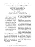
Fig. 1. Caseinolytic activity (A) and Suc-Leu-Leu-Val-Tyr-MCA-cleav-
ing activity (B) of ovarian fluid carrying embryos at various develop-
mental stages (from stage 11 to stage 31) and after embryo
delivery, D. Black circles and white squares indicate the activities
of the fluid preincubated without and with 20 m
M EDTA, respec-
tively. Caseinolytic and MCA-cleaving activities are expressed as
DA
280
30 min
)1
and nmolÆmin
)1
, respectively.
M. Kawaguchi et al. Hatching enzyme of ovoviviparous black rockfish
FEBS Journal 275 (2008) 2884–2898 ª 2008 The Authors Journal compilation ª 2008 FEBS 2885
protease with the substrate specificity similar to that of
known HCEs is present specifically in the stage 31
ovarian fluid.
Choriolytic activity in stage 31 ovarian fluid and
morphological changes of the chorion
As stage 31 of black rockfish embryos is the stage
immediately before hatching, it is conceivable that
metalloprotease(s) present in the stage 31 ovarian fluid
are the hatching enzyme(s) of black rockfish. When the
stage 31 ovarian fluid was incubated with chorion frag-
ments, the amount of liberated peptides was increased
up to 30 min and became constant thereafter
(Fig. 2A). Most of the peptides were not liberated after
the treatment with EDTA, suggesting that metallopro-
tease efficiently digesting the chorion is present in the
stage 31 ovarian fluid. After 30 min of incubation, the
chorion was swollen (Fig. 2D), and the thickness of
the chorion was increased about four times when com-
pared with that of the control chorion (Fig. 2B,C).
Eighty minutes later, the inner layer of the chorion
was completely digested, and the thin outer layer
remained undigested (Fig. 2E).
The fine structure of the black rockfish chorion before
or after incubation with ovarian fluid was observed with
an electron microscope. The control chorion was com-
posed of a thick inner layer and a thin outer layer. The
inner layer seems to be composed of two layers, which
are morphologically distinct (Fig. 3A). No significant
change of the chorion was observed after the incubation
with stage 24 ovarian fluid (data not shown). On the
other hand, stage 31 ovarian fluid swelled both of the
inner layers of the isolated chorion (Fig. 3B), and fine
fibrillar structures were observed in the outer region of
the inner layer (Fig. 3C). This structural change was
similar to that of the chorion isolated from stage 31
embryos (Fig. 3D). The chorion-digesting property of
the stage 31 ovarian fluid was similar to that of HCEs
that have been previously reported in medaka and Fund-
ulus [8,13]. This observation suggests that an HCE-like
activity, rather than an LCE-like activity, exists in
stage 31 ovarian fluid.
Identification of HCE from stage 31 ovarian fluid
The protease(s) in stage 31 ovarian fluid was par-
tially purified by successive HPLC steps through a gel
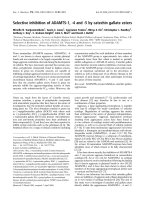
Fig. 2. (A) Time course of chorion solubilization by stage 31 ovarian
fluid. Black circles and white squares indicate the activities of the
fluid preincubated without and with 20 m
M EDTA, respectively. The
activity is expressed as the value of DA
595
. Black rockfish chorion
isolated from stage 11 embryos was incubated for 0 min (B, C),
30 min (D) and 80 min (E). Scale bars: 100 lm. Arrows indicate
thickness of chorion.
Fig. 3. Electron microscopic observation of morphological change
of the chorion by stage 31 ovarian fluid. The chorion isolated from
stage 11 embryos was incubated with only the buffer (A) and with
stage 31 ovarian fluid (B). (C) High magnification of the part shown
in the box in (B). The bar indicates the outer layer. (D) The chorion
isolated from a stage 31 embryo. Scale bars: 1 lm (A, B, D) and
0.5 lm (C).
Hatching enzyme of ovoviviparous black rockfish M. Kawaguchi et al.
2886 FEBS Journal 275 (2008) 2884–2898 ª 2008 The Authors Journal compilation ª 2008 FEBS
filtration column, S-Sepharose column and Source 15S
column. Figure 4 shows the chromatogram of the
Source 15S column. Most of the proteins were
adsorbed to the column, and the proteolytic activity
was eluted as two peaks just after a large protein peak.
Then, the fraction containing the two peaks was sub-
jected to reversed-phase column chromatography. The
five protein peaks thus obtained were analyzed by
SDS ⁄ PAGE. The major peak, containing a 23 kDa
protein, the molecular mass of which was anticipated
to be the molecular mass of other euteleostean HCEs,
was subjected to MALDI-TOF MS analysis (Fig. 4).
The values (m ⁄ z 22 789.68 and 23 075.27) were almost
identical to the relative molecular masses calculated
from two black rockfish HCE cDNAs (SsHCE1,
M
r
= 22 584; SsHCE2, M
r
= 23 056) cloned in the
present study (described later). These results strongly
suggest that the chorion-swelling activity in the
stage 31 ovarian fluid is responsible for the action of
HCEs, the genes of which are orthologous to those of
other euteleostean HCEs.
Cloning of black rockfish hatching enzyme
cDNAs
It has been suggested that both HCE and LCE genes
are present in euteleostean fishes [10]. However, only
HCE was identified in stage 31 ovarian fluid. Whether
black rockfish possess both the HCE and LCE genes
or not remains unclear. First, we performed cloning of
hatching enzyme cDNAs by RT-PCR and RACE PCR
from the RNA of black rockfish embryos. As a result,
the 1009 bp and 1088 bp cDNAs were cloned from
black rockfish embryos. Figure 5 shows the phyloge-
netic tree constructed from the previously cloned
hatching enzyme cDNAs of fishes belonging to the
Elopomorpha (Japanese eel) and the Euteleostei
(medaka, Fundulus, fugu, and Tetraodon), together
with the cDNAs cloned in the present study. The tree
clearly shows that euteleostean hatching enzymes are
divided into HCE and LCE clades with high probabil-
ity (92% for the maximum likelihood tree, 100% for
the neighbor-joining tree, and 100% for the Bayesian
tree). On the basis of the tree, the two cloned cDNAs
were named black rockfish Seb. schlegelii HCEs,
SsHCE1 and SsHCE2.
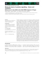
Fig. 4. Elution pattern of cation exchange Source 15S chromatogra-
phy with a linear gradient from 0 to 1
M NaCl. Solid line, absor-
bance at 280 nm; dashed line, Suc-Leu-Leu-Val-Tyr-MCA-cleaving
activity shown as nmolÆmin
)1
. The inset shows the MALDI-TOF MS
spectrum obtained from the major peak by RP-HPLC with the
range of m ⁄ z values from 21 716 to 24 768. Ions at m ⁄ z 22 789.68
and 23 075.27 were identified as the black rockfish HCE.
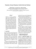
Fig. 5. A 55% majority rule consensus phylogenetic tree con-
structed by the maximum likelihood method. The tree was con-
structed using nucleotide sequences at the mature enzyme portion
of hatching enzymes of arowana (AwHE, AB276000), bony tongue
(BtHE, AB360712), Japanese eel (EHE, AB071423–9), Fundulus
(FHCE, AB210813; and FLCE, AB210814), medaka (MHCE,
M96170; and MLCE, M96169), Tetraodon (TnHCE, AB246043; and
TnLCE, AB246044), fugu (FgHCE, AB246041; and FgLCE,
AB246042), stickleback (GaHCE, AB353108–9; and GaLCE,
AB353110), Set. guentheri (SgHCE, AB353105–6; and SgLCE,
AB353107), H. hilgendorfi (HhHCE, AB353102–3; and HhLCE,
AB353104), and black rockfish (SsHCE, AB353099–100; and
wSsLCE, AB353101). Numbers at the nodes indicate bootstrap val-
ues for the maximum likelihood tree and neighbor-joining tree, and
Bayesian posterior probabilities, shown as percentages.
M. Kawaguchi et al. Hatching enzyme of ovoviviparous black rockfish
FEBS Journal 275 (2008) 2884–2898 ª 2008 The Authors Journal compilation ª 2008 FEBS 2887
To obtain evolutionary information, we amplified
HCE genes from genomic DNAs of Helicolenus hil-
gendorfi and Setarches guentheri, which belong to the
same subfamily (Sebastinae) as that of black rockfish
[15]. From both the species, SsHCE1 and SsHCE2 or-
thologs (HhHCE1 and HhHCE2 for H. hilgendorfi,
and SgHCE1 and SgHCE2 for Set. guentheri) were
cloned (Fig. 5). HCE (GaHCE1 and GaHCE2)
cDNAs were also cloned from the stickleback Gaster-
osteus aculeatus, belonging to the Gasterosteiformes
[15], which is an order different from the Scorpaenifor-
mes. Both the orders belong to the same series, the
Percomorpha.
The amino acid sequences of HCEs deduced from
the newly cloned cDNAs are shown in Fig. 6A. All
of them possessed two active site consensus
sequences of the astacin family proteases: HExxHxx-
GFxHExxRxDR (zinc-binding site) and SxMHY
(methionine turn) [17–19]. In addition, six cysteines,
which are present in all of the previously cloned
fish hatching enzymes [9], were conserved among
them.
Fig. 6. (A) A multiple alignment of amino acid sequences of hatching enzymes. White and black triangles indicate putative signal sequence
cleavage sites and N-terminals of mature enzymes, respectively. Arrows indicate intron insertion sites of LCE genes. Identical residues are
boxed. Dashes represent gaps. Two active site consensus sequences of the astacin family protease are given in dark (zinc-binding site) and
light (methionine turn) gray boxes, and conserved cysteine residues are in black boxes. (B) Exon–intron structures of black rockfish
(wSsLCE), H. hilgendorfi (HhLCE), Set. guentheri (SgLCE) and stickleback (GaLCE) LCE and HCE genes. The exons and introns are indicated
by boxes and solid lines, respectively. Numbers in parentheses indicate intron phases.
Hatching enzyme of ovoviviparous black rockfish M. Kawaguchi et al.
2888 FEBS Journal 275 (2008) 2884–2898 ª 2008 The Authors Journal compilation ª 2008 FEBS
The gene structures of all the HCE genes were deter-
mined to be intron-less (Fig. 6B), which is characteris-
tic of HCE genes [10]. Southern blot analysis showed
that the SsHCE1 probe hybridized with at least four
EcoRI fragments of 4.4, 3.8, 3.4 and 3.2 kbp of black
rockfish genomic DNA (Fig. 7A), indicating that the
black rockfish HCE gene is a multicopy gene, like
other euteleostean HCE genes examined so far [10].
As no LCE cDNA fragments were obtained from
the black rockfish by the above strategy, we employed
another strategy: that is, primers were generated from
the sequence of stickleback LCE (GaLCE) cDNA. Six
different-size cDNAs (600–2 kbp) were cloned from
black rockfish embryos, and five of the six were the
transcripts that would be formed by abnormal splicing
(see later). The other one (929 bp, SsLCE1) was well
aligned with other known LCE cDNAs, but its ORF
was incomplete. Thus, the black rockfish LCE gene is
transcribed, but the gene is not translated into a func-
tional protein. The LCE gene is predicted to be a
pseudogene. We named it black rockfish pseudo-LCE
gene (wSsLCE). These results support the finding from
the protein level experiment that only HCE activity,
not the cooperative activity of HCE and LCE, is pres-
ent in stage 31 ovarian fluid.
LCE genes were cloned from H. hilgendorfi
(HhLCE) and Set. guentheri (SgLCE). Their ORFs
were predicted to be complete. Figure 8 shows nucle-
otide and deduced amino acid sequences of
wSsLCE1 and HhLCE cDNAs. The identity of the
nucleotide sequences of the ORF between them was
95%. When compared with HhLCE cDNA,
wSsLCE1 cDNA possessed a pretermination stop
codon due to nucleotide substitution of 262G to
262T, and a frameshift mutation due to one nucleo-
tide deletion (288delA) (Fig. 8).
The gene structure of wSsLCE was determined using
the nucleotide sequence of wSsLCE1 cDNA. The
wSsLCE gene was composed of eight exons and seven
introns; its structure, including the positions of exon–
intron boundaries and intron phases, was the same as
that of other euteleostean LCE genes (Fig. 6B) [10].
Southern blot analysis was performed using genomic
DNA digested with BamHI, HindIII, ScaI and BglII.
The wSsLCE1 DNA probe hybridized with a single
fragment in each digest (Fig. 7B), suggesting that the
wSsLCE gene is a single-copy gene, like other euteleos-
tean LCE genes examined so far [10].
As described above, in addition to wSsLCE1 cDNA,
five different-size cDNAs were cloned from black rock-
fish embryos using primers designed from the 5¢-UTR
and 3¢-UTR for wSsLCE1 cDNA. The wSsLCE2
(724 bp) and wSsLCE3 (606 bp) cDNAs were shorter
than wSsLCE1 cDNA (870 bp), whereas wSsLCE4
(1033 bp), wSsLCE5 (2036 bp) and wSsLCE6
(1852 bp) cDNAs were longer than wSsLCE1 cDNA
(Fig. 9A). wSsLCE2 and wSsLCE3 cDNAs lacked the
entire region of exon 4 (146 bp) and exon 4⁄ 5 (264 bp)
of the wSsLCE gene, respectively. Considering that the
wSsLCE gene is a single-copy gene, wSsLCE2 and
wSsLCE3 cDNAs are predicted to be the products
resulting from exon skipping by aberrant splicing. As
the pretermination stop codon and the nucleotide dele-
tion are present in exon 4, wSsLCE2 and w
SsLCE3
cDNAs have complete ORFs. However, their trans-
lated products lack the N-terminal region of the
mature enzyme encoded by exon 4, and are considered
to be nonfunctional. On the other hand, wSsLCE4 and
wSsLCE5 cDNAs possessed the entire intron 1
(163 bp) and intron 5 (1166 bp) sequences, respec-
tively, showing cancellation of splicing of intron 1 and
intron 5, respectively. wSsLCE6 cDNA was 184 bp
shorter than wSsLCE5 cDNA, due to partial deletion
of exon 5 and partial inclusion of intron 5. wSsLCE6
cDNA is considered to be the transcript that appears
as a result of imprecise splicing.
As shown in Fig. 9B, intron regions including the
5¢-splicing boundary of intron 5 also showed the simi-
larity among the black rockfish, H. hilgendorfi and
Set. guentheri. When we focused on the 5¢-splicing con-
sensus sequence (gtragt) [20], we found a G to A sub-
stitution in the +5 site of the wSsLCE gene (gtra
gt to
gtga
at), whereas those of the HhLCE and SgLCE
genes were well conserved. An experiment has demon-
strated that +5 site mutation causes the exon skipping
[21]. These results suggest that the mutation found in
the wSsLCE gene probably results in intron 5 being
Fig. 7. Southern blot analysis of SsHCE1 (A) and wSsLCE (B)
genes. The restriction enzymes are shown at the top. Numbers on
the left refer to the positions of size markers.
M. Kawaguchi et al. Hatching enzyme of ovoviviparous black rockfish
FEBS Journal 275 (2008) 2884–2898 ª 2008 The Authors Journal compilation ª 2008 FEBS 2889
retained by the cancellation of splicing, as seen in
wSsLCE5 cDNA, and in the exon deletion, as seen in
wSsLCE3 cDNA (Fig. 9A).
Half of the wSsLCE cDNAs cloned in the present
study had one nucleotide deletion (73delG) located at
the 5¢-end of exon 2 (Fig. 8). The region including the
exon–intron boundary between intron 1 and exon 2
was amplified by PCR from the genomic DNA.
Sequence analysis revealed that the gene is heterozy-
gous, and that a nucleotide substitution-destroying
splicing acceptor consensus sequence (A
GtoAA;
Fig. 9B) is present in one of the alleleic wSsLCE genes.
One of the alleles used the original AG acceptor
sequence, and the other mutated allele used a pseudo-
AG acceptor sequence by shifting one nucleotide to
the 3¢-site; that is, )1A in the intronic sequence and
73G in the exonic sequence were used as the acceptor
sites. The occurrence of 73delG in wSsLCE cDNA can
be explained if the 73G was spliced out for use as a
pseudo-AG acceptor sequence (Fig. 9B). The substi-
tution might also cause the intron 1 retention, as seen
in wSsLCE4 cDNA (Fig. 9A).
Expression of black rockfish hatching enzyme
genes
First, the gene expression of SsHCE and wSsLCE was
analyzed by northern blot analysis. An SsHCE1 DNA
probe was used for detecting the HCE transcript. This
probe probably detects both the SsHCE1 and SsHCE2
transcripts, because of their high level of similarity
(88%). The hybridization of this probe with 10 lgof
total RNA did not show any signal. This amount of
RNA, 10 lg, is known to be enough for detecting the
HCE transcripts of medaka and Fundulus [8,22]. The
result suggests that the expression of SsHCE genes is
much weaker than that in other fish species, and there-
fore, poly(A)-rich RNA purified from 100 lg of total
Fig. 8. Nucleotide and predicted amino acid sequences of wSsLCE1 and HhLCE. Arrows indicate intron insertion sites with intron numbers.
Boxes indicate mutation sites found in the wSsLCE gene as described in the text.
Hatching enzyme of ovoviviparous black rockfish M. Kawaguchi et al.
2890 FEBS Journal 275 (2008) 2884–2898 ª 2008 The Authors Journal compilation ª 2008 FEBS
RNA was employed. The SsHCE1 probe hybridized
with about 1 kb of transcript; this size was consistent
with that of the cDNAs. The transcripts were detected
in stage 17 ⁄ 18 embryos, decreased in amount towards
stage 25, and disappeared thereafter (Fig. 10A). We
failed to detect the positive signal of the wSsLCE gene
transcript by northern blot analysis.
Next, gene expression was determined by RT-PCR
(Fig. 10B). After 28 cycles of PCR, sufficient expres-
sion of the SsHCE1 and SsHCE2 genes was
detected, and the band intensity of SsHCE2 tran-
scripts was about half that of SsHCE1. For the
wSsLCE gene, the 33 cycles of RT-PCR gave these
bands at about 700 bp, 800 bp, 1 kbp, and 1.2 kbp,
corresponding to wSsLCE3, wSsLCE2, wSsLCE1 and
wSsLCE4 cDNAs, respectively. The expression pat-
tern of the wSsLCE gene through the developmental
stages was similar to that of the SsHCE genes, but
the expression was much weaker than that of the
SsHCE genes.
As shown in Fig. 11, whole-mount in situ hybrid-
ization using an antisense RNA probe for the
SsHCE1 gene revealed a distribution of cells express-
ing SsHCE transcripts in developing black rockfish
embryos. It is well known that the fish hatching
gland cells differentiate at the anterior end of the
hypoblast layer, called the pillow, in the late gastrula
embryos, and until hatching, the gland cells migrate
to the final destination in a species-dependent man-
ner [5,22]. In stage 17 embryos of the black rockfish,
positive cells were first observed along the edge of
the anterior head. These cells seem to make a start
in migration from the pillow (Fig. 11A). From
stage 18 to stage 22, the cells migrated posteriorly
(Fig. 11B), and they were finally distributed widely
in the epidermis of both lateral sides of the head
Fig. 9. (A) A schematic representation of the splicing variants of the wSsLCE gene. The black triangle indicates putative N-terminals of
mature enzymes. The structures of the normally spliced form (w SsLCE1) and the alternatively spliced forms (wSsLCE2–6) are shown.
wSsLCE2, wSsLCE3, wSsLCE4, wSsLCE5 and wSsLCE6 have an exon 4 deletion, an exon 4 and 5 deletion, an intron 1 inclusion, an intron 5
inclusion, and partial deletion of exon 5 and partial inclusion of intron 5, respectively. (B) Nucleotide mutations found on the splice site con-
sensus sequence at intron 5 and intron 1. The upper part gives a comparison of the exon–intron boundary between exon 5 and intron 5
among the wSsLCE, HhLCE and SgLCE genes. The consensus sequence of splicing donor site is shown at the top. The lower part is an
electropherogram of the PCR product around the boundary between intron 1 and exon 2. The splicing acceptor consensus sequence and
pseudo-AG consensus sequence are indicated by red boxes on the upper and lower lines, respectively, together with each cDNA product.
The regions of the exon and intron are indicated by upper-case and lower-case letters, respectively.
M. Kawaguchi et al. Hatching enzyme of ovoviviparous black rockfish
FEBS Journal 275 (2008) 2884–2898 ª 2008 The Authors Journal compilation ª 2008 FEBS 2891
(Fig. 11C,D). In stage 24 and stage 25 embryos, the
signals in positive cells became weak and their num-
bers were decreased. No signals were observed in
stage 29 and stage 31 embryos and posthatching fry,
and nor were signals from sense RNA observed in
any embryos.
Fig. 10. Expression analysis of the SsHCE1,
SsHCE2 and wSsLCE genes. (A) Northern
blot analysis of expression of the SsHCE
gene during development. Arrowheads indi-
cate the positions of 28S and 18S rRNA. (B)
RT-PCR analysis of SsHCE1, SsHCE2 and
wSsLCE during development. b-Actin was
used as a control. PCR cycles were 28 for
SsHCE1 and SsHCE2, 33 for wSsLCE, and
24 for b-actin. Developmental stages are
shown at the top. Fry, posthatching
embryos. The 200 bp (SsHCE1, SsHCE2,
and wSsLCE) and 100 bp (b-actin) ladder
markers are shown in the left lane.
Fig. 11. Whole-mount in situ hybridization
of SsHCE gene during the development of
black rockfish embryos. The SsHCE1 RNA
probe was hybridized with stage 17 (A),
stage 18 (B), stage 22 (C, D), stage 24 (E)
and stage 25 (F) embryos. (A, B) Dorsal
views of head regions. Upper, the anterior-
most. (C, E, F) Lateral views. Upper, dorsal.
(D) Dorsal view of the head region. Right,
the anterior-most. Yolk was removed from
stage 22 embryos (C, D). Scale bars:
200 lm. (G) Average number of hatching
gland cells per embryo. The values are
expressed as the mean of five embryos.
Error bars indicate the standard deviation.
Hatching enzyme of ovoviviparous black rockfish M. Kawaguchi et al.
2892 FEBS Journal 275 (2008) 2884–2898 ª 2008 The Authors Journal compilation ª 2008 FEBS
Throughout the developmental stages, the total
number of SsHCE-expressing cells per embryo seemed
to be less than in other fishes. The number of hatching
gland cells in hybridized embryos was counted, and
the average number per embryo was determined at
each developmental stage (Fig. 11G). In stage 17 and
stage 22 embryos, about 100 cells were observed, and
the number was decreased to about one-half at
stage 24, to about one-quarter at stage 25, and to zero
at stage 29. These results were consistent with the
developmental expression profile obtained by northern
blot analysis. In comparison, we counted the numbers
of hatching gland cells of rainbow trout, ayu or loach
embryos at the middle to late stages of somitogenesis.
There were about 3000 (loach), 2000 (rainbow trout)
and 1000 (ayu) per embryo. Thus, black rockfish
hatching gland cells were about 10–30 times fewer in
number than those of other fish species. Summing up
the results, the black rockfish hatching enzyme gene is
actively expressed, but its expression stops at the ear-
lier stages. In addition, the expression level is consid-
ered to be suppressed to a greater extent than in other
fishes.
Discussion
We investigated the hatching of an ovoviviparous
black rockfish. The EDTA-sensitive protease activity
with a substrate specificity similar to that of known
HCEs was detected in the ovarian fluid carrying
embryos immediately before hatching stage (stage 31).
Furthermore, the protease was found to swell the inner
layer of the egg envelope (chorion) and to release some
water-soluble peptides from the chorion. HCE, one of
the euteleostean hatching enzymes, is well known to
swell the chorion by its proteolytic action. The prote-
ases in the stage 31 ovarian fluid were partially puri-
fied, and a proteolytically active fraction containing
proteins had a molecular mass corresponding to the
cloned SsHCE1 and SsHCE2 cDNAs according to
MALDI-TOF MS analysis. Therefore, these results
strongly suggest that HCEs are secreted from black
rockfish embryos immediately before the hatching
stage. This is the first demonstration of hatching
enzymes in ovoviviparous fish.
At the natural hatching of medaka and Fundulus
embryos, the chorion is efficiently solubilized, and no
swelling of the chorion has been observed, due to the
concurrent and cooperative action of LCE and HCE
[8,13]. The morphological change of the chorion
observed in black rockfish embryos implies that its
chorion digestion mechanism is different from that of
other euteleostean fishes. In addition, the present study
revealed that HCE cDNAs were cloned and their gene
expression was observed specifically in the hatching
gland cells of embryos, whereas the LCE gene was
pseudogenized. These results suggest that the chorion
digestion at black rockfish hatching is performed by
HCE alone. The intact chorion of the black rockfish
was thin and fragile when compared with the medaka
and Fundulus chorions (Fig. 2B), and had about one-
fourth the thickness of the medaka chorion [23].
According to in vitro experiments, the chorion was
completely digested by a long period of incubation
(80 min) with stage 31 ovarian fluid. Considering that
the hatching enzyme stays with the chorion for a long
time in the ovarian cavity, HCE alone would be suffi-
cient for chorion digestion.
The northern blot analysis and in situ hybridization
experiment showed that expression of the HCE gene
was suppressed to a very low extent when compared
with that of other euteleostean HCE genes. In addi-
tion, the hatching enzyme synthesis of the black rock-
fish ceased around the middle of somitogenesis,
whereas that of other teleostean fishes, such as
medaka, zebrafish, Japanese eel and ayu, could be
detected at stages from the beginning of its expression
to immediately before hatching [5,7,9,22]. These results
imply that the black rockfish embryo synthesizes an
amount sufficient for, but limited to, chorion digestion.
Such an amount would not be harmful for embryos,
as embryos might be damaged by a long period of
incubation with a high concentration of the protease.
Thus, the hatching enzyme system in oviparous fish
embryos is conserved in the ovoviviparous black
rockfish, with adaptations to their specific hatching
environment.
According to the teleostean phylogenetic tree pro-
posed by Nelson, the ovoviviparous black rockfish and
oviparous H. hilgendorfi belong to the same tribe
(Sebastinae) but different genera, and oviparous
Set. guentheri belongs to the same subfamily (Sebasti-
nae) but a different tribe [15]. The mitochondrial
DNA-based phylogenetic tree indicates that the genus
Helicolenus is sister to Sebastes, which includes the
black rockfish [24]. The nucleotide sequences of black
rockfish hatching enzyme cDNAs indicated high simi-
larity (93% and 97% for HCE1 and HCE2, respec-
tively, and 95% for LCE) to those of H. hilgendorfi,
and the phylogenetic analysis (Fig. 5) agreed well with
the mitochondrial phylogenetic tree. Despite this phy-
logenetically close relationship, the LCE genes of
H. hilgendorfi and Set. guentheri had complete ORFs,
whereas that of the black rockfish was incomplete. The
Sebastes fossils can be traced back to the late Miocene
(about 6–10 million years ago, MYA) [25]. This time
M. Kawaguchi et al. Hatching enzyme of ovoviviparous black rockfish
FEBS Journal 275 (2008) 2884–2898 ª 2008 The Authors Journal compilation ª 2008 FEBS 2893
agrees well with the divergence time of Sebastes, about
8 MYA, obtained by molecular clock estimation [26].
These results suggest that the pseudogenization
occurred within about 8 MYA of the evolutionary
pathway to Sebastes. Considering that the expression
of the wSsLCE gene was very low, the wSsLCE gene is
presumed to be on the way to becoming completely
silent.
Splicing processes are known to be catalyzed by
spliceosomes including small nuclear ribonucleoprotein
particles. These factors are responsible for the accurate
positioning of the intronic sequence elements consist-
ing of the 5¢-splice site, branchpoint sequence, polypy-
rimidine tract, and 3¢-splice site [27–29]. The consensus
sequences at the exon–intron boundaries are essential
for specifying the splicing sites. More than 90% of
abnormal splicing products have been reported to be
due to the mutation(s) of such consensus sequences
[30]; several splicing mutations causing exon skipping
and intron retention has been reported [30] [21]. For
example, the pseudo-cytochrome P4502D7 gene, con-
taining a frameshift mutation in its ORF, which is
expressed in human brain, has abnormal alternated
spliced variants resulting from exon deletion or intron
inclusion [31]. In the present study, we cloned abnor-
mal spliced variants of five different lengths of
wSsLCE cDNAs from black rockfish embryos. Such
products have never been cloned from other fish spe-
cies [4,5,8–10], suggesting that the aberrant splicing of
the wSsLCE gene occurred only in the black rockfish
lineage.
We found some nucleotide substitutions in the splice
site consensus sequences of the wSsLCE gene, as
shown in Fig. 9B. One possible evolutionary pathway
to the occurrence of aberrant splicing is as follows.
After the black rockfish LCE gene had became
untranslated into a functional protein by mutation in
the ORF, the intronic region responsible for splicing
would become free from selective pressure. Then,
several mutations would have been accumulated by
neutral evolution, and nucleotide substitutions in the
consensus sequence would give rise to abnormal
alternative splicing.
Recently, it has been reported that nonsense muta-
tions can activate nonsense-associated altered splicing
to yield a stable mRNA lacking the mutations [29,32].
It is possible that nonsense-associated altered splicing
occurred in the wSsLCE gene to produce wSsLCE2
and wSsLCE3 cDNAs, which possess a long ORF, by
skipping exon 4 containing the pretermination stop
codon and nucleotide deletion. However, neither tran-
script is dominant, and their expression was much
lower than that of wSsLCE1 cDNA. These aberrant
splicings did not produce functional products, and the
black rockfish established a single enzyme hatching
system.
The present study has shown that various types of
alternative splicing could arise due to nucleotide sub-
stitution(s) at the intronic sequence. Alternative splic-
ing is known to play important roles in generating
variations in protein function [33], and at least 35–
59% of human genes are alternatively spliced [34]. The
present investigation on the pseudogenized LCE gene
gives us an idea of the evolutionary process generating
alternative splicing, i.e. the mutations of the intronic
sequences of the genes and their subsequent natural
selection.
Experimental procedures
Fish
Black rockfish (Seb. schlegelii) were maintained in an
indoor culturing system at Miyako Fisheries Research Sta-
tion, Japan. As black rockfish females usually fertilize their
eggs from the beginning to the middle of April in the sys-
tem, the developing embryos were ordinarily collected by
canulation into the ovary from the end of April to the mid-
dle of June. Developmental stages of embryos were deter-
mined according to the criteria proposed by Kusakari [16],
and eggs and ovarian fluid were collected separately.
Stage 17, 18, 21, 22, 24, 25, 29 and 31 prehatching
embryos, and posthatching fry, were fixed with 4% parafor-
maldehyde in NaCl ⁄ P
i
at 4 °C overnight. The fixed
embryos were dehydrated gradually through a methanol
series (25%, 50% and 75% methanol in NaCl ⁄ P
i
, and then
100% methanol). The dehydrated embryos were stored at
)30 °C in methanol until use for whole-mount in situ
hybridization. RNAs were extracted from embryos of
stages 17 ⁄ 18, 19 ⁄ 20, 21, 25, 30 and 31 and posthatching fry
with Isogen (Nippon Gene, Tokyo, Japan), following the
manufacturer’s instructions. After being treated with
RNase-free DNase I (Takara, Tokyo, Japan), the extracted
RNAs were dissolved in RNase-free water, and stored at
)30 °C.
Estimation of caseinolytic activity
The caseinolytic activity of ovarian fluid was measured
using a 375 lL reaction mixture consisting of 83 mm
Tris ⁄ HCl (pH 8.0), 0.128 m NaCl, 3.3 mgÆmL
)1
casein, and
the ovarian fluid. The reaction mixture was incubated for
30 min at 30 °C. After the reaction was stopped by adding
125 lL of 20% perchloric acid, the mixture was allowed to
stand in an ice-cold water bath for 10 min and then centri-
fuged at 18 500 g for 10 min at 4 °C. The absorbance at
280 nm (A
280
) of the supernatant was measured.
Hatching enzyme of ovoviviparous black rockfish M. Kawaguchi et al.
2894 FEBS Journal 275 (2008) 2884–2898 ª 2008 The Authors Journal compilation ª 2008 FEBS
Estimation of MCA-peptide-cleaving activity
MCA-peptides, peptidyl-7-amino-4-methylcoumarins (Pep-
tide Institute, Inc., Osaka, Japan), were employed for evalu-
ating the substrate specificity of the enzyme activity in
ovarian fluid. A 250 lL reaction mixture containing 100 lm
Suc-Leu-Leu-Val-Tyr-MCA or Suc-Ala-Pro-Ala-MCA,
50 mm Tris ⁄ HCl buffer (pH 8.0), 0.128 m NaCl and ovar-
ian fluid was incubated at 30 °C for 30 min. After the reac-
tion was stopped by adding 500 lL of 20% acetic acid, the
fluorescence was measured with a Hitachi 204 fluorescence
spectrophotometer at 380 nm excitation and 460 nm
emission.
Estimation of choriolytic activity
The choriolytic activity of ovarian fluid was measured using
a30lL reaction mixture consisting of 0.128 m NaCl,
10 mm Tris ⁄ HCl buffer (pH 8.0), 0.3 lL of ovarian fluid
and three chorions isolated from stage 11 embryos. After
incubation at 30 °C for 60 min, the mixture was spun down
(at 1000 g for 10s), and 2 lL aliquots of the supernatant
were added to 60 lL of Bradford reagent (Sigma, St Louis,
MO, USA). The protein amount was determined by absor-
bance at 595 nm.
Inhibition of enzyme activity
The effect of EDTA on caseinolytic, MCA-peptide-cleaving
or choriolytic activity was examined. Ovarian fluid was
preincubated with 20 mm EDTA at 30 °C for 10 min in the
buffer, and substrates were then added. The enzyme activi-
ties were measured by the protocol described above.
Electron microscopy
The chorion was fixed in 2.5% glutaraldehyde in 0.1 m cac-
odylate buffer (pH 7.4) at 4 °C overnight. After being
rinsed with the 0.1 m cacodylate buffer, the chorion was
fixed with 1% osmium tetroxide in the same buffer, dehy-
drated in acetone, and embedded in epoxy resin.
Identification of SsHCE from ovarian fluid
Ammonium sulfate powder was added to about 2 mL of
stage 31 ovarian fluid (60% saturation). The precipitate was
collected by centrifugation (at 18 500 g for 10 min), and dis-
solved in a small amount of 50 mm bicarbonate buffer
(pH 10.2) containing 0.2 m NaCl and 0.1% Tween-20. The
solution was applied to a Superdex 75 (Amersham Pharma-
cia Biotech, Uppsala, Sweden) column for the HPLC system
(Gilson, Middleton, WI, USA), equilibrated with the same
buffer. The fractions with proteolytic activities were collected
and applied to an S-Sepharose (Amersham Pharmacia Bio-
tech) column previously equilibrated with 50 mm Tris ⁄ HCl
buffer (pH 7.2) with 0.1% Tween-20. Then, the column was
washed with the same buffer, and adsorbed protein was
eluted once with the same buffer containing 1 m NaCl. After
being dialyzed against 20 mm Tris ⁄ HCl buffer (pH 7.2) with
0.1% Tween-20, the samples were applied to a Source 15S
column (Amersham Pharmacia Biotech) for the HPLC sys-
tem and eluted with a linear gradient of 0–1 m NaCl in
20 mm Tris ⁄ HCl buffer (pH 7.2) with 0.1% Tween-20. The
fraction with proteolytic activity was collected and applied to
a YMC-Pack ODS-A (YMC Co., Ltd, Kyoto, Japan) col-
umn and eluted with a linear gradient of 0–90% acetonitrile
containing 0.1% trifluoroacetic acid under the HPLC system.
The protein amount was monitored by measuring absor-
bance at 210 nm. The relative molecular masses of proteins
were determined with a Voyager-DESTR (Applied Bio-
systems, Foster City, CA, USA) mass spectrometer.
Cloning of hatching enzyme cDNAs from black
rockfish embryos
For cloning of black rockfish orthologs of HCE (SsHCE1
and SsHCE2), cDNA fragments were obtained by the RT-
PCR method using four forward and one reverse primers
designed from the conserved regions including active site
consensus sequences of astacin family proteases as previ-
ously described [9]. Then, 5¢-RACE and 3¢-RACE PCR and
nested PCR were performed from cDNAs synthesized from
RNAs extracted from prehatching embryos with the
SMART RACE cDNA Amplification Kit (Clontech,
Mountain View, CA, USA). The following primers were
used: 5¢-RACE (for first PCR), 5¢-AAGTTGTAGGCCTTC
TGCGGGTTGATGTTC-3¢;5¢-RACE (for nested PCR),
5¢-GAGCATGGTTGAT CTCGTGCTGGATGATGC-3¢;
3¢-RACE (for first PCR), 5¢-GTACGACTACATCAGCA
TCGAGAACAGAGC-3¢; and 3¢-RACE (for nested PCR),
5¢-ATGTTTCTCCTCTCTGGGCAGAACTGGAGG-3¢.
Two and one fragments were obtained by 5¢-RACE and
3¢-RACE PCR, respectively. The nucleotide sequences of
overlapping regions of one of the 5¢-RACE fragments were
identical to the 3¢-RACE PCR product, whereas those of
the other were not. The 3¢-RACE PCR and its nested PCR
were performed to obtain the full-length cDNAs for the
other 5¢-RACE PCR product. The following primers were
used: 3¢-RACE (for first PCR), 5¢-CATCCTCTCATGGA
GGAAGGAAGCGGAGCC-3¢; and 3¢-RACE (for nested
PCR), 5¢-GAGCCGAGGCCCAAGAGGACGAAGATG
ACG-3¢.
Nucleotide sequences were determined by a 377 DNA
sequencer (Applied Biosystems), using a BigDye Termina-
tor Cycle Sequencing Kit.
For LCE (w SsLCE) cDNA, primers were generated from
nucleotide sequences of stickleback LCE cDNA. The nuc-
leotide sequences of forward and reverse primers were
M. Kawaguchi et al. Hatching enzyme of ovoviviparous black rockfish
FEBS Journal 275 (2008) 2884–2898 ª 2008 The Authors Journal compilation ª 2008 FEBS 2895
5¢-GAGCACGAGCTGCTGCACGCGCTGGGCTTC-3¢
and 5¢ -GTAGTGCATCACGGATGAGTAATCATACG-
G-3¢, respectively. After a cDNA fragment was obtained,
5¢-RACE and 3¢-RACE PCR and their nested PCR were
performed to clone full-length LCE cDNA using the fol-
lowing primers: 5¢-RACE (for first PCR), 5¢-GGGAATTA-
TGGTCTCCGATCGAC-3¢;5¢-RACE (for nested PCR),
5¢-TCGTGT CCTGCTTTCGGAAGTT GTACACGG-3¢;
3¢-RACE (for first PCR), 5¢-TCATCCCACGTGAAGCT
CAGAGG-3¢; and 3¢-RACE (for nested PCR), 5¢-GCTGT
CACTGCAGAGGTTCGGCTGCGTACG-3¢.
One cDNA fragment was amplified by 3¢-RACE PCR,
and three fragments were obtained by 5¢-RACE PCR. They
were considered to be the products of splicing variants
(wSsLCE1, wSsLCE5 and wSsLCE6). To confirm their
sequences, the full-length LCE cDNAs were amplified. The
primers were designed from the 5¢-UTR and 3¢-UTR
sequences common to the three cDNAs: forward, 5¢-CCG
GTGTGACAAGTCTCAGAAGTGAC-3¢; and reverse, 5¢-
GTCATACTGTCACATTGAAACATACAGTAATAC-3¢.
In this procedure, three cDNAs that were regarded as
the products of aberrant splicing were newly obtained in
addition to the former three. Finally, six LCE cDNAs
(wSsLCE1–6) were cloned.
Stickleback hatching enzyme genes were cloned in silico
using the Ensembl genome database (embl.
org/Gasterosteus_aculeatus/index.html), and then full-
length cDNAs for HCEs and LCE were cloned by RT-PCR
from RNA extracted from prehatching embryos.
Phylogeny
A multiple sequence alignment of amino acid sequences of
mature enzyme portions was performed using the clus-
tal x program [35], and the codon-based alignment of their
nucleotide sequences was done using the codonalign 2.0
program. Trees were constructed according to the maxi-
mum likelihood method in the program phyml [36], the
Bayesian inference in the program mrbayes 3.1.2 [37,38]
with the HKY [39] +I+G model, and the neighbor-joining
method with the distance matrix calculated using the HKY
[39] model in the program paup* 4.0b [40]. The arowana
hatching enzyme (AwHE) gene was used as an outgroup.
The reliability of the tree was assessed by bootstrap values
obtained with 2000 pseudoreplicates for maximum likeli-
hood and neighbor-joining trees.
Gene amplification
Genomic DNAs were obtained by proteinase K digestion
followed by phenol ⁄ chloroform extraction and ethanol pre-
cipitation. The black rockfish hatching enzyme genes were
amplified from the genomic DNA using primers designed
from nucleotide sequences of the 5¢- and 3¢-ends of each
full-length cDNA.
Hatching enzyme genes for H. hilgendorfi and Set. guen-
theri were cloned by PCR from the genomic DNA of each
species, using primers generated from nucleotide sequences
of the 5¢-UTR and 3¢-UTR for SsHCE1, SsHCE2 and
wSsLCE cDNAs.
Southern blot analysis
Digoxigenin (DIG)-labeled DNA probes were synthesized
with a PCR DIG Probe Synthesis Kit (Roche, Indianapolis,
IN, USA), using full-length SsHCE1 and w SsLCE1 cDNAs
as templates. One hundred micrograms of genomic DNA
was digested with EcoRI, BamHI, HindIII, ScaI and BglII,
fractionated by electrophoresis on 0.7% agarose gel, and
transferred to a nylon membrane (Hybond N
+
; Amersham,
Piscataway, NJ, USA). After the membrane had been pre-
hybridized in DIG Easy Hyb (Roche) at 42 °C for 2 h,
hybridization was performed at 42 °C overnight in the same
buffer with the DIG-labeled DNA probe. The membrane
was washed twice with 2· standard NaCl ⁄ Cit (1· NaCl ⁄ Cit
consists of 150 mm NaCl and 15 mm sodium citrate)
(pH 7.0) and 0.1% SDS for 5 min at room temperature,
and three times with 0.2· NaCl ⁄ Cit (pH 7.0) and 0.1%
SDS for 15 min at 60 °C. The membrane was incubated
with 0.2% blocking reagent in NaCl ⁄ P
i
containing 0.1%
Tween-20 for 30 min at room temperature, and with
1 : 5000-diluted alkaline phosphatase-conjugated antibodies
to digoxigenin in the same buffer for 1 h. After three 5 min
washes with NaCl ⁄ P
i
containing 0.3% Tween-20, the mem-
brane was incubated in a reaction buffer consisting of 0.1%
diethanolamine and 1 mm MgCl
2
for 5 min at room
temperature. The membrane was incubated with 1% 3-[4-
methoxyspiro{1,2-dioxetane-3,2¢-(5¢-chloro)tricyclo[3.3.1.1
3,7
]
decan}-4-yl]phenyl phosphate in the buffer and exposed to
scientific imaging film (Kodak, Rochester, NY, USA) in the
dark.
Northern blot analysis
Poly(A)-rich RNA was extracted from total RNA
(100 lg) using a PolyATract mRNA Isolation System
(Promega, Madison, WI, USA), electrophoresed on 1%
formaldehyde–agarose gel, and transferred to a nylon
membrane (Hybond N; Amersham). Hybridization was
performed using the same protocol as for the Southern
blot analysis.
Semiquantitative estimation of expression of
hatching enzyme genes by RT-PCR
RT-PCR was performed using 0.1 lg of RNA with a One-
Step RT-PCR kit (Qiagen, Valencia, CA, USA), according
to the manufacturer’s instructions. b-Actin was used as a
control, and the primers were designed from the nucleotide
Hatching enzyme of ovoviviparous black rockfish M. Kawaguchi et al.
2896 FEBS Journal 275 (2008) 2884–2898 ª 2008 The Authors Journal compilation ª 2008 FEBS
sequence of black rockfish b-actin (accession number:
AY166590). PCR was performed for 28 cycles (SsHCE1
and SsHCE2), 33 cycles (wSsLCE) or 24 cycles (b-actin),
using the following profile: (a) denaturing for 30 s at 94 °C;
(b) primer annealing for 30 s at 60 °C; and (c) extension
for 1 min at 72 °C. A final extension was performed for
10 min at 72 °C.
Whole-mount in situ hybridization
After chorions were removed from fixed embryos, the
embryos were rehydrated through a reversed methanol
series (75%, 50% and 25% methanol in NaCl ⁄ P
i
contain-
ing 0.1% Tween-20), washed for 3 · 5 min in NaCl ⁄ P
i
containing 0.1% Tween-20, and prehybridized in a
hybridization buffer consisting of 50% formamide, 5 ·
NaCl ⁄ Cit (pH 6.0), 0.1% Tween-20, 50 lgÆmL
)1
tRNA
and 50 lgÆmL
)1
heparin for 2 h at 55 °C. Hybridization
was performed overnight at 55 °C in the hybridization
buffer with a DIG-labeled antisense probe or a sense
RNA probe for SsHCE1. After four 30 min washes with
a solution consisting of 50% formamide and 2 · NaCl ⁄
Cit (pH 6.0) containing 0.1% Tween-20 (SSCT) at 68 °C,
the embryos were incubated for 4 · 15 min in 2 · SSCT
at 68 °C, washed for 3 · 20 min in 0.2 · SSCT at 68 °C,
transferred to NaCl ⁄ P
i
containing 0.1% Tween-20, and
washed for 3 · 5 min at room temperature. The embryos
were incubated for 90 min with 1% blocking reagent in
NaCl ⁄ P
i
containing 0.1% Tween-20, and then with
1 : 8000-diluted alkaline phosphatase-conjugated antibod-
ies to digoxigenin in NaCl ⁄ P
i
containing 0.1% Tween-20
at 4 °C overnight. After eight 30 min washes in NaCl ⁄ P
i
containing 0.1% Tween-20, the embryos were incubated
in a staining buffer consisting of 100 mm Tris ⁄ HCl
(pH 9.5), 50 mm MgCl
2
, 100 mm NaCl and 0.1% Tween-
20 for 2 · 5 min, and stained with 1 : 50 (v ⁄ v) Nitro Blue
tetrazolium ⁄ 5-bromo-4-chloroindol-2-yl phosphate in the
staining buffer. After the reaction, the embryos were
washed with NaCl ⁄ P
i
.
Acknowledgements
We express our thanks to Professor F. S. Howell,
Department of Chemistry, Faculty of Science and
Technology, Sophia University, Tokyo, for reading
the manuscript, and to Dr K. Yamagami, former
Professor of Developmental Biology, Life Science
Institute, Sophia University, Tokyo, for giving us
valuable advice and reading the manuscript. We
thank Dr T. P. Satoh for providing samples of
H. hilgendorfi and Set. guentheri. The present study
was supported in part by Grants-in-Aid for Scientific
Research (C) from JSPS to I. Iuchi (No. 17570189)
and to S. Yasumasu (No. 15570102).
References
1 Yamagami K (1972) Isolation of a choriolytic enzyme
(hatching enzyme) of the teleost, Oryzias latipes. Dev
Biol 29, 343–348.
2 Yamagami K (1973) Some enzymological properties of
a hatching enzyme (chorionase) isolated from the fresh-
water teleost, Oryzias latipes. Comp Biochem Physiol
46B, 603–616.
3 Yamagami K, Hamazaki TS, Yasumasu S, Masuda K
& Iuchi I (1992) Molecular and cellular basis of forma-
tion, hardening, and breakdown of the egg envelope
in fish. Int Rev Cytol 136, 51–92.
4 Yasumasu S, Yamada K, Akasaka K, Mitsunaga K,
Iuchi I, Shimada H & Yamagami K (1992a) Isolation
of cDNAs for LCE and HCE, two constituent proteases
of the hatching enzyme of Oryzias latipes, and concur-
rent expression of their mRNAs during development.
Dev Biol 153, 250–258.
5 Inohaya K, Yasumasu S, Araki K, Naruse K, Yama-
zaki K, Yasumasu I, Iuchi I & Yamagami K (1997)
Species-dependent migration of fish hatching gland cells
that express astacin-like proteases in common. Dev
Growth Differ 39, 191–197.
6 Olivotto I, Yasumasu S, Gioacchini G, Maradonna F,
Cionna C & Carnevali O (2004) Cloning and expression
of high choriolytic enzyme, a component of the hatch-
ing enzyme system, during embryonic development of
the marine ornamental fish Chrysiptera prasema. Mar
Biol 145, 1235–1241.
7 Hiroi J, Maruyama K, Kawazu K, Kaneko T,
Ohtani-Kaneko R & Yasumasu S (2004) Structure and
developmental expression of hatching enzyme genes of
the Japanese eel Anguilla japonica: an aspect of the
evolution of fish hatching enzyme gene. Dev Genes Evol
214, 176–184.
8 Kawaguchi M, Yasumasu S, Shimizu A, Hiroi J,
Yoshizaki N, Nagata K, Tanokura M & Iuchi I (2005)
Purification and gene cloning of Fundulus heteroclitus
hatching enzyme. A hatching enzyme system composed
of high choriolytic enzyme and low choriolytic enzyme
is conserved between two different teleosts, Fundulus
heteroclitus and medaka Oryzias latipes. FEBS J 272,
4315–4326.
9 Kawaguchi M, Yasumasu S, Hiroi J, Naruse K, Inoue
M & Iuchi I (2006) Evolution of teleostean hatching
enzyme genes and their paralogous genes. Dev Genes
Evol 216, 769–784.
10 Kawaguchi M, Yasumasu S, Hiroi J, Naruse K, Suzuki
T & Iuchi I (2007) Analysis of the exon–intron struc-
tures of fish, amphibian, bird and mammalian hatching
enzyme genes, with special reference to the intron loss
evolution of hatching enzyme genes in Teleostei. Gene
392, 77–88.
M. Kawaguchi et al. Hatching enzyme of ovoviviparous black rockfish
FEBS Journal 275 (2008) 2884–2898 ª 2008 The Authors Journal compilation ª 2008 FEBS 2897
11 Yasumasu S, Iuchi I & Yamagami K (1988) Medaka
hatching enzyme consists of two kinds of proteases
which act cooperatively. Zool Sci 5, 191–195.
12 Yasumasu S, Iuchi I & Yamagami K (1989) Purification
and partial characterization of high choriolytic enzyme
(HCE), a component of the hatching enzyme of the tele-
ost, Oryzias latipes. J Biochem 105, 204–211.
13 Yasumasu S, Iuchi I & Yamagami K (1989) Isolation
and some properties of low choriolytic enzyme (LCE), a
component of the hatching enzyme of the teleost, Oryz-
ias latipes. J Biochem 105, 212–218.
14 Bond JS & Beynon RJ (1995) The astacin family of
metalloendopeptidases. Protein Sci 4, 1247–1261.
15 Nelson JS (2006) Fishes of the World, 4th edn. Wiley,
New York, NY.
16 Kusakari M (1995) Studies on the reproductive biology
and artificial juvenile production of kurosoi Sebastes
schlegeli. Sci Rep Hokkaido Fish Exp Stn 47, 41–124.
17 Jiang W & Bond JS (1992) Families of metalloendopep-
tidases and their relationships. FEBS Lett 312, 110–114.
18 Bode W, Gomis-Ruth FX & Stockler W (1993) Astac-
ins, serralysins, snake venom and matrix metalloprotein-
ases exhibit identical zinc-binding environments
(HEXXHXXGXXH and Met-turn) and topologies and
should be grouped into a common family, the ‘metzinc-
ins’. FEBS Lett 331, 134–140.
19 Stocker W, Grams F, Baumann U, Reinemer P, Gomis-
Ruth FX, McKay DB & Bode W (1995) The metzinc-
ins – topological and sequential relations between the
astacins, adamalysins, serralysins, and matrixins (colla-
genases) define a superfamily of zinc-peptidases. Protein
Sci 4, 823–840.
20 Shapiro MB & Senapathy P (1987) RNA splice junc-
tions of different classes of eukaryotes: sequence statis-
tics and functional implications in gene expression.
Nucleic Acids Res 15, 7155–7174.
21 Baralle D & Baralle M (2005) Splicing in action: assess-
ing disease causing sequence changes. J Med Genet 42,
737–748.
22 Inohaya K, Yasumasu S, Ishimaru M, Ohyama A,
Iuchi I & Yamagami K (1995) Temporal and spatial
patterns of gene expression for the hatching enzyme in
the teleost embryo, Oryzias latipes. Dev Biol 171,
374–385.
23 Yamamoto M & Yamagami K (1975) Electron
microscopic studies on choriolysis by the hatching
enzyme of the teleost, Oryzias latipes. Dev Biol 43,
313–321.
24 Smith WL & Wheeler WC (2004) Polyphyly of the
mail-cheeked fishes (Teleostei: Scorpaeniformes):
evidence from mitochondrial and nuclear sequence data.
Mol Phylogenet Evol 32, 627–646.
25 Barsukov VV (1989) The upper Miocene rockfishes
(Scorpaenidae, Sebastinae) from California. Proc Zool
Inst Len 201, 73–109.
26 Hyde JR & Vetter RD (2007) The origin, evolution,
and diversification of rockfishes of the genus Sebastes
(Cuvier). Mol Phylogenet Evol 44, 790–811.
27 Staley JP & Guthrie C (1998) Mechanical devices of the
spliceosome: motors, clocks, springs, and things. Cell
92, 315–326.
28 Reed R (2000) Mechanisms of fidelity in pre-mRNA
splicing. Curr Opin Cell Biol 12, 340–345.
29 Hastings ML & Krainer AR (2001) Pre-mRNA splicing
in the new millennium. Curr Opin Cell Biol 13, 302–309.
30 Nakai K & Sakamoto H (1994) Construction of a novel
database containing aberrant splicing mutations of
mammalian genes. Gene 141, 171–177.
31 Pai HV, Kommaddi RP, Chinta SJ, Mori T, Boyd MR
& Ravindranath V (2004) A frameshift mutation and
alternate splicing in human brain generate a functional
form of the pseudogene cytochrome P4502D7 that
demethylates codeine to morphine. J Biol Chem 279,
27383–27389.
32 Moore MJ (2002) RNA events. No end to nonsense.
Science 298, 370–371.
33 Black DL (2003) Mechanisms of alternative pre-messen-
ger RNA splicing. Annu Rev Biochem 72, 291–336.
34 Modrek B & Lee C (2002) A genomic view of alterna-
tive splicing. Nat Genet 30, 13–19.
35 Thompson JD, Gibson TJ, Plewniak F, Jeanmougin F
& Higgins DG (1997) The CLUSTAL_X windows
interface: flexible strategies for multiple sequence align-
ment aided by quality analysis tools. Nucleic Acids Res
25, 4876–4882.
36 Guindon S & Gascuel O (2003) A simple, fast, and
accurate algorithm to estimate large phylogenies by
maximum likelihood. Syst Biol 52, 696–704.
37 Huelsenbeck JP & Ronquist F (2001) MRBAYES:
Bayesian inference of phylogenetic trees. Bioinformatics
17, 754–755.
38 Ronquist F & Huelsenbeck JP (2003) MrBayes 3:
Bayesian phylogenetic inference under mixed models.
Bioinformatics 19, 1572–1574.
39 Hasegawa M, Kishino H & Yano T (1985) Dating of
the human–ape splitting by a molecular clock of mito-
chondrial DNA. J Mol Evol 22, 160–174.
40 Swofford DL (2003) PAUP*. Phylogenetic Analysis
Using Parsimony (*and Other Methods). Version 4.
Sinauer Associates, Sunderland, MA.
Hatching enzyme of ovoviviparous black rockfish M. Kawaguchi et al.
2898 FEBS Journal 275 (2008) 2884–2898 ª 2008 The Authors Journal compilation ª 2008 FEBS