Báo cáo khoa học: Investigations on the evolutionary conservation of PCSK9 reveal a functionally important protrusion pot
Bạn đang xem bản rút gọn của tài liệu. Xem và tải ngay bản đầy đủ của tài liệu tại đây (1023.46 KB, 13 trang )
Investigations on the evolutionary conservation of PCSK9
reveal a functionally important protrusion
Jamie Cameron
1
, Øystein L. Holla
1
, Knut Erik Berge
1,
*, Mari Ann Kulseth
1
, Trine Ranheim
1
,
Trond P. Leren
1
and Jon K. Laerdahl
2
1 Medical Genetics Laboratory, Department of Medical Genetics, Rikshospitalet University Hospital, Oslo, Norway
2 Centre for Molecular Biology and Neuroscience (CMBN), Institute of Medical Microbiology, Rikshospitalet University Hospital, Oslo,
Norway
An elevated level of plasma low-density lipoprotein
(LDL) cholesterol is a major risk factor for coronary
heart disease. The key factor regulating the level of
LDL cholesterol is the cell surface LDL receptor
(LDLR) [1]. The number of LDLRs is regulated at the
transcriptional level [1] but is also post-transcription-
ally regulated by proprotein convertase subtilisin⁄ kexin
type 9 (PCSK9) [2], also known as NARC-1 [3]. Over-
expression of PCSK9 in mice leads to reduced levels of
LDLR and increased levels of LDL cholesterol [2,4,5],
whereas mice with no functional PCSK9 have
increased levels of LDLR and reduced levels of LDL
cholesterol [6].
Some aspects of the mechanism by which PCSK9
regulates the number of LDLRs have recently been
identified. Secreted PCSK9 binds to the epidermal
growth factor-like repeat A (EGF-A) of the extra-
cellular domain of the LDLR [7]. PCSK9 bound to the
Keywords
evolutionary conservation; LDL cholesterol;
LDL receptor; PCSK9; structural
bioinformatics
Correspondence
J. K. Laerdahl, Centre for Molecular Biology
and Neuroscience (CMBN), Institute of
Medical Microbiology, Rikshospitalet
University Hospital, NO-0027 Oslo, Norway
Fax: +47 22 84 47 82
Tel: +47 22 84 47 84
E-mail:
(Received 3 April 2008, revised 9 May 2008,
accepted 16 June 2008)
doi:10.1111/j.1742-4658.2008.06553.x
Proprotein convertase subtilisin ⁄ kexin type 9 (PCSK9) interferes with the
recycling of low-density lipoprotein (LDL) receptor (LDLR). This leads to
LDLR degradation and reduced cellular uptake of plasma LDL. Naturally
occurring human PCSK9 loss-of-function mutations are associated with
low levels of plasma LDL cholesterol and a reduced risk of coronary heart
disease. PCSK9 gain-of-function mutations result in lower LDL clearance
and increased risk of atherosclerosis. The exact mechanism by which
PCSK9 disrupts the normal recycling of LDLR remains to be determined.
In this study, we have assembled homologs of human PCSK9 from 20 ver-
tebrates, a cephalochordate and mollusks in order to search for conserved
regions of PCSK9 that may be important for the PCSK9-mediated degra-
dation of LDLR. We found a large, conserved protrusion on the surface of
the PCSK9 catalytic domain and have performed site-directed mutagenesis
experiments for 13 residues on this protrusion. A cluster of residues that is
important for the degradation of LDLR by PCSK9 was identified. Another
cluster of residues, at the opposite end of the conserved protrusion, appears
to be involved in the physical interaction with a putative inhibitor of
PCSK9. This study identifies the residues, sequence segments and surface
patches of PCSK9 that are under strong purifying selection and provides
important information for future studies of PCSK9 mutants and for inves-
tigations on the function of this regulator of cholesterol homeostasis.
Abbreviations
CRD, cysteine-rich domain of PCSK9, i.e. the C-terminal domain; EGF-A, epidermal growth factor-like repeat A of LDLR; EST, expressed
sequence tag; LDL, low-density lipoprotein; LDLR, low-density lipoprotein receptor; PC, proprotein convertase; PCSK9, proprotein
convertase subtilisin ⁄ kexin type 9; WT, wild-type.
*[Correction added on 16 July 2008, after first online publication: the author name has been amended]
FEBS Journal 275 (2008) 4121–4133 ª 2008 The Authors Journal compilation ª 2008 FEBS 4121
LDLR is internalized by endocytosis [7,8], and bound
PCSK9 somehow disrupts the recycling of the LDLR.
As a consequence, the LDLR is transferred to the
lysosomes for degradation [7].
PCSK9 belongs to a superfamily of subtilisin-like
serine proteases and is the ninth mammalian member
identified in the proprotein convertase (PC) family [3].
The PC zymogens have an N-terminal signal sequence,
a prodomain, a catalytic subtilisin-like domain, and a
C-terminal domain [9]. They undergo autocatalytic
cleavage in the endoplasmic reticulum, but the prodo-
main remains noncovalently bound to the catalytic
domain. In PCSK9, the backbone is cut between
Gln152 and Ser153 [4], and autocatalysis, as well as
correct folding of the protein, is necessary for secretion
of PCSK9 [3]. Unlike other convertases, PCSK9 does
not appear to undergo a second autocatalytic event
resulting in the release of an active protease [4,10].
Instead, the prodomain remains tightly bound to the
mature, cleaved PCSK9 after secretion. It has been
shown that the enzymatic activity of PCSK9 is not
necessary for its regulation of the LDLR [11,12]. The
finding that individuals without any detectable plasma
PCSK9 are healthy and develop normally [13,14] sug-
gests that drugs targeting PCSK9 might represent a
promising new class of LDL cholesterol-lowering
drugs.
The crystal structure of free PCSK9 has recently
been determined, and showed the catalytic domain to
have high structural similarity to other subtilisin-like
serine proteases [10,15,16]. The prodomain (resi-
dues 31–152) is tightly bound to the catalytic domain
(residues 153–449), hindering access to the active site
catalytic triad, Asp186, His226, and Ser386. The N-ter-
minal part of the prodomain (residues 31–60) is struc-
turally disordered. The C-terminal cysteine-rich
domain (CRD) is built from three modules arranged
with quasi-three-fold rotational symmetry, where each
module forms a two-sheet b-sandwich comprising six
b-strands. Each b-sandwich of this pseudo-propeller
fold is structurally homologous to the C-terminal
region of resistin [15] and is held together by three
structurally conserved disulfide bonds.
In humans, various mutations in the PCSK9 gene
have been found to cause autosomal dominant hypo-
cholesterolemia or hypercholesterolemia [4,13,17–24].
For mutations that do not affect PCSK9 folding or
secretion, these effects appear to be largely mediated
by different affinities of the mutant PCSK9s for the
LDLR [11,25]. However, another level of complexity
has been added with the recent finding that PCSK9
itself is cleaved by the PC furin, and, to a lesser extent,
by PC5 ⁄ 6A [26]. PCSK9 is cleaved between resi-
dues 218 and 219 in what has been shown to be a
structurally disordered loop on the surface of the
PCSK9 catalytic domain [10,15,16]. Furin-cleaved
PCSK9 is inactive in degrading LDLR, and naturally
occurring gain-of-function mutations such as R215H
[24], F216L and R218S [17,27] are likely to be gain-of-
function mutations due to reduced furin cleavage.
The exact mechanism by which PCSK9 binds to the
LDLR and disrupts the normal recycling of the LDLR
remains to be determined. One strategy to elucidate
the underlying mechanism is to study how mutations
in the PCSK9 gene affect the PCSK9-mediated degra-
dation of the LDLR. Candidate residues for being of
functional importance for macromolecular interactions
involving PCSK9 are those that are highly conserved
between different species, especially conserved residues
that are solvent-exposed in unbound PCSK9 and that
do not appear to be important for protein folding.
Specific and functionally important protein–protein
interactions between PCSK9 and other macromole-
cules are likely to be mediated through a contact area
with complementary shape, hydrophobicity and
charges for the two protein surfaces. Mutations that
change the properties of the interacting PCSK9 surface
will result in altered, usually weakened and less specific
interaction with LDLR or another binding partner.
Consequently, residues involved in protein–protein
interactions will be more conserved during evolution
than other surface-exposed residues. We therefore
extracted the sequences of homologs of human PCSK9
from public sequence databases in order to search for
functional regions by mapping phylogenetic informa-
tion onto the known protein structure.
PCSK9 is present in the proteome of most verte-
brates as well as in the invertebrate Branchiostoma flor-
idae. Whereas most residues exposed on the PCSK9
surface appear to be under limited selective pressure in
vertebrates, a large protrusion on the catalytic domain
contains a number of absolutely conserved residues.
This protrusion could play an important role in
specific macromolecular interactions, e.g. for the inter-
action with and degradation of the LDLRs. We have
therefore performed site-directed mutagenesis of 13
residues within this protrusion in order to study how
the mutant PCSK9s affect uptake of LDL. We found
that the conserved residues cluster in two groups: one
group causes PCSK9 gain-of-function mutations,
whereas the remaining residues are located in a small
patch giving rise to mutations of the loss-of-function
type. Our data suggest that the conserved protrusion is
involved in two separate specific macromolecular inter-
actions of importance for the PCSK9-mediated degra-
dation of the LDLRs.
Conserved protrusion on PCSK9 J. Cameron et al.
4122 FEBS Journal 275 (2008) 4121–4133 ª 2008 The Authors Journal compilation ª 2008 FEBS
Results
PCSK9 has homologs in chordates and mollusks
Homologs of human PCSK9 were extracted from a
number of public databases, including the NCBI non-
redundant protein and expressed sequence tag (EST)
databases [28], uniprot [29], the ensembl resources
[30], and some sequencing project databases. Protein
sequences homologous to full-length human PCSK9
from many vertebrates were found, including primates,
rat, mouse, squirrel (Spermophilus tridecemlineatus),
and a number of other placental mammals, opossum
(Monodelphis domestica), chicken, the Carolina anole
lizard (Anolis carolinensis), frogs (Xenopus tropicalis ⁄
Xenopus laevis), and the fish species Oryzias latipes,
Danio rerio, Tetraodon nigroviridis, and Takifugu rubri-
pes (see supplementary Doc. S1). No vertebrate with
more than a single PCSK9 homolog was found.
Data from the Florida lancelet (B. floridae) sequenc-
ing project, containing both genomic and EST
sequences, indicate at least two potential homologs of
full-length PCSK9 in this organism. B. floridae is a
representative of the cephalochordates, one of the
three chordate subphyla, the other two being verte-
brates and urochordates. No homologs of full-length
PCSK9 were detected in any urochordate, e.g. in the
fairly well-studied Ciona intestinalis, or in any other
invertebrates. The C-terminal domain of PCSK9, the
CRD, was not found in any vertebrate protein apart
from PCSK9 itself. However, homologs of the CRD
appear to be present in proteins in the marine Califor-
nia sea slug (Aplysia californica) and in the freshwater
snail Biomphalaria glabrata. These CRD homologs
were extracted from ESTs from the Aplysia EST
project and the Biomphalaria sequencing project (see
supplementary Doc. S1).
The sequence data for the PCSK9 homologs are
given in the supplementary Table S1. Multiple
sequence alignments for all PCSK9 homologs were
generated (Fig. 1 and supplementary Figs S1 and S2).
Bovines might be lacking a functional PCSK9
Sequence searching with human PCSK9 in the NCBI
EST databases gave highly significant hits in human,
mouse, rat, dog, chicken, frog, fish and lancelet ESTs.
However, there was not a single detected PCSK9
homolog in 1.3 million bovine EST sequences. In a
recent Bos taurus genome assembly (Btau_3.1) from
the Baylor College of Medicine Human Genome
Sequencing Center, we detected a genomic sequence on
Bos taurus chromosome 3 with high similarity to
human PCSK9 exons 8–12. These putative PCSK9
exons have insertions ⁄ deletions and nonsynonymous
mutations in regions that are absolutely conserved in
all other vertebrates, including fish, and there appears
to be a premature stop codon in putative bovine
exon 10. Traces sequenced in both directions on the
genome are available in the NCBI Trace Archive that
supports the stop codon in exon 10. The Btau_3.1 ver-
sion of the bovine genome is a preliminary assembly
based on approximately 26 million reads and 7·
sequence coverage. Close to 95% of bovine ESTs were
contained in the assembled contigs, indicating that less
than one in 20 Bos taurus protein-coding genes are
missing in this assembly.
The above findings suggest that the region on bovine
chromosome 3 with homology to PCSK9 is a remnant
of a PCSK9 pseudogene, and that extant Bos taurus
might be lacking functional PCSK9.
Site-directed mutagenesis of residues in a
conserved protrusion on PCSK9
On the basis of a multiple sequence alignment of 18
vertebrate PCSK9 homologs, residue conservation
was mapped onto a PCSK9 structural model with the
consurf tool [31,32] (Fig. 2). Residue conservation
on the solvent-exposed PCSK9 surface is limited. The
exception is a large protrusion on the catalytic
domain with a surface area of roughly 1500 A
˚
2
(Fig. 2B). Approximately half of this protrusion, the
part closest to the prodomain, is built from the struc-
turally disordered loop Gly213–Arg218 (Fig. 2A) and
residues partially covered by this loop (Fig. 2C). Evo-
lutionarily conserved regions on protein surfaces are
likely to be of functional importance, such as being
involved in specific interactions with other macro-
molecules. We therefore performed site-directed muta-
genesis of the 13 most conserved residues in this
region (i.e. residues on yellow, blue, pink and green
background in Fig. 2C) and investigated how these
mutations affected PCSK9 secretion and the internali-
zation of LDL.
To study whether the mutant PCSK9s were autocat-
alytically cleaved and secreted in a normal fashion,
HepG2 cells were transiently transfected with mutant
PCSK9 plasmids harboring each of the 13 different
mutations. The amounts of pro-PCSK9 and mature
PCSK9 in cell lysates were determined by western blot
analysis using an antibody to PCSK9. In cells express-
ing wild-type (WT) PCSK9, two bands of 73 kDa and
64 kDa were observed, which correspond to pro-
PCSK9 and the mature form of PCSK9, respectively
(Fig. 3). Unlike the catalytically inactive mutant
J. Cameron et al. Conserved protrusion on PCSK9
FEBS Journal 275 (2008) 4121–4133 ª 2008 The Authors Journal compilation ª 2008 FEBS 4123
S386A-PCSK9, the 13 new PCSK9 mutants appeared
to be autocatalytically cleaved in a fashion similar to
that of WT-PCSK9. Moreover, all 13 mutant PCSK9s,
except for C375A-PCSK9 and C378A-PCSK9, were
secreted in a normal fashion (Fig. 3). The amount of
C378A-PCSK9 in the culture media was markedly
reduced, whereas no C375A-PCSK9 was observed in
the media. It is likely that C375A-PCSK9 and C378A-
PCSK9 are completely or partially, respectively,
retained in the endoplasmic reticulum due to abnormal
protein folding caused by disruption of the disulfide
bond bridging the residues Cys375 and Cys378.
Effect of PCSK9 mutants on the internalization
of LDL and on PCSK9 cleavage by furin
To study the effects of the 13 PCSK9 mutants on the
PCSK9-mediated degradation of the LDLR, we used
transiently transfected HepG2 cells and studied the
amount of LDL internalization by flow cytometry.
HepG2 cells transfected with WT-PCSK9, empty plas-
mid, the catalytically inactive S386A-PCSK9 plasmid
[3,23] or one of the two gain-of-function plasmids,
S127R-PCSK9 and D374Y-PCSK9 [23], were used as
controls (Fig. 4). Internalization of LDL by cells
A
B
C
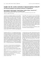
Fig. 1. Multiple sequence alignments of human PCSK9 homologs from vertebrates, a cephalochordate (B. floridae) and the mollusks Aplysia
and Biomphalaria, showing the signal sequence and N-terminus of the prodomain (A), two segments of the catalytic domain (B), and the full
CRD, the C-terminal domain (C). Conserved residues are indicated by numbering referring to human PCSK9. The catalytic triad residues are
marked with an asterisk. The full alignment and sequence data are given in supplementary Figs S1, S2 and supplementary Table S1.
Conserved protrusion on PCSK9 J. Cameron et al.
4124 FEBS Journal 275 (2008) 4121–4133 ª 2008 The Authors Journal compilation ª 2008 FEBS
expressing these control plasmids was comparable to
previous findings [23,24].
Cells expressing the two mutants C375A-PCSK9
and C378A-PCSK9 internalized 19% and 14% more
LDL, respectively, than cells expressing WT-PCSK9.
Thus, as expected for PCSK9 mutants that are secreted
at markedly reduced levels, the two mutants present as
loss-of-function type. Cells expressing R194A-PCSK9,
D238A-PCSK9, T377A-PCSK9 or F379A-PCSK9
also internalized more LDL than cells expressing
WT-PCSK9. The amounts of LDL internalized by
these cells were higher or similar to those of cells
expressing C375A-PCSK9 or C378A-PCSK9 (Fig. 4).
Thus, we also consider these four to be loss-of-func-
tion mutants.
Cells expressing R237A-PCSK9 did not show any
significant difference in LDL internalization as com-
pared with cells expressing WT-PCSK9 (Fig. 4).
R237A-PCSK9 is therefore a neutral variant. Cells
expressing one of the remaining six PCSK9 mutants
AC
B
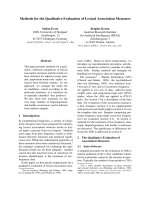
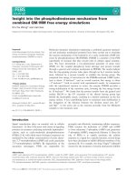
Fig. 2. Structural model of human PCSK9 with the conserved protrusion. (A) The structurally disordered loop Gly213–Arg218 (orange) with
the furin recognition motif is located on the catalytic domain (green). Also shown is the prodomain (gray) blocking the active site and the
CRD, the C-terminal domain (pink). (B) Amino acid residue conservation in 18 vertebrate PCSK9 homologs mapped, employing
CONSURF [31],
onto the space-filling representation of the PCSK9 model. The spatial orientation is identical to (A) (upper), and rotated 180° around a vertical
axis (lower). The color scale extends from cyan (highly variable residues), through white (intermediate) to magenta (highly conserved). Yellow
residues are of intermediate variability, but with low statistical confidence [31]. The conserved protrusion is visible in the panel as an
extended patch in magenta (upper part, right-hand side). (C) Magnification of the conserved protrusion showing the 13 residues of the cur-
rent study giving mutants with a low level of protein secretion (yellow background), loss-of-function mutants (blue), gain-of-function mutants
(pink), no change (green), as well as three residues of the disordered loop Gly213–Arg218 previously shown to give rise to gain-of-function
mutants (orange). The model is rotated 50° around a vertical axis with respect to (B), upper panel.
J. Cameron et al. Conserved protrusion on PCSK9
FEBS Journal 275 (2008) 4121–4133 ª 2008 The Authors Journal compilation ª 2008 FEBS 4125
S153A-PCSK9, Q190A-PCSK9, D204A-PCSK9,
K222A-PCSK9, D374A-PCSK9 and S376A-PCSK9
internalized less LDL than cells expressing WT-PCSK9
(Fig. 4). The amounts of LDL internalized by cells
expressing these mutants were in the same range or
lower than in cells expressing the gain-of-function
mutant S127R-PCSK9. Thus, we consider these
mutants to be gain-of-function mutants.
The four loss-of-function mutants involving Arg194,
Asp238, Thr377 or Phe379 are located close together
on the conserved protrusion (Fig. 2C). Four gain-of-
function mutants, involving Gln190, Lys222, Asp374
and Ser376, are located together in a separate region
between the loss-of-function patch and the disordered
loop Gly213–Arg218. The two remaining gain-of-func-
tion mutants, involving Ser153 and Asp204, are
located on opposing edges of the conserved protrusion
(Fig. 2C).
Five of the six gain-of-function mutant residues are
clustered in the vicinity of the disordered loop consist-
ing of residues Gly213–Arg218 (Fig. 2C). This loop
contains the furin cleavage site RFHR
218
[26], and
cleavage by furin at this site results in PCSK9 that is
inactive in degrading the LDLR [26]. To determine
whether the gain-of-function mutants had reduced
furin cleavage, the amounts of furin-cleaved PCSK9 in
the media of HEK293 cells transiently transfected with
the different PCSK9 plasmids were determined by wes-
tern blot analysis. HEK293 cells were chosen for these
analyses because truncated PCSK9 due to cleavage by
furin is more prominent than in the medium of HepG2
cells. R215H-PCSK9 was included as a negative con-
trol. Furin-cut PCSK9 was present in small amounts
in the media of cells transfected with gain-of-function
plasmids as well as in the media of cells transfected
with WT-PCSK9 plasmid or with loss-of-function
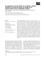
Fig. 4. Internalization of LDL by HepG2 cells transiently transfected with mutant PCSK9 plasmids. The effects of mutants S153A-PCSK9,
Q190A-PCSK9, R194A-PCSK9, D204A-PCSK9, K222A-PCSK9, R237A-PCSK9, D238A-PCSK9, D374A-PCSK9, C375A-PCSK9, S376A-PCSK9,
T377A-PCSK9, C378A-PCSK9 and F379A-PCSK9 on the internalization of fluorescently labeled LDL (10 lgÆmL
)1
) were studied in transiently
transfected HepG2 cells by flow cytometry. WT-PCSK9 plasmid, empty plasmid, and the catalytically inactive S386A-PCSK9 plasmid, as well
as D374Y-PCSK9 and S127R-PCSK9, were used as controls. The values relative to WT-PCSK9 are given as the mean from three experi-
ments (± SEM). The amount of LDL internalized by cells transfected with WT-PCSK9 was assigned a value of 100.
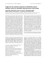
Fig. 3. Autocatalytic cleavage of the mutants S153A-PCSK9, Q190A-PCSK9, R194A-PCSK9, D204A-PCSK9, K222A-PCSK9, R237A-PCSK9,
D238A-PCSK9, D374A-PCSK9, C375A-PCSK9, S376A-PCSK9, T377A-PCSK9, C378A-PCSK9 and F379A-PCSK9 was determined by western
blot analysis of cell lysates from HepG2 cells transiently transfected with the mutant PCSK9 plasmids. WT-PCSK9 plasmid, empty plasmid
and the catalytically inactive S386A-PCSK9 plasmid were used as controls. Uncleaved, pro-PCSK9 and mature, cleaved PCSK9 are indicated
(upper panel). The lower panel shows the amount of mature, cleaved PCSK9 in the media. Three separate experiments were performed;
one representative experiment is shown.
Conserved protrusion on PCSK9 J. Cameron et al.
4126 FEBS Journal 275 (2008) 4121–4133 ª 2008 The Authors Journal compilation ª 2008 FEBS
plasmids (Fig. 5). Clearly, the relative levels of full-
length mature and furin-cut PCSK9 in the media do
not correlate with mutants being gain-of-function
or not.
R194A-PCSK9 and D204A-PCSK9 are
post-translationally modified
As can be seen from Figs 3 and 5, abnormal migration
of mature, cleaved PCSK9 was observed in lysates and
media of HepG2 cells and HEK293 cells transfected
with the R194A-PCSK9 plasmid or the D204A-PCSK9
plasmid. However, the corresponding uncleaved pro-
PCSK9 (Fig. 3) and the furin-cleaved PCSK9 (Fig. 5)
appeared to migrate normally. To study whether the
abnormal migration of the mature forms of R194A-
PCSK9 and D204A-PCSK9 was due to altered auto-
catalytic cleavage, western blot analyses of media from
transfected HepG2 cells were performed using an anti-
body against the prodomain of PCSK9. The prodo-
mains of R194A-PCSK9 and D204A-PCSK9 migrated
normally (Fig. 6). Thus, the two mutants were autocat-
alytically cleaved in a normal fashion.
To study whether the abnormal migration of
mature, cleaved R194A-PCSK9 and D204A-PCSK9
was due to abnormal glycosylation, the sensitivities of
R194A-PCSK9 and D204A-PCSK9 to an enzyme mix
designed to remove all sugars were determined in cell
lysates of transiently transfected HepG2 cells. The
results showed that the differences in the migration of
mature PCSK9 remained after the enzyme treatment
(Fig. 7). Thus, an abnormal post-translational modifi-
cation other than glycosylation appears to be respon-
sible for the abnormal migration of R194A-PCSK9
and D204A-PCSK9.
Discussion
Vertebrate genome sequencing projects are currently
supplying the research community with sequence data
from a large number of species that have varying evo-
lutionary relationships with humans. The data from
these projects make it possible to study in detail the
level of evolutionary residue conservation in proteins,
including estimates of statistical significance. In the
present study, we have extracted protein sequence data
from 21 chordate proteomes and mapped the degree of
residue conservation onto the surface of a PCSK9
structure model. We found that most of the residues
on the surface of this LDLR-degrading protein appear
to be tolerant to substitutions. However, a single large
protrusion on the catalytic domain contains a number
of residues that are highly conserved (Fig. 2B). We
have performed site-directed mutagenesis of 13 resi-
dues contributing to this protrusion (Fig. 2C), and
show that most mutants have either increased or
decreased ability to degrade the LDLR and internalize
Fig. 5. Amounts of furin-cleaved PCSK9 in the media of HEK293 cells transiently transfected with the mutant PCSK9 plasmids
S153A-PCSK9, Q190A-PCSK9, R194A-PCSK9, D204A-PCSK9, K222A-PCSK9, R237A-PCSK9, D238A-PCSK9, D374A-PCSK9, C375A-PCSK9,
S376A-PCSK9, T377A-PCSK9, C378A-PCSK9 and F379A-PCSK9 were determined by western blot analysis. WT-PCSK9 plasmid, and the
gain-of-function plasmids D374Y-PCSK9 and R215H-PCSK9, were used as controls. R215H-PCSK9 is not cleaved by furin. Three separate
experiments were performed; one representative experiment is shown.
WT R194A
D204A
PCSK9 prodomain
Fig. 6. Western blot analysis using an antibody to PCSK9 recogniz-
ing the prodomain was used to identify the prodomains of
WT-PCSK9, R194A-PCSK9 and D204A-PCSK9 in the media of
HepG2 cells.
Fig. 7. Western blot analysis was performed for deglycosylated cell
lysates. The figure shows a representative western blot of cell
lysates of HepG2 cells transiently transfected with WT plasmid or
plasmids containing R194A-PCSK9 or D204A-PCSK9 with or with-
out prior treatment with the Glycoprotein Deglycosylation Kit. A
horizontal dotted line is included to show that all the mature
PCSK9s after deglycosylation have increased mobility due to degly-
cosylation.
J. Cameron et al. Conserved protrusion on PCSK9
FEBS Journal 275 (2008) 4121–4133 ª 2008 The Authors Journal compilation ª 2008 FEBS 4127
LDL as compared to WT-PCSK9 (Fig. 4). Only one
of these residues, Asp374, has previously been associ-
ated with hypercholesterolemia in human populations
[18,19,33].
Previous studies have described a number of natu-
rally occurring loss-of-function mutations in PCSK9
that result in proteins that are not autocatalytically
cleaved and ⁄ or not folded properly [4,10,13,23,24].
Apart from the previously studied active site mutant
S386A-PCSK9 [23], the only mutants of the present
study that clearly have impaired secretion are C375A-
PCSK9 and C378A-PCSK9 (Fig. 3). Both residues are
absolutely conserved in all chordates (Fig. 1B and sup-
plementary Fig. S1), demonstrating their importance
for the formation of a disulfide bridge stabilizing the
conformation and overall shape of the conserved
protrusion (Fig. 2).
Four of the mutants, R194A-PCSK9, D238A-
PCSK9, T377A-PCSK9, and F379A-PCSK9, were
secreted in a similar fashion as WT-PCSK9 (Fig. 3),
but nevertheless present as loss-of-function mutants
(Fig. 4). The residues involved are located close
together on the conserved protrusion, at the far end
from both the prodomain and the disordered loop
Gly213–Arg218 (Fig. 2C). All four residues are highly
conserved in chordates (Fig. 1B and supplementary
Fig. S1), and are clearly under strong purifying selec-
tive pressure; that is to say, there is substantial nega-
tive natural selection against amino acid replacements
for these residues. Arg194 is conserved in all verte-
brates, but is replaced by Glu in B. floridae, whereas
Asp238 is conserved in all vertebrates except for the
fish Fugu (T. rubripes), where it is replaced by Glu,
a residue with similar properties to Asp. Thr377 is
conserved in every single chordate, whereas Phe379 is
conserved in all vertebrates except for the rat, where it
is substituted by another aromatic residue, Tyr. As
discussed above, conservation of these residues is not
necessary for protein expression, folding, autocatalysis
or secretion, strongly indicating that this patch on the
PCSK9 surface is, instead, of importance for the direct
physical protein–protein interactions leading to
PCSK9-mediated degradation of the LDLR.
During the preparation of this manuscript, Kwon
et al. [34] published the crystal structure of the protein
complex formed between PCSK9 and the EGF-A
domain of the LDLR. They found that the interaction
between PCSK9 and EGF-A was primarily hydropho-
bic, with some additional specific polar interactions.
Phe379 was in the center of the hydrophobic surface,
and Arg194, Asp238 and Thr377 were involved in the
polar interactions with EGF-A [34]. Thus, the surface
region of PCSK9 involved in this interaction coincides
with the part of the conserved protrusion associated
with loss-of-function mutants in the present study. Our
findings that mutations R194A, D238A, T377A and
F379A were loss-of-function mutations are in agree-
ment with the notion that they diminish the binding of
PCSK9 to EGF-A.
The crystal structure obtained by Kwon et al. [34]
shows that the N-terminal amine of mature PCSK9
Ser153 forms a salt bridge with a residue in EGF-A,
but that the Ser153 side-chain does not directly contact
the binding partner. Correspondingly, our results
showed that S153A-PCSK9 is not a loss-of-function
mutant. Instead, S153A-PCSK9 appears to lead to
decreased internalization of LDL as compared to WT-
PCSK9. One might speculate that this could be due to
a slight change in the ability of residue 153 to form a
salt bridge to EGF-A, e.g. through an inductive effect.
All the other residues that were associated with
gain-of-function mutations in the present study were
located either between the EGF-A binding patch and
the disordered loop Gly213–Arg218 (Gln190, Lys222,
Asp374, and Ser376), or between the disordered loop
and the prodomain (Asp204) (Fig. 2C). These residues
are under selective pressure, with Asp204 and Asp374
being conserved in all vertebrates. Lys222 is conserved
in nonfish vertebrates, whereas the conservation of
Ser376 appears to be slightly lower (Fig. 1B and sup-
plementary Fig. S1). Mutations of the disordered loop
residues, Arg215 [24], Phe216, and Arg218 [26], located
in this part of the conserved protrusion, have previ-
ously been shown to be associated with resistance to
furin cleavage. We therefore investigated whether the
gain-of-function mutants of the present study showed
reduced cleavage by furin. However, we did not find
any difference in the amounts of the furin-cleaved
bands when investigating the media of cells transfected
with loss-of-function mutants as compared with gain-
of-function mutants (Fig. 5.).
Some caution should be exercised when interpreting
these data, as in our study overexpressed PCSK9 has
to be cut by endogenous furin, and these conditions
may not be physiologically relevant for the in vivo
situation. Interestingly, the basic residues in the furin
recognition sequence RFHR
218
of the disordered loop
Gly213–Arg218 are conserved in all vertebrates, but
not in the pufferfish T. rubripes and T. nigroviridis or
in the opossum (Fig. 1B). The cephalochordate homo-
logs have a deletion of four residues in this loop as
compared with the human PCSK9, and appear to
completely lack the disordered loop. It appears that
PC regulation of PCSK9 is a vertebrate invention and
that this level of regulation has subsequently been lost
in some vertebrate subgroups.
Conserved protrusion on PCSK9 J. Cameron et al.
4128 FEBS Journal 275 (2008) 4121–4133 ª 2008 The Authors Journal compilation ª 2008 FEBS
If the gain-of-function character of the mutants of
the present study is not due to reduced furin affinity,
another possibility is that a different, unknown, mac-
romolecule is interacting in a specific manner with the
relevant part of the conserved protrusion of PCSK9.
This macromolecule may be competing with EGF-A
binding or may inhibit the PCSK9-mediated degrada-
tion of the LDLR by another mechanism. Fan et al.
[35] have recently suggested that multimerization of
PCSK9 is important for its LDLR-regulating activity.
They found that mutation of Asp374, a residue in the
conserved protrusion, affected PCSK9 self-association.
It is, however, not obvious that PCSK9 self-association
is important in vivo when PCSK9 is secreted at low
concentrations. Previous studies have found no indica-
tions of multimerization for mature PCSK9 [3].
Earlier studies have shown that the naturally occur-
ring mutant D374Y-PCSK9 binds LDLR more effi-
ciently than WT-PCSK9 [8,10,25], and Kwon et al. [34]
suggested that this was due to an additional hydrogen
bond between PCSK9 Tyr374 and His306 of the EGF-
A. We now show that D374A-PCSK9, which results in
a residue Ala374 that clearly cannot form any hydro-
gen bonds with its side-chain, is also a gain-of-function
mutant, although it is only half as potent as D374Y-
PCSK9. This may indicate that the naturally occurring
D374Y-PCSK9 is a gain-of-function mutant due to two
different mechanisms: one is to strengthen the interac-
tion between PCSK9 and EGF-A, and the other is to
disrupt the binding to PCSK9 of a putative inhibitory
macromolecule.
The sequence data that were gathered for the present
study reveal interesting phylogenetic relationships in
addition to the identification of the conserved protru-
sion discussed above. Homologs of full-length PCSK9
were found in a single copy in a number of verte-
brates, and at least in duplicate in the cephalochordate
B. floridae. PCSK9 thus appears to be restricted to
chordates, and possibly limited to the Cephalochordata
and Vertebrata. On the basis of the presumed diver-
gence of the chordate subphyla, one might speculate
on a Cambrian or late Proterozoic origin of PCSK9.
Interestingly, we were unable to find any bovine
PCSK9 homolog that appears to be functional, but the
Bos taurus genome does appear to contain a PCSK9-
like pseudogene. One might speculate that the cow,
with its diet of mainly grasses and plant material,
might be thriving without PCSK9, as do some human
individuals without functional PCSK9 [13,14].
The CRD, whose function still appears to be a
mystery, is the C-terminal PCSK9 domain. It does not
appear to occur in any vertebrate protein apart from
PCSK9 itself, but was detected in sequence data from
two mollusk proteins of unknown function. Although
the available data are limited, these mollusk CRDs do
not appear to be present in proteins that contain pro-
tease domains (see supplementary Doc. S1). This might
indicate that mollusks employ the CRD for a different
purpose than vertebrates do in PCSK9. It is possible
that investigations on mollusk CRD-containing pro-
teins might give indications on the function of the
PCSK9 CRD.
The multiple sequence alignments of the PCSK9
homologs (Fig. 1 and supplementary Figs S1 and S2)
clearly show the catalytic domain to be more con-
served than the CRD. This is also the case for PCSK9
conservation within the group of primates [36]. Resi-
due identities between human and opossum are 76%
and 53% for the catalytic domain and CRD, respec-
tively. The prodomain is also fairly well conserved,
apart from the structurally disordered region compris-
ing the N-terminal 30 residues (Fig. 1A). This segment
is very rich in acidic residues, with seven of 10 N-ter-
minal residues of human PCSK9 being Asp or Glu.
This is immediately followed by five small aliphatic
residues and a segment with five more acidic residues.
The N-terminal region of the prodomain will clearly
interact strongly and nonspecifically with a positively
charged moiety. The signal sequence is not conserved,
except for a Leu-rich segment.
The three catalytic residues are absolutely conserved
in all PCSK9 homologs (Fig. 1B), as is the last residue
of the prodomain, Gln152, supporting the notion that
these residues are essential for autocatalysis and effi-
cient secretion of PCSK9. The 18 Cys residues of the
PCSK9 CRD are conserved in all chordates, as well as
in the mollusk CRDs (Fig. 1C and supplementary
Fig. S2). This clearly demonstrates that the nine disul-
fide bridges covalently stabilizing the three modules of
this domain are essential for its processing and func-
tion. The CRD also contains a number of conserved
Ser, Thr and small aliphatic residues. These are mainly
located deep in the structure, and are most likely
essential for correct folding of the CRD. There is a
single patch of conserved residues on the surface of the
CRD, comprising Arg458, Thr459, Trp461, and
Glu481. These residues all contribute to the part of the
CRD surface that is interacting with the catalytic
domain. The evolutionary conservation of these resi-
dues indicates that although this interaction might be
weak [16], it appears to be of functional importance.
Piper et al. [16] have noted a large number of His
residues in the CRD. With a pK
a
value for His
between 6 and 7, it is likely that the net charge of the
CRD will become substantially more positive at
endosomal pH 5–5.5 than at pH 7.4 at the plasma
J. Cameron et al. Conserved protrusion on PCSK9
FEBS Journal 275 (2008) 4121–4133 ª 2008 The Authors Journal compilation ª 2008 FEBS 4129
membrane. It is tempting to speculate that this could
result in an altered interaction with the strongly acidic
N-terminal region of the prodomain. This might be the
reason why PCSK9 binds more strongly to the LDLR
in the endosomal ⁄ lysosomal compartments than in the
plasma [7,10]. However, the positions and number of
His residues are not particularly conserved in the CRD
(Fig. 1C). Whereas human PCSK9 has 15 His residues
out of a total of 245 CRD residues (6.1%), the propor-
tions are 5.3% for rat, 5.0% for opossum, 3.0% for
medaka fish, and 1.3% for the mollusk Aplysia.
Asn533, which is glycosylated in human PCSK9, has
been shown not to be essential for PCSK9 secretion
[13,26]. It is conserved in placental mammals only. The
corresponding residue is not likely to be glycosylated
in other vertebrates.
In conclusion, there is a single, large, evolutionarily
conserved protrusion on the surface of the catalytic
domain of PCSK9. The lack of other residue conserva-
tion on the PCSK9 surface makes it less likely that
there are other parts of PCSK9 that interact with high
specificity with other macromolecules as part of the
PCSK9-mediated degradation of the LDLR. A cluster
of residues on the conserved protrusion is involved in
the binding of PCSK9 to the EGF-A domain of the
LDLR, and mutations of these residues lead to loss of
function, as found in our study and in the study of
Kwon et al. [34]. The part of the protrusion located
around the disordered loop Gly213–Arg218 contains a
number of conserved residues for which site-directed
mutagenesis produced gain-of-function mutants. These
residues appear to be involved in some form of inhibi-
tion of the PCSK9-mediated degradation of the
LDLR. However, our data do not clearly support a
model that solely involves reduced cleavage by furin.
Thus, further studies are needed to clarify whether
these residues are involved in the binding of a different
macromolecule that inhibits the degradation of the
LDLR by PCSK9.
Experimental procedures
Data collection and bioinformatics analysis
Database resources provided by the NCBI [28], uniprot
[29], the ensembl project [30], the DOE Joint Genome
Institute (), the Baylor College of
Medicine Human Genome Sequencing Center (http://
www.hgsc.bcm.tmc.edu/projects/bovine), the Aplysia EST
project () and the B. glabrata
Genome Initiative ( />genome) were searched for homologs of human PCSK9. A
major proportion of the extracted 24 PCSK9 homologs from
23 species is due to automatic gene searching in genomic
data from early-stage sequencing projects. This necessitated
some manual trimming and manipulation of the sequences
(see supplementary Doc. S1 and supplementary Table S1).
Multiple sequence alignments were generated with mus-
cle [37], and the multiple sequence alignments were viewed
and manipulated with jalview [38]. A PCSK9 structural
model was generated from a published experimental struc-
ture [10] as described previously [24]. Amino acid conserva-
tion in all vertebrate PCSK9 homologs, but excluding the
two pufferfish species, was mapped onto the PCSK9 model
employing consurf [31,32]. The protein structure illustra-
tions were generated with pymol [39].
Cell cultures
HepG2 cells and HEK293 cells, obtained from the Euro-
pean Collection of Cell Cultures (Porton Down, UK), were
cultured in MEM (Gibco, Carlsbad, CA, USA) containing
penicillin (50 UÆ mL
)1
), streptomycin (50 lgÆmL
)1
), l-gluta-
mine (2 mm) and 10% fetal bovine serum (Invitrogen,
Carlsbad, CA, USA), in a humidified atmosphere (37 °C,
5% CO
2
).
Mutagenesis, cloning and expression of PCSK9
Mutations S153A, Q190A, R194A, D204A, K222A,
R237A, D238A, D374A, C375A, S376A, T377A, C378A or
F379A were introduced into a pCMV–PCSK9–FLAG plas-
mid kindly provided by J. D. Horton (University of Texas
Southwestern Medical Center, Dallas, TX, USA), using
QuickChange XL Mutagenesis Kit (Stratagene, La Jolla,
CA, USA) according to the manufacturer’s instructions.
The primer sequences used for the mutagenesis are given in
supplementary Table S2. The resulting mutant plasmids are
referred to as S153A-PCSK9, Q190A-PCSK9, R194A-
PCSK9, D204A-PCSK9, K222A-PCSK9, R237A-PCSK9,
D238A-PCSK9, D374A-PCSK9, C375A-PCSK9, S376A-
PCSK9, T377A-PCSK9, C378A-PCSK9, and F379A-
PCSK9. The integrity of each plasmid was confirmed by
DNA sequencing. An empty plasmid, pcDNA3.1 ⁄ myc his-c
(Invitrogen), as well as four previously published mutant
PCSK9 plasmids containing mutations S386A, S127R,
R215H or D374Y [23,24], were used as controls in the
transfection experiments together with WT-PCSK9 plasmid.
Transient transfections of HepG2 cells and HEK293 cells
with WT-PCSK9 plasmid or mutant PCSK9 plasmids were
performed as described by Cameron et al. [24].
Western blot analysis of transfected HepG2 and
HEK293 cells
Western blot analyses of cell lysates and culture media of
transiently transfected HepG2 cells or HEK293 cells were
Conserved protrusion on PCSK9 J. Cameron et al.
4130 FEBS Journal 275 (2008) 4121–4133 ª 2008 The Authors Journal compilation ª 2008 FEBS
performed as previously described [23]. A rabbit anti-
PCSK9 IgG (Cayman Chemical Company, Ann Arbor,
MI, USA) that recognizes the epitope spanning resi-
dues 490–502 was used to detect pro-PCSK9 and mature,
cleaved PCSK9. A custom-made rabbit polyclonal antibody
directed against residues 46–62 of human PCSK9 (Bethyl
Laboratories, Montgomery, TX, USA) was used to identify
the prodomain of PCSK9.
Analyses of internalization of LDL cholesterol in
transfected HepG2 cells
Analyses of the amounts of LDL cholesterol internalized in
HepG2 cells transiently transfected with different PCSK9-
containing plasmids were performed as previously described
[23]. Briefly, 24 h after transfection, the media were
replaced with serum-free OptiMEM (Gibco) and incubated
for 24 h to increase the expression of the LDLR. The cells
were then incubated with 10 lgÆmL
)1
fluorescently labeled
LDL for 2 h at 37 °C before the amounts of LDL internal-
ized were determined by flow cytometry.
Analysis of glycosylation
Cell lysates were collected from HepG2 cells 48 h after
transfection with mutant or WT-PCSK9 plasmids as pre-
viously described [23]. Twenty micrograms of each cell
lysate was treated with Glycoprotein Deglycosylation Kit
(Calbiochem, Darmstadt, Germany) according to the
manufacturer’s instructions. The treated lysates were sub-
jected to western blot analysis using the antibody to
PCSK9 directed at residues 490–502, as previously
described [23].
Acknowledgements
This work was supported by the Research Council of
Norway.
References
1 Goldstein JL, Hobbs HH & Brown MS (2001) Familial
hypercholesterolemia. In The Metabolic and Molecular
Bases of Inherited Disease (Scriver CR, Beaudet AL, Sly
WS & Valle D, eds), pp. 2863. McGraw-Hill, New
York.
2 Park SW, Moon Y-A & Horton JD (2004) Post-tran-
scriptional regulation of low density lipoprotein recep-
tor protein by proprotein convertase subtilisin ⁄ kexin
type 9a in mouse liver. J Biol Chem 279, 50630–50638.
3 Seidah NG, Benjannet S, Wickham L, Marcinkiewicz J,
Jasmin SB, Stifani S, Basak A, Prat A & Chretien M
(2003) The secretory proprotein convertase neural apop-
tosis-regulated convertase 1 (NARC-1): liver regenera-
tion and neuronal differentiation. Proc Natl Acad Sci
USA 100, 928–933.
4 Benjannet S, Rhainds D, Essalmani R, Mayne J,
Wickham L, Jin W, Asselin M-C, Hamelin J, Varret M,
Allard D et al. (2004) NARC-1 ⁄ PCSK9 and its natural
mutants. Zymogen cleavage and effects on the low
density lipoprotein (LDL) receptor and LDL choles-
terol. J Biol Chem 279, 48865–48875.
5 Maxwell KN, Fisher EA & Breslow JL (2005) Overex-
pression of PCSK9 accelerates the degradation of the
LDLR in a post-endoplasmic reticulum compartment.
Proc Natl Acad Sci USA 102, 2069–2074.
6 Rashid S, Curtis DE, Garuti R, Anderson NN, Bash-
makov Y, Ho YK, Hammer RE, Moon Y-A & Horton
JD (2005) Decreased plasma cholesterol and hypersensi-
tivity to statins in mice lacking Pcsk9. Proc Natl Acad
Sci USA 102, 5374–5379.
7 Zhang D-W, Lagace TA, Garuti R, Zhao Z, McDon-
ald M, Horton JD, Cohen JC & Hobbs HH (2007)
Binding of proprotein convertase subtilisin ⁄ kexin
type 9 to epidermal growth factor-like repeat A of
low density lipoprotein receptor decreases receptor
recycling and increases degradation. J Biol Chem 282,
18602–18612.
8 Lagace TA, Curtis DE, Garuti R, McNutt MC, Park
SW, Prather HB, Anderson NN, Ho YK, Hammer RE
& Horton JD (2006) Secreted PCSK9 decreases the
number of LDL receptors in hepatocytes and in livers
of parabiotic mice. J Clin Invest 116, 2995–3005.
9 Seidah NG, Khatib AM & Prat A (2006) The propro-
tein convertases and their implication in sterol and ⁄ or
lipid metabolism. Biol Chem 387, 871–877.
10 Cunningham D, Danley DE, Geoghegan KF, Griffor
MC, Hawkins JL, Subashi TA, Varghese AH, Ammirati
MJ, Culp JS, Hoth LR et al. (2007) Structural and bio-
physical studies of PCSK9 and its mutants linked to
familial hypercholesterolemia. Nat Struct Mol Biol 14 ,
413–419.
11 McNutt MC, Lagace TA & Horton JD (2007) Catalytic
activity is not required for secreted PCSK9 to reduce
low density lipoprotein receptors in HepG2 cells. J Biol
Chem 282, 20799–20803.
12 Li J, Tumanut C, Gavigan J-A, Huang W-J, Hampton
EN, Tumanut R, Suen KF, Trauger JW, Spraggon G,
Lesley SA et al. (2007) Secreted PCSK9 promotes LDL
receptor degradation independently of proteolytic activ-
ity. Biochem J 406, 203–207.
13 Zhao Z, Tuakli-Wosornu Y, Lagace TA, Kinch L,
Grishin NV, Horton JD, Cohen JC & Hobbs HH
(2006) Molecular characterization of loss-of-function
mutations in PCSK9 and identification of a compound
heterozygote. Am J Hum Genet 79
, 514–523.
14 Hooper AJ, Marais AD, Tanyanyiwa DM & Burnett
JR (2007) The C679X mutation in PCSK9 is present
J. Cameron et al. Conserved protrusion on PCSK9
FEBS Journal 275 (2008) 4121–4133 ª 2008 The Authors Journal compilation ª 2008 FEBS 4131
and lowers blood cholesterol in a Southern African
population. Atherosclerosis 193, 445–448.
15 Hampton EN, Knuth MW, Li J, Harris JL, Lesley SA
& Spraggon G (2007) The self-inhibited structure of
full-length PCSK9 at 1.9 A
˚
reveals structural homology
with resistin within the C-terminal domain. Proc Natl
Acad Sci USA 104, 14604–14609.
16 Piper DE, Jackson S, Liu Q, Romanow WG, Shetterly
S, Thibault ST, Shan B & Walker NPC (2007) The
crystal structure of PCSK9: a regulator of plasma LDL-
cholesterol. Structure 15, 545–552.
17 Abifadel M, Varret M, Rabe
`
s J-P, Allard D, Ouguer-
ram K, Devillers M, Cruaud C, Benjannet S, Wickham
L, Erlich D et al. (2003) Mutations in PCSK9 cause
autosomal dominant hypercholesterolemia. Nat Genet
34, 154–156.
18 Leren TP (2004) Mutations in the PCSK9 gene in Nor-
wegian subjects with autosomal dominant hypercholes-
terolemia. Clin Genet 65, 419–422.
19 Timms KM, Wagner S, Samuels ME, Forbey K, Gold-
fine H, Jammulapati S, Skolnick MH, Hopkins PN,
Hunt SC & Shattuck DM (2004) A mutation in PCSK9
causing autosomal-dominant hypercholesterolemia in a
Utah pedigree. Hum Genet 114, 349–353.
20 Kotowski IK, Pertsemlidis A, Luke A, Cooper RS,
Vega GL, Cohen JC & Hobbs HH (2006) A spectrum
of PCSK9 alleles contributes to plasma levels of low-
density lipoprotein cholesterol. Am J Hum Genet 78,
410–422.
21 Cohen JC, Boerwinkle E, Mosley TH Jr & Hobbs HH
(2006) Sequence variations in PCSK9, low LDL, and
protection against coronary heart disease. N Engl J
Med 354, 1264–1272.
22 Berge KE, Ose L & Leren TP (2006) Missense muta-
tions in the PCSK9 gene are associated with hypo-
cholesterolemia and possibly increased response to
statin therapy. Arterioscler Thromb Vasc Biol 26,
1094–1100.
23 Cameron J, Holla ØL, Ranheim T, Kulseth MA, Berge
KE & Leren TP (2006) Effect of mutations in the
PCSK9 gene on the cell surface LDL receptors. Hum
Mol Genet 15, 1551–1558.
24 Cameron J, Holla ØL, Laerdahl JK, Kulseth MA, Ran-
heim T, Rognes T, Berge KE & Leren TP (2008) Char-
acterization of novel mutations in the catalytic domain
of the PCSK9 gene. J Intern Med 263, 420–431.
25 Fisher TS, Surdo PL, Pandit S, Mattu M, Santoro
JC, Wisniewski D, Cummings RT, Calzetta A, Cub-
bon RM, Fischer PA et al. (2007) Effects of pH and
low density lipoprotein (LDL) on PCSK9-dependent
LDL receptor regulation. J Biol Chem 282, 20502–
20512.
26 Benjannet S, Rhainds D, Hamelin J, Nassoury N &
Seidah NG (2006) The proprotein convertase (PC)
PCSK9 is inactivated by furin and ⁄ or PC5
⁄ 6A.
Functional consequences of natural mutations and
post-translational modifications. J Biol Chem 281,
30561–30572.
27 Allard D, Amsellem S, Abifadel M, Trillard M,
Devillers M, Luc G, Krempf M, Reznik Y, Girardet
J-P, Fredenrich A et al. (2005) Novel mutations of
the PCSK9 gene cause variable phenotype of auto-
somal dominant hypercholesterolemia. Hum Mutat 26,
497, doi: 10.1002/humu.9383.
28 Wheeler DL, Barrett T, Benson DA, Bryant SH, Canese
K, Chetvernin V, Church DM, DiCuccio M, Edgar R,
Federhen S et al. (2008) Database resources of the
National Center for Biotechnology Information. Nucleic
Acids Res 36, D13–D21.
29 The UniProt Consortium (2008) The Universal Protein
Resource (UniProt). Nucleic Acids Res 36, D190–
D195.
30 Flicek P, Aken BL, Beal K, Ballester B, Caccamo M,
Chen Y, Clarke L, Coates G, Cunningham F, Cutts T
et al. (2008) Ensembl 2008. Nucleic Acids Res 36,
D707–D714.
31 Landau M, Mayrose I, Rosenberg Y, Glaser F, Martz
E, Pupko T & Ben-Tal N (2005) ConSurf 2005: the pro-
jection of evolutionary conservation scores of residues
on protein structures. Nucleic Acids Res 33, W299–
W302.
32 Mayrose I, Graur D, Ben-Tal N & Pupko T (2004)
Comparison of site-specific rate-inference methods for
protein sequences: empirical Bayesian methods are supe-
rior. Mol Biol Evol 21 , 1781–1791.
33 Naoumova RP, Tosi I, Patel D, Neuwirth C, Horswell
SD, Marais AD, van Heyningen C & Soutar AK (2005)
Severe hypercholesterolemia in four British families with
the D374Y mutation in the PCSK9 gene: long-term fol-
low-up and treatment response. Arterioscler Thromb
Vasc Biol 25, 2654–2660.
34 Kwon HJ, Lagace TA, McNutt MC, Horton JD &
Deisenhofer J (2008) Molecular basis for LDL receptor
recognition by PCSK9. Proc Natl Acad Sci USA 105,
1820–1825.
35 Fan D, Yancey PG, Qiu S, Ding L, Weeber EJ, Linton
MF & Fazio S (2008) Self-association of human PCSK9
correlates with its LDLR-degrading activity. Biochemis-
try 47, 1631–1639.
36 Ding K, McDonough SJ & Kullo IJ (2007) Evidence
for positive selection in the C-terminal domain of the
cholesterol metabolism gene PCSK9 based on phyloge-
netic analysis in 14 primate species. PLoS One 2, e1098,
doi: 10.1371/journal.pone.0001098.
37 Edgar RC (2004) MUSCLE: multiple sequence align-
ment with high accuracy and high throughput. Nucleic
Acids Res 32, 1792–1797.
38 Clamp M, Cuff J, Searle SM & Barton GJ (2004)
The Jalview Java alignment editor. Bioinformatics 20,
426–427.
Conserved protrusion on PCSK9 J. Cameron et al.
4132 FEBS Journal 275 (2008) 4121–4133 ª 2008 The Authors Journal compilation ª 2008 FEBS
39 Delano WL (2002) The PyMOL Molecular Graphics
System. DeLano Scientific, Palo Alto, CA.
Supplementary material
The following supplementary material is available
online:
Doc. S1. Supplementary materials and methods:
sequence data collection.
Fig. S1. Multiple sequence alignment of the signal
sequence, the prodomain and the catalytic domain of
PCSK9 homologs.
Fig. S2. Multiple sequence alignment of the C-terminal
domain for PCSK9 homologs.
Table S1. Sequence data for PCSK9 homologs.
Table S2. Primer sequences used to generate mutant
PCSK9 plasmids.
This material is available as part of the online article
from
Please note: Blackwell Publishing is not responsible
for the content or functionality of any supplementary
materials supplied by the authors. Any queries (other
than missing material) should be directed to the corre-
sponding author for the article.
J. Cameron et al. Conserved protrusion on PCSK9
FEBS Journal 275 (2008) 4121–4133 ª 2008 The Authors Journal compilation ª 2008 FEBS 4133