Báo cáo khoa học: Structural requirements for antimicrobial versus chemoattractant activities for dermaseptin S9 pptx
Bạn đang xem bản rút gọn của tài liệu. Xem và tải ngay bản đầy đủ của tài liệu tại đây (613.16 KB, 18 trang )
Structural requirements for antimicrobial versus
chemoattractant activities for dermaseptin S9
Constance Auvynet
1,2
, Chahrazade El Amri
1
, Claire Lacombe
1
, Francine Bruston
1
, Julie Bourdais
2
,
Pierre Nicolas
1
and Yvonne Rosenstein
2
1 UPMC Universite
´
de Paris 06, CNRS FRE 2852, Peptidome de la Peau des Amphibiens, Paris Cedex 5, France
2 Departamento de Medicina Molecular y Bioprocesos, Instituto de Biotecnologia, Universidad Nacional Auto
´
noma de Me
´
xico, Cuernavaca,
Morelos, Mexico
Antimicrobial peptides that play a role in host defence
against competing or pathogenic microorganisms are
small proteins, typically 10–50 residues long, interact
with lipid bilayers to alter cell-membrane permeability,
which often leads to cell death. Structural studies have
revealed that the peptide secondary structures include
a-helices, b-sheet structures stabilized by two or three
disulfide bonds, and extended structures with over-
representation of one or two amino acids (W, P, H or
G) [1]. However, all these peptides, regardless of the
secondary structure or length, are cationic and exhibit
amphipathic properties upon interaction with lipid
bilayers, with apolar amino acid residues segregating
from the hydrophilic residues on opposite sides of the
3D structure. These structural elements are believed to
play a crucial role in the binding of cationic host
Keywords
antimicrobial peptide; chemotaxis;
dermaseptin; frog skin; infrared
spectroscopy
Correspondence
P. Nicolas, UPMC University Paris 06, CNRS
FRE 2852, Peptidome de la Peau des
Amphibiens, Tour 43, 2 place Jussieu,
75251 Paris cedex 5, France
Fax: +33 1 44 27 59 94
Tel: +33 1 44 27 95 36
E-mail:
Y. Rosenstein, Departamento de Medicina
Molecular y Bioprocesos, Instituto de
Biotecnologia, Universidad Nacional
Auto
´
noma de Me
´
xico, Avenida Universidad
2001, Col Chamilpa, Cuernavaca, Mor.
62270, Mexico
Fax: +52 73172388
Tel: +52 5 55 66 22 76 63
E-mail:
(Received 29 April 2008, revised 11 June
2008, accepted 16 June 2008)
doi:10.1111/j.1742-4658.2008.06554.x
Dermaseptin S9 (Drs S9), GLRSKIWLWVLLMIWQESNKFKKM, iso-
lated from frog skin, does not resemble any of the cationic and amphipathic
antimicrobial peptides identified to date, having a highly hydrophobic core
sequence flanked at either side by cationic termini. Previous studies [Lequin
O, Ladram A, Chabbert A, Bruston F, Convert O, Vanhoye D, Chassaing
G, Nicolas P & Amiche M (2006) Biochemistry 45, 468–480] demonstrated
that this peptide adopted a non-amphipathic a-helical conformation in
trifluoroethanol ⁄ water mixtures, but was highly aggregated in aqueous
solutions and in the presence of sodium dodecyl sulfate micelles. Circular
dichroism, FTIR and attenuated total reflectance FTIR spectroscopies,
combined with a surface plasmon resonance study, show that Drs S9 forms
stable and ordered b-sheet aggregates in aqueous buffers or when bound to
anionic or zwitterionic phospholipid vesicles. These structures slowly assem-
bled into amyloid-like fibrils in aqueous environments via spherical inter-
mediates, as revealed by electron microscopy and Congo red staining.
Drs S9 induced the directional migration of neutrophils, T lymphocytes
and monocytes. Interestingly, the antimicrobial and chemotactic activities
of Drs S9 are modulated by its amyloid-like properties. Whereas spherical
oligomers of Drs S9 exhibit antimicrobial activity, the soluble, weakly self-
associated forms of Drs S9 act on human leukocytes to promote chemotaxis
and ⁄ or immunological response activation in the same range of concentra-
tion as amyloidogenic peptides Ab(1–42), the most fibrillogenic isoform of
amyloid beta peptides, and the prion peptide PrP(106–126).
Abbreviations
ATR, attenuated total reflectance; DMPC, 1,2-dimyristoyl-sn- glycero-3-phosphatidylcholine; DMPG, 1,2-dimyristoyl-sn-glycero-3-
phosphatidylglycerol; Drs B2, dermaseptin B2; Drs B4, dermaseptin B4; Drs S9, dermaseptin S9; ERK, extracellular signal-regulated kinase;
fMLP, formyl methionyl-leucyl-phenylalanine; FPRL-1, formyl peptide receptor like 1; PTX, pertussis toxin; SPR, surface plasmon resonance;
TFA, trifluoroacetic acid.
4134 FEBS Journal 275 (2008) 4134–4151 ª 2008 The Authors Journal compilation ª 2008 FEBS
defence peptides to the negatively charged outer leaflet
of bacterial bilayers. Once bound, the hydrophobic
face of the amphipathic peptide allows the peptide to
enter the membrane interior, thereby triggering local
fusion of the membrane leaflets, pore formation,
cracks and membrane disruption [2–7].
Frog skin is by far the most important source of
antimicrobial peptides, with more than half of the pep-
tides described to date isolated from South American
Hylidae or European, Asian or North American Rani-
dae [1]. Among these peptides, dermaseptins S and B,
from the skin of the South American tree frogs Phyllo-
medusa bicolor and P. sauvagei, form a family of
amphipathic, a-helical, closely related antimicrobial
peptides with broad-spectrum microbicidal activities at
micromolar concentrations against Gram-positive and
Gram-negative bacteria, fungi, yeasts and protozoa [8].
In previous work, we have used the conservation of
the preproregion sequences of the preprodermaseptin
transcripts to identify a new member of the
dermaseptin S family, dermaseptin S9 (Drs S9),
GLRSKIWLWVLLMIWQESNKFKKM [9]. The
structure of this peptide does not resemble that of any
antimicrobial peptide identified to date, having a
hydrophobic central core flanked by positively charged
termini. This structure is similar to that of synthetic
peptides originally designed as transmembrane mimetic
models that spontaneously become inserted into mem-
branes [10]. We have shown that Drs S9 adopted a
non amphipathic a-helical conformation in the pres-
ence of trifluoroethanol ⁄ water mixtures, which are
known to promote helical structures, but that it aggre-
gated in aqueous solutions and in the presence of SDS
micelles [9]. Other antimicrobial peptides of the derm-
aseptin S family, particularly Drs S4 [11], have been
reported to form aggregates, leading to the proposal
that the state of aggregation of a peptide in solution
might be an important determinant for selective cyto-
toxicity as well as other biological events [12]. In the
present study, we characterized the self-organization of
Drs S9 in aqueous solutions and determined how this
might affect its biological activity. Drs S4 forms amor-
phous aggregates, but Drs S9 is a tryptophan-rich
peptide that forms ordered aggregates in aqueous
solutions, two characteristics that are reminiscent of
amyloid peptides. We thus determined whether Drs S9
could have amyloidogenic properties. Our data provide
evidence that, similar to amyloids, Drs S9 folds into a
b-sheet structure that assembles into amyloid-like
fibrils via spherical oligomeric intermediates in a time-
and temperature-dependent manner, and that, unlike
several antimicrobial peptides that form amyloid-like
fibers in the presence of acidic phospholipids [13–15],
Drs S9 forms amyloid fibrils in an aqueous envir-
onment.
Antimicrobial peptides are fundamental components
of the innate immune response; in addition to killing
invading microorganisms, they modulate several
immune functions such as chemotaxis of immune cells
[16] through specific cell-surface receptors, such as for-
myl peptide receptor-like 1 (FPRL-1) [17]. Interest-
ingly, amyloid peptides, which are associated with
neuroinflammatory disorders such as Alzheimer’s,
prion or Parkinson’s diseases, also interact with
FPRL-1 and promote chemotaxis, favouring inflamma-
tion [18–21]. The neurotoxin prion peptide fragment
PrP(106–126) is a chemotactic agonist for the G pro-
tein-coupled receptor FPRL-1 [18]. Moreover, the vari-
ous structural states of the amyloidal peptide Ab(1–42)
modulate its biological activities: the low-molecular-
weight form of the peptide seems to be chemotactic,
while the oligomeric form seems to be neurotoxic
[21,22]. With this in mind, we also tested the chemo-
tactic potential of Drs S9 and determined whether the
amyloidogenic properties of Drs S9 could modulate its
biological activities. Maximum antimicrobial activity
of Drs S9 was detected in its oligomeric form, while,
similar to amyloid-like peptides, Drs S9 was chemotac-
tic for human leukocytes in its soluble, low-molecular-
weight, self-associated form. Together, the data pre-
sented here establish that dermaseptin S9 is the first
antimicrobial frog skin peptide to exhibit amyloido-
genic properties with potent chemotactic and antimi-
crobial activities.
Results
Dermaseptin S9 is highly aggregated and forms
b-structures
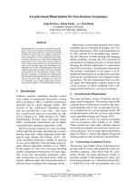
Amide H ⁄ D exchange kinetics monitored by attenu-
ated total reflectance (ATR) FTIR provided informa-
tion on the solvent accessibility of the peptide NH
amide groups. The solid state was taken as 100% NH
content for the NH ⁄ ND exchange analysis. After
15 min, lower NH ⁄ ND exchanges were observed for
Drs S9 dissolved in NaCl ⁄ P
i
(18%) than for Drs S9
dissolved in D
2
O (50%) (Fig. 1A), which is concomi-
tant with an increase in b-sheet content (see below)
that underlies the self-associative propensity of the
peptide in the saline buffer.
The kinetics of interactions between a peptide and
an adsorbing surface can be monitored in real time by
surface plasmon resonance (SPR). A shift in the reso-
nance signal during the adsorption step may provide
information about the concentration of the peptide
C. Auvynet et al. Antimicrobial and chemotactic dermaseptin S9
FEBS Journal 275 (2008) 4134–4151 ª 2008 The Authors Journal compilation ª 2008 FEBS 4135
on the adsorbing surface. For the hydrophobic HPA
biosensor, consisting of long chain alkanethiol mole-
cules attached directly to the gold film, the SPR signals
(resonance units) plotted as a function of peptide con-
centration (1–300 lm) further suggest a self-associative
propensity of Drs S9 (Fig. 1B). The resulting curve
was biphasic, in contrast to monophasic curves
obtained with another well-known dermaseptin,
Drs B2 [23]. The density of Drs S9 peptide adsorbed
onto HPA was quantified under continuous flow,
assuming the manufacturer’s characteristics for the
detection surface (1.2 mm
2
) [24]. The surface area
occupied by a 23-residue peptide molecule differs
depending on its conformation. When in a-helix for-
mation, its contact surface may be regarded as a rect-
angle measuring 3.3 nm long by 1.5 nm wide, giving a
surface area of approximately 5 nm
2
; in the b-structure
formation, the peptide has a contact area of 10–
12 nm
2
[25]. Consequently, the hydrophobic surface
could bind 0.9 ngÆmm
)2
of helical and 0.4 ngÆmm
)2
of
b-sheet peptide, respectively. The measured adsorption
densities for 30 lm peptide were thus compatible with
a b-structure for Drs S9 (Table 1). Figure 1B shows
that, in the range of 5–30 lm, a monolayer of Drs S9
in b-sheet formation was adsorbed (480 resonance
units, see Table 1). From 30–300 lm, the resonance
unit values were 2- to 4-fold greater, corresponding to
various types of peptide–peptide association. Further-
0 5 10 15
0
20
40
60
80
100
D
2
O
PBS
NH content (%)
Time (min)
0 50 100 150 200 250 300
0
250
500
750
1000
1250
1500
1750
Resonance units (RU)
PS9 concentration (µM)
200 220 240
–40
–20
0
20
40
60
PBS
PB
H
2
O
Δε
(M
–1
·cm
–1
)
Wavelength (nm)
1500 1600 1700 18001550 1650 1750
0.00
0.01
0.02
0.03
0.04
0.05
1624 cm
–1
Absorbance (a.u.)
Wavenumber (cm
–1
)
0
10
20
30
40
1624 (β-sheet)
1636
16501665
1685
(β-hairpin)
(β-turn)
(β-turnandbent)
(disordered)
A
CD
E
B
Fig. 1. Dermaseptin S9 is highly aggregated
and forms b-structures. (A) NH content
kinetics obtained by ATR FTIR. (B) Absorp-
tion density (resonance units) versus con-
centration (1–300 l
M) obtained by SPR. (C)
CD spectra of Drs S9 (30 l
M) freshly dis-
solved in water, phosphate buffer (PB) or
NaCl ⁄ P
i
(PBS). (D) ATR FTIR spectra in the
1500–1800 cm
)1
region indicating the adop-
tion of b-sheet structure in NaCl ⁄ P
i
medium.
(E) Distribution of component band contents
for Drs S9 in NaCl ⁄ P
i
(grey) or D
2
O (open).
Table 1. Adsorption potencies of Drs S9 on the hydrophobic HPA
biosensor as function of peptide concentration.
50 l
M HPA 100 lM HPA 300 lM HPA
SPR response
(resonance units)
480 750 1630
Adsorption density
(ngÆmm
)2
)
0.4 0.6 1.3
Adsorbed molecule
no. ⁄ mm
2
(· 10
12
)
0.07 0.12 0.26
Complex density
(ngÆmm
)2
)
a
0.4 0.6 1.3
a
The complex density (ng per mm
2
) is evaluated from values of
the SPR response taken at 180 s in the desorption step.
Antimicrobial and chemotactic dermaseptin S9 C. Auvynet et al.
4136 FEBS Journal 275 (2008) 4134–4151 ª 2008 The Authors Journal compilation ª 2008 FEBS
more, for peptide concentrations between 30 and
300 lm, the density of the complex evaluated from
values observed at 180 s during the desorption step
were identical to adsorption densities, suggesting that,
in contrast to some frog antimicrobial peptides [26],
the rate of interaction between Drs S9 and the hydro-
phobic support is not limited by the surface.
Dichroic spectra typical of b-sheet conformation
(Fig. 1C), with a negative band at 215–218 nm (amide
n fi p* transition) and a positive band at 190–200 nm
(amide p fi p* transition) were obtained from freshly
dissolved Drs S9 in phosphate buffer or NaCl ⁄ P
i
. The
spectrum profile of Drs S9 dissolved in H
2
O exhibited
a blue shift that can be attributed to a distorted b-sheet
conformation, underlining the impact of the environ-
ment on peptide structure stability as well as on the
degree of peptide self-association. In addition, the neg-
ative band became broader in phosphate buffer at
230 nm, probably due to contributions from aromatic
side chains [27]. The effect of temperature on the stabil-
ity of the peptide structure was also tested by subjecting
a solution of Drs S9 in 50 mm phosphate buffer to tem-
peratures ranging from 5 to 70 °C. No clear transition
from an ordered structure, i.e. a b-sheet conformation,
to a random coil structure was observed, demonstrating
that Drs S9 was quite stable, with a multimeric
arrangement (supplementary Fig. S1).
The 1500–1800 cm
)1
region of the ATR FTIR spec-
trum of Drs S9 dissolved in deuterated NaCl ⁄ P
i
showed a peak at 1624 ± 2 cm
)1
, corresponding to an
intermolecular b-sheet structure (Fig. 1D). Comparison
between the distribution of the Drs S9 amide I¢ com-
ponent bands in the two buffers (NaCl ⁄ P
i
or D
2
O)
(Fig. 1E) confirmed a higher content of ordered struc-
ture in NaCl⁄ P
i
medium than in aqueous medium as
evaluated by second-derivative analysis. Moreover,
Drs S9 freshly dissolved in H
2
O or in NaCl ⁄ P
i
migrated with apparent molecular masses of approxi-
mately 6 and 12 kDa under SDS–PAGE (supplemen-
tary Fig. S2), suggesting that Drs S9 could already be
self-associated as dimers or tetramers.
Dermaseptin S9 has b-amyloid-like properties
As, similar to amyloidogenic proteins, Drs S9 exhib-
ited a b-sheet-rich conformation and a high propensity
to aggregate in an aqueous environment, we assessed
its ability to form amyloid fibrils using classical
approaches [28]: (a) detection of yellow–green bi-refrin-
gence under polarized light upon staining with Congo
red, (b) identification of fibrils obtained by negative
stain transmission electron microscopy (TEM), and (c)
amide I¢ analysis by infrared spectroscopy. After
7 days of incubation at 37 °C, Congo red-treated
Drs S9 exhibited a red colour under normal light
(Fig. 2Aa), and the yellow–green bi-refringence charac-
teristic of amyloidogenic peptides under polarized light
(Fig. 2Ab). In contrast, at day 0 or after 3 days of
incubation at 37 °C, no characteristic bi-refringence
was observed under polarized light (data not shown).
Dried phosphate buffer, or a 7-day-old solution of
Drs B2, an a-helical linear antimicrobial peptide [23],
stained with Congo red in phosphate buffer did not
display the characteristic red staining under normal
light (Fig. 2Ac,Ae) nor yellow–green bi-refringence
under polarized light (Fig. 2Ad,Af). Neither Congo
red nor Drs S9 alone exhibited the red colour or the
yellow–green bi-refringence. At day 3, aggregates with
a granular appearance that looked like spherical oligo-
mers could be visualized (Fig. 2Ba). Consistent with
Congo red staining data, electron microscopy revealed
that, at day 7, the same solutions of Drs S9 exhibited,
like amyloid fibrils, a long filamentous structure of
8 nm diameter (Fig. 2Bb). As expected, at day 0, nei-
ther spherical oligomers nor fibrillar structures could
be observed (data not shown). Thus, three structural
states can be defined for Drs S9: day 0 (D0), low
molecular weight; day 3 (D3), oligomeric; day 7 (D7),
fibrillar. A kinetic study of the structure evolution
Drs S9 was monitored using CD (supplementary
Fig. S1). The amyloid-like arrangement of the peptide
was confirmed by submitting a 7-day-old solution of
Drs S9 stained with Congo red and dried onto a glass
slide to ATR FTIR spectroscopy. Maximum absorp-
tion of the spectrum was registered at 1624 cm
)1
, with
shoulders at 1665 and 1685 cm
)1
(Fig. 3A). Second-
derivative analysis of the spectrum (Fig. 3B) showed a
15% gain in the proportion of b-structures (Fig. 3C)
compared to freshly dissolved Drs S9 with 75%
b-structure (Fig. 1E). In addition, 1624 cm
)1
and
1685 cm
)1
individual component bands were identified
as fingerprints of intermolecular b-sheet aggregates
and b-amyloid structures [29]. In summary, these data
show that Drs S9 exhibits the biophysical and morpho-
logical characteristics of amyloidogenic peptides.
Dermaseptin S9 induces chemotaxis via a
seven-transmembrane G protein-coupled cell
surface receptor
Amyloidogenic peptides have been shown to partici-
pate in inflammatory processes by inducing cell
migration [30]. We investigated whether, similar to
amyloid-like peptides, Drs S9 was also chemotactic.
We evaluated the capacity of Drs S9 to induce the
migration of human peripheral blood neutrophils
C. Auvynet et al. Antimicrobial and chemotactic dermaseptin S9
FEBS Journal 275 (2008) 4134–4151 ª 2008 The Authors Journal compilation ª 2008 FEBS 4137
and T lymphocytes, as well as that of human acute
monocytic leukaemia cells, THP-1. Drs S9 triggered the
migration of human neutrophils (Fig. 4A), T lympho-
cytes (Fig. 4C) and THP-1 monocytes (data not shown)
with a typical bell-shaped dose–response curve at D0.
For all cell types, maximal response was observed at a
concentration of 50 lm. Equivalent concentrations of
Drs B2 had no effect on leukocyte motility (Fig. 4C).
Drs S9-induced cell migration reflected a chemotactic
rather than a chemokinetic movement, as addition of
various concentrations of Drs S9 to the upper wells of
the chamber abrogated neutrophil migration towards
the Drs S9-loaded lower wells (Fig. 4B), suggesting
that the cell locomotion we detected was the result of
chemotactic movement rather than enhanced random
movement.
To determine whether Drs S9-induced neutrophil
chemotaxis was mediated through a G protein-coupled
receptor, as is the case for Ab(1–42) peptides [30], we
examined whether this chemotactic activity could be
inhibited by pertussis toxin (PTX), a toxin that specifi-
cally inhibits G
ia
protein-coupled receptor signalling
[31]. Incubation of neutrophils with PTX for 30 min at
37 °C prior to the start of the chemotaxis assay inhib-
ited the migration of neutrophils by 50% compared
with freshly dissolved Drs S9 (Fig. 5A). The ability of
neutrophils to migrate towards formyl methionyl-
leucyl-phenylalanine (fMLP) (100 nm) was abrogated
by PTX. Together, these data suggest that, in addition
to a G
i
protein-coupled receptor, Drs S9-dependent
chemotactic signals are mediated by a non-PTX-
sensitive receptor.
a
A
B
b
a
b
cd
e
f
Fig. 2. Dermaseptin S9 forms amyloid-like
fibrils. (A) Drs S9 binds Congo red and
exhibits yellow–green bi-refringence; Drs S9
(500 l
M) (a, b) or Drs B2 (500 lM) (c, d),
both suspended in 50 m
M phosphate buffer
incubated for 7 days at 37 °C, or phosphate
buffer alone (e, f) were stained with Congo
red and observed under normal (a, c, e) or
polarized light (b, d, f). (B) Negative staining
electron microscopy micrograph of Drs S9
(500 l
M)in50mM phosphate buffer incu-
bated at 37 °C for (a) 3 days (bar, 1.1 nm) or
(b) 7 days (bar, 50 nm).
Antimicrobial and chemotactic dermaseptin S9 C. Auvynet et al.
4138 FEBS Journal 275 (2008) 4134–4151 ª 2008 The Authors Journal compilation ª 2008 FEBS
A variety of agonistic ligands have been described
for the low-affinity FPRL-1 receptor, including
fMLP at high concentrations (approximately 100 nm)
and amyloid Ab(1–42) peptides [32–34]. To assess
whether Drs S9-induced chemotaxis could be medi-
ated through this receptor, experiments were per-
formed in which neutrophils were pre-incubated with
Ab(1–42) (50 lm) or fMLP (100 nm) for 30 min
before testing their ability to respond to a gradient
of Drs S9, Ab(1–42) or fMLP in a chemotaxis assay.
Preincubating the cells with fMLP reduced the che-
motactic activity of Drs S9 by approximately 50%
(Fig. 5B), similar to what we had observed with
PTX. Comparable results were observed in response
to Ab(1–42) or fMLP when the cells were preincu-
bated with Ab(1–42), Drs S9 or fMLP (Fig. 5B). All
these data suggest that the chemotactic activity of
Drs S9 could be partially mediated through a recep-
tor similar to the one used by high concentrations of
fMLP or by Ab(1–42) (FPRL-1).
Activation of a G protein-coupled receptor by a
chemoattractant can lead to activation of the MAPK
pathway [35,36]. Incubation of neutrophils with freshly
0
10
20
30
40
1624 (β-sheet)
1636
16501665
1685
C
1590 1620 1650 1680 1710
Wavenumber (cm
–1
)
1624
1665
1685
1675
1665
1650
1624
1685
B
A
(β-hairpin)
(Amyloid
characteristic)
(disordered)
(Amyloid
characteristic)
2
1
0
–1
–2
–3
0.04
0.03
0.02
0.01
0.00
Absorbance (a.u.)
Absorbance 10
4
(a.u.)
1590 1620
1650
1680 1710
Fig. 3. Drs S9 exhibits aggregated b -sheet structures with an amy-
loid-like arrangement as demonstrated by ATR FTIR. (A) Amide I¢
band (1580–1720 cm
)1
) for Drs S9 (500 lM) incubated for 7 days
and stained with Congo Red. (B) Second-derivative analysis of the
spectrum in (A) indicating a strong individual band at 1685 cm
)1
that is present in b-amyloid structures. (C) Distribution of the indi-
vidual band content of amide I¢ from the spectrum in (A).
Drs S9 day 0
Drs S9 day 3
Drs S9 day 7
Drs S9 in the lower wells
Drs S9 in the lower and upper wells
fMLP (100 n
M) in the lower and
upper wells
Drs S9 day 0
Drs B 2day 0
Concentration (µM)
0 0.05 0.5 5 15 50 150 350 fMLP (100 n
M)
400
A
B
C
350
300
250
200
150
100
50
0
400
350
300
250
200
150
100
50
0
250
200
150
100
50
0
Chemotaxis index
0 0.05 0.5 5 50 350 fMLP (100 nM)
0 0.05 0.5 5 15 30 150 250 350 500 700 fMLP (100 n
M)
Fig. 4. Dermaseptin S9 is a potent chemoattractant. (A) Neutrophil
migration induced by freshly prepared Drs S9, or Drs S9 incubated
for 3 or 7 days at 37 °C, or by fMLP as a positive control and med-
ium as a negative control. (B) Abolition of the chemotactic effect
by adding the same concentrations of Drs S9 or fMLP in the upper
and the lower wells of the chemotaxis chamber compared with
addition of Drs S9 or fMPL in the lower wells only. (C) Induction of
migration of T lymphocytes by Drs S9, Drs B2 or fMLP. Similar
results were obtained from three different experiments.
C. Auvynet et al. Antimicrobial and chemotactic dermaseptin S9
FEBS Journal 275 (2008) 4134–4151 ª 2008 The Authors Journal compilation ª 2008 FEBS 4139
dissolved Drs S9 (100 lm) for 5 or 10 min or fMLP
for 5 min induced the phosphorylation of the extra-
cellular signal-regulated kinase ERK1 ⁄ 2, compared to
neutrophils incubated for the same time period with
medium alone (Fig. 5C). Preincubation with PTX,
PD98059 (a specific inhibitor of MEK1 and MEK2,
the ERK MAPK kinases) or both, before adding
Drs S9 or fMLP, prevented ERK1 ⁄ 2 phosphorylation.
These data suggest that Drs S9 is sensed in part
through a seven-transmembrane G protein-coupled
receptor, probably FPRL-1, coupled to the ERK1 ⁄ 2
MAPK kinase pathway.
The b-amyloid-like behaviour of dermaseptin S9
modulates its chemotactic and antimicrobial
activities
Taking advantage of better knowledge of the aggre-
gative and amyloid properties of Drs S9, we investi-
gated whether the fibrillization process influenced the
chemotactic and antibacterial activities of the pep-
tide. The activity of Drs S9 freshly dissolved in che-
motaxis medium was compared to that of Drs S9
incubated for 3 or 7 days at 37 °C. In all cases,
Drs S9 induced cell migration with a peak response
at 50 lm, but with maximum efficiency for freshly
dissolved Drs S9 (Fig. 4A). Interestingly, the a-helical
Drs B2 freshly dissolved in the same medium or
incubated for 3 or 7 days at 37 °C was not a
chemoattractant for T lymphocytes (Fig. 4C), neutro-
phils or THP-1 monocytes (data not shown) in the
same range of concentration (0.05–350 lm), indicat-
ing the importance of the peptide structure for
selected biological properties.
Drs S9 dissolved in NaCl ⁄ P
i
or RPMI-1640 exhib-
ited antimicrobial activity against all Gram-negative
bacteria strains tested at day 0. The antibacterial activ-
ity of 3-day-old Drs S9 in NaCl ⁄ P
i
(Table 2) or
RPMI-1640 ⁄ 1% BSA (data not shown) was stronger
than that of Drs S9 at day 0. After 7 days of incuba-
tion in NaCl ⁄ P
i
at 37 °C, Drs S9 exhibited little or no
antibacterial activity. No significant differences were
observed when Drs B2, a potent a-helical antibacterial
peptide, was dissolved in NaCl⁄ P
i
or RPMI-1640 ⁄ 1%
BSA and tested, or incubated for 3 or 7 days at 37 °C
prior to testing its antibacterial activity. Moreover,
fMLP (100 nM)
300
250
200
150
100
50
0
0 200 40
Peptides
Concentrations of Drs S9 0 day aged (µ
M)
Chemotaxis index
Neutrophils pre-incubated with medium for 30 min
Neutrophils pre-incubated with PTX (200ng. mL
–1
) for 30 min
Chemotaxis index
250
200
150
100
50
0
Medium Drs S9 (50 µ
M)A
β
(1–42) (50 µm) fMLP (100 nM)
Pre-incubation with Drs S9
Pre-incubation with fLMP
Pre-incubation with medium
Pre-incubation with Aβ(1–42)
A
B
C
RPMI Drs S9
5′ 10′
PTX
PD98059
–– – –
––––
––
––
+++ +
++++
Drs S9fMLP fMLP
pERK1/2
ERK1/2
Fig. 5. Dermaseptin S9 induces cell migration through a seven-
transmembrane G protein-coupled receptor, presumed to be the
FRLP-1 receptor. (A) Pre-incubating neutrophils with PTX
(200 ng mL
)1
) partially inhibits Drs S9-induced migration. fMLP was
used as a positive control. (B) Pre-incubating neutrophils with
Ab(1–42), Drs S9 or fMLP reduces the migration induced by freshly
dissolved Drs S9, Ab(1–42) or fMLP. (C) ERK1 ⁄ 2 phosphorylation in
response to Drs S9 or fMLP is abolished by pre-incubating human
neutrophils with PTX and ⁄ or PD98059. The same membrane was
stripped and blotted with anti-ERK1 ⁄ 2. Similar results were
obtained from three separate experiments.
Table 2. Effect of Drs S9 oligomerization state on the antimicrobial
activity of freshly prepared Drs S9 and Drs B2 or after incubation
for 3 or 7 days in NaCl ⁄ P
i
or H
2
Oat37°C.
Bacterial strains
Minimal inhibitory concentration
a
(lM)
Drs S9 (NaCl ⁄ P
i
or RPMI)
Drs B2
(NaCl ⁄ P
i
)
0 day 3 days 7 days
0, 3 and
7 days
Citrobacter rodentium 12.5 6.5 50 –
Salmonella typhimurium 25 6.5 100 3.1
Escherichia coli EPEC 50 12.5 100 3.1
Escherichia coli JPN15 50 25 R 0.2
a
The antimicrobial activity is expressed as the minimal inhibitory
concentration (l
M), which is the minimal peptide concentration
required for total inhibition of cell growth in liquid medium. Strains
were considered resistant (R) when their growth was not inhibited
by peptide concentrations > 100 l
M.
Antimicrobial and chemotactic dermaseptin S9 C. Auvynet et al.
4140 FEBS Journal 275 (2008) 4134–4151 ª 2008 The Authors Journal compilation ª 2008 FEBS
after removing the Drs S9 from the wells and replacing
it with fresh LB medium overnight, none of the sensi-
tive strains were capable of resuming growth (data not
shown), suggesting that Drs S9 is a bactericidal agent.
Together, these data indicate that Drs S9 is a more
potent bactericidal agent in the oligomeric spherical
form detected at 3 days of incubation at 37 °C, and is
a potent chemoattractant in its low-molecular-weight
oligomeric form (day 0).
Dermaseptin S9 differently affects vesicle lipid
assemblies
Bacterial membranes contain substantial (up to 30%)
amounts of negatively charged lipids such as phosphat-
idylglycerol, cardiolipin and phosphatidylserine [37],
thus 1,2-dimyristoyl-sn-glycero-3-phosphatidylglycerol
(DMPG) was used as a model system for determination
of the relationships between antimicrobial activity,
structure and membrane disturbances in the FTIR
study. Phosphatidylcholine is widely recognized
as representative for mammalian cell membranes,
thus 1,2-dimyristoyl-sn-glycero-3-phosphatidylcholine
(DMPC) was used as the lipid model to assess the inter-
actions of Drs S9 with neutral eukaryotic membranes.
We examined the conformation of Drs S9 in the
presence of DMPG or DMPC vesicles, and evaluated
the impact of freshly dissolved Drs S9 on the lipid
bilayer assembly by transmission FTIR spectroscopy.
Maximum absorbance of the amide I¢ bands was
observed at 1624 ± 1 cm
)1
in NaCl ⁄ P
i
, DMPC and
DMPG vesicles (Fig. 6A). Analysis of the amide I¢
bands using a decomposition procedure described
recently [25] allows band-to -band c omparisons (Fig. 6B).
The coil ⁄ helix contents (25%) were the same in aque-
ous NaCl ⁄ P
i
buffer and in the presence of DMPG and
DMPC, but phospholipid vesicles promoted intercon-
version from b-hairpin to b-sheet structure with better
efficiency.
The stretching vibration mode of the DMPG
carbonyl groups [m(CO)] was used to check the hydra-
tion and conformational changes of the membrane
interface region (Fig. 7A). As shown in Table 3, Drs S9
caused redistribution of the two [m(CO)] components
(hydrogen bond at 1726 cm
)1
and dehydration at
1743 cm
)1
), characterized by a higher value (2.1) of the
1726 ⁄ 1743 ratio compared to pure DMPG (1.8). Thus,
the interaction of Drs S9 with DMPG vesicles decreased
the water content associated with phospholipid head-
groups (CO), probably due to hydrogen bonding inter-
actions between the lipid head groups and the peptide
[26,38,39]. To evaluate the perturbations generated by
Drs S9 in the hydrocarbon core of the DMPG bilayer,
we analysed spectra in the 2800–3000 cm
)1
region
(Fig. 7B). The symmetric m
S
(CH
2
) stretching band at
2852 cm
)1
and the antisymmetric m
AS
(CH
2
) stretching
band at 2923 cm
)1
were shifted towards higher wave
numbers. These displacements and redistributions could
be due to a gel to liquid crystalline-phase transition [39].
Drs S9 strongly affected the antisymmetric m
AS
(CH
3
)
stretching modes at 2956 cm
)1
, indicating enhancement
of the alkyl chain flexibility, as confirmed by the deter-
mination of the bilayer core disruption (Table 3).
In contrast, Drs S9 did not perturb the DMPC bilayer
as neither the m(CO) lipid carbonyl groups nor the
m
S
(CH
2
-CH
3
)orm
AS
(CH
2
-CH
3
) stretching vibration-
mode were affected (Fig. 7B). This suggests that no
noticeable interactions were established between the
zwitterionic vesicles and the peptide. These data are
corroborated by adsorption density measurements by
SPR on hybrid bilayers of DMPC and HPA, the results
DMPG PBS DMPC
0
20
40
60
80
100
β
-hairpin
β
-hairpin
β
-hairpin
coil/helix
coil/helix
coil/helix
β
-sheet
β
-sheet
β
-sheet
Component content (%)
Absorbance (a.u.)
0.002
0.015
0.010
0.005
0.000
Buffer
A
B
DMPC
DMPG
1500 1600 17001550 1650
Wavenumber (cm
–1
)
Fig. 6. Conformations of dermaseptin S9 in the presence of anionic
DMPG and zwitterionic DMPC vesicles. (A) Transmission FTIR
spectra in the 1500–1700 cm
)1
region for Drs S9 in NaCl ⁄ P
i
or
DMPC or DMPG vesicles. (B) Individual band contents (± 1%)
resolved in the amide I¢ domain (lipid : peptide ratio = 10).
C. Auvynet et al. Antimicrobial and chemotactic dermaseptin S9
FEBS Journal 275 (2008) 4134–4151 ª 2008 The Authors Journal compilation ª 2008 FEBS 4141
of which were found to be very low and inferior to those
obtained on hydrophobic surfaces (Table 1). The
polar head groups of DMPC prevent strong peptide
adsorption.
Discussion
The dermaseptin superfamily includes peptides with
very different structural characteristics: (a) the derm-
aseptins stricto sensu (dermaseptins S and B) from
Phyllomedusa sauvagei and P. bicolor, amphipathic a-
helical peptides that all have a conserved tryptophan
residue at position 3 and a positive net charge attribut-
able to the presence of lysine residues between alter-
nating hydrophobic and hydrophilic sequences [8],
(b) the plasticins, which are rich in glycine residues
arranged in regular pentamer motifs GXXXG (where
X is any amino acid residue) and are characterized by
a high structural malleability [40], and (c) dermaseptin
S9, from Phyllomedusa sauvagei, a highly aggregated
and non-amphipathic peptide that has a hydrophobic
core sequence flanked at both termini by several posi-
tively charged residues [9]. Taking advantage of earlier
observations by Lequin et al. [9] regarding the aggre-
gative properties of Drs S9, we evaluated the peptide
self-organization properties in aqueous solutions. We
found that Drs S9 self-assembled via spherical interme-
diates into amyloid-like fibrils as evidenced by elec-
tronic microscopy and Congo red staining (Fig. 2).
Antimicrobial peptides and amyloids are known to
play a role in chemotaxis and to ultimately promote
inflammation [16]. We therefore evaluated the antimi-
crobial and chemotactic potential of the various struc-
tural forms of Drs S9, i.e. monomeric and⁄ or weakly
self-associated, oligomeric or protofibrillar, and fibril-
lar forms. Interestingly, these structural intermediates
resulted in various biological properties (Fig. 8).
Amyloid-like properties of dermaseptin S9
Assembly of proteins or peptides into amyloid-like
fibrils is a multistep process initiated by conforma-
tional changes, during which intermediate aggregation
states such as oligomers, protofibrils and filaments are
seen [41]. Here we have shown that Drs S9 possesses
most amyloidogenic characteristics [28]. Drs S9 has a
b-sheet-rich structure as shown by ATR FTIR, shows
1680 1700 1720 1740 1760 1780 1800
–0.01
0.00
0.01
0.02
0.03
0.04
0.05
0.06
0.07
1739
1733
A
B
Absorbance (a.u.)
Wavenumber (cm
–1
)
DMPG + Drs S9
DMPG
2800 2850 2900 2950 3000
DMPC
DMPD + Drs S9
DMPG
DMPG + Drs S9
Wavenumber (cm
–1
)
Fig. 7. Dermaseptin S9 interacts with anionic DMPG and zwitter-
ionic DMPC vesicles. (A) DMPG CO ester spectra with or without
Drs S9. (B) Normalized FTIR spectra (2800–3000 cm
)1
) of Drs S9
with zwitterionic DMPC or anionic DMPG vesicles.
Table 3. Disturbance of anionic lipid assembly by Drs S9; assignment and distribution of component bands (± 1%) in lipid m(CO) and m(CH)
stretching vibration modes in FTIR spectra obtained with 2 m
M Drs S9 in the presence of 20 mM DMPG or DMPC vesicles suspended in
NaCl ⁄ P
i
.
Interface: peptide ⁄ DMPG lipid CO (%) Bilayer core: peptide ⁄ DMPG alkyl chain (%)
1726 cm
)1
hydrogen bond
1743 cm
)1
dehydrated
1726 ⁄ 1743
ratio
2852 cm
)1
m
S
(CH
2
)
2875 cm
)1
m
S
(CH
3
)
2923 cm
)1
m
AS
(CH
2
)
2956 cm
)1
m
AS
(CH
3
)
Without peptide 65 35 1.8 21 6 68 4
With Drs S9 68 32 2.1 21 1 63 15
Antimicrobial and chemotactic dermaseptin S9 C. Auvynet et al.
4142 FEBS Journal 275 (2008) 4134–4151 ª 2008 The Authors Journal compilation ª 2008 FEBS
green bi-refringence as observed by microscopy after
Congo red staining, and produces spherical oligomers
and fibrils visualized by electron microscopy after 3
and 7 days of incubation at 37 °C, respectively
(Fig. 2). FTIR allowed clear distinction between amy-
loidogenic peptides and simple b-aggregated peptides
by the observation of specific 1665 and 1685 cm
)1
bands as reported previously [29]. Multiple b-structures
were observed, and were consistent with the formation
of oligomeric species (3 days) and long fibrils (7 days)
(Fig. 3). Measurement of aggregation by physicochem-
ical methods is a difficult task. We have demonstrated
that the SPR technique can provide information on
the bound peptide structure through determination of
its adsorption density on the sensor chip surface
[25,42]. Calculated adsorption densities on the HPA
hydrophobic surface as a function of peptide concen-
tration were in agreement with a b-structure for
Drs S9 and suggested b-oligomer elongation (Table 1).
This is in contrast with neutral plasticins, which also
aggregate and form b-structures, but differ in their
adsorption mode on the HPA hydrophobic support,
which is governed by the surface-limited rate [25]. Our
data are in agreement with previous observations indi-
cating that the rate of adsorption of amyloidogenic
Ab(1–42) peptide to lipid and sensor chip surfaces is a
strictly diffusion-limited process [43]. Furthermore, the
self-associating properties of Drs S9 were also corrobo-
rated by slow NH ⁄ ND exchange kinetics monitored by
ATR FTIR (Fig. 1A).
Amyloidogenic peptides self-assemble, probably
through specific molecular interaction patterns, gener-
ating ordered mature fibrils. It is thought that, due to
the multiple aromatic residues present in amyloid-like
peptides, p–p stacking plays a crucial role in the fibril-
lization process, by reducing the energetic barrier
[44,45]. As Drs S9 has a hydrophobic core sequence
(residues 8–15) that is rich in aromatic residues, the
peptide can potentially establish p–p interactions [46]
that could be at the origin of its amyloid-like behav-
iour. The structural properties of most of the amyloi-
dogenic peptides are environment-dependent. For
example, Ab(1–42) is a-helical in the presence of triflu-
oroethanol [47], but is poorly structured and aggre-
gated in water and shows b-sheet formation in
phosphate buffer solutions [28,48]. Similarly, Drs S9
was shown to fold into a non-amphipathic a-helical
conformation in trifluoroethanol⁄ water mixtures [9].
Amyloid peptides and proteins can be divided into
two main categories: those associated with pathological
D3
D7
Amyloid-like fibrils
R
E
C
E
P
T
O
R
G-PROTEIN
Chemotactic activity on eukaryotes
D0
Antimicrobial activity on prokaryotes
Fig. 8. Summary of the main oligomeriza-
tion states adopted by Drs S9 in relation to
its biological activities. D0, freshly dissolved
Drs S9 is mainly dimeric or tetrameric and
exhibits maximal chemotactic activity. D3,
after 3 days, Drs S9 has formed self-associ-
ated oligomers with potent antimicrobial
activity. D7, after 7 days, Drs S9 has formed
amyloid-like fibrils.
C. Auvynet et al. Antimicrobial and chemotactic dermaseptin S9
FEBS Journal 275 (2008) 4134–4151 ª 2008 The Authors Journal compilation ª 2008 FEBS 4143
processes, and those with functional implications in vivo
[49]. For example, both human Pmel17, which plays an
important role in the biosynthesis of the pigment mela-
nin, and the factor XII protein of the haemostatic sys-
tem are activated by amyloid structures [50]. Drs S9
does not seem fall into either these two categories.
However, several examples of amyloidogenic peptides
with biological activities in vitro, such as salmon calci-
tonin, have been reported [51]. Drs S9 could thus be a
member of this third group of amyloidogenic peptides.
Chemotactic properties of dermaseptin S9
Many antimicrobial peptides are multifunctional endog-
enous effectors of host innate defences against patho-
genic microorganisms [16,52,53]. Only a few studies
dealing with chemoattractive properties have been
reported for frog antimicrobial peptides. They all relate
to amphipathic a-helical peptides such as Drs S1 [54] or
temporin A and its analogues [55]. Drs S1 and tempo-
rin A were shown to be chemotactic at 10 and 250 nm,
respectively, contrasting with Drs S9, which was found
to induce the directional migration of neutrophils,
monocytes and T lymphocytes within the same range of
concentration reported for Ab(1–42), i.e. 10–50 lm [30].
Moreover, Drs S9-induced chemotaxis results from the
b-structure of the peptide, whereas a-helical Drs B2
had a poor chemotactic potential under similar experi-
mental conditions (Fig. 4C). On the other hand, the
chemotactic activity of Drs S9 decreased as it acquired
an oligomeric or fibrillar conformation, probably
because of a reduced ability to diffuse and generate a
concentration gradient as the fibrillar conformation
develops (Fig. 4A). Amyloid peptides have been shown
to favour a pro-inflammatory response, which includes
the ability to directly induce chemotaxis of mononu-
clear cells [30]. Drs S9 chemotactic activity seemed to
be only partially sensitive to PTX and to ERK1 ⁄ ERK2-
dependent signalling, indicating that it was capable of
chemoattracting leukocytes via a seven-transmembrane
G
ia
protein-coupled receptor as well as a non-PTX-sen-
sitive receptor. Amyloid peptide Ab(1–42), the neuro-
toxic prion peptide fragment PrP(106–126) and high
concentrations of fMLP have been identified as potent
agonists of a seven-transmembrane G
a
protein-coupled
receptor, FPRL-1. Moreover, these receptors are over
expressed by inflammatory cells damaged by amyloid
deposits [30]. De-sensitization of the cell to Drs S9 fol-
lowing pretreatment with Ab(1–42) or fMLP suggests
that Drs S9 could partially induce chemotaxis through
FPRL-1 and an additional receptor. Interestingly, the
linear frog skin peptide temporin A and its analogues
selectively stimulate chemotaxis of phagocytes via
FPRL-1 [55]. In addition, SPR data obtained using
hybrid HPA ⁄ DMPC bilayers mimicking eukaryotic
cells showed that Drs S9 is poorly adsorbed and does
not perturb the zwitterionic lipid surface (data not
shown). This is consistent with interaction of Drs S9
with the immune cell membrane through a receptor
(Fig. 7).
Antimicrobial activity of dermaseptin S9 and
interaction with anionic membranes
The antimicrobial activity spectrum of Drs S9 has been
previously reported for peptide samples freshly dis-
solved (D0) with or without trifluoroacetic acid (TFA)
counter-ions [9]. The minimal inhibitory concentrations
were in the range 3–10 lm for the trifluoroacetic acid-
containing peptide sample, and 12–50 lm for trifluor-
oacetic acid-free peptide (Table 2). Trifluoroacetic acid
counter-ions slow down b-sheet structure formation of
several amyloidogenic peptides (supplementary Fig. S3
and Doc. S1), and this may explain discrepancies
between the minimal inhibitory concentration results.
In our study, performed using a trifluoroacetic acid-
free form of Drs S9, the antimicrobial activity of the
peptide was found to be higher in its spherical oligo-
meric form at day 3 than in its low-molecular-weight,
self-associated form, with a potency similar to that of
Drs B2, one of the most potent frog antimicrobial pep-
tides [56]. In contrast, the antimicrobial activity of
fibrillar Drs S9 was strongly reduced (Table 2). Thus,
the amyloidogenic properties of Drs S9 influence its
antibacterial activity, suggesting different modes of
membrane permeabilization.
Drs S9 was shown to have a high affinity for anionic
model membranes that mimic bacterial membranes [9].
FTIR data indicated that the anionic phospholipid ves-
icles promoted structural interconversion of Drs S9
from a b-hairpin into a b-sheet structure (Fig. 6B).
The presence of an anionic membrane interface was
early shown to accelerate b-sheet formation and Ab
aggregation [57]. As a consequence, interaction of the
peptide with alkyl chains of DMPG phospholipids
resulted in a noticeable disturbance of the alkyl chain
order of the fluid bilayer, probably as a consequence
of Drs S9 insertion (Fig. 7 and Table 3). Together,
these data suggest that oligomeric Drs S9 in the
b-sheet formation exerted its microbicidal activity by
perturbing both the membrane interface and the
hydrophobic core of the bacterial membrane.
The possible analogy between the mechanism of
action of Drs S9 against bacteria and the patho-
genic mechanisms leading to cytotoxicity mediated by
amyloidogenic proteins remains to be investigated. It
Antimicrobial and chemotactic dermaseptin S9 C. Auvynet et al.
4144 FEBS Journal 275 (2008) 4134–4151 ª 2008 The Authors Journal compilation ª 2008 FEBS
has been demonstrated that the islet amyloid polypep-
tide (IAPP) and a-synuclein protofibrils have the ability
to permeabilize synthetic vesicles by a pore-like mecha-
nism [58]. Interactions of amyloid Ab(25–35) peptide
with phospholipids were also shown to be based on
electrostatic interactions. These interactions are
thought to favour aggregation of the peptides, and the
presence of the aggregates may disturb the lipid–water
interface of the membrane [59]. Interactions between
the amyloidogenic peptide Ab(1–40) and membranes
have been extensively studied. While monomeric or
weakly self-associated Ab(1–40) binds rapidly but
reversibly to liposomes, its aggregated form binds
slowly but essentially irreversibly [43] according to a
two-step model: partial reversible adsorption of mono-
meric Ab(1–40), followed by acceleration of peptide
aggregation at the membrane interface, leading to a
degree of irreversible adsorption. Similar behaviour
could be attributed in hindsight to Drs S9 on DMPG
vesicles, as assessed by SPR experiments using an L1
sensor chip [9]. In contrast, Drs S9 was irreversibly
adsorbed on the hydrophobic HPA support whatever
its aggregation state. This behaviour is in contrast to
that of peptides forming supramolecular protein-lipid
amyloid-like fibers upon binding to negatively charged
phospholipid-containing membranes [13–15]. For
example, LL-37 exerts its antimicrobial effects by com-
promising the membrane barrier properties of the tar-
get microbes through a mechanism involving cytotoxic
oligomers, producing amyloid-like fibers in the presence
of acidic phospholipids [60]. On the other hand, derm-
aseptins S have been shown to be active against bacte-
ria and to form aggregates at high peptide ⁄ lipid ratios,
whereas dermaseptins B are peculiarly active against
fungi, forming aggregates at low peptide ⁄ lipid ratios
[61]. This led to the proposal that the peptide aggrega-
tion state in solution could be an important factor
affecting selective membrane disruption and cytoxicity
[11].
In conclusion, the data provided here indicate out
that antimicrobial peptides represent interesting lead
molecules that could boost innate immune responses
and selectively modulate pathogen-induced inflamma-
tory responses [52]. Interestingly, our data indicate that
structural requirements for chemotactic effects of
Drs S9 and Ab(1–42) peptide are similar. Analogues of
Drs S9 retaining antimicrobial activity but with no
effect on phagocytic leukocytes, or analogues main-
taining both antimicrobial and immune cell-activating
capabilities, could be developed. These dual abilities,
killing bacterial pathogens and activating the immuno-
logical response, could be of great interest in the
design of immunotherapeutic tools.
Experimental procedures
Peptide synthesis and purification
Resin and Fmoc amino acids were obtained from PerSeptive
Biosystems France Ltd. (Voisins-le-Bretonneux, France).
Trifluoroacetic acid, dimethyl formamide and other reagents
for peptide synthesis were purchased from SdS (Aix en
Provence, France), and used as supplied. Drs S9 and Drs B2
were synthesized by solid-phase peptide synthesis using an
Applied Biosystems 433A automated peptide synthesizer
(Applera, Courtaboeuf, France) and Fmoc chemistry [9].
Briefly, synthesis products were cleaved from the Wang PS
resin (Applera) by a 15 mL mixture of trifluoroacetic acid
(95%), H
2
O (2.5%) and tri-isopropylsilan (2.5%), precipi-
tated in ether, centrifuged at 5000 g for 10 min and then
lyophilized. Peptides were purified by HPLC (Millipore,
Billerica, MA, USA) using a Nucleosil C18 reverse-phase
column (5 lm, 10 · 250 mm) (Sigma-Aldrich, Lyon,
France) using a solvent system composed of water contain-
ing 0.l% trifluoroacetic acid as solvent A and acetonitrile
containing 0.07% trifluoroacetic acid as solvent B. The col-
umn was eluted with a 0–60% linear gradient of solvent B
at flow rates of 4 mLÆmin
)1
and a detection wavelength of
280 nm. Purity was controlled by MALDI-TOF mass spec-
trometry (Voyager DE RP; PerSeptive Biosystems). Trifluo-
roacetate (CF
3
COO
)
) counter-ions strongly associated with
the peptides were exchanged for chloride ions by lyophiliz-
ing the synthesis products in 80 mm HCl. This exchange
eliminated the strong C=O stretching band of CF
3
COO
)
anions centred at 1673 cm
)1
from the peptide amide I¢
absorption range [62] for transmission FTIR experiments
(supplementary Fig. S1 and Table S1). Trifluoroacetic acid-
free Drs S9 was used in all experiments.
Preparation of phospholipid vesicles
DMPC and DMPG were purchased from Avanti Polar
Lipids (Coger, Paris, France). Phospholipid large unilamel-
lar vesicles were prepared as previously described [9].
Circular dichroism spectroscopy
The CD spectra of Drs S9 were recorded on a Jasco J810
spectropolarimeter (Jasco Corp., Tokyo, Japan). Spectral
measurements were performed at 25 °C over the range 190–
250 nm, with a scan speed of 20 nmÆmin
)1
, 1 nm bandwidth
and using a quartz optical cell with 1 mm path length. Typ-
ically, five scans were accumulated and averaged. All spec-
tra were corrected by subtracting the background obtained
for peptide-free mixtures. CD measurements are reported
as De (m
)1
Æcm
)1
). Drs S9 was freshly dissolved in 50 mm
phosphate buffer (pH 7), 50 mm (NaCl ⁄ P
i
,pH7)orH
2
O
at a final peptide concentration of 30 lm. If required, each
C. Auvynet et al. Antimicrobial and chemotactic dermaseptin S9
FEBS Journal 275 (2008) 4134–4151 ª 2008 The Authors Journal compilation ª 2008 FEBS 4145
dilution was incubated at 37 °C for 3 or 7 days. Thermal
denaturation of Drs S9 in phosphate buffer was also inves-
tigated between 5 and 70 °C (supplementary Fig. S2).
FTIR measurements
For transmission experiments, lyophilized Drs S9 was
freshly dissolved either in deuterated phosphate buffer
(50 mm Na
2
DPO
4
), adjusted to pD 7 with DCl, or with
20 mm DMPC or DMPG vesicles extruded in the same buf-
fer at a final concentration of 2 mm. Spectra were recorded
on a Perkin-Elmer (Courtaboeuf, France) model 1720 Fou-
rier transform spectrometer at room temperature in dried
air, with a resolution of 2 cm
)1
. Transmission spectra were
obtained using an IR cell with CaF
2
windows and a 50 lm
spacer. Subtracting a computed monodeuterated water
spectrum minimized the monodeuterated water contribu-
tion. Drs S9–lipid interactions in the 1500–1800 cm
)1
and
2800–3000 cm
)1
domains were established by stepwise spec-
tral subtractions. ATR FTIR spectra were acquired using a
Bruker (Champs sur Marne, France) Equinox 95 IR 850
spectrometer equipped with a crystal support and an
mercury-cadmium-telluride (MCT) detector. For each spec-
trum, 100 co-added scans, at a resolution of 2 cm
)1
with
one level of zero filling and boxcar apodization, were col-
lected. Spectra obtained against air at room temperature
were used as reference and subtracted. For the amide H ⁄ D
exchange kinetics experiments, lyophilized Drs S9 was
hydrated using D
2
O or deuterated NaCl ⁄ P
i
for 15 min, and
spectra were recorded after 1, 5, 10 and 15 min [63]. For
amyloid structure experiments, 30 lL of a 500 lm Drs S9
suspension in phosphate buffer, pH 7, was incubated for
7 days at 37 °C with or without Congo red and dried on a
microscope glass slide. The necessary ATR FTIR control
spectra (glass slide, glass slide with phosphate buffer, glass
slide with dried Congo red + ethanol) were determined
and stepwise spectral subtraction performed.
Secondary structure determination
Structure–spectra correlations of the amide I¢ band with the
secondary structure have been proposed in numerous studies
[64,65]. Second-derivative analyses revealed that the
1580–1720 cm
)1
region consisted of five overlapping
component bands (1624, 1637, 1650 and 1685 cm
)1
), repre-
senting various states of peptide carbonyl hydration (hydro-
gen bonding in b-strands, weak hydrogen bonding, hydrated,
weakly hydrated, and unsolvated) and specific peptide sec-
ondary structures (b-sheet, b-hairpin, random coil, a-helix,
turn or bent). The lipid CO ester in the spectral decomposi-
tion of the 1680–1780 cm
)1
domain gave maximum compo-
nents at 1727 and 1742 cm
)1
for DMPC and 1726 and
1743 cm
)1
for DMPG, close to published data [26,38,39].
These two main bands have been attributed to hydrogen-
bonded (hydrated) and free (anhydrous) carbonyl groups,
respectively [66]. The lipid m(CH) stretching contribution in
the 2800–3000 cm
)1
spectral region was characterized by
four bands: two strong bands (2852 and 2923 cm
)1
) assigned
to the symmetric and antisymmetric methylene stretching
modes, and weaker bands (2875 and 2956 cm
)1
) assigned to
the symmetric and antisymmetric methyl stretching modes
[39]. The spectral range of conformational interest, the ami-
de I¢ band around 1580–1720 cm
)1
, the lipid CO ester of the
1680–1780 cm
)1
domain and the lipid CH stretching bands
in the 2800–3000 cm
)1
region, were analysed using asrel ⁄
pamir programs to calculate the relative abundance of each
secondary structure element in the various experiments
(ATR and transmission), as previously described [25].
Binding analysis by surface plasmon resonance
Biosensor experiments were carried out at 25 °C using a
Biacore 2000 biosensor (Biacore, Uppsala, Sweden) with
the hydrophobic HPA monolayer surface. The HPA sensor
chip was composed of octadecanethiol covalently bonded
to the gold surface to provide a hydrophobic monolayer
[67]. All experiments were performed in running NaCl ⁄ P
i
(50 mm phosphate buffer at pH 7.4 plus 150 mm NaCl),
degassed and filtered out through a 0.22 lm filter. Peptide
was diluted to between 1 and 300 lm. The kinetics were
measured using a 3 min adsorption step followed by a
3 min desorption step. Sensorgrams, were obtained at flow
rate of 20 lL min
)1
to avoid the limitation by mass trans-
port. To regenerate the biosensor chip surface, complete
dissociation of bound peptide was achieved by injection of
100 lL of the non-ionic detergent 40 mm n-octyl-d-gluco-
pyranoside at flow rate of 10 lL min
)1
(return to initial
baseline). The SPR response (a change in resonance signal),
expressed as resonance units, depends on the peptide-
adsorbed density on the membrane surface.
Congo red staining
A 500 lm suspension of Drs S9 in 50 mm phosphate buffer,
pH 7, was prepared and incubated for 0, 3 or 7 days at
37 °C. Then 30 lL of each preparation were air-dried on a
microscope glass slide and stained with 200 lL of a solution
of 1 mm Congo red in 80% v ⁄ v ethanol. After a few seconds,
excess Congo red was removed with a filter paper and the
preparation was air-dried. Bi-refringence was detected under
polarized light using a Leitz laborlux 12 PolS microscope at
20· magnification (Leitz, Wetzlar, Germany). Slides with a
1mm solution of Drs B2 or 50 mm phosphate buffer were
also prepared in parallel.
Transmission electron microscopy
Copper grids (200-mesh) covered by a carbon-stabilized
Formvar film were hydrated with bacitracin for 10 min.
Antimicrobial and chemotactic dermaseptin S9 C. Auvynet et al.
4146 FEBS Journal 275 (2008) 4134–4151 ª 2008 The Authors Journal compilation ª 2008 FEBS
Excess fluid was removed and the grids were placed in
10 lL Drs S9 samples for 10 min. After wiping the grids,
samples were fixed with paraformaldehyde (2% v ⁄ v) for
2 min and rinsed with 0.2 m ammonium acetate. After
removing excess fluid, grids were negatively stained with 1%
uranyl acetate in water for 10 min. Samples were observed
with a Philips (Eindhoven, the Netherlands) CM12 electron
microscope and micrographs were taken using Kodak
(Structure Probe Inc., West Chester, PA, USA) EM4489
electron microscope films. The solutions of Drs S9 used
were the same as those used for Congo red staining.
Cell preparation and culture
Neutrophils were isolated from heparinized peripheral
blood from healthy adult donors using dextran sedimenta-
tion, Ficoll–Hypaque (Pharmacia Biotech AB, Uppsala,
Sweden) gradient centrifugation and hypotonic lysis [68].
Peripheral-blood T cells were isolated from leukocyte con-
centrates obtained from healthy adult donors from the
blood bank of the Hospital de Zona of the (Instituto
Mexicano del Seguro Social) in Cuernavaca (Mexico).
T lymphocytes were purified as described previously [69].
THP-1 cells, a pre-monocytic leukaemia cell line, were cul-
tured in RPMI-1640 (Hyclone, South Logan, UT, USA)
supplemented with 5% fetal calf serum (Hyclone) and 5%
bovine iron-supplemented calf serum (Hyclone), 2 mm
l-glutamine, 50 U mL
)1
penicillin, 50 lgmL
)1
strepto-
mycin and 50 lm b-mercaptoethanol. All cells were
washed and resuspended in chemotaxis medium (RPMI-
1640, 1% BSA).
Chemotaxis assays
The chemotactic activity of Drs S9 was tested on calcein-
labelled neutrophils, T lymphocytes or THP-1 cells using a
48-well microchemotaxis chamber (Neuro Probe, Gaithers-
burg, MD, USA) as described previously [70]. Briefly, vari-
ous concentrations of Drs S9 or Drs B2, each diluted in
chemotaxis medium and incubated for 0, 3 or 7 days at
37 °C before the experiment, were placed in the bottom
wells of the chamber; fMLP (100 nm) was used a positive
control and chemotaxis medium as a negative control. The
chamber was incubated for 45 min (neutrophils), 60 min
(THP-1 cells) or 180 min (T lymphocytes) at 37 °Cina
humidified atmosphere. Cell migration was assessed by
measuring the fluorescence of the lower face of the mem-
branes using an Alpha Innotech image analyser and the
Alpha Innotech fluorchem 8800 (San Leandro, CA, USA)
software. To differentiate between chemotaxis and chemo-
kinesis, experiments were carried out in which the same
concentrations of Drs S9 present in the lower wells were
added simultaneously to the upper wells. For some experi-
ments, neutrophils were pre-incubated with PTX
(200 ng mL
)1
) (Sigma, St Louis, MO, USA), Ab(1–42)
(50lm), Drs S9 (50 lm) or fMLP (100 nm) for 30 min at
37 °C prior to evaluation of their migration towards Drs S9
or Ab(1–42).
Cell activation and immunoblotting
Purified human neutrophils (2 · 10
6
per sample) resus-
pended in RPMI-1640 were incubated with Drs S9
(100 lm), fMLP (100 nm) or chemotaxis medium for the
indicated durations at 37 °C, 5% CO
2
. In some cases, neu-
trophils were pre-treated for 30 min at 37 °C with PTX
(200 ng mL
)1
) (Sigma) or PD98059 (30 lm) (Calbiochem,
San Diego, CA, USA) or fMLP (100 nm). Activation was
terminated by adding ice-cold NaCl ⁄ P
i
and centrifugation
at 100 000 g for 10 s. The supernatant was then removed
and the cell pellet was lysed by adding 50 lL of lysis buffer
(25 mm Hepes pH 7.5, 25% Triton X-100, 1.5 mm MgCl
2
,
150 mm NaCl, 0.2 mm EDTA, 200 mm phenylmethane-
sulfonyl fluoride, 1 lgmL
)1
leupeptin, 1 lgmL
)1
pep-
statin, 1 lgmL
)1
aprotinin, 1 m Na
3
VO
4
, 0,5 mm
dithiothreitol). Tubes were agitated at 4 °C for 15 min and
centrifuged for 10 min at 100 000 g,4°C. Total cell lysates
were separated by SDS–PAGE. Proteins were then
transferred to nitrocellulose membranes, and the phosphor-
ylation of ERK1 ⁄ ERK2 was detected using anti-phospho-
ERK1 ⁄ 2 (Santa Cruz Biotechnology, Santa Cruz, CA,
USA) by enhanced chemiluminescence (Amersham, Saclay,
France). Membranes were re-probed for ERK1 ⁄ 2 (Santa
Cruz Biotechnology).
Antimicrobial assays
Gram-negative bacterial strains Escherichia coli EPEC,
E. coli JPN15, Salmonella typhimurium and Citrobact-
er rodentitium were cultured as described previously [71].
Briefly, antimicrobial activity was monitored by incubating
10 lL of Drs S9 (0.2–100 lm), each previously incubated
for 0, 3 or 7 days in NaCl ⁄ P
i
or RPMI-1640, 1% BSA,
with 90 lL of an inoculum of each bacterial strain in the
appropriate liquid medium. After incubating overnight at
37 °C with agitation, bacterial growth was monitored by
measuring the absorbance at 630 nm using an ELISA
reader (BioTek Instruments, Winooski, VT, USA) [72].
Minimal inhibitory concentrations were determined. To
evaluate whether Drs S9 was bacteriostatic or bactericidal,
plates were centrifuged at 3000 g for 10 min, and the med-
ium in the wells was removed and replaced by fresh med-
ium. These plates were then incubated overnight before
assessing bacterial growth. Data shown were obtained from
two independent assays, each performed in duplicate.
Acknowledgements
This work was supported by grants from the Ministe
`
re
des Affaires Etrange
`
res (France), the Centre National
C. Auvynet et al. Antimicrobial and chemotactic dermaseptin S9
FEBS Journal 275 (2008) 4134–4151 ª 2008 The Authors Journal compilation ª 2008 FEBS 4147
de la Recherche Scientifique (CNRS), the Universite
´
Pierre et Marie Curie, the Secretaria de Relaciones
Exteriores (Mexico) and Consejo Nacional de Cienciay
Tecnologia (CONACYT) (Mexico). The authors thank
Dr Anne Chabas (Faculte
´
des Sciences et Technologie,
Cre
´
teil, France) for use of the polarized-light micro-
scope, Denis Baron (Laboratoire de Dynamique, Inter-
actions et Reactivite, Unversite Paris 6, France) for
creation of the ASREL ⁄ PAMIR software, the Labora-
toire de Dynamique, Interactions et Re
´
activite
´
of the
Universite
´
Pierre et Marie Curie for support and use
of infrared equipment, Dr Jose Luis Puente for the
bacterial strains, Dr Rosana Sanchez (both from Insti-
tuto de Biotecnologia, Universidad Nacional Autono-
made de Mexico, Mexico) for technical help with the
electron microscope, and Dr Irma Aguilar (National
Institute of Genomic Medicine, Mexico) for help with
the chemotaxis assays.
References
1 Nicolas P & Mor A (1995) Peptides as weapons against
microorganisms in the chemical defense system of
vertebrates. Annu Rev Microbiol 49, 277–304.
2 Bechinger B & Lohner K (2006) Detergent-like actions
of linear amphipathic cationic antimicrobial peptides.
Biochim Biophys Acta 1758, 1529–1539.
3 Huang HW (2006) Molecular mechanism of antimicro-
bial peptides: the origin of cooperativity. Biochim
Biophys Acta 1758, 1292–1302.
4 Papo N, Oren Z, Pag U, Sahl HG & Shai Y (2002) The
consequence of sequence alteration of an amphipathic
alpha-helical antimicrobial peptide and its diastereo-
mers. J Biol Chem 277, 33913–33921.
5 Hancock RE (1999) Host defence (cationic) peptides:
what is their future clinical potential? Drugs 57, 469–473.
6 Epand RM & Vogel HJ (1999) Diversity of antimicro-
bial peptides and their mechanisms of action. Biochim
Biophys Acta 1462, 11–28.
7 Shai Y (1999) Mechanism of the binding, insertion and
destabilization of phospholipid bilayer membranes by
a-helical antimicrobial and cell non-selective membrane-
lytic peptides. Biochim Biophys Acta 1462, 55–70.
8 Vanhoye D, Bruston F, Nicolas P & Amiche M (2003)
Antimicrobial peptides from hylid and ranin frogs
originated from a 150-million-year-old ancestral
precursor with a conserved signal peptide but a hyper-
mutable antimicrobial domain. Eur J Biochem 270,
2068–2081.
9 Lequin O, Ladram A, Chabbert L, Bruston F, Convert
O, Vanhoye D, Chassaing G, Nicolas P & Amiche M
(2006) Dermaseptin S9, an alpha-helical antimicrobial
peptide with a hydrophobic core and cationic termini.
Biochemistry 45, 468–480.
10 Liu LP & Deber CM (1998) Guidelines for membrane
protein engineering derived from de novo designed
model peptides. Biopolymers 47, 41–62.
11 Ghosh JK, Shaool D, Guillaud P, Ciceron L, Mazier
D, Kustanovich I, Shai Y & Mor A (1997) Selective
cytotoxicity of dermaseptin S3 toward intraerythrocytic
Plasmodium falciparum and the underlying molecular
basis. J Biol Chem 272, 31609–31616.
12 Feder R, Dagan A & Mor A (2000) Structure–activity
relationship study of antimicrobial dermaseptin S4
showing the consequences of peptide oligomerization
on selective cytotoxicity. J Biol Chem 275, 4230–4238.
13 Sood R, Domanov Y & Kinnunen PK (2007) Fluores-
cent temporin B derivative and its binding to liposomes.
J Fluoresc 17, 223–234.
14 Zhao H, Sood R, Jutila A, Bose S, Fimland G, Nissen-
Meyer J & Kinnunen PK (2006) Interaction of the anti-
microbial peptide pheromone plantaricin A with model
membranes: implications for a novel mechanism of
action. Biochim Biophys Acta 1758, 1461–1474.
15 Zhao H, Jutila A, Nurminen T, Wickstrom SA, Keski-
Oja J & Kinnunen PK (2005) Binding of endostatin to
phosphatidylserine-containing membranes and forma-
tion of amyloid-like fibers. Biochemistry 44, 2857–2863.
16 Bowdish DM, Davidson DJ & Hancock RE (2005) A
re-evaluation of the role of host defence peptides in
mammalian immunity. Curr Protein Pept Sci 6, 35–51.
17 Zasloff M (2002) Antimicrobial peptides of multicellular
organisms. Nature 415, 389–395.
18 Le Y, Yazawa H, Gong W, Yu Z, Ferrans VJ, Murphy
PM & Wang JM (2001) The neurotoxic prion peptide
fragment PrP(106–126) is a chemotactic agonist for the
G protein-coupled receptor formyl peptide receptor-
like 1. J Immunol 166 , 1448–1451.
19 Du Yan S, Zhu H, Fu J, Yan SF, Roher A, Tourtel-
lotte WW, Rajavashisth T, Chen X, Godman GC, Stern
D et al. (1997) Amyloid-beta peptide-receptor for
advanced glycation endproduct interaction elicits neuro-
nal expression of macrophage-colony stimulating factor:
a proinflammatory pathway in Alzheimer disease. Proc
Natl Acad Sci USA 94, 5296–5301.
20 London JA, Biegel D & Pachter JS (1996) Neurocyto-
pathic effects of beta-amyloid-stimulated monocytes: a
potential mechanism for central nervous system damage
in Alzheimer disease. Proc Natl Acad Sci USA 93,
4147–4152.
21 Badolato R, Johnston JA, Wang JM, McVicar D, Xu
LL, Oppenheim JJ & Kelvin DJ (1995) Serum amy-
loid A induces calcium mobilization and chemotaxis of
human monocytes by activating a pertussis toxin-sensi-
tive signaling pathway. J Immunol 155, 4004–4010.
22 Lambert MP, Barlow AK, Chromy BA, Edwards C,
Freed R, Liosatos M, Morgan TE, Rozovsky I, Trom-
mer B, Viola KL et al. (1998) Diffusible, nonfibrillar
Antimicrobial and chemotactic dermaseptin S9 C. Auvynet et al.
4148 FEBS Journal 275 (2008) 4134–4151 ª 2008 The Authors Journal compilation ª 2008 FEBS
ligands derived from Abeta1-42 are potent central ner-
vous system neurotoxins. Proc Natl Acad Sci USA 95,
6448–6453.
23 Noinville S, Bruston F, El Amri C, Baron D & Nicolas
P (2003) Conformation, orientation, and adsorption
kinetics of dermaseptin B2 onto synthetic supports at
aqueous ⁄ solid interface. Biophys J 85, 1196–1206.
24 Stenberg E, Persson B, Roos H & Urbaniczsky C
(1991) Quantitative determination of surface concentra-
tion of protein with surface plasmon resonance using
radiolabeled proteins. J Colloid Interface Sci 143,
513–526.
25 El Amri C, Lacombe C, Zimmerman K, Ladram A,
Amiche M, Nicolas P & Bruston F (2006) The plastic-
ins: membrane adsorption, lipid disorders, and biologi-
cal activity. Biochemistry 45, 14285–14297.
26 Wantyghem J, Baron MH, Picquart M & Lavialle F
(1990) Conformational changes of Robinia pseudo-
acacia lectin related to modifications of the environ-
ment: FTIR investigation. Biochemistry 29, 6600–
6609.
27 Woody RW (1994) Contributions of tryptophan side
chains to the far-ultraviolet circular dichroism of pro-
teins. Eur Biophys J 23, 253–262.
28 Nilsson MR (2004) Techniques to study amyloid fibril
formation in vitro. Methods 34, 151–160.
29 Zandomeneghi G, Krebs MR, McCammon MG &
Fandrich M (2004) FTIR reveals structural differences
between native beta-sheet proteins and amyloid fibrils.
Protein Sci 13, 3314–3321.
30 Le Y, Gong W, Tiffany HL, Tumanov A, Nedospasov
S, Shen W, Dunlop NM, Gao JL, Murphy PM, Oppen-
heim JJ et al. (2001) Amyloid (beta)42 activates a
G-protein-coupled chemoattractant receptor, FPR-
like-1. J Neurosci 21, RC123, 1–5.
31 Kaslow HR & Burns DL (1992) Pertussis toxin and tar-
get eukaryotic cells: binding, entry, and activation.
FASEB J 6, 2684–2690.
32 He R, Sang H & Ye RD (2003) Serum amyloid A
induces IL-8 secretion through a G protein-coupled
receptor, FPRL1 ⁄ LXA4R. Blood 101, 1572–1581.
33 Cui Y, Le Y, Yazawa H, Gong W & Wang JM (2002)
Potential role of the formyl peptide receptor-like 1
(FPRL1) in inflammatory aspects of Alzheimer’s
disease. J Leukoc Biol 72, 628–635.
34 Su SB, Gong W, Gao JL, Shen W, Murphy PM,
Oppenheim JJ & Wang JM (1999) A seven-transmem-
brane, G protein-coupled receptor, FPRL1, mediates
the chemotactic activity of serum amyloid A for human
phagocytic cells. J Exp Med 189, 395–402.
35 Sodhi A & Biswas SK (2002) Monocyte chemoattrac-
tant protein-1-induced activation of p42 ⁄ 44 MAPK and
c-Jun in murine peritoneal macrophages: a potential
pathway for macrophage activation. J Interferon Cyto-
kine Res 22, 517–526.
36 Majka M, Ratajczak J, Lee B, Honczarenko M, Doug-
las R, Kowalska MA, Silberstein L, Gewirtz AM &
Ratajczak MZ (2000) The role of HIV-related chemoki-
ne receptors and chemokines in human erythropoiesis
in vitro. Stem Cells 18, 128–138.
37 Yount NY, Bayer AS, Xiong YQ & Yeaman MR
(2006) Advances in antimicrobial peptide immuno-
biology. Biopolymers 84, 435–458.
38 Blume A, Hubner W & Messner G (1988) Fourier
transform infrared spectroscopy of 13C = O-labeled
phospholipids hydrogen bonding to carbonyl groups.
Biochemistry 27, 8239–8249.
39 Cameron DG, Casal HL & Mantsch HH (1980)
Characterization of the pretransition in 1,2-dipalmi-
toyl-sn-glycero-3-phosphocholine by Fourier transform
infrared spectroscopy. Biochemistry 19, 3665–
3672.
40 El Amri C & Nicolas P (2008) Plasticins: membrane-
damaging peptides with ‘chameleon-like’ properties. Cell
Mol Life Sci 65, 895–909.
41 Fandrich M (2007) On the structural definition of amy-
loid fibrils and other polypeptide aggregates. Cell Mol
Life Sci 64, 2066–2078.
42 El Amri C, Bruston F, Joanne P, Lacombe C & Nicolas
P (2007) Intrinsic flexibility and structural adaptability
of plasticins membrane-damaging peptides as a
strategy for functional versatility. Eur Biophys J 36,
901–909.
43 Kremer JJ & Murphy RM (2003) Kinetics of adsorp-
tion of beta-amyloid peptide Abeta(1–40) to lipid bilay-
ers. J Biochem Biophys Methods 57, 159–169.
44 Waters ML (2004) Aromatic interactions in peptides:
impact on structure and function. Biopolymers 76, 435–
445.
45 Zanuy D, Porat Y, Gazit E & Nussinov R (2004) Pep-
tide sequence and amyloid formation; molecular simula-
tions and experimental study of a human islet amyloid
polypeptide fragment and its analogs. Structure 12,
439–455.
46 Tjernberg L, Hosia W, Bark N, Thyberg J & Johansson
J (2002) Charge attraction and beta propensity are
necessary for amyloid fibril formation from tetrapep-
tides. J Biol Chem 277, 43243–43246.
47 Crescenzi O, Tomaselli S, Guerrini R, Salvadori S,
D’Ursi AM, Temussi PA & Picone D (2002) Solution
structure of the Alzheimer amyloid beta-peptide (1–42)
in an apolar microenvironment. Similarity with a virus
fusion domain. Eur J Biochem 269, 5642–5648.
48 Malavolta L, Pinto MR, Cuvero JH & Nakaie CR
(2006) Interpretation of the dissolution of insoluble
peptide sequences based on the acid–base properties of
the solvent. Protein Sci 15, 1476–1488.
49 Fowler DM, Koulov AV, Balch WE & Kelly JW (2007)
Functional amyloid – from bacteria to humans. Trends
Biochem Sci 32, 217–224.
C. Auvynet et al. Antimicrobial and chemotactic dermaseptin S9
FEBS Journal 275 (2008) 4134–4151 ª 2008 The Authors Journal compilation ª 2008 FEBS 4149
50 Fowler DM, Koulov AV, Alory-Jost C, Marks MS,
Balch WE & Kelly JW (2006) Functional amyloid
formation within mammalian tissue. PLoS Biol 4, e6.
51 Diociaiuti M, Polzi LZ, Valvo L, Malchiodi-Albedi F,
Bombelli C & Gaudiano MC (2006) Calcitonin forms
oligomeric pore-like structures in lipid membranes.
Biophys J 91, 2275–2281.
52 Mookherjee N & Hancock RE (2007) Cationic host
defence peptides: innate immune regulatory peptides as
a novel approach for treating infections. Cell Mol Life
Sci 64, 922–933.
53 Otvos L Jr (2005) Antibacterial peptides and proteins
with multiple cellular targets. J Pept Sci 11, 697–706.
54 Ammar B, Perianin A, Mor A, Sarfati G, Tissot M,
Nicolas P, Giroud JP & Roch-Arveiller M (1998)
Dermaseptin, a peptide antibiotic, stimulates microbi-
cidal activities of polymorphonuclear leukocytes. Bio-
chem Biophys Res Commun 247, 870–875.
55 Chen Q, Wade D, Kurosaka K, Wang ZY, Oppenheim
JJ & Yang D (2004) Temporin A and related frog anti-
microbial peptides use formyl peptide receptor-like 1 as
a receptor to chemoattract phagocytes. J Immunol 173,
2652–2659.
56 Lequin O, Bruston F, Convert O, Chassaing G &
Nicolas P (2003) Helical structure of dermaseptin B2
in a membrane-mimetic environment. Biochemistry 42,
10311–10323.
57 Terzi E, Holzemann G & Seelig J (1995) Self-associa-
tion of beta-amyloid peptide (1–40) in solution and
binding to lipid membranes. J Mol Biol 252, 633–642.
58 Lashuel HA & Lansbury PT Jr (2006) Are amyloid dis-
eases caused by protein aggregates that mimic bacterial
pore-forming toxins? Q Rev Biophys 39, 167–201.
59 Del Mar Martinez-Senac M, Villalain J & Gomez-Fer-
nandez JC (1999) Structure of the Alzheimer beta-amy-
loid peptide (25–35) and its interaction with negatively
charged phospholipid vesicles. Eur J Biochem 265, 744–
753.
60 Sood R, Domanov Y, Pietiainen M, Kontinen VP &
Kinnunen PK (2008) Binding of LL-37 to model bio-
membranes: insight into target vs host cell recognition.
Biochim Biophys Acta 1778, 983–996.
61 Shai Y (1995) Molecular recognition between mem-
brane-spanning polypeptides. Trends Biochem Sci 20,
460–464.
62 Laczko I, Hollosi M, Urge L, Ugen KE, Weiner DB,
Mantsch HH, Thurin J & Otvos L Jr (1992) Synthesis
and conformational studies of N-glycosylated analogues
of the HIV-1 principal neutralizing determinant.
Biochemistry 31, 4282–4288.
63 Bruston F, Lacombe C, Zimmermann K, Piesse C,
Nicolas P & El Amri C (2007) Structural malleability
of plasticins: preorganized conformations in solution
and relevance for antimicrobial activity. Biopolymers 86,
42–56.
64 Tamburro AM, Pepe A, Bochicchio B, Quaglino D
& Ronchetti IP (2005) Supramolecular amyloid-like
assembly of the polypeptide sequence coded by
exon 30 of human tropoelastin. J Biol Chem 280,
2682–2690.
65 Fandrich M & Dobson CM (2002) The behaviour
of polyamino acids reveals an inverse side chain effect
in amyloid structure formation. EMBO J 21, 5682–
5690.
66 Castano S, Desbat B, Laguerre M & Dufourcq J (1999)
Structure, orientation and affinity for interfaces and lip-
ids of ideally amphipathic lytic LiKj(i = 2j) peptides.
Biochim Biophys Acta 1416, 176–194.
67 Cooper MA, Try AC, Carroll J, Ellar DJ & Williams
DH (1998) Surface plasmon resonance analysis at a
supported lipid monolayer. Biochim Biophys Acta 1373,
101–111.
68 Boyum A (1968) Isolation of leucocytes from human
blood. Further observations. Methylcellulose, dextran,
and ficoll as erythrocyte aggregating agents. Scand J
Clin Lab Invest Suppl 97 , 31–50.
69 Pedraza-Alva G, Merida LB, Burakoff SJ & Rosenstein
Y (1996) CD43-specific activation of T cells induces
association of CD43 to Fyn kinase. J Biol Chem 271,
27564–27568.
70 Deng X, Ueda H, Su S, Gong W, Dunlop N, Gao J,
Murphy P & Wang J (1999) A synthetic peptide derived
from human immunodeficiency virus type 1 gp 120
downregulates the expression and function of chemoki-
ne receptor CCR5 and CXCR4 in monocytes by acti-
vating the 7-transmembrane G-protein-coupled receptor
FPRL1 ⁄ LXA4R. Blood 94, 1165–1173.
71 Vanhoye D, Bruston F, El Amri S, Ladram A, Amiche
M & Nicolas P (2004) Membrane association, electro-
static sequestration, and cytotoxicity of Gly-Leu-rich
peptide orthologs with differing functions. Biochemistry
43, 8391–8409.
72 Casteels P, Ampe C, Jacobs F & Tempst P (1993) Func-
tional and chemical characterization of hymenoptaecin,
an antibacterial polypeptide that is infection-inducible
in the honeybee (Apis mellifera). J Biol Chem 268,
7044–7054.
Supplementary material
The following supplementary material is available
online:
Doc. S1. Influence of trifluoroacetic acid counterions
on b-sheet structure Drs S9.
Fig. S1. CD spectroscopic measurements of the effect
of temperature on the stability of Drs S9.
Fig. S2. SDS–PAGE of Drs S9.
Fig. S3. Influence of trifluoroacetic acid counter-ions
on b-sheet structure of Drs S9.
Antimicrobial and chemotactic dermaseptin S9 C. Auvynet et al.
4150 FEBS Journal 275 (2008) 4134–4151 ª 2008 The Authors Journal compilation ª 2008 FEBS
Table S1. Structure assignments and individual band
contents (%) resolved in the amide I¢ domain of ATR
FTIR spectra obtained in NaCl ⁄ P
i
in the presence or
absence of trifluoroacetic acid.
This material is available as part of the online article
from
Please note: Blackwell Publishing are not responsible
for the content or functionality of any supplementary
materials supplied by the authors. Any queries (other
than missing material) should be directed to the corre-
sponding author for the article.
C. Auvynet et al. Antimicrobial and chemotactic dermaseptin S9
FEBS Journal 275 (2008) 4134–4151 ª 2008 The Authors Journal compilation ª 2008 FEBS 4151