TẮC NGHẼN ĐƯỜNG TIẾT NIỆU
Bạn đang xem bản rút gọn của tài liệu. Xem và tải ngay bản đầy đủ của tài liệu tại đây (4.04 MB, 17 trang )
TẮC NGHẼN ĐƯỜNG TIẾT NIỆU
• Giới thiệu
Tắc nghẽn đường tiết niệu là bệnh lí gây cản trở sự lưu thơng nước tiểu, có thể xảy ra ở thận,
niệu quản, bàng quang, niệu đạo.
Bình thường dịng nước tiểu có áp lực rất nhỏ. Khi đường tiểu bị tắc nghẽn, nước tiểu bị
chặn lại và đẩy ngược lên trên từ vị trí tắc nghẽn và làm giãn niệu quản, bồn thận, các đài
thận. Tình trạng ứ nước thận có thể làm giảm hoặc mất chức năng của thận, giảm nhu động
niệu quản. Đồng thời tăng nguy cơ hình thành sỏi niệu và nhiễm trùng đường tiết niệu.
Ở trẻ em, nguyên nhân chủ yếu do bất thường bẩm sinh của đường tiết niệu. Ở người
nam>60 tuổi, nguyên nhân chủ yếu do tăng sản lành tính tiền liệt tuyến, K TLT. Khoảng 4%
bệnh nhân suy thận giai đoạn cuối do tắc nghẽn đường tiết niệu.
Basic sciences
1. Metanephric blastema(mesoderm) tạo các nephron.
Ureteric bub mọc ra từ mesonephric duct(mesoderm)
tạo niệu quản, bồn thận, các đài thận lớn&bé, ống góp.
Khúc nối bồn thận-niệu quản được canalize sau cùng->vị
trí dễ bị tắc nghẽn nhất. Urogenital sinus có nguồn gốc từ
cloaca(hindgut-endoderm) tạo bàng quang và 1/3 dưới
âm đạo ở nữ; tạo bàng quang, tiền liệt tuyến, niệu đạo ở
nam.
2. Đường tiết niệu được chia thành: trên(thận và niệu
quản) và dưới(bàng quang và niệu đạo). Niệu quản có 3
chỗ hẹp: khúc nối bồn thận-niệu quản, bắt chéo ĐM
chậu và nội thành bàng quang. Niệu đạo ở nam có thể
chia thành: niệu đạo tlt, niệu đạo màng và niệu đạo
dương vật. Niệu đạo ở nữ tương ứng với niệu đạo tiền
liệt tuyến và niệu đạo màng ở nam. Cơ tròn trong điều
khiển cổ bàng quang được chi phối bởi tk autonomic(thụ
thể α1-adrenergic).
Basic sciences
3. Đơn vị cấu trúc và chức năng của thận là nephron.
GFR là 125mL/min, nước tiểu tạo thành 1mL/min. Net
filtration pressure=P GC- P BS+Ԉ BS. JGA duy trì GFR
nhờ vào RAAS. ≥50% tổng số nephron hoạt động đủ
đảm nhận chức năng của thận. Nhu động niệu quản
bắt đầu từ bể thận dọc xuống khắp niệu quản đưa
nước tiểu từ thận xuống bàng quang(mỗi 10-30s).
4. Đường tiết niệu khơng có vi trùng thường
trú(normal flora). Thận ứ nước tạo điều kiện cho vi
trùng phát triển và không bị cuốn trôi. E. coli là nguyên
nhân thường gặp nhất do khả năng bám dính vào bề
mặt biểu mơ của đường tiết niệu(P-fimbriae) và tăng
số lượng. Klebsiella do nhiễm trùng bệnh viện. Proteus
do bội nhiễm.
Tắc khúc nối bồn thận-niệu quản
(Ureteropelvic Junction Obstruction-UPJO)
1.
Bệnh căn(etiology):
I. Nguyên phát:
A.
B.
Instrinsic UPJO: chủ yếu ở trẻ sơ sinh, nhũ nhi.
Extrinsic UPJO: chủ yếu ở trẻ nhỏ, vị thành niên.
II. Thứ phát
High-grade VUR
III. Lower pole UPJO
Tắc khúc nối bồn thận-niệu quản
(Ureteropelvic Junction Obstruction-UPJO)
2. Bệnh nguyên(pathogenesis):
A.
B.
Instrinsic UPJO: Sự hình thành của các sợi cơ vịng niệu
quản ở khúc nối bồn thận-niệu quản bị khiếm khuyết>mất liên tục trong sự nhu động niệu quản, từ đó giảm
khả năng dẫn nước tiểu từ bồn thận xuống niệu quản.
Extrinsic UPJO: khúc nối bồn thận-niệu quản vắt qua
ĐM phụ nuôi đài dưới. Chưa chứng minh được có hay
khơng sự tham gia của instrinsic factor.
Tắc khúc nối bồn thận-niệu quản
(Ureteropelvic Junction Obstruction-UPJO)
3. Sinh lý bệnh(pathophysiology):
-
-
Chủ mô thận bị teo(atrophy) do 2 nguyên nhân:
compression atrophy do tăng áp lực trong bồn
thận&ischemic atrophy do các nhánh mạch máu nuôi
thận bị nén giữa bao thận và áp lực trong bồn thận, các
đài thận.
↓epithelial growth factor gene expression và
↑monocyte chemotactic protein-1 nRNA ->glomerular
monocyte infiltration, giảm RBF và GFR.
Sự kích hoạt hệ RAAS cũng góp phần làm giảm GFR.
Tắc khúc nối bồn thận-niệu quản
(Ureteropelvic Junction Obstruction-UPJO)
Triệu chứng:
-
Thường được phát hiện trong thai kỳ bởi siêu âm bụng.
-
Chạm thận(+), bập bềnh thận(+).
Đau âm ỉ hông lưng.
Nôn.
Đái máu. LUTS, sốt cao, ấn đau góc sườn-cột sống khi
viêm bồn thận-thận.
Chẩn đốn: urea máu, creatinin máu. TPTNT.
Siêu âm bụng+KUB+MSCT/UIV. Siêu âm bụng+MRI ở phụ nữ
mang thai.
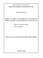
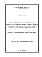
Điều trị: Phẫu thuật tạo hình khúc nối bồn thận-niệu quản
Phim UIV sau tiêm thuốc 70ph. Tắc khúc nối bồn thậnniệu quản(P) và (T).
Tắc ngõ ra bàng quang
(Bladder Outlet Obstruction-BOO)
1. Bệnh căn(etiology):
-. Nam giới: BPH, urethral stricture(trauma> urethritis), prostate cancer, dysfunctional
voiding(chronic prostitis), neurogenic-based detrusor-sphincter dyssynergia (DSD), and
primary bladder neck obstruction.
-. Ở nữ: Iatrogenic, pelvic prolapse, primary bladder neck obstruction, dysfuctional voiding,
DSD.
Tắc ngõ ra bàng quang
(Bladder Outlet Obstruction-BOO)
2.
3.
Bệnh nguyên(pathogenesis): Giảm hoặc tắc hồn
tồn dịng nước tiểu đi ra khỏi bàng quang.
Sinh lý bệnh(Pathophysiology): detrusor muscle
hypertrophy and collagen deposition(overflowed
bladder)->↓bladder compliance. Hydronephrosis.
Tắc ngõ ra bàng quang
(Bladder Outlet Obstruction-BOO)
Triệu chứng: Các triệu chứng cơ năng đường tiểu
dưới(LUTS).
Chẩn đoán: Phải loại trừ viêm bàng quang. Niệu động
học(urodynamics). Có thể kết hợp nội soi niệu đạo-bàng
quang, chụp niệu đạo-bàng quang ngược dòng, đo
lượng nước tiểu tồn lưu.
Điều trị: Mục tiêu giải quyết nguyên nhân gây tắc
nghẽn. 80% BPH điều trị thuốc thành công. Hẹp niệu
đạo điều trị bằng nong hẹp hoặc tạo hình niệu đạo.
Megaureter
1.
Bệnh căn(etiology)&bệnh
nguyên(pathogenesis):
- Primary Vesicoureteral Reflux: primary maturation
abnormality of the vesicoureteral junction or a
short distal ureteric submucosal tunnel in the
bladder-> alters the function of the valve
mechanism.
-. Primary megaureter:
A. obstructed primary megaureter:
aperistalic(neural crest migration defect).
B. refluxing primary megaureter: a short or absent
intravesical ureter, congenital paraureteric
diverticulum, or other derangement of the
vesicoureteral junction.
C. nonrefluxing unobstructed primary megaureter:
unknown.
Megaureter
1.
Bệnh căn(etiology)&bệnh
nguyên(pathogenesis):
- Ectopic Ureter: abnormal ureteral bud
migration(cauda ectopia).
-. Ureterocele:
A. Simple: stenosis of the distal end of the
normally positioned ureteral orifice leads to
ballooning of the segment immediately above it,
forming the ureterocele.
B. Ectopic: dilated submucosal distal portion of
an ectopic ureter.
2. Sinh lí bệnh(pathophysiology):
Hydronephrosis->decrease renal function.
Urinary tract infection.
Megaureter
Symptoms: Prenetal US.
Diagnosis: US. Real-time US.
-
VUR: VUCG.
Nuclear cystography(lower radiation dosage). Echo-enhanced cystosonography.
-
Primary megaureter: R/O VUR, urethral valves or neuropathic bladder dysfunction.
Real-time US+/-MRI for POM.
- Ectopic ureter: US, VCUG, and IVU.
- Ureterocele: US, VCUG, or IVU.
Treatment: Surgery.
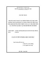
Simple ureterocele. (a) IVU image shows the typical cobra head appearance at the end of both ureters
Primary
megaureter
in athe
with
hydroureteronephrosis
atlef
prenatal
US.
(a)
Transverse
US
image
through
the (arrow). (b)
ureterocele
inneonate
a (b)
3-month-old
(a)
IVU
image
shows
a duplex
kidney.
There
is
nonvisualization
the
upperof
moiety
Prolapsing ureterocele.
(a) OnEctopic
an IVU
image,
bladder
isboy.
filled
with
a large
ureterocele
(u) that
projects
through
neck
the bladder
(arrowheads).
★
= bladder.
IVU image
shows
the
contrast
material-filled
bladder
(★
) with of
athe
and
of the
lower pole
pelvicaliceal
( ∗(u).
). Note
theIVU
increased
the spinal
column
and the
bladder in
(★a)moderate
shows dilatation
a markedly
dilated
and
tortuoussystem
ureter
imagedistance
showsbetween
the
and
tortuous
lefthe perineal orifice.
Clinical photograph obtained
different
patient shows
a ureterocele
as(b)an
( ★) dilated
that
prolapses
through
negative filling
defect (arrowheads)
that
representsmanifesting
a ureterocele.
(c) interlabial
Transversemass
US image
of the
lef
kidney
produced
by
the
upper
moiety.
A
large
filling
defect
(u)
is
seen
in
the
bladder,
a
finding
that
represents
a
ureterocele.
(b)
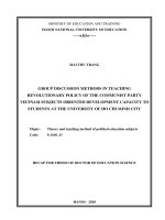
Ectopic ureter draining into the seminal vesicles. (a) IVU image shows an abdominal mass displacing the bladder ( ★) to the
ureter (u) with slight hydronephrosis. ★ = bladder. (c) Photograph of the gross specimen shows the dilated ureter (u)
bladder (B)
demonstrates
a sonolucent
cystic
structure
an echogenic
(U)represents
that projects
intoureterocele.
the
Longitudinal
US scan through
the bladder
shows
a rounded,with
well-defined
cystic masswall
(u) that
an ectopic
(c)
lef. The lef ureter (arrowheads) is also displaced by the mass. The right renal pelvis is seen (arrows), but not the right
and a narrowed
juxtavesicular
ureteral
segment
(arrow).
VCUG
image
shows
bilateral
grade
III
reflux
(moderate
dilatation
of
the
photograph
shows the ureterocele
★) protruding into the open bladder.
bladder, aClinical
finding
that represents
a simple (ureterocele.
ureter.
(b) Photograph
of theingross
specimenboy
shows
right ureter
(u) draining
into a widely
dilated
seminal
vesicle
(SV),
Ectopic ureter.
(a) VCUG
image obtained
a 3-week-old
withthe
wetting
demonstrates
single-ureter
ectopia
in the
posterior
urethra
ureters and of both renal pelves [p] and calices) during filling of the
Primary(arrow).
megaureter
in
a 7-month-old
boy.
Longitudinal
US image
the bladder
( ★) demonstrates
a dilated
distal(u)
ureter
andinto
a
produced
the
mass
effect
at
★ =which
bladder.
(b) Lateral
VCUG
imageobserved
obtained
inIVU.
athrough
4-month-old
girl shows
reflux into an ectopic
ureter
that (u)
drains
Bilateral VUR in an 8-year-old bladder
girl. (a) (Transverse
US image through the bladder ( ★) shows dilatation of both ureters ( u).
★).
narrowed
ureteral
to the aperistaltic segment (arrow).
thejuxtavesical
neck (arrow)
of thesegment
bladder (corresponding
★).
Tài liệu tham khảo:
1. Ureteropelvic Junction Obstruction, Campbell-Walsh Urology(12th edition), p. 3698-3702
2. Urinary Obstruction & Stasis, Smith and Tanagho's General Urology(19th Edition), p. 177188
3. />obstructive-uropathy
4. />5. />6. />7. />8. />9. />10. />