ARTERY BY PASS ppt
Bạn đang xem bản rút gọn của tài liệu. Xem và tải ngay bản đầy đủ của tài liệu tại đây (32.39 MB, 546 trang )
ARTERY BYPASS
Edited by Wilbert S. Aronow
Artery Bypass
/>Edited by Wilbert S. Aronow
Contributors
Lester A.H. Critchley, Inna Kammerer, Tagreed Altaei, Imad Jamal, Diyar Dilshad, Mohammed A Balghith, Rainer G. H.
Moosdorf, Maseeha Khaleel, Tracy Dorheim, Daniel Anderson, Michael Duryee, Geoffrey Thiele, Takao Kato, Benetti,
Haralabos Parissis, Alan Soo, Bassel Al-Alao, Aditya M Sharma, Herbert Aronow, Oguzhan Yıldız, Melik Seyrek,
Husamettin Gul, Cheng-Xiong Gu, Yang Yu, Chuan Wang, AC Zago, Eduardo K Saadi, Rui M. Almeida, Wilbert S.
Aronow, Sean Maddock, Gilbert L. Tang, Ramin Malekan, Yuki Igarashi, Takeo Igarashi, Ryo Haraguchi, Kazuo
Nakazawa, Jiri Mandak, Martin Šimek, Martin Kalab, Martin Molitor, Patrick Tobbia, Vladimír Lonský, Marcel A. Beijk,
Ralf Harskamp, Luminita Iliuta, Faisal Latif, Muhammad A. Chaudhry, Zainab Omar, Philippe Dubois, Maximilien
Gourdin, Tsuyoshi Kaneko, Sary Aranki, J D Schwalm, Michael Tsang, Andrea Székely, Zsuzsanna Cserép, Masaki
Yamamoto, Kazumasa Orihashi, Takayuki Sato, Kim Houlind, Johnny Christensen
Published by InTech
Janeza Trdine 9, 51000 Rijeka, Croatia
Copyright © 2013 InTech
All chapters are Open Access distributed under the Creative Commons Attribution 3.0 license, which allows users to
download, copy and build upon published articles even for commercial purposes, as long as the author and publisher
are properly credited, which ensures maximum dissemination and a wider impact of our publications. After this work
has been published by InTech, authors have the right to republish it, in whole or part, in any publication of which they
are the author, and to make other personal use of the work. Any republication, referencing or personal use of the
work must explicitly identify the original source.
Notice
Statements and opinions expressed in the chapters are these of the individual contributors and not necessarily those
of the editors or publisher. No responsibility is accepted for the accuracy of information contained in the published
chapters. The publisher assumes no responsibility for any damage or injury to persons or property arising out of the
use of any materials, instructions, methods or ideas contained in the book.
Publishing Process Manager Viktorija Zgela
Technical Editor InTech DTP team
Cover InTech Design team
First published March, 2013
Printed in Croatia
A free online edition of this book is available at www.intechopen.com
Additional hard copies can be obtained from
Artery Bypass, Edited by Wilbert S. Aronow
p. cm.
ISBN 978-953-51-1025-5
free online editions of InTech
Books and Journals can be found at
www.intechopen.com
Contents
Preface IX
Section 1 Basic Science and Physiology 1
Chapter 1 Impact of Ischemia on Cellular Metabolism 3
Maximilien Gourdin and Philippe Dubois
Chapter 2 Inflammation and Vasomotricity During Reperfusion 19
Maximilien Gourdin and Philippe Dubois
Chapter 3 Ventricular Arrhythmias and Myocardial
Revascularization 37
Rainer Moosdorf
Chapter 4 Minimally Invasive Cardiac Output Monitoring in the
Year 2012 45
Lester Augustus Hall Critchley
Chapter 5 Intraoperative Indocyanine Green Imaging Technique in
Cardiovascular Surgery 81
Masaki Yamamoto, Kazumasa Orihashi and Takayuki Sato
Chapter 6 Peripheral Tissue Oxygenation During Standard and
Miniaturized Cardiopulmonary Bypass (Direct Oxymetric Tissue
Perfusion Monitoring Study) 99
Jiri Mandak
Section 2 Coronary Artery Bypass Graft Surgery 117
Chapter 7 Total Arterial Revascularization in Coronary Artery Bypass
Grafting Surgery 119
Sean Maddock, Gilbert H. L. Tang, Wilbert S. Aronow and Ramin
Malekan
Chapter 8 MINI OPCABG 135
Federico Benetti, Natalia Scialacomo, Jose Luis Ameriso and Bruno
Benetti
Chapter 9 Saphenous Vein Conduit in Coronary Artery Bypass Surgery —
Patency Rates and Proposed Mechanisms for Failure 149
Maseeha S. Khaleel, Tracy A. Dorheim, Michael J. Duryee, Geoffrey
M. Thiele and Daniel R. Anderson
Chapter 10 The Impact of Arterial Grafts in Patients
Undergoing GABG 161
Haralabos Parissis, Alan Soo and Bassel Al-Alao
Chapter 11 Complex Coronary Artery Disease 173
Tsuyoshi Kaneko and Sary Aranki
Chapter 12 Aspirin Therapy Resistance in Coronary Artery Bypass
Grafting 187
Inna Kammerer
Chapter 13 Treatment of Coronary Artery Bypass Graft Failure 193
M.A. Beijk and R.E. Harskamp
Chapter 14 The Cardioprotection of Silymarin in Coronary Artery Bypass
Grafting Surgery 239
D. Tagreed Altaei, D. Imad A. Jamal and D. Diyar Dilshad
Chapter 15 Pharmacology of Arterial Grafts for Coronary Artery
Bypass Surgery 251
Oguzhan Yildiz, Melik Seyrek and Husamettin Gul
Chapter 16 Surgical Treatment for Diffuse Coronary Artery Diseases 277
Cheng-Xiong Gu, Yang Yu and Chuan Wang
Chapter 17 The Antiagregant Treatment After Coronary Artery Surgery
Depending on Cost – Benefit Report 291
Luminita Iliuta
ContentsVI
Section 3 Percutaneous Coronary Intervention 315
Chapter 18 Multivessel Disease in the Modern Era of Percutaneous
Coronary Intervention 317
Michael Tsang and JD Schwalm
Chapter 19 Artery Bypass Versus PCI Using New Generation DES 353
Mohammed Balghith
Chapter 20 Generating Graphical Reports on Cardiac
Catheterization 367
Yuki Igarashi, Takeo Igarashi, Ryo Haraguchi and Kazuo Nakazawa
Section 4 Peripheral and Cerebral Vascular Disease Intervention 385
Chapter 21 Management of Carotid Artery Disease in the Setting of
Coronary Artery Disease in Need of Coronary Artery
Bypass Surgery 387
Aditya M. Sharma and Herbert D. Aronow
Chapter 22 Infected Aneurysm and Inflammatory Aorta: Diagnosis and
Management 405
Takao Kato
Chapter 23 Endovascular Treatment of Ascending Aorta: The Last
Frontier? 413
Eduardo Keller Saadi, Rui Almeida and Alexandre do Canto Zago
Chapter 24 The Role of The Angiosome Model in Treatment of Critical
Limb Ischemia 425
Kim Houlind and Johnny Christensen
Chapter 25 Impact of Renal Dysfunction and Peripheral Arterial Disease on
Post-Operative Outcomes After Coronary Artery Bypass
Grafting 437
Muhammad A. Chaudhry, Zainab Omar and Faisal Latif
Section 5 Miscellaneous Cardiac Surgical Topics 461
Chapter 26 Short and Long Term Effects of Psychosocial Factors on the
Outcome of Coronary Artery Bypass Surgery 463
Zsuzsanna Cserép, Andrea Székely and Bela Merkely
Contents VII
Chapter 27 Current Challenges in the Treatment of Deep Sternal Wound
Infection Following Cardiac Surgery 493
Martin Šimek, Martin Molitor, Martin Kaláb, Patrick Tobbia and
Vladimír Lonský
ContentsVIII
Preface
The latest diagnostic and therapeutic modalities in the management of coronary artery dis‐
ease by coronary artery bypass graft surgery and by percutaneous coronary intervention
with stenting and in the interventional management of other atherosclerotic vascular disease
have led to a reduction in cardiovascular mortality and morbidity. This book entitled Artery
Bypass provides an excellent update on these advances which every physician seeing pa‐
tients with atherosclerotic vascular disease should be familiar with. This book includes 27
chapters written by experts in their topics.
The first section of this book discusses basic science and physiology and includes 6 chapters.
The second section of this book discusses coronary artery bypass graft surgery and includes
11 chapters. The third section of this book discusses percutaneous coronary intervention
with stenting and includes 3 chapters. The fourth section of this book discusses peripheral
and cerebral vascular disease intervention and includes 5 chapters. The fifth section of this
book discusses miscellaneous cardiac surgical topics and includes 2 chapters. Another
strength of thisbook is that unresolved issues are also discussed.
I would like to thank all of the contributors for their outstanding work. Finally, I would like
to thank you, the reader, for your commitment to providing the best possible care to your
patients with atherosclerotic vascular disease. I hope you will find this book a valuable re‐
source in providing excellent care to your patients with atherosclerotic vascular disease.
Wilbert S. Aronow, MD, FACC, FAHA, FCCP, FACP
Professor of Medicine, New York Medical College
Valhalla, NY, USA
Section 1
Basic Science and Physiology
Chapter 1
Impact of Ischemia on Cellular Metabolism
Maximilien Gourdin and Philippe Dubois
Additional information is available at the end of the chapter
/>1. Introduction
As in all aerobic eukaryotic cells, oxygen is essential for homeostasis in human cells. The in‐
terruption of blood flow to tissues results in an arrested oxygen supply and disrupts the bio‐
chemical reactions that ensure the smooth functioning, integrity and survival of the cells.
The limited oxygen reserves that are dissolved in the interstitial fluid and are bound to he‐
moglobin, myoglobin and neuroglobin do not maintain efficient, long-term metabolism.[1,2]
Lack of oxygen affects all functions within the cell. Table 1 summarizes the main cellular
consequences of ischemia.
(1) cellular acidosis;
(2) loss of sarcoplasmic membrane potential;
(3) cellular swelling;
(4) cytoskeleton disorganization;
(5) reduction of adenosine-5’-triphospate (ATP)
and phosphocreatine is more than reduction in
the energy substrates;
(6) reduction of glutathione, of a-tocopherol;
(7) increasing expression of leukocyte adhesion
molecules;
(8) secretion of cytokines/chemokines
- Tumor Necrosis Factor (TNF-α)
- Interleukins (IL-) -1, 6, 8
Table 1. Major cellular consequences of ischemia
© 2013 Gourdin and Dubois; licensee InTech. This is an open access article distributed under the terms of the
Creative Commons Attribution License ( which permits
unrestricted use, distribution, and reproduction in any medium, provided the original work is properly cited.
2. Adenosine triphosphate depletion
Eukaryotic cells contain mitochondria, organelles whose main function is to produce adeno‐
sine triphosphate (ATP). ATP is an essential energy substrate, as its hydrolysis provides en‐
ergy for many metabolic and biochemical reactions involved in development, adaptation
and cell survival. ATP production in an aerobic cell is particularly effective when the degra‐
dation of key nutrients such as glucose and fatty acids is coupled to a supramolecular com‐
plex located in the inner membrane of mitochondria to drive oxidative phosphorylation.
Oxidative phosphorylation is mediated by an electron transport chain that consists of four
protein complexes and establishes a transmembrane electrochemical gradient by supporting
the accumulation of protons in the intermembrane space of the mitochondria. This gradient
is used as an energy source by ATP synthase during the synthesis of an ATP molecule from
a molecule of adenosine diphosphate (ADP) and an inorganic phosphate (Figure 1). Without
oxygen, oxidative phosphorylation stops: the proton gradient between the intermembrane
space and the inner mitochondria is abolished, and ATP synthesis is interrupted. The ensu‐
ing rapid fall in intracellular ATP induces a cascade of events leading to reversible cell dam‐
age. However, over time, the damage increases and gradually becomes irreversible, which
may lead to cell death and destruction of the parenchymal tissue.
Figure 1. Hydrolysis of Adenosine-triphosphate provides energy (30.5 kJ per mole) for biochemical reactions
When devoid of ATP, the cell derives its energy from the pyrophosphate bonds of ADP as
they are degraded to adenosine monophosphate (AMP) and then to adenosine. Adenosine
diffuses freely out of the cell, dramatically reducing the intracellular pool of adenine nucleo‐
tides, the precursors for ATP.
3. Changes in metabolism (Figure 2)
In the presence of oxygen, human cells respire and derive their energy from the complete
degradation of food (fats, carbohydrates and amino acids) by specific oxidative processes
that fuel oxidative phosphorylation. A lack of oxygen completely changes these metabolic
pathways, disrupting glycolysis and inhibiting the degradation pathways of lipids (beta-oxi‐
dation), amino acids and oxidative phosphorylation.
Artery Bypass4
3.1. Glucose metabolism
During ischemia, the cell will change not only its glucose supply routes but also its glycoly‐
sis pathways and transition from aerobic glycolysis to anaerobic glycolysis. When this hap‐
pens, the available cytosolic glucose is metabolized by anaerobic glycolysis and becomes the
main source of ATP. The efficiency of this process is much lower than that of aerobic glycol‐
ysis coupled to oxidative phosphorylation; the anaerobic degradation of one molecule of
glucose produces 2 ATP molecules compared to the 36 ATP molecules that are produced un‐
der aerobic conditions. Consumption quickly exceeds production, and the intracellular con‐
centration of ATP decreases. For example, in the heart, the degree of glycolysis inhibition is
directly proportional to the severity of coronary flow restriction.[3]-[5]
3.1.1. Glucose supply
With the complete interruption of or decrease in blood flow, the extracellular concentration
of glucose drops very quickly. First, the cell optimizes the uptake of glucose from the inter‐
stitial space by improving glucose transmembrane transport by increasing the sarcoplasmic
expression of the high-affinity glucose transporters GLUT-1 and GLUT-4. [6]-[8] This protec‐
tive mechanism temporarily compensates for the decrease in extracellular glucose concen‐
tration. Next, the cell uses its intracellular glucose stores of glycogen. [9] The decrease in
intracellular ATP and glucose-6-phosphate, the rising lactate/pyruvate ratio and the increase
in intracellular AMP and the inorganic phosphate concentration activate a phosphorylase
kinase, which catalyses the conversion of glycogene phosphorylase b to its active form, gly‐
cogene phosphorylase a. This cascade reaction leads to an intense and rapid consumption of
glycogen. [10]-[14]
3.1.2. Glycolysis pathways
The inhibition of oxidative phosphorylation caused by lack of oxygen does not allow the
pyruvate produced by glycolysis to be degraded. Under aerobic conditions, pyruvate is
transported into the mitochondria and feeds into the Krebs cycle, which provides the nicoti‐
namide adenine dinucleotide (NADH, H
+
) and flavine adenine dinucleotide (FADH
2
) cofac‐
tors for oxidative phosphorylation, significantly increasing the yield of glycolysis.
Ischemia modulates the activity of the following two key enzymes of anaerobic glycolysis:
phosphofructo-1-kinase (PF1K) and glyceraldehyde-3-phosphate dehydrogenase (GAPDH).
Following the onset of ischemia, or during moderate ischemia, the activation of glycogenol‐
ysis accelerates glycolysis.[15]-[17] The decrease in both intracellular ATP and creatine phos‐
phate, along with increases in the intracellular concentrations of AMP, inorganic phosphate
and fructose-1,6-bisphosphate, intensify the activity of PF1K and GAPDH. [17]-[20]
During prolonged or sustained ischemia, the low intracellular glucose concentration, the
disappearance of glycogen and severe intracellular acidosis eventually inhibit PF1K. Fur‐
thermore, high concentrations of lactate and protons in ischemic tissues also inhibit
GAPDH. [21],[22]
Impact of Ischemia on Cellular Metabolism
/>5
Moreover, the lactate/pyruvate ratio, intracellular acidosis and the absence of regenerated
essential cofactors, such as NADH,H
+
, affect the catalytic activity of the other enzymes in‐
volved in the initial step of glycolysis and prevent the optimal performance of anaerobic
glycolysis. [23]
3.2. Lipid metabolism (Figure 2)
The importance of oxygen in functional oxidative phosphorylation leads to a significant
reduction in ATP production from the beta-oxidation of fatty acids that is proportional
to the degree of ischemia. In mild to moderate ischemia, the rate of fatty acid oxidation
decreases but still fuels oxidative phosphorylation. [4],[24] In more severe ischemia, the
lack of the cofactors NADH,H
+
and FAD
+
, which are normally regenerated through oxi‐
dative phosphorylation, completely inhibits acyl-CoenzymeA (acyl-CoA) dehydrogenase
and 3-hydroxyacyl-CoA dehydrogenase, which are key beta-oxidation enzymes.[4],[25]
The cytosolic concentrations of fatty acids, acyl-CoA and acylcarnitine rise gradually.
[26]-[28] The accumulation of these amphiphilic compounds in ischemic tissues has ma‐
jor functional implications. They dissolve readily in cell membranes and affect the func‐
tional properties of membrane proteins. Decreased activity of Na
+
/K
+
-ATPase and the
sarcoplasmic and endoplasmic reticulum Ca
2+
-ATPase pumps, as well as the activation of
ATP-dependent potassium channels, reduces the inwardly rectifying potassium current
and prolongs the opening of Na
+
channels, delaying their inactivation.[29]-[31] The accu‐
mulation of amphiphilic compounds produces a time-dependent reversible reduction in
gap-junction conductance. [31]
3.3. Metabolite detoxification pathways
Reducing the intracellular concentration of ATP inhibits the hexose phosphate cycle.
This metabolic pathway regenerates glutathione, ascorbic acid and tocopherol, which
are involved in the detoxification of metabolites from the cytosol and the sarcoplasmic
membrane.
4. Intracellular acidosis
Intracellular acidosis is a cardinal feature of cellular ischemia. The increased production of
protons due to metabolic modifications very quickly saturates the buffering capacity of the
cell. Intracellular acidosis interferes directly and indirectly with the optimal functioning of
the cell by increasing intracellular Na
+
through the activation of Na
+
/H
+
exchangers and by
Ca
2+
activation of Na
+
/Ca
2+
exchangers, increasing the production of free radicals; changing
the affinity of different proteins, such as enzymes and troponin C, to Ca
2+
; modifying terti‐
ary protein structures; inhibiting enzymes; and disrupting the function of sarcoplasmic
pumps and carriers.[29]
Artery Bypass6
dehydrogenase and 3-hydroxyacyl-CoA dehydrogenase, which are key beta-oxidation enzymes.
[4],[25]
The cytosolic concentrations
of fatty acids, acyl-CoA and acylcarnitine rise gradually.
[26]-[28]
The accumulation of these amphiphilic compounds in ischemic
tissues has major functional implications. They dissolve readily in cell membranes and affect the functional properties of
membrane proteins. Decreased activity of Na
+
/K
+
-ATPase and the sarcoplasmic and endoplasmic reticulum Ca
2+
-ATPase pumps, as
well as the activation of ATP-dependent potassium channels, reduces the inwardly rectifying potassium current and prolongs the
opening of Na
+
channels, delaying their inactivation.
[29]-[31]
The accumulation of amphiphilic compounds produces a time-
dependent reversible reduction in gap-junction conductance.
[31]
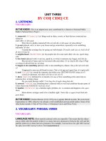
Figure 2. This figure shows schematically oxidative metabolism, ATP production and the consequences of oxygen deprivation. GLUT-1 and
GLUT-4: glucose transporters; GP: Glycogene phosphorylase; HK: Hexokinase; PF1K: Phosphofructo-1-kinase; GADPH: glyceraldehyde-3-
phosphate dehydrogenase; NADH, H
+
: nicotinamide adenine dinucleotide; FADH
2
: flavine adenin dinucleotide; P: phosphate;AMP, adenosine
monophosphate; adenosine diphosphate;ADP: adenosine diphosphate ATP: adenosine triphosphate; CO
2
: carbon dioxide; O
2
Oxygen; - :
inhibition; + activation; H
+
: proton; e
-
: electron.
3.3. Metabolite detoxification pathways
H
+
H
+
H
+
H
+
H
+
H
+
H
+
H
+
H
+
H
+
H
+
H
+
H
+
H
+
H
+
H
+
H
+
NADH+H
+
Pyruvate
Acetyl-Co A
H
+
H
+
H
+
H
+
H
+
H
+
H
+
H
+
ATP
ATP
ADP
P
i
ATP
H
+
ADP
P
i
ATP
H
+
Fatty Acid
Fatty Acid
CO
2
NADH+H
+
FADH
2
CO
2
CO
2
ATP
2e
-
-ox
y
dation
Electron transport chain
and
creation of H+ gradient
Krebs
cycle
Hypoxia
-
GLUT-1 GLUT-4
Glucose
Glucose
Glucose
Glycogen
Fasting
Hypoxia
+
Glucose-6-P
-
Fructose-6-P
Fructose-1-6-P
1,3-diphosphoglycerate
Hypoxia, AMP, ADP
Insulin
+
ATP, citrate, free fatty acid
p
H
-
Pyruvate
-
Lactate, NADPH
2
NADPH+H
+
Lactate
-
Hypoxia
Hypoxia
+
Sarcoplasmic membrane
mitochondrion
Inner membrane
Intermembrane space
Outer membrane
in
out
Interstitium
GP
HK
GADPH
PF1K
metabolism
Figure 2. This figure shows schematically oxidative metabolism, ATP production and the consequences of oxygen
deprivation. GLUT-1 and GLUT-4: glucose transporters; GP: Glycogene phosphorylase; HK: Hexokinase; PF1K: Phospho‐
fructo-1-kinase; GADPH: glyceraldehyde-3-phosphate dehydrogenase; NADH, H
+
: nicotinamide adenine dinucleotide;
FADH
2
: flavine adenin dinucleotide; P: phosphate;AMP, adenosine monophosphate; adenosine diphosphate;ADP: ad‐
enosine diphosphate ATP: adenosine triphosphate; CO
2
: carbon dioxide; O
2
Oxygen; - : inhibition; + activation; H
+
:
proton; e
-
: electron.
Impact of Ischemia on Cellular Metabolism
/>7
The main source of protons during ischemia comes from the production of lactate from pyr‐
uvate by lactate dehydrogenase. The accumulation of extracellular lactate greatly reduces
the effectiveness of the lactate/proton cotransporter, preventing the removal of protons. Ad‐
ditionally, the residual metabolic activity also contributes to acidosis, as the hydrolysis of an
ATP molecule releases a proton.
5. Changes in the ionic cellular equilibrium (Figure 3)
Ischemia induces a profound disturbance of the ionic homeostasis of a cell. The two major
changes are the loss of ionic transmembrane gradients, which causes membrane depolariza‐
tion, and increased intracellular sodium ([Na
+
]
i
), which is responsible for inducing a rise in
the intracellular calcium ([Ca
2+
]
i
) levels, leading to cellular edema.
Cellular depolarization occurs very rapidly after the onset of ischemia, and these mecha‐
nisms are not fully understood. However, it is recognized that both the inhibition of the Na
+
/K
+
-ATPase and the opening of ATP-dependent K
+
channels play a crucial role. Cellular de‐
polarization is characterized by a negative outgoing current and a decrease in the extracellu‐
lar concentrations of Na
+
, Cl
-
and Ca
2+
, as well as an increase in the extracellular
concentration of K
+
. Progressive depolarization of the cell also promotes prolonged activa‐
tion of voltage-dependent sodium channels. [29]
The accumulation of sodium in the cytosol is multifactorial. Acidosis stimulates Na
+
/H
+
ex‐
changers to purge cellular H
+
, which results in increased intracellular Na
+
.[32]-[34] This net
movement of Na
+
is accompanied by osmotic water movement. Moreover, inhibition of the
Na
+
/K
+
-ATPase due to a lack of ATP prevents the removal of excess intracellular Na
+
. The
high intracellular concentration of Na
+
affects the function of other membrane transporters,
such as the Na
+
/Ca
2+
antiporter, an accelerator. This allows the extrusion of sodium from the
cell at the expense of an intracellular accumulation of Ca
2+
. The massive entry of calcium in‐
to the cell disrupts the mechanisms that regulate its intracellular concentration and induces
the release of calcium from the intracellular endoplasmic reticulum stores.[35] The lack of
ATP prevents calcium excretion into the interstitium and its sequestration in the endoplas‐
mic reticulum. The accumulation of cytosolic calcium induces degradation of membrane
phospholipids and cytoskeletal proteins, alters the both the calcium affinity and the efficien‐
cy of proteins involved in contractility, activates nitric oxide synthase (NOS) and proteases
such as calpains and caspases, promotes the production of free radicals and alters the terti‐
ary structure of enzymes such as xanthine dehydrogenase, which is converted to xanthine
oxidase. [36]-[38]
6. Mitochondria
The mitochondrion plays a central role in ischemic injury. Not only is it the site of critical
biochemical reactions in the cell, such as oxidative phosphorylation, beta-oxidation and the
Artery Bypass8
citric acid cycle, but it also occupies a unique position in the cellular balance between life
and death. Inhibition of the mitochondrial respiratory chain as a result of oxygen depriva‐
tion is the cornerstone of metabolic disturbances.
Figure 3. This figure summarizes the ionic perturbations in an ischemic cell.
6.1. Disturbance of ATP synthesis.
Without the respiratory chain oxidation-reduction reactions, proton accumulation in the mitochondrial intermembrane space is
interrupted, disrupting the electrochemical gradient that allows ATP synthase to synthesize ATP. During ischemia, the proton-
translocating F0F1-ATP synthase, which normally produces ATP, becomes an F0F1-ATPase and consumes ATP in order to pump
protons from the matrix to the intermembrane space and maintain the mitochondrial membrane potential.
[39],[40]
The mitochondria
therefore become a site of ATP consumption produced by anaerobic glycolysis.
6.2. An increase in free radical production
Free radical oxygen species (ROS) are highly reactive chemical compounds because they have unpaired electrons in their electron
cloud. ROS are capable of oxidizing cellular constituents such as proteins, deoxyribonucleic acid (DNA), membrane phospholipids
and other adjacent biological structures. In addition to their role in ischemia, ROS are constitutively generated during metabolic
processes and have an important role in cell signaling. Mitochondrial respiration constitutively produces a small amount of ROS,
primarily the superoxide anion O
2
-●
at complexes I and III of the electron transport chain. The anion is rapidly converted to
hydrogen peroxide (H
2O2) by metallo-enzymes and superoxide dismutase (SOD).
[41]-[43]
Cellular stress, particularly oxidative stress,
dramatically increases mitochondrial ROS production by disrupting and later inhibiting oxidative phosphorylation. Moreover, the
rise in mitochondrial calcium increases ROS production and greatly decreases the antioxidant capacity of mitochondria by
decreasing the glutathione peroxidase concentration and SOD activity.
6.3. Intramitochondrial calcium overload
The mitochondrial calcium concentration is in equilibrium between its cytosolic concentration and the proton gradient on either
side of the inner membrane of mitochondria. The loss of this gradient due to the inhibition of the respiratory chain, as well as the
elevated cytosolic calcium that results from ischemia, allows for the accumulation of calcium in the mitochondria and promotes
mitochondrial swelling and the opening of the permeability transition pore.
6.4. Opening of the mitochondrial permeability transition pore
Ischemic disturbances within mitochondria, such as calcium overload, loss of membrane potential, oxidative stress, mass
production of free radicals, low NADPH/NADP
+
and reduced glutathione to oxidized glutathione ratios (GSH/GSSG), low intra-
mitochondrial concentration of ATP or high inorganic phosphate, will promote opening of the permeability transition pore (mPTP)
upon reperfusion, a major player in I/R injury-mediated cell lethality.
[42],[44]
mPTP is a nonspecific channel, and its opening
out
in
Anaerobic metabolism
[Ca
++
]
i
[Na
+
]i
ATP
[H
+
]
i
↓
O2
×
Cellular edema
Ca
++
Can.L
ATP
ADP+Pi
K
+
Na
+
I
I
I
V
H
+
Na
+
Ca
++
Na
+
m
inhibition
A
cceleration
A
cceleration Loss of membrane
p
otential
- protein degradation
- Protein structure modifications
-Plasmic phospholipids degradation
-Mitochondrial dysfunction
Figure 3. This figure summarizes the ionic perturbations in an ischemic cell.
6.1. Disturbance of ATP synthesis.
Without the respiratory chain oxidation-reduction reactions, proton accumulation in the mi‐
tochondrial intermembrane space is interrupted, disrupting the electrochemical gradient
that allows ATP synthase to synthesize ATP. During ischemia, the proton-translocating
F0F1-ATP synthase, which normally produces ATP, becomes an F0F1-ATPase and con‐
sumes ATP in order to pump protons from the matrix to the intermembrane space and
maintain the mitochondrial membrane potential.[39],[40] The mitochondria therefore be‐
come a site of ATP consumption produced by anaerobic glycolysis.
6.2. An increase in free radical production
Free radical oxygen species (ROS) are highly reactive chemical compounds because they
have unpaired electrons in their electron cloud. ROS are capable of oxidizing cellular con‐
stituents such as proteins, deoxyribonucleic acid (DNA), membrane phospholipids and oth‐
er adjacent biological structures. In addition to their role in ischemia, ROS are constitutively
generated during metabolic processes and have an important role in cell signaling. Mito‐
chondrial respiration constitutively produces a small amount of ROS, primarily the superox‐
Impact of Ischemia on Cellular Metabolism
/>9
ide anion O
2
-●
at complexes I and III of the electron transport chain. The anion is rapidly
converted to hydrogen peroxide (H
2
O
2
) by metallo-enzymes and superoxide dismutase
(SOD). [41]-[43] Cellular stress, particularly oxidative stress, dramatically increases mito‐
chondrial ROS production by disrupting and later inhibiting oxidative phosphorylation.
Moreover, the rise in mitochondrial calcium increases ROS production and greatly decreases
the antioxidant capacity of mitochondria by decreasing the glutathione peroxidase concen‐
tration and SOD activity.
6.3. Intramitochondrial calcium overload
The mitochondrial calcium concentration is in equilibrium between its cytosolic concentra‐
tion and the proton gradient on either side of the inner membrane of mitochondria. The loss
of this gradient due to the inhibition of the respiratory chain, as well as the elevated cytosol‐
ic calcium that results from ischemia, allows for the accumulation of calcium in the mito‐
chondria and promotes mitochondrial swelling and the opening of the permeability
transition pore.
6.4. Opening of the mitochondrial permeability transition pore
Ischemic disturbances within mitochondria, such as calcium overload, loss of membrane po‐
tential, oxidative stress, mass production of free radicals, low NADPH/NADP
+
and reduced
glutathione to oxidized glutathione ratios (GSH/GSSG), low intra-mitochondrial concentra‐
tion of ATP or high inorganic phosphate, will promote opening of the permeability transi‐
tion pore (mPTP) upon reperfusion, a major player in I/R injury-mediated cell lethality.[42],
[44] mPTP is a nonspecific channel, and its opening suddenly increases the permeability of
the inner mitochondrial membrane to both water and various molecules of high molecular
weight (> 1,500 kDa). The opening of mPTPs abolishes the mitochondrial membrane poten‐
tial and uncouples oxidative phosphorylation, which empties the mitochondria of its matrix
and induces apoptosis by releasing the intra-mitochondrial proteins cytochrome c, endonu‐
clease G, Smac/Diablo and apoptosis-inducing factor into the cytosol. [44
]-[52]
7. Structural and functional modifications
The cytoskeleton, the internal structural organization of a cell, is composed of a highly regu‐
lated complex network of organized structural proteins, including actin, microtubules and
lamins. The cytoskeleton performs multiple functions. It maintains internal cellular com‐
partmentalization and mediates the transmission of mechanical forces within the cell to ad‐
jacent cells and the extracellular matrix, the distribution of organelles, the movement of
molecules or components and the docking of proteins such as membrane receptors or ion
channels. Ischemia deconstructs the cytoskeleton. [53]-[56] The high intracellular concentra‐
tions of Ca
2+
that are associated with ischemia activate multiple phosphorylases and proteas‐
es that disassemble and degrade the cytoskeleton, thereby eliminating the functions that rely
on its integrity, such as phagocytosis, exocytosis, myofilament contraction, intercellular
Artery Bypass10
communication and cell anchorage. Destruction of the internal architecture worsens I/R inju‐
ries and leads to apoptosis. [53],[56],[57] During ischemia, all elements of the cytoskeleton
are affected, but with different kinetics.[54],[55] Moreover, the accumulation of osmotically
active particles, including lactate, sodium, inorganic phosphate and creatine, induces cellu‐
lar oedema.[38]
Regulatory cellular mechanisms provide intracellular homeostasis that enables optimal en‐
zyme function in a relatively narrow range of environmental conditions. The conditions cre‐
ated by ischemia, such as acidosis and calcium overload, modify or inhibit the activity of
many enzymes due to changes in the pH and tertiary structures, affecting cellular metabo‐
lism. For example, ischemia induces the conversion of xanthine dehydrogenase to xanthine
oxidase.[36]-[38] These two enzymes catalyze the same reactions, converting hypoxanthine
to xanthine and xanthine to uric acid. The first reaction uses NAD
+
as a cofactor, whereas the
second uses oxygen and produces O
2
-●
, a free radical.
8. Protein synthesis and sarcoplasmic protein expression in an ischemic
cell
Protein synthesis is a complex process that requires continuous and adequate energy intake,
strict control of ionic homeostasis of the cell and the smooth functioning of many other pro‐
teins. Ischemia disrupts these necessary conditions and therefore profoundly affects protein
synthesis beyond acute injury. However, the transcription of several genes is initiated at the
onset of ischemia, and the mechanisms underlying this phenomenon are not fully under‐
stood. Nevertheless, it appears that the mass production of free radicals, the high concentra‐
tion of calcium, acidosis and the activation of the family of mitogen-activated protein
kinases (MAP kinases) play an important role. Nuclear factor heat shock transcription fac‐
tor-1 (HSF-1) activates the expression of heat shock proteins (HSPs), a family of chaperone
proteins, and inhibits the expression of other proteins. HSPs are synthesized in different sit‐
uations of stress, including hyperthermia, ischemia, hypoxia and mechanical stress, and are
intended to prevent the structural modifications of key metabolic and cytoskeletal enzymes
and inhibit the activity of caspases. [58]-[60]
The low oxygen partial pressure during ischemia activates other nuclear factors, such as hy‐
poxia-inducible factor-1alpha (HIF-1α). HIF-1α stimulates the transcription of many genes
involved in cellular defense, such as those encoding NOS and GLUT-1, and other enzymes
involved in glucose metabolism.[61]
In addition, ischemia activates innate immunity by stimulating sarcoplasmic receptors, such
as the Toll-like receptors (TLR) TLR-2 and TLR-6, the synthesis and sarcoplasmic expression
of which are increased. Receptor stimulation supports the synthesis of chemokines and cyto‐
kines and contributes to I/R injury.[61]-[66]
At the onset of ischemia, many substances are secreted by the cell. For example, ischemic
cardiomyocytes secrete bradykinin, norepinephrine, angiotensin, adenosine, acetylcholine
Impact of Ischemia on Cellular Metabolism
/>11
and opioids.[67]-[69] In addition, ischemia stimulates the expression of adhesion molecules,
such as P-selectins, L-selectins, intercellular adhesion molecule-1 (ICAM-1) and platelet-en‐
dothelial cell adhesion molecules (PECAM), on the surface of endothelial cells, leukocytes
and other ischemic cells. [62],[63],[70],[71] Furthermore, many cytokines, such as tumor ne‐
crosis factor-α, interleukin (IL)-1, IL-6 and IL-8, and vasoactive agents, such as endothelins
and thromboxane A2, are secreted by cells in response to ischemia. [62],[70],[72] Cytokines
and chemokines, the production of which dramatically increases during reperfusion, initiate
the local inflammatory response and prepare for the recruitment of inflammatory cells into
the injured area, respectively.
Author details
Maximilien Gourdin
*
and Philippe Dubois
*Address all correspondence to:
Université de Louvain (UCL), University Hospital CHU UCL Mont-Godinne – Dinant,
Yvoir, Belgium
References
[1] Jennings RB, Murry CE, Steenbergen C, Jr., Reimer KA: Development of cell injury in
sustained acute ischemia. Circulation 1990; 82: II2-12
[2] Kloner RA, Jennings RB: Consequences of brief ischemia: stunning, preconditioning,
and their clinical implications: part 1. Circulation 2001; 104: 2981-9
[3] Neely JR, Liedtke AJ, Whitmer JT, Rovetto MJ: Relationship between coronary flow
and adenosine triphosphate production from glycolysis and oxidative metabolism.
Recent Adv Stud Cardiac Struct Metab 1975; 8: 301-21
[4] Neely JR, Morgan HE: Relationship between carbohydrate and lipid metabolism and
the energy balance of heart muscle. Annu Rev Physiol 1974; 36: 413-59
[5] Neely JR, Whitmer JT, Rovetto MJ: Effect of coronary blood flow on glycolytic flux
and intracellular pH in isolated rat hearts. Circ Res 1975; 37: 733-41
[6] Sun D, Nguyen N, DeGrado TR, Schwaiger M, Brosius FC, 3rd: Ischemia induces
translocation of the insulin-responsive glucose transporter GLUT4 to the plasma
membrane of cardiac myocytes. Circulation 1994; 89: 793-8
[7] Tian R, Abel ED: Responses of GLUT4-Deficient Hearts to Ischemia Underscore the
Importance of Glycolysis. Circulation 2001; 103: 2961-2966
Artery Bypass12
[8] Young LH, Renfu Y, Russell R, Hu X, Caplan M, Ren J, Shulman GI, Sinusas AJ: Low-
flow ischemia leads to translocation of canine heart GLUT-4 and GLUT-1 glucose
transporters to the sarcolemma in vivo. Circulation 1997; 95: 415-22
[9] Stanley WC, Hall JL, Stone CK, Hacker TA: Acute myocardial ischemia causes a
transmural gradient in glucose extraction but not glucose uptake. Am J Physiol 1992;
262: H91-6
[10] Begum N, Graham AL, Sussman KE, Draznin B: Role of cAMP in mediating effects of
fasting on dephosphorylation of insulin receptor. Am J Physiol 1992; 262: E142-9
[11] Dobson JG, Jr., Mayer SE: Mechanisms of activation of cardiac glycogen phosphory‐
lase in ischemia and anoxia. Circ Res 1973; 33: 412-20
[12] Morgan HE, Parmeggiani A: Regulation of Glycogenolysis in Muscle. Ii. Control of
Glycogen Phosphorylase Reaction in Isolated Perfused Heart. J Biol Chem 1964; 239:
2435-9
[13] Schaefer S, Ramasamy R: Glycogen utilization and ischemic injury in the isolated rat
heart. Cardiovasc Res 1997; 35: 90-8
[14] Schulze W, Krause EG, Wollenberger A: On the fate of glycogen phosphorylase in
the ischemic and infarcting myocardium. J Mol Cell Cardiol 1971; 2: 241-51
[15] Kubler W, Spieckermann PG: Regulation of glycolysis in the ischemic and the anoxic
myocardium. J Mol Cell Cardiol 1970; 1: 351-77
[16] Rovetto MJ, Whitmer JT, Neely JR: Comparison of the effects of anoxia and whole
heart ischemia on carbohydrate utilization in isolated working rat hearts. Circ Res
1973; 32: 699-711
[17] Wollenberger A, Krause EG: Metabolic control characteristics of the acutely ischemic
myocardium. Am J Cardiol 1968; 22: 349-59
[18] Francis SH, Meriwether BP, Park JH: Interaction between adenine nucleotides and 3-
phosphoglyceraldehyde dehydrogenase. II. A study of the mechanism of catalysis
and metabolic control of the multi-functional enzyme. J Biol Chem 1971; 246: 5433-41
[19] Oguchi M, Meriwether BP, Park JH: Interaction between adenosine triphosphate and
glyceraldehyde 3-phosphate dehydrogenase. 3. Mechanism of action and metabolic
control of the enzyme under simulated in vivo conditions. J Biol Chem 1973; 248:
5562-70
[20] Williamson JR: Glycolytic control mechanisms. II. Kinetics of intermediate changes
during the aerobic-anoxic transition in perfused rat heart. J Biol Chem 1966; 241:
5026-36
[21] Rovetto MJ, Lamberton WF, Neely JR: Mechanisms of glycolytic inhibition in ische‐
mic rat hearts. Circ Res 1975; 37: 742-51
Impact of Ischemia on Cellular Metabolism
/>13
[22] Williamson JR: Effects of insulin and diet on the metabolism of L-lactate and glucose
by the perfused rat heart. Biochem J 1962; 83: 377-83
[23] Fleet WF, Johnson TA, Graebner CA, Gettes LS: Effect of serial brief ischemic epi‐
sodes on extracellular K+, pH, and activation in the pig. Circulation 1985; 72: 922-32
[24] Calvani M, Reda E, Arrigoni-Martelli E: Regulation by carnitine of myocardial fatty
acid and carbohydrate metabolism under normal and pathological conditions. Basic
Res Cardiol 2000; 95: 75-83
[25] Neely JR, Feuvray D: Metabolic products and myocardial ischemia. Am J Pathol
1981; 102: 282-91
[26] Ford DA, Gross RW: Differential accumulation of diacyl and plasmalogenic diglycer‐
ides during myocardial ischemia. Circ Res 1989; 64: 173-7
[27] Jaswal JS, Keung W, Wang W, Ussher JR, Lopaschuk GD: Targeting fatty acid and
carbohydrate oxidation a novel therapeutic intervention in the ischemic and failing
heart. Biochim Biophys Acta; 1813: 1333-50
[28] van der Vusse GJ, Glatz JF, Stam HC, Reneman RS: Fatty acid homeostasis in the nor‐
moxic and ischemic heart. Physiol Rev 1992; 72: 881-940
[29] Martin C, Riou B, Vallet B: Physiologie humaine appliquée. 2006: First Edition, Chap‐
ter 17, pp 217-227
[30] McHowat J, Yamada KA, Wu J, Yan GX, Corr PB: Recent insights pertaining to sarco‐
lemmal phospholipid alterations underlying arrhythmogenesis in the ischemic heart.
J Cardiovasc Electrophysiol 1993; 4: 288-310
[31] Yamada KA, McHowat J, Yan GX, Donahue K, Peirick J, Kleber AG, Corr PB: Cellu‐
lar uncoupling induced by accumulation of long-chain acylcarnitine during ischemia.
Circ Res 1994; 74: 83-95
[32] Lazdunski M, Frelin C, Vigne P: The sodium/hydrogen exchange system in cardiac
cells: its biochemical and pharmacological properties and its role in regulating inter‐
nal concentrations of sodium and internal pH. J Mol Cell Cardiol 1985; 17: 1029-42
[33] Nawada R, Murakami T, Iwase T, Nagai K, Morita Y, Kouchi I, Akao M, Sasayama S:
Inhibition of sarcolemmal Na+,K+-ATPase activity reduces the infarct size-limiting
effect of preconditioning in rabbit hearts. Circulation 1997; 96: 599-604
[34] Tani M, Neely JR: Role of intracellular Na+ in Ca2+ overload and depressed recovery
of ventricular function of reperfused ischemic rat hearts. Possible involvement of H+-
Na+ and Na+-Ca2+ exchange. Circ Res 1989; 65: 1045-56
[35] Silver IA, Erecinska M: Ion homeostasis in rat brain in vivo: intra- and extracellular
[Ca2+] and [H+] in the hippocampus during recovery from short-term, transient is‐
chemia. J Cereb Blood Flow Metab 1992; 12: 759-72
Artery Bypass14
[36] Chambers DE, Parks DA, Patterson G, Roy R, McCord JM, Yoshida S, Parmley LF,
Downey JM: Xanthine oxidase as a source of free radical damage in myocardial is‐
chemia. J Mol Cell Cardiol 1985; 17: 145-52
[37] Granger DN: Role of xanthine oxidase and granulocytes in ischemia-reperfusion in‐
jury. Am J Physiol 1988; 255: H1269-75
[38] Maxwell SR, Lip GY: Reperfusion injury: a review of the pathophysiology, clinical
manifestations and therapeutic options. Int J Cardiol 1997; 58: 95-117
[39] Grover GJ, Atwal KS, Sleph PG, Wang FL, Monshizadegan H, Monticello T, Green
DW: Excessive ATP hydrolysis in ischemic myocardium by mitochondrial F1F0-AT‐
Pase: effect of selective pharmacological inhibition of mitochondrial ATPase hydro‐
lase activity. Am J Physiol Heart Circ Physiol 2004; 287: H1747-55
[40] Murphy E, Steenbergen C: Mechanisms underlying acute protection from cardiac is‐
chemia-reperfusion injury. Physiol Rev 2008; 88: 581-609
[41] Balaban RS, Nemoto S, Finkel T: Mitochondria, oxidants, and aging. Cell 2005; 120:
483-95
[42] Di Lisa F, Canton M, Menabo R, Kaludercic N, Bernardi P: Mitochondria and cardio‐
protection. Heart Fail Rev 2007; 12: 249-60
[43] Turrens JF: Mitochondrial formation of reactive oxygen species. J Physiol 2003; 552:
335-44
[44] Hausenloy DJ, Yellon DM: Preconditioning and postconditioning: united at reperfu‐
sion. Pharmacol Ther 2007; 116: 173-91
[45] Akao M, O'Rourke B, Teshima Y, Seharaseyon J, Marban E: Mechanistically distinct
steps in the mitochondrial death pathway triggered by oxidative stress in cardiac
myocytes. Circ Res 2003; 92: 186-94
[46] Costantini P, Chernyak BV, Petronilli V, Bernardi P: Selective inhibition of the mito‐
chondrial permeability transition pore at the oxidation-reduction sensitive dithiol by
monobromobimane. FEBS Lett 1995; 362: 239-42
[47] Costantini P, Chernyak BV, Petronilli V, Bernardi P: Modulation of the mitochondrial
permeability transition pore by pyridine nucleotides and dithiol oxidation at two
separate sites. J Biol Chem 1996; 271: 6746-51
[48] Crompton M: The mitochondrial permeability transition pore and its role in cell
death. Biochem J 1999; 341 (Pt 2): 233-49
[49] Danial NN, Korsmeyer SJ: Cell death: critical control points. Cell 2004; 116: 205-19
[50] Gustafsson AB, Gottlieb RA: Bcl-2 family members and apoptosis, taken to heart. Am
J Physiol Cell Physiol 2007; 292: C45-51
Impact of Ischemia on Cellular Metabolism
/>15