Báo cáo khoa học: Calpain involvement in the remodeling of cytoskeletal anchorage complexes potx
Bạn đang xem bản rút gọn của tài liệu. Xem và tải ngay bản đầy đủ của tài liệu tại đây (527.17 KB, 12 trang )
MINIREVIEW
Calpain involvement in the remodeling of cytoskeletal
anchorage complexes
Marie-Christine Lebart and Yves Benyamin
UMR5539, EPHE-CNRS-UM2, cc107, Universite
´
de Montpellier II, France
Introduction
The importance of cytoskeletal anchorages and their
renewal is evident in both physiological and pathologi-
cal situations. During fast processes, such as cell shape
modification, adhesion to extracellular matrix, cell
migration, and growth factor-induced signaling path-
ways, the turnover of anchorage complexes is involved
in the rapidity of the response to cell polarization and
directional movements. On the other hand, adhesive
contacts of muscle cells need stabilization of the
cytoskeleton to resist long-term forces induced by
acto–myosin interactions. Coupling between actin
microfilaments and organized integrin complexes must
also include a regulatory mechanism able to disassem-
ble these structures with minimal inertia, thus with a
limited number of participants, to ensure convenient
timing during motile progression. Calcium-dependent
proteolysis is this ubiquitous mechanism, based on
calpain 1 and calpain 2, designed to modulate key
aspects of adhesion and migration phenomena, inclu-
ding spreading, membrane protrusion, integrin cluster-
ing, and cytoskeleton detachment.
Transitory adhesion complexes
Motile cells (for review see [1]) assemble transient
adhesions at the leading edge, called focal complexes
[2]. In fibroblasts, focal complexes are highly transient
structures and some of them mature into more stable
adhesions called focal adhesions (FAs) [3]. FAs are
clustered integrins that mediate cell adhesion and sign-
aling in association with numerous proteins ( 50) [4],
some of which participate in anchorage of actin stress
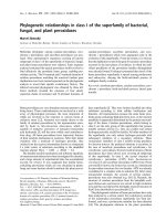
fibers. These structures are the sites of multiple interac-
tions (Fig. 1) of low affinity [5], which may facilitate
protein exchange dynamics. FAs have been shown to
be motile in stationary cells, whereas the vast majority
Keywords
adhesion; calpain; cytoskeleton; focal
complexes; ischemia; muscle
Correspondence
M C. Lebart, UMR5539, EPHE-CNRS-UM2,
cc107, Universite
´
de Montpellier II, place E.
Bataillon, 34095 Montpellier cedex 5, France
Fax: +33 0467144727
Tel: +33 0467143889
E-mail:
(Received 23 March 2006, accepted 31 May
2006)
doi:10.1111/j.1742-4658.2006.05350.x
Cells offer different types of cytoskeletal anchorages: transitory structures
such as focal contacts and perennial ones such as the sarcomeric cytoskele-
ton of muscle cells. The turnover of these structures is controlled with dif-
ferent timing by a family of cysteine proteases activated by calcium, the
calpains. The large number of potential substrates present in each of these
structures imposes fine tuning of the activity of the proteases to avoid
excessive action. This phenomenon is thus guaranteed by various types of
regulation, ranging from a relatively high calcium concentration necessary
for activation, phosphorylation of substrates or the proteases themselves
with either a favorable or inhibitory effect, possible intervention of phos-
pholipids, and the presence of a specific inhibitor and its possible degrada-
tion before activation. Finally, formation of multiprotein complexes
containing calpains offers a new method of regulation.
Abbreviations
FA, focal adhesion; FAK, focal adhesion kinase; MARCKS, myristoylated alanine-rich C-kinase substrate; MAP, microtubule-associated
protein; PKC, protein kinase C.
FEBS Journal 273 (2006) 3415–3426 ª 2006 The Authors Journal compilation ª 2006 FEBS 3415
of FAs in migrating cells do not move [6], consistent
with a role for these sites as traction points (associated
with the presence of myosin in stress fibers). As the cell
moves forward, FAs are located inside the cell and dis-
appear from the rear.
The formation of FAs obeys a consensus model
according to which integrin engagement with extracel-
lular matrix initiates the activation of focal adhesion
kinase (FAK), recruited from the cytosol, followed by
one of the actin and cytoskeletal proteins. In the past
two years, there have been a large number of studies
of the regulation of FA dynamics. In particular, from
live cell imaging of fluorescently labeled FA compo-
nents, it appeared that the cytoskeletal protein, talin
[7], in addition to kinases and adaptor molecules,
including FAK [8], Src, p130CAS, paxillin, extracellu-
lar signal-regulated kinase and myosin light-chain kin-
ase (MLCK), are critical for adhesion turnover [9].
Moreover, FAs have been shown to be sensitive (disas-
sembly) to calcium increase [10,11].
Calpain involvement in FA originates with a study
showing that inhibitors of calpain are responsible for a
decrease in the number of FAs with stabilization of the
peripheral contacts [12,13]. These studies were con-
firmed with calpain null cells (regulatory subunit),
which also showed a decreased number of FAs [14]. The
calcium-activated protease was in fact first identified in
FAs by Beckerle et al. [15], with colocalization of talin
with the catalytic subunit of calpain. More recently, the
mechanism necessary to recruit calpain 2 to peripheral
adhesion sites was shown to involve FAK [16].
It now seems clear that calpains not only act on the
destabilization of adhesion to the extracellular matrix
which is necessary at the rear of the cell to allow
migration, but also play an important function in the
formation and turnover of adhesion complexes. The
importance of these proteases at this particular place
is highlighted by the impressive list of potential sub-
strates of calpains found in adhesive structures
(Table 1).
Assembly ⁄ disassembly of FAs
The importance of FAs in assembly was highlighted
by integrin-containing clusters, which are present at
the very early stages of cell spreading [17]. These struc-
tures, which have been proposed to precede the focal
complexes that mature into FAs, were shown to form
in a calpain-dependent mechanism and are character-
ized by the presence of b3 integrin subunit and spec-
trin, both cleaved by calpain [17,18]. The authors
suggest that such cleavages could have active roles,
such as regulation of the recruitment of other proteins
in these clusters and decreasing the tension associated
with microfilament contacts to allow better clustering
of the integrins [18]. Furthermore, it has been sugges-
ted that talin cleavage by calpain may contribute to
the effects of the protease on the clustering and activa-
tion of integrins [19,20]. The importance of calpain in
FA assembly during myoblast fusion has also been
proposed [21]. As inhibition of calpains following cal-
pastatin overexpression is responsible for a decrease in
Fig. 1. Schematic representation of the various contacts established by calpain substrates in adhesion structures. Contacts are indicated by
double arrows. Proteins with kinase or phosphatase activity are noted in bold; those that have been demonstrated to interact with calpain
are circled in black; calpain regulators appear in grey boxes. Phosphorylation (and dephosphorylation) events are indicated by dashed arrows.
Calpain in cytoskeletal anchorage complex modeling M C. Lebart and Y. Benyamin
3416 FEBS Journal 273 (2006) 3415–3426 ª 2006 The Authors Journal compilation ª 2006 FEBS
adhesiveness, the authors propose that, in such situ-
ation, the formation of new FAs could be altered.
They also observed, as a consequence of calpain inhi-
bition, a marked decrease in myristoylated alanine-rich
C-kinase substrate (MARCKS) proteolysis, adding a
new substrate to the list of potential calpain substrates
(Table 1).
The proposition of calpain participation in the dis-
assembly of FAs is more straightforward and origi-
nates with the studies of Huttenlocher et al. [12]
showing that inhibiting calpain stabilizes peripheral
adhesive complexes. Then, using live cell imaging,
Huttenlocher’s group further demonstrated that cal-
pain action on the disassembly of adhesive complex
sites could be the result of influencing a-actinin–zyxin
colocalization [22], as inhibition of calpain disrupts
a-actinin localization to zyxin-containing focal con-
tacts. Finally, considering that microtubules promote
the disassembly of adhesive contact sites [23], the
group analyzed the effect of the protease in the context
of nocodazole treatment. They observed that recovery
of focal complex turnover after nocodazole wash-out
Table 1. Calpain substrates found in adhesion structures (focal adhesion, focal complexes, podosomes or integrin containing clusters).
Comments References
Structural proteins of cytoskeleton
a-Actinin Difference site of cleavage depending on the isoforms generating [39,86]
cleavage in the COOH terminal
Filamins For the c isoform (specific for muscle), cleavage in the hinge 2 region [32]
phosphorylation of the filamin C-terminus domain by PKCa protects the ABP against proteolysis
L-Plastin The cleavage separates the N-terminal domain from the core of the molecule Lebart et al.
(unpublished)
Vinculin In platelets, the major fragment is 95 kDa, corresponding to the head of the molecule [87]
Talin The cleavage separates the talin N-terminal from the C- terminal domains and unmasks the
integrin-binding site
[20]
Paxillin In vivo proteolysis inhibited by ALLN; [7,88]
Proteolysis inhibited by siRNA of calpain 2
MARCKS Phosphorylated MARCKS is a good substrate for calpains, [30,89]
The cleavage reveals an actin-binding site
Cortactin Cleavage by calpain 2 regulates cell migration [29,90]
Phosphorylation increases its sensitivity to calpain
Spectrin Phosphorylation decreases spectrin sensitivity to calpain in vitro [18,31]
Exclusive presence of the cleaved form in integrin-containing clusters
P130Cas Cleavage appears in vitro [91]
Tensin Cleavage in vitro and inhibition of protein cleavage in vivo by calpain inhibitor [92]
Gelsolin Cleavage between the G1-3 and the G4-6; localization in podosomes C. Roustan (personal
communication)
WASP
family proteins
WASP (essential component of podosomes) and WAVE are substrates [93–95]
Signal transduction proteins
Pp60Src Possible cleavage by calpain as demonstrated in vivo using calcium ionophore and inhibition
of proteolysis using calpeptin as inhibitor
[96]
FAK In vivo and in vitro cleavage, responsible for the loss of [88]
association of FAK with paxillin, vinculin, and p130cas
PKC In vitro proteolysis of three isoforms, a, b, c; [97–99]
Phosphorylated PKCl translocates to the membrane where there is a distinction between PKCa
and d and the calpain isoforms (l versus m) involved in the cleavage
RhoA Cleavage (in vivo and in vitro) responsible for the creation of a dominant negative form of RhoA;
identification of the cleavage site
[100]
PTPs The phosphorylated form of SHP-1 is protected against proteolysis by calpain [101]
PTP-1B is cleaved by calpain in spreading platelets [102,103]
MLCK Proposed cleavage by calcium-activated protease depending on the presence of CaM [104]
Tubulin Possible cleavage of a tubulin [105]
Better action of the protease before microtubule formation [106]
MAPs MAP1 and 2 are substrates [106]
Phosphorylation of MAP2 protects from calpain 2 cleavage [107]
Dynamin Isoform 1 (synaptic vesicles) would be cleaved [108]
M C. Lebart and Y. Benyamin Calpain in cytoskeletal anchorage complex modeling
FEBS Journal 273 (2006) 3415–3426 ª 2006 The Authors Journal compilation ª 2006 FEBS 3417
was inhibited in the presence of calpain inhibitors, sug-
gesting that calpain is required for this mechanism.
More recently, another study, also based on live cell
imaging, proposed a role for calpain in disassembly of
adhesive structures. The very elegant work using a
mutant of talin in the calpain cleavage site shows that
direct talin proteolysis is the key mechanism by which
calpain influences the disassembly of talin from adhe-
sion and by doing so regulates the dynamics of other
adhesion components, such us paxillin, vinculin and
zyxin [24]. The authors discuss the eventual role of the
proteolytic fragment in intracellular signaling. The idea
of a calpain fragment having specific functions is very
interesting. It underlies the fact that the protease has a
very small number of sites in the target molecule with
a particular way to generate complete structural
domains. In favor of this hypothesis are the results
that we have obtained with an actin crosslinking pro-
tein, l-plastin, found in FAs and podosomes [25]. We
have found that this actin-binding protein is a new
substrate of calpain 1 separating the core domain,
able to bind actin and the N-terminal domain which
supports the protein regulation (calcium and phos-
phorylation) (unpublished work). As synthetic peptide
containing the N-terminal sequence of l-plastin (fused
with a penetrating sequence) has been shown to acti-
vate integrins [26,27], it is tempting to speculate that
the N-terminal domain, being free from the rest of the
molecule, has a specific role.
Regulation of cleavage activity
Because the calcium concentration necessary to acti-
vate these proteases does not exist normally in the cell,
except under pathological conditions, researchers have
focused on the idea that other regulatory mechanisms
may lower this requirement. They identified phos-
phorylation and phospholipids as possibly having an
important role in adhesion. The latter were proposed
after in vitro demonstration that certain combinations
of phospholipids considerably lower the calcium con-
centration required for calpain activation [28], but this
field of investigation is poorly supported by in vivo
experiments.
Phosphorylation of the substrates has been shown to
regulate both positively and negatively the proteolytic
activity of calpain. The first example found in the lit-
erature concerns cortactin for which the phosphoryla-
tion of several unidentified Tyr residues by pp60Src
would accelerate the cleavage by calpain 1 [29]. Simi-
larly, it was recently shown that MARCKS proteolysis
by calpain is positively influence by its phosphoryla-
tion [30]. On the other hand, another French group
identified a Tyr residue located in the calpain cleavage
site of a II-spectrin as an in vitro substrate for Src kin-
ase and further demonstrated that phosphorylation of
this residue decreases spectrin sensitivity to calpain
in vitro [31]. Finally, in our laboratory, Raynaud et al.
[32] showed that phosphorylation of the filamin C-ter-
minus domain by protein kinase C (PKC) a protected
c-filamin against proteolysis by calpain 1 in COS cells.
They further illustrated their idea using myotubes,
showing that the stimulation of PKC activity prevents
c-filamin proteolysis by calpain, resulting in an
increase in myotube adhesion.
An alternative mode of regulation of protease activ-
ity in the adhesive context may involve phosphoryla-
tion of calpain itself. Again, both activating and
inhibiting roles of calpain phosphorylation have been
reported with an isoform-specific action. In particular,
this was discovered using different effectors, namely
epidermal growth factor and a chemokine (IP-9), both
inducing loss of FA plaques [33]. The significant
result comes from the fact that when these effectors
are used on the same cells, they induce different acti-
vation of calpain 1 and 2 [33,34]. In this context, epi-
dermal growth factor was shown to utilize the
microtubule-associated protein (MAP) kinase signaling
pathway with phosphorylation of calpain 2 by extra-
cellular signal-regulated kinase and activation of the
protease in the absence of calcium [34,35]. On the
other hand, calpain inactivation can be achieved when
calpain 2 is phosphorylated by protein kinase A [36].
Activation of the protease activity, as followed by
FAK cleavage and FA disruption, can also be associ-
ated with the degradation of the specific inhibitor of
calpain, calpastatin. Indeed, Carragher et al. [37] have
identified a positive feedback loop whereby activation
of v-Src promotes calpain 2 synthesis, which in turn
promotes calpastatin degradation, further enhancing
calpain activity. Moreover, a new way of activating
calpain was proposed with the discovery of the pres-
ence of an ion channel (TRPM7) in adhesion com-
plexes. This channel may be able to activate calpain 2,
although independently of an increase in the global
calcium concentration [38].
Finally, one should keep in mind that calpain may
interact with a potential target without proteolysis.
This introduces the notion of recognition without pro-
teolysis. This concept emerged in our laboratory in
2003, with the discovery that a-actinin could interact
in vitro with calpain 1 in the absence of any proteolysis
[39]. We have observed the same phenomenon with
l-plastin (our unpublished data). Moreover, it is now
clear that multimeric complexes containing calpain can
exist, which is particularly true in the adhesion context
Calpain in cytoskeletal anchorage complex modeling M C. Lebart and Y. Benyamin
3418 FEBS Journal 273 (2006) 3415–3426 ª 2006 The Authors Journal compilation ª 2006 FEBS
[16,40,41]. These complexes may be an alternative way
of recruiting calpain to FAs, thereby positioning the
protease at the very place needed for action.
In conclusion, calpains have much to do (and do
much) in adhesive structures. Control of their activities
is guaranteed by a high calcium concentration asso-
ciated with a multitude of factors varying from
phospholipids to phosphorylation, including phos-
phorylation of potential substrates (with either a favo-
rable or inhibitory effect) or even phosphorylation of
the protease itself. Association with a specific inhibitor,
possible control of degradation of the inhibitor, and
association with a potential substrate are security
measures to avoid anarchic action of the proteases.
Perennial structures
Role of calpain in myofibril disassembly
Muscle cell renewal involves elimination of useless
myofibrils before replacement during growth or after
tissue damage [42–44]. The role of ubiquitous calpains
has been highlighted in the disassembly of sarcomeres
upstream of proteasomal degradation [45,46]. Investi-
gations on muscle wasting [47] induced by hindlimb
unloading [48], food deprivation [49], or during various
pathologies [50] showed cleavage and dissociation of
proteins to be essential preliminary steps in sarcomeric
cytoskeleton stability. The involvement of calpains 1
and 2 in this muscle damage was clearly demonstrated
by overexpressing calpastatin in transgenic mice, which
reduced muscle atrophy by 30% during the unloading
period [48,51]. On the other hand, calpain 3 (p94), the
muscle-specific isoform which is insensitive to calpasta-
tin inhibition and is affected in atrophy processes,
should also be considered [52].
Myofibril organization appears as a dense bundle of
three classes of filaments (thin, thick and elastic) in the
long axis associated with desmin filaments and con-
necting proteins in the transverse direction [53]. The
early dissociation events in which calpains participate
[54] pointed to the I–Z–I complex of sarcomeres and
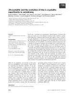
the costameric region (Fig. 2A). Sarcolemmal invagina-
tions (transverse tubules) and sarcoplasmic reticulum
(terminal cysternae) are closely associated with the
I–Z–I structure [53,55] to trigger muscle contractions
in a Ca
2+
-dependent fashion [56]. The first signs of
degradation are nebulin disappearance and emergence
of a large titin fragment of 1200 kDa, which covers
the region I-band to the A–I junction, followed by
continuous release of a-actinin (Z-filament) and degra-
dation products from cleavages of desmin, filamin
and dystrophin [57,58]. During this early stage, no
solubilized myosin or its related degradation products
are observed. Electron microscopic observations show
a decreased density of the I–Z–I region associated with
detachment of sarcolemma from the myofibril core
[59,60]. The kinetics of these degradations are closely
related to muscle type: red versus white muscle [61,62].
Calpain location in the I–Z–I structure
Similar amounts of calpains 1 and 2 were generally
found in mammal skeletal muscle, mainly associated
with subcellular elements [54,63]. Previous immunoloc-
alizations have shown that the two proteases are essen-
tially concentrated in the myofibrils near the Z-disk
and, to a lesser extent, in the I-band [64–66]. Their
presence has also been reported under the sarcolemma
membrane [43] closer to the cytoskeletal anchorage
sites [59], which roughly corresponds to the calpastatin
position [66]. Furthermore, calpain 3 was detected in
the I-band at the N2-line, in the M-band, and also at
the Z-line [67,68]; for more details, see Dugnez et al.
[68a] in this minireview series. Recently [32], calpain 1
was located between the Z-line and N1-line on each
side of the Z-disk and in the N2-line vicinity (Fig. 2B).
At least three proteins in this region, titin, a-actinin
and c-filamin, are able to bind calpain 1 with increas-
ing affinity in the presence of calcium [32,39,69]. Speci-
fic binding sites have been identified in the C-terminal
EF-hand part of a-actinin [39], the Z8–I5 N-terminal
titin region [69], in the titin I-band section near the
PEVK region [69], and in the C-terminal region
(hinge 2) of c-filamin [32].
Sequence of I–Z–I disorganization
The role of calpain has been mainly explored during
the postmortem stage of progression or on isolated
myofibrils [43,58–60]. Analysis of protein cleavage, tis-
sue imaging and the involvement of calpain isoforms
have been explored simultaneously [57,59,70]. Muscle
ischemia leads, in a few hours in fish white muscle
[71] and in 1–2 days in red muscle models, to ATP
depletion and Ca
2+
ion release into the cytosol, fol-
lowed by a decrease in pH to 5.5, which induces
intense myofibril contraction (rigor mortis). Early cal-
cium-dependent proteolysis affects the cytoskeletal
anchorages at the costameric junctions, where filamin
isoforms and dystrophin are quickly cleaved
[57,61,62], as well as desmin filaments [58], leading to
dissociation of the myofibril network with loss of
register and delamination of the sarcolemmal mem-
brane [59,61]. In contrast with mammalian red muscle
[59], Z-disks are quickly dissociated in fish white
M C. Lebart and Y. Benyamin Calpain in cytoskeletal anchorage complex modeling
FEBS Journal 273 (2006) 3415–3426 ª 2006 The Authors Journal compilation ª 2006 FEBS 3419
A
B
0
05
–2
60
70
80
90
100
110
120
130
3 5 13 15 23 25
10 15 20 25 30 35 40
50
100
150
200
250
Calpain in cytoskeletal anchorage complex modeling M C. Lebart and Y. Benyamin
3420 FEBS Journal 273 (2006) 3415–3426 ª 2006 The Authors Journal compilation ª 2006 FEBS
muscle with a concomitant release of a-actinin [61,72].
The fact that white muscle represents a simpler organ-
ization, with a single sheet of Z-filaments (a-actinin)
which connects elastic and thin filaments [73], prob-
ably explains the different observations. During rigor
mortis in red muscles, myofibril fractures are often
observed in the I-band at the N1-line and N2-line
close to calpain positions [69]. This was attributed to
the intense muscle contraction associated with calpain
cleavage. At the end of this calcium-dependent proteo-
lysis process [59,61], myofibrils appear dissociated and
fragmented into pieces mainly composed of A-bands
with large blank spaces (I–Z–I structures).
Regulation of calpains during I–Z–I
disorganization
As in the case of adhesion complexes, Ca
2+
concentra-
tions above 10 lm are nonphysiological but can be
reached during severe ischemia, calcium channel
deregulation, or cell membrane injury [56,74]. The
intracellular pH, which falls to acidic values in post-
mortem conditions, only partially (40%) decreases cal-
pain 1 activity [57]. It has also been shown using p94
knock-out mice that, in these extreme conditions, cal-
pain 3 would not play an active role, in contrast with
calpain 1 [75]. On the other hand, lower Ca
2+
concen-
trations (1–5 lm), reached during excessive exercise
[42,76] or experimentally applied to skinned fibers [77],
induce a loss of the excitation–contraction coupling
associated with a decrease in the passive force produc-
tion related to titin proteolysis [77]. This response can
be inhibited by leupeptin, a powerful cysteine protease
inhibitor, but not by calpastatin, which neutralizes ubi-
quitous calpains and not p94 [77]. Thus, damage
observed during a Ca
2+
-rigor period would be a dele-
terious effect of calpain 3.
The presence of phospholipids in the sarcolemma
and reticulum membranes [63,78] or in Z-disks [79]
could decrease the Ca
2+
concentration requirements
for autolysis of calpain 1 to levels found in the rigor
state [80]. Such regulation implies release of calpain 1
from its potential inhibitor molecule, calpastatin [81],
or cytoskeletal proteins such as titin [69] and c-filamin
[32] which can bind calpains as stable complexes. A
recent study [82] has highlighted a possible regulation
of the ubiquitous calpain system by p94, which is able
to cleave calpastatin and also titin and c-filamin
[68,83] in regions close to calpain 1-binding sites
[32,69]. Thus, activation of p94 may lead to the release
of calpain 1 from its regulators and phospholipid acti-
vation [84]. Validation of such a model would involve
identification of p94 in the activation process [47].
Conclusion
A growing body of evidence indicates that the two
calpain isoforms perform vital operations in cell
motility and tissue renewal. However, this potential is
sometimes deviated from the normal physiological
benefits to pathological behaviors such as invasive
properties of cells [85] or ischemia and genetic dis-
eases which affect calcium homeostasis [50]. Control
of calpain activity by treatment with inhibitory drugs
may limit the invasive properties of metastasis and
tissue injury. Such investigations involve searching for
efficient competitive inhibitors of cellular substrates as
well as modeling of the domain II active conforma-
tion in calpain 1 and calpain 2 to optimize specifici-
ties. The concept of a cell-diffusive molecule able to
tie up calpains in their inactive conformation, as cal-
pastatin does, would be another option. The numer-
ous possible targets in cells (Table 1), the broad
spectrum of the cleaved sequences, and the fact that
the two ubiquitous isoforms can substitute for each
other in differentiated cells are serious problems. A
way of perturbing communication between domains
IV and III or maintaining domain I anchorage within
domain VI, thus locking the open conformation
regardless of the calcium concentration, would be an
Fig. 2. Location of calpain 1 and its targets in the myofibril. (A) Schematic representation of a peripheral myofibril [53] in skeletal muscle (I–
Z–I part), representing calpain 1 location (pink area) as well as several of its main targets (red double arrow) assumed to be essential for cell
adhesion and membrane stability (b-integrin, dystrophin), thin filament cohesion (nebulin, capZ), myofibril–cytoskeleton linkage (c-filamin, des-
min) and the passive tension in sarcomeres (titin). Connections between myofibrils and the sarcolemma were drawn by using peripheral
actin cytoskeleton anchored in a costameric structure. The triad complex including transverse tubule (tt) and terminal cysternea (tc) was
located near the Z–line in the interaction with T-cap [55]. Intermediary filaments (desmin) that maintain sarcomere alignment are suggested
by a dashed line towards the myofibril core. (B) Immunofluorescent (a,b) and immunoperoxidase (c,d) patterns of calpain 1 in longitudinal
(a,c,d) and transverse (b) sections of mouse (a,b) and bovine (c,d) muscle fibers. The Z-line was expanded and scanned for density (e,f) to
compare the control muscle strip treated with the secondary peroxidase-labeled antibody alone (c,e) with the one treated with calpain 1 anti-
body (d,f). Note the increased intensity of the N2-line (d) and the doublet (arrowhead) at the Z-line edges (f). S, sarcolemmal membrane;
Z, Z-line; N, nucleus; TC, triad complex; M, M-line; N2, N2-line. Experimental conditions for calpain 1 location were previously described
[32,69].
M C. Lebart and Y. Benyamin Calpain in cytoskeletal anchorage complex modeling
FEBS Journal 273 (2006) 3415–3426 ª 2006 The Authors Journal compilation ª 2006 FEBS 3421
exciting breakthrough in pharmacological investiga-
tions.
References
1 Webb DJ, Zhang H & Horwitz AF (2005) Cell migra-
tion: an overview. Methods Mol Biol 294, 3–11.
2 Nobes CD & Hall A (1995) Rho, rac, and cdc42
GTPases regulate the assembly of multimolecular focal
complexes associated with actin stress fibers, lamellipo-
dia, and filopodia. Cell 81, 53–62.
3 Rottner K, Hall A & Small JV (1999) Interplay
between Rac and Rho in the control of substrate con-
tact dynamics. Curr Biol 9, 640–648.
4 Miranti CK & Brugge JS (2002) Sensing the environ-
ment: a historical perspective on integrin signal trans-
duction. Nat Cell Biol 4, E83–E90.
5 Goldmann WH (2000) Kinetic determination of focal
adhesion protein formation. Biochem Biophys Res
Commun 271, 553–557.
6 Smilenov LB, Mikhailov A, Pelham RJ, Marcantonio
EE & Gundersen GG (1999) Focal adhesion motility
revealed in stationary fibroblasts. Science 286, 1172–
1174.
7 Franco S, Perrin B & Huttenlocher A (2004) Isoform
specific function of calpain 2 in regulating membrane
protrusion. Exp Cell Res 299, 179–187.
8 Hamadi A, Bouali M, Dontenwill M, Stoeckel H,
Takeda K & Ronde P (2005) Regulation of focal adhe-
sion dynamics and disassembly by phosphorylation of
FAK at tyrosine 397. J Cell Sci 118, 4415–4425.
9 Webb DJ, Donais K, Whitmore LA, Thomas SM, Tur-
ner CE, Parsons JT & Horwitz AF (2004) FAK-Src
signalling through paxillin, ERK and MLCK regulates
adhesion disassembly. Nat Cell Biol 6, 154–161.
10 Giannone G, Ronde P, Gaire M, Beaudouin J, Haiech
J, Ellenberg J & Takeda K (2004) Calcium rises locally
trigger focal adhesion disassembly and enhance resi-
dency of focal adhesion kinase at focal adhesions.
J Biol Chem 279, 28715–28723.
11 Giannone G, Ronde P, Gaire M, Haiech J & Takeda
K (2002) Calcium oscillations trigger focal adhesion
disassembly in human U87 astrocytoma cells. J Biol
Chem 277, 26364–26371.
12 Huttenlocher A, Palecek SP, Lu Q, Zhang W, Mellgren
RL, Lauffenburger DA, Ginsberg MH & Horwitz AF
(1997) Regulation of cell migration by the calcium-
dependent protease calpain. J Biol Chem 272, 32719–
32722.
13 Kulkarni S, Saido TC, Suzuki K & Fox JE (1999)
Calpain mediates integrin-induced signaling at a point
upstream of Rho family members. J Biol Chem 274,
21265–21275.
14 Dourdin N, Bhatt AK, Dutt P, Greer PA, Arthur JS,
Elce JS & Huttenlocher A (2001) Reduced cell migra-
tion and disruption of the actin cytoskeleton in cal-
pain-deficient embryonic fibroblasts. J Biol Chem 276,
48382–48388.
15 Beckerle MC, Burridge K, DeMartino GN & Croall
DE (1987) Colocalization of calcium-dependent pro-
tease II and one of its substrates at sites of cell adhe-
sion. Cell 51, 569–577.
16 Carragher NO, Westhoff MA, Fincham VJ, Schaller
MD & Frame MC (2003) A novel role for FAK as a
protease-targeting adaptor protein: regulation by p42
ERK and Src. Curr Biol 13, 1442–1450.
17 Bialkowska K, Du Kulkarni SX, Goll DE, Saido TC
& Fox JE (2000) Evidence that beta3 integrin-induced
Rac activation involves the calpain-dependent forma-
tion of integrin clusters that are distinct from the focal
complexes and focal adhesions that form as Rac and
RhoA become active. J Cell Biol 151, 685–696.
18 Bialkowska K, Saido TC & Fox JE (2005) SH3
domain of spectrin participates in the activation of Rac
in specialized calpain-induced integrin signaling com-
plexes. J Cell Sci 118, 381–395.
19 Calderwood DA (2004) Integrin activation. J Cell Sci
117, 657–666.
20 Yan B, Calderwood DA, Yaspan B & Ginsberg MH
(2001) Calpain cleavage promotes talin binding to the
beta 3 integrin cytoplasmic domain. J Biol Chem 276,
28164–28170.
21 Dedieu S, Poussard S, Mazeres G, Grise F, Dargelos
E, Cottin P & Brustis JJ (2004) Myoblast migration is
regulated by calpain through its involvement in cell
attachment and cytoskeletal organization. Exp Cell Res
292, 187–200.
22 Bhatt A, Kaverina I, Otey C & Huttenlocher A (2002)
Regulation of focal complex composition and disas-
sembly by the calcium-dependent protease calpain.
J Cell Sci 115, 3415–3425.
23 Kaverina I, Krylyshkina O & Small JV (1999)
Microtubule targeting of substrate contacts promotes
their relaxation and dissociation. J Cell Biol 146,
1033–1044.
24 Franco SJ, Rodgers MA, Perrin BJ, Han J, Bennin
DA, Critchley DR & Huttenlocher A (2004) Calpain-
mediated proteolysis of talin regulates adhesion
dynamics. Nat Cell Biol 6, 977–983.
25 Babb SG, Matsudaira P, Sato M, Correia I & Lim SS
(1997) Fimbrin in podosomes of monocyte-derived
osteoclasts. Cell Motil Cytoskeleton 37, 308–325.
26 Jones SL, Wang J, Turck CW & Brown EJ (1998) A
role for the actin-bundling protein 1-plastin in the reg-
ulation of leukocyte integrin function. Proc Natl Acad
Sci USA 95, 9331–9336.
27 Wang J, Chen H & Brown EJ (2001) l-plastin peptide
activation of alpha (v) beta (3)-mediated adhesion
requires integrin conformational change and actin fila-
ment disassembly. J Biol Chem 276, 14474–14481.
Calpain in cytoskeletal anchorage complex modeling M C. Lebart and Y. Benyamin
3422 FEBS Journal 273 (2006) 3415–3426 ª 2006 The Authors Journal compilation ª 2006 FEBS
28 Chakrabarti AK, Dasgupta S, Gadsden RHSr, Hogan
EL & Banik NL (1996) Regulation of brain m calpain
Ca
2+
sensitivity by mixtures of membrane lipids: acti-
vation at intracellular Ca
2+
level. J Neurosci Res 44,
374–380.
29 Huang C, Tandon NN, Greco NJ, Ni Y, Wang T &
Zhan X (1997) Proteolysis of platelet cortactin by cal-
pain. J Biol Chem 272, 19248–19252.
30 Dulong S, Goudenege S, Vuillier-Devillers K, Manenti
S, Poussard S & Cottin P (2004) Myristoylated ala-
nine-rich C kinase substrate (MARCKS) is involved in
myoblast fusion through its regulation by protein
kinase C alpha and calpain proteolytic cleavage.
Biochem J 382, 1015–1023.
31 Nicolas G, Fournier CM, Galand C, Malbert-Colas L,
Bournier O, Kroviarski Y, Bourgeois M, Camonis JH,
Dhermy D, Grandchamp et al. (2002) Tyrosine phos-
phorylation regulates alpha II spectrin cleavage by cal-
pain. Mol Cell Biol 22, 3527–3536.
32 Raynaud F, Jond-Necand C, Marcilhac A, Fu
¨
rst D &
Benyamin Y (2006) Calpain 1-c filamin interaction in
muscle cells: a possible in situ regulation by PKC-a. Int
J Biochem Cell Biol 38, 404–413.
33 Satish L, Blair HC, Glading A & Wells A (2005) Inter-
feron-inducible protein 9 (CXCL11)-induced cell moti-
lity in keratinocytes requires calcium flux-dependent
activation of mu-calpain. Mol Cell Biol 25, 1922–1941.
34 Glading A, Bodnar RJ, Reynolds IJ, Shiraha H, Satish
L, Potter DA, Blair HC & Wells A (2004) Epidermal
growth factor activates m-calpain (calpain II), at least
in part, by extracellular signal-regulated kinase-
mediated phosphorylation. Mol Cell Biol 24, 2499–
2512.
35 Cuevas BD, Abell AN, Witowsky JA, Yujiri T, John-
son NL, Kesavan K, Ware M, Jones PL, Weed SA,
DeBiasi RL, et al. (2003) MEKK1 regulates calpain-
dependent proteolysis of focal adhesion proteins for
rear-end detachment of migrating fibroblasts. EMBO J
22, 3346–3355.
36 Shiraha H, Glading A, Chou J, Jia Z & Wells A (2002)
Activation of m-calpain (calpain II) by epidermal
growth factor is limited by protein kinase A phosphor-
ylation of m-calpain. Mol Cell Biol 22, 2716–2727.
37 Carragher NO, Westhoff MA, Riley D, Potter DA, Dutt
P, Elce JS, Greer PA & Frame MC (2002) v-Src-induced
modulation of the calpain-calpastatin proteolytic system
regulates transformation. Mol Cell Biol 22, 257–269.
38 Su LT, Agapito MA, Li M, Simonson WT, Huttenlo-
cher A, Habas R, Yue L & Runnels LW (2006) Trpm7
regulates cell adhesion by controlling the calcium-
dependent protease calpain. J Biol Chem 281 11260–
11270.
39 Raynaud F, Bonnal C, Fernandez E, Bremaud L,
Cerutti M, Lebart MC, Roustan C, Ouali A & Benya-
min Y (2003) The calpain 1–alpha–actinin interaction.
Resting complex between the calcium-dependent prote-
ase and its target in cytoskeleton. Eur J Biochem 270,
4662–4670.
40 Lebart M-C, Le Goff E, Hubert F, Herrada-Aldrian
G, Rebie
`
re B, Roustan C & Benyamin Y (2006) Evi-
dence for l-plastin-beta integrin complex and regula-
tion by microcalpain cleavage. Biochem J, in press.
41 Westhoff MA, Serrels B, Fincham VJ, Frame MC &
Carragher NO (2004) SRC-mediated phosphorylation
of focal adhesion kinase couples actin and adhesion
dynamics to survival signaling. Mol Cell Biol 24, 8113–
8133.
42 Belcastro AN, Shewchuk LD & Raj DA (1998) Exer-
cise-induced muscle injury: a calpain hypothesis. Mol
Cell Biochem 179, 135–145.
43 Goll DE, Thompson VF, Taylor RG & Christiansen
JA (1992) Role of the calpain system in muscle growth.
Biochimie 74, 225–237.
44 Hasselgren PO & Fischer JE (2001) Muscle cachexia:
current concepts of intracellular mechanisms and mole-
cular regulation. Ann Surg 233, 9–17.
45 Jagoe RT & Goldberg AL (2001) What do we really
know about the ubiquitin-proteasome pathway in mus-
cle atrophy? Curr Opin Clin Nutr Metab Care 4, 183–
190.
46 Bassaglia Y, Cebrian J, Covan S, Garcia M & Foucrier J
(2005) Proteasomes are tightly associated to myofibrils
in mature skeletal muscle. Exp Cell Res 302, 221–232.
47 Bartoli M & Richard I (2005) Calpains in muscle wast-
ing. Int J Biochem Cell Biol 37, 2115–2133.
48 Tidball JG & Spencer MJ (2002) Expression of a cal-
pastatin transgene slows muscle wasting and obviates
changes in myosin isoform expression during murine
muscle disuse. J Physiol 545, 819–828.
49 Nakashima K, Komatsu T, Yamazaki M & Abe H
(2005) Effects of fasting and refeeding on expression of
proteolytic-related genes in skeletal muscle of chicks.
J Nutr Sci Vitaminol (Tokyo). 51, 248–253.
50 Zatz M & Starling A (2005) Calpains and disease.
N Engl J Med 352, 2413–2423.
51 Kent MP, Spencer MJ & Koohmaraie M (2004) Post-
mortem proteolysis is reduced in transgenic mice over-
expressing calpastatin. J Anim Sci 82, 794–801.
52 Kramerova I, Kudryashova E, Venkatraman G &
Spencer MJ (2005) Calpain 3 participates in sarcomere
remodeling by acting upstream of the ubiquitin-protea-
some pathway. Hum Mol Genet 14, 2125–2134.
53 Squire JM, Al-Khayat HA, Knupp C & Luther PK
(2005) Molecular architecture in muscle contractile
assemblies. Adv Protein Chem 71, 17–87.
54 Goll DE, Thompson VF, Li H, Wei W & Cong J
(2003) The calpain system. Physiol Rev 83, 731–801.
55 Furukawa T, Ono Y, Tsuchiya H, Katayama Y, Bang
ML, Labeit D, Labeit S, Inagaki N & Gregorio CC
(2001) Specific interaction of the potassium channel
M C. Lebart and Y. Benyamin Calpain in cytoskeletal anchorage complex modeling
FEBS Journal 273 (2006) 3415–3426 ª 2006 The Authors Journal compilation ª 2006 FEBS 3423
beta-subunit minK with the sarcomeric protein T-cap
suggests a T-tubule-myofibril linking system. J Mol
Biol 313, 775–784.
56 Berchtold MW, Brinkmeier H & Muntener M (2000)
Calcium ion in skeletal muscle: its crucial role for mus-
cle function, plasticity, and disease. Physiol Rev 80,
1215–1265.
57 Huff-Lonergan E, Mitsuhashi T, Beekman DD, Parrish
FC Jr, Olson DG & Robson RM (1996) Proteolysis of
specific muscle structural proteins by mu-calpain at low
pH and temperature is similar to degradation in post-
mortem bovine muscle. J Anim Sci 74, 993–1008.
58 Geesink GH & Koohmaraie M (1999) Postmortem
proteolysis and calpain ⁄ calpastatin activity in callipyge
and normal lamb biceps femoris during extended post-
mortem storage. J Anim Sci 77, 1490–1501.
59 Taylor RG, Geesink GH, Thompson VF, Koohmaraie
M & Goll DE (1995) Is Z-disk degradation responsible
for postmortem tenderization? J Anim Sci 73 , 1351–
1367.
60 Taylor RG, Papa I, Astier C, Ventre F, Benyamin Y &
Ouali A (1997) Fish muscle cytoskeleton integrity is
not dependent on intact thin filaments. J Muscle Res
Cell Motil 18, 285–294.
61 Papa I, Taylor RG, Astier C, Ventre F, Lebart MC,
Roustan C, Ouali A & Benyamin Y (1997) Dystrophin
cleavage and sarcolemma detachment are early post
mortem changes on bass (Dicentrarchus labrax) white
muscle. J Food Sci. 62, 917–921.
62 Bonnal C, Raynaud F, Astier C, Lebart MC, Marci-
lhac A, Coves D, Corraze G, Gelineau A, Fleurence J,
Roustan C, et al. (2001) Postmortem degradation of
white fish skeletal muscle (sea bass, Dicentrarchus lab-
rax): fat diet effects on in situ dystrophin proteolysis
during the prerigor stage. Mar Biotechnol (NY) 3,
172–180.
63 Hood JL, Logan BB, Sinai AP, Brooks WH & Rosz-
man TL (2003) Association of the calpain ⁄ calpastatin
network with subcellular organelles. Biochem Biophys
Res Commun 310, 1200–1212.
64 Dayton WR & Schollmeyer JV (1981) Immunocyto-
chemical localization of a calcium-activated protease in
skeletal muscle cells. Exp Cell Res 136, 423–433.
65 Yoshimura N, Murachi T, Heath R, Kay J, Jasani B
& Newman GR (1986) Immunogold electron-micro-
scopic localisation of calpain I in skeletal muscle of
rats. Cell Tissue Res 244, 265–270.
66 Kumamoto T, Kleese WC, Cong JY, Goll DE, Pierce
PR & Allen RE (1992) Localization of the Ca(
2+
)-
dependent proteinases and their inhibitor in normal,
fasted, and denervated rat skeletal muscle. Anat Rec
232, 60–77.
67 Sorimachi H, Kinbara K, Kimura S, Takahashi M,
Ishiura S, Sasagawa N, Sorimachi N, Shimada H,
Tagawa K, Maruyama K, et al. (1995) Muscle-specific
calpain, p94, responsible for limb girdle muscular
dystrophy type 2A, associates with connectin through
IS2, a p94-specific sequence. J Biol Chem 270, 31158–
31162.
68 Taveau M, Bourg N, Sillon G, Roudaut C, Bartoli M
& Richard I (2003) Calpain 3 is activated through
autolysis within the active site and lyses sarcomeric
and sarcolemmal components. Mol Cell Biol 23, 9127–
9135.
68a Duguez S, Bartoli M, Richard I (2006) Calpain 3: a
key regulator of the sarcomere? FEBS J 273, 000–000.
69 Raynaud F, Fernandez E, Coulis G, Aubry L, Vignon
X, Bleimling N, Gautel M, Benyamin Y & Ouali A
(2005) Calpain 1–titin interactions concentrate calpain
1 in the Z-band edges and in the N2-line region within
the skeletal myofibril. FEBS J 272, 2578–2590.
70 Delgado EF, Geesink GH, Marchello JA, Goll DE &
Koohmaraie M (2001) Properties of myofibril-bound
calpain activity in longissimus muscle of callipyge and
normal sheep. J Anim Sci 79, 2097–2107.
71 Astier C, Labbe JP, Roustan C & Benyamin Y (1991)
Sarcomeric disorganization in post-mortem fish mus-
cles. Comp Biochem Physiol B 100, 459–465.
72 Papa I, Alvarez C, Verrez-Bagnis V, Fleurence J &
Benyamin Y (1996) Post mortem release of fish white
muscle a-actinin as a marker of disorganisation. J Sci
Food Agric 72, 63–70.
73 Luther PK (1991) Three-dimensional reconstruction of
a simple Z-band in fish muscle. J Cell Biol 113, 1043–
1055.
74 Spencer MJ, Croall DE & Tidball JG (1995) Calpains
are activated in necrotic fibers from mdx dystrophic
mice. J Biol Chem 270, 10909–10914.
75 Geesink GH, Taylor RG & Koohmaraie M (2005) Cal-
pain 3 ⁄ p94 is not involved in postmortem proteolysis.
J Anim Sci 83, 1646–1652.
76 Gissel H & Clausen T (2001) Excitation-induced Ca
2+
influx and skeletal muscle cell damage. Acta Physiol
Scand 171, 327–334.
77 Verburg E, Murphy RM, Stephenson DG & Lamb
GD (2005) Disruption of excitation-contraction cou-
pling and titin by endogenous Ca
2+
-activated proteases
in toad muscle fibres. J Physiol 564, 775–790.
78 Hood JL, Brooks WH & Roszman TL (2004) Differen-
tial compartmentalization of the calpain ⁄ calpastatin
network with the endoplasmic reticulum and Golgi
apparatus. J Biol Chem 279, 43126–43135.
79 Takahashi K, Shimada K, Ahn DH & Ji JR (2001) Iden-
tification of lipids as the main component of skeletal
muscle Z-discs. J Muscle Res Cell Motil 22, 353–360.
80 Suzuki K & Sorimachi H (1998) A novel aspect of cal-
pain activation. FEBS Lett 433, 1–4.
81 Nori SL, Pompili E, De Santis E, De Renzis G, Bondi
A, Collier WL, Ippoliti F & Fumagalli L (1993) Immu-
nogold ultrastructural localization of calpastatin, the
Calpain in cytoskeletal anchorage complex modeling M C. Lebart and Y. Benyamin
3424 FEBS Journal 273 (2006) 3415–3426 ª 2006 The Authors Journal compilation ª 2006 FEBS
calpain inhibitor, in rabbit skeletal muscle. Cell Mol
Biol (Noisy-le-grand) 39, 729–737.
82 Ono Y, Kakinuma K, Torii F, Irie A, Nakagawa K,
Labeit S, Abe K, Suzuki K & Sorimachi H (2004)
Possible regulation of the conventional calpain system
by skeletal muscle-specific calpain, p94 ⁄ calpain 3.
J Biol Chem 279, 2761–2771.
83 Guyon JR, Kudryashova E, Potts A, Dalkilic I, Brosi-
us MA, Thompson TG, Beckmann JS, Kunkel LM &
Spencer MJ (2003) Calpain 3 cleaves filamin C and
regulates its ability to interact with gamma- and delta-
sarcoglycans. Muscle Nerve 28, 472–483.
84 Tompa P, Emori Y, Sorimachi H, Suzuki K & Frie-
drich P (2001) Domain III of calpain is a Ca
2+
-regula-
ted phospholipid-binding domain. Biochem Biophys
Res Commun 280, 1333–1339.
85 Benetti R, Copetti T, Dell’Orso S, Melloni E, Branco-
lini C, Monte M & Schneider C (2005) The calpain sys-
tem is involved in the constitutive regulation of beta-
catenin signaling functions. J Biol Chem 280, 22070–
22080.
86 Selliah N, Brooks WH & Roszman TL (1996) Proteo-
lytic cleavage of alpha-actinin by calpain in T cells
stimulated with anti-CD3 monoclonal antibody.
J Immunol 156, 3215–3221.
87 Serrano K & Devine DV (2004) Vinculin is proteolyzed
by calpain during platelet aggregation: 95 kDa cleavage
fragment associates with the platelet cytoskeleton. Cell
Motil Cytoskeleton 58, 242–252.
88 Carragher NO, Levkau B, Ross R & Raines EW
(1999) Degraded collagen fragments promote rapid dis-
assembly of smooth muscle focal adhesions that corre-
lates with cleavage of pp125 (FAK), paxillin, and talin.
J Cell Biol 147, 619–630.
89 Tapp H, Al-Naggar IM, Yarmola EG, Harrison A,
Shaw G, Edison AS & Bubb MR (2005) MARCKS is
a natively unfolded protein with an inaccessible actin-
binding site: evidence for long-range intramolecular
interactions. J Biol Chem 280, 9946–9956.
90 Perrin BJ, Amann KJ & Huttenlocher A (2006) Proteo-
lysis of cortactin by calpain regulates membrane protru-
sion during cell migration. Mol Biol Cell 17, 239–250.
91 Shim SR, Kook S, Kim JI & Song WK (2001)
Degradation of focal adhesion proteins paxillin and
p130cas by caspases or calpains in apoptotic rat-1
and L929 cells. Biochem Biophys Res Commun 286,
601–608.
92 Chen H, Ishii A, Wong WK, Chen LB & Lo SH
(2000) Molecular characterization of human tensin.
Biochem J 351, 403–411.
93 Calle Y, Jones GE, Jagger C, Fuller K, Blundell MP,
Chow J, Chambers T & Thrasher AJ (2004) WASp
deficiency in mice results in failure to form osteoclast
sealing zones and defects in bone resorption. Blood
103, 3552–3561.
94 Oda A, Miki H, Wada I, Yamaguchi H, Yamazaki D,
Suetsugu S, Nakajima M, Nakayama A, Okawa K,
Miyazaki H, et al. (2005) WAVE ⁄ Scars in platelets.
Blood 105, 3141–3148.
95 Shcherbina A, Miki H, Kenney DM, Rosen FS, Tak-
enawa T & Remold-O’Donnell E (2001) WASP and
N-WASP in human platelets differ in sensitivity to
protease calpain. Blood 98, 2988–2991.
96 Oda A, Druker BJ, Ariyoshi H, Smith M & Salzman
EW (1993) pp60src is an endogenous substrate for cal-
pain in human blood platelets. J Biol Chem 268,
12603–12608.
97 Kennett SB, Roberts JD & Olden K (2004) Require-
ment of protein kinase C micro activation and calpain-
mediated proteolysis for arachidonic acid-stimulated
adhesion of MDA-MB-435 human mammary carci-
noma cells to collagen type IV. J Biol Chem 279, 3300–
3307.
98 Kishimoto A, Mikawa K, Hashimoto K, Yasuda I,
Tanaka S, Tominaga M, Kuroda T & Nishizuka Y
(1989) Limited proteolysis of protein kinase C subspe-
cies by calcium-dependent neutral protease (calpain).
J Biol Chem 264, 4088–4092.
99 Liang YC, Yeh JY, Forsberg NE & Ou BR (2006)
Involvement of mu- and m-calpains and protein kinase
C isoforms in L8 myoblast differentiation. Int J Bio-
chem Cell Biol 38, 662–670.
100 Kulkarni S, Goll DE & Fox JE (2002) Calpain cleaves
RhoA generating a dominant-negative form that inhi-
bits integrin-induced actin filament assembly and cell
spreading. J Biol Chem 277, 24435–24441.
101 Falet H, Pain S & Rendu F (1998) Tyrosine unpho-
sphorylated platelet SHP-1 is a substrate for calpain.
Biochem Biophys Res Commun 252, 51–55.
102 Frangioni JV, Oda A, Smith M, Salzman EW &
Neel BG (1993) Calpain-catalyzed cleavage and sub-
cellular relocation of protein phosphotyrosine phos-
phatase 1B (PTP-1B) in human platelets. EMBO J
12, 4843–4856.
103 Yuan Y, Dopheide SM, Ivanidis C, Salem HH & Jack-
son SP (1997) Calpain regulation of cytoskeletal signal-
ing complexes in von Willebrand factor-stimulated
platelets. Distinct roles for glycoprotein Ib-V–IX and
glycoprotein IIb-IIIa (integrin alphaIIbbeta3) in von
Willebrand factor-induced signal transduction. J Biol
Chem 272, 21847–21854.
104 Ito M, Tanaka T, Nunoki K, Hidaka H & Suzuki K
(1987) The Ca
2+
-activated protease (calpain) modu-
lates Ca
2+
⁄ calmodulin dependent activity of smooth
muscle myosin light chain kinase. Biochem Biophys Res
Commun 145, 1321–1328.
105 Santella L, Kyozuka K, Hoving S, Munchbach M,
Quadroni M, Dainese P, Zamparelli C, James P
& Carafoli E (2000) Breakdown of cytoskeletal
proteins during meiosis of starfish oocytes and pro-
M C. Lebart and Y. Benyamin Calpain in cytoskeletal anchorage complex modeling
FEBS Journal 273 (2006) 3415–3426 ª 2006 The Authors Journal compilation ª 2006 FEBS 3425
teolysis induced by calpain. Exp Cell Res 259, 117–
126.
106 Billger M, Wallin M & Karlsson JO (1988) Proteolysis
of tubulin and microtubule-associated proteins 1 and 2
by calpain I and II. Difference in sensitivity of
assembled and disassembled microtubules. Cell Calcium
9, 33–44.
107 Alexa A, Tompa P, Baki A, Vereb G & Friedrich P
(1996) Mutual protection of microtubule-associated
protein 2 (MAP2) and cyclic AMP-dependent protein
kinase II against mu-calpain. J Neurosci Res 44,
438–445.
108 Kelly BL, Vassar R & Ferreira A (2005) Beta-amyloid-
induced dynamin 1 depletion in hippocampal neurons.
A potential mechanism for early cognitive decline in
Alzheimer disease. J Biol Chem 280, 31746–31753.
Calpain in cytoskeletal anchorage complex modeling M C. Lebart and Y. Benyamin
3426 FEBS Journal 273 (2006) 3415–3426 ª 2006 The Authors Journal compilation ª 2006 FEBS