Báo cáo khoa học: Critical roles of conserved carboxylic acid residues in pigeon cytosolic NADP+-dependent malic enzyme docx
Bạn đang xem bản rút gọn của tài liệu. Xem và tải ngay bản đầy đủ của tài liệu tại đây (565.75 KB, 10 trang )
Critical roles of conserved carboxylic acid residues in
pigeon cytosolic NADP
+
-dependent malic enzyme
Shuo-Chin Chang
1
*, Kuan-Yu Lin
1
*, Yu-Jung Chen
1
, Chin-Hung Lai
1
, Gu-Gang Chang
2
and Wei-Yuan Chou
1
1 Department of Biochemistry, National Defense Medical Center, Taipei, Taiwan
2 Faculty of Life Sciences, Institute of Biochemistry, Structural Biology Program, National Yang-Ming University, Taipei, Taiwan
Cytosolic NADP
+
-dependent malic enzyme (EC
1.1.1.40) catalyses the decarboxylation of l-malate to
pyruvate with oxaloacetate as intermediate and is asso-
ciated with the reduction of NADP
+
to NADPH in
the presence of a bivalent metal ion. Malic enzyme is a
tetramer of identical subunits. The 3D structures of
malic enzymes have been studied extensively [1]. Since
the first crystal structure was solved for the human
mitochondrial NAD(P)
+
-dependent malic enzyme
complexed with Mn
2+
and ATP [2], 14 structures from
various species, including the human, pigeon, the
roundworm Ascaris suum, and the bacterium Thermo-
toga maritima, have been deposited in the protein data-
bank. All these structures, except that of T. maritima,
have similar topology. These structures are classified
into open and closed forms, depending on the presence
of the substrate, l-malate, or its analogues [3]. It has
been proposed that the closed form is the catalytically
active form of the enzyme.
Based on pH profiles and isotope studies of malic
enzyme, it was proposed that its catalysis involves a
general acid ⁄ base mechanism [4–7]. A general base is
involved in deprotonating the C2 hydroxy group to
form an oxaloacetate intermediate and in facilitating
the hydride transfer from C2 to NADP
+
. After
decarboxylation of oxaloacetate, a general acid partici-
pates in the enol–keto tautomerization of pyruvate.
Site-directed mutagenesis and kinetic results suggest
that K199 in Ascaris (K162 in pigeon) [8] and D295
(D258 in pigeon) [9] function as the general acid and
base, respectively. Our previous studies indicated that
the K162 residue of pigeon NADP
+
-dependent malic
Keywords
chemical rescue; general acid ⁄ base; malic
enzyme; metal ion binding; site-directed
mutagenesis
Correspondence
W Y. Chou, Department of Biochemistry,
National Defense Medical Center,
161 MinQuan E. Road Sec 6, Taipei,
Taiwan 11490
Fax: +886 2 8792 3106
Tel: +886 2 8791 0776
E-mail:
*These authors contributed equally to the
experimental work.
(Received 19 May 2006, revised 2 July
2006, accepted 7 July 2006)
doi:10.1111/j.1742-4658.2006.05409.x
Malic enzyme catalyses the reduction of NADP
+
to NADPH and the
decarboxylation of l-malate to pyruvate through a general acid ⁄ base mech-
anism. Previous kinetic and structural studies differ in their interpretation
of the amino acids responsible for the general acid ⁄ base mechanism. To
resolve this discrepancy, we used site-directed mutagenesis and kinetic ana-
lysis to study four conserved carboxylic amino acids. With the D257A
mutant, the K
m
for Mn
2+
and the k
cat
decreased relative to those of the
wild-type by sevenfold and 28-fold, respectively. With the E234A mutant,
the K
m
for Mg
2+
and l-malate increased relative to those of the wild-type
by 87-fold and 49-fold, respectively, and the k
cat
remained unaltered, which
suggests that the E234 residue plays a critical role in bivalent metal ion
binding. The k
cat
for the D235A and D258A mutants decreased relative to
that of the wild-type by 7800-fold and 5200-fold, respectively, for the over-
all reaction, by 800-fold and 570-fold, respectively, for the pyruvate reduc-
tion partial reaction, and by 371-fold and 151-fold, respectively, for the
oxaloacetate decarboxylation. The activities of the overall reaction and the
pyruvate reduction partial reaction of the D258A mutant were rescued by
the presence of 50 mm sodium azide. In contrast, small free acids did not
have a rescue effect on the activities of the E234A, D235A, and D257A
mutants. These data suggest that D258 may act as a general base to extract
the hydrogen of the C2 hydroxy group of l-malate with the aid of D235-
chelated Mn
2+
to polarize the hydroxyl group.
4072 FEBS Journal 273 (2006) 4072–4081 ª 2006 The Authors Journal compilation ª 2006 FEBS
enzyme is a general acid that donates a proton in
enol–keto tautomerization [10]. However, the crystal
structure of human mitochondrial NAD(P)
+
-depend-
ent malic enzyme revealed that the oxygen of the carb-
oxy group of D279 (D258 in the pigeon, D295 in
A. suum) is structurally too distant to extract the pro-
ton from the C2 hydroxy group of l-malate and would
not play a role in the general acid ⁄ base mechanism
[11]. Therefore, the authors proposed that K183 (K162
in the pigeon, K199 in A. suum) and Y112 (Y91 in the
pigeon, Y126 in A. suum) are the general base and
acid, respectively. Similar geometry was observed in
A. suum mitochondrial NAD-dependent malic enzyme
[12]. Recently, Cook and his colleagues [13] re-exam-
ined the contribution of residues Y126, K199, and
D294 (D257 in the pigeon, D274 in the human) to
pH–rate profiles. They proposed that these three resi-
dues form a catalytic triad, with K199 as the general
base and Y126 as the general acid in the enzymatic
mechanism.
The crystal structure of pigeon malic enzyme showed
that the metal ion is co-ordinated with the carboxylic
group on the side chain of E234, D235, D258, the C1
carboxy group and the C2 hydroxy group of l-malate,
and a free water molecule to form an octahedral con-
formation [14]. The metal-binding roles of E234 and
D235 have been confirmed in metal-protected urea-
denaturation studies [15]. However, the K
m
value for
Mn
2+
decreased 100-fold with E234Q, but was unal-
tered with D235N. This prompted us to examine the
contribution of these three amino-acid residues to
metal ion binding and enzymatic catalysis.
In this study, we sought to delineate the possible
roles of these conserved residues in the active site of
pigeon cytosolic NADP
+
-dependent malic enzyme by
site-directed mutagenesis and detailed enzyme kinetic
studies.
Results
Purification and structural characterization of
wild-type and mutant malic enzymes
To evaluate the possible roles of the conserved carb-
oxylic amino acids at the active site, E234, D235,
D257, and D258 in pigeon NADP
+
-dependent malic
enzyme were replaced by alanine using site-directed
mutagenesis. The mutated enzymes were expressed in
Escherichia coli BL21(DE3) and purified. All recom-
binant enzymes were shown to be homogeneous by
SDS ⁄ PAGE (see Supplementary material Fig. S1). CD
spectra of all recombinant enzymes were measured
to evaluate whether the secondary structures of the
mutant enzymes were altered. The CD spectra of the
four mutant enzymes were very similar to that of the
wild-type (Fig. S2). Differences in absorption intensity
were caused by differences in protein concentration.
All enzymes had similar contents of a-helix and
b-sheet secondary structures. Most of the kinetic vari-
ation in the mutant enzymes was caused by a lack of
functional groups and not by global conformational
changes.
To determine whether malic enzyme endogenous
to E. coli was present in our purified recombinant
enzymes, an alternative construct of these mutants
was expressed in the pET15b plasmid. These mutant
enzymes, which contained a His
6
tag at the N-termi-
nus, were purified using a Ni
2+
-chelating column to
exclude endogenous malic enzyme. The enzymatic
activities of these constructs were similar to those of
enzymes that were purified using an ADP–Sepharose
column (data not shown). This suggests that the
amount of endogenous enzyme in our preparations
was negligible.
Steady-state kinetic properties of wild-type and
mutant malic enzymes
Preliminary kinetic studies showed that none of the
mutants had an appreciable effect on the apparent K
m
for NADP
+
. Because the metal ion K
m
and k
cat
dif-
fered between mutants, we performed detailed initial
velocity studies in which both the metal ion and the
l-malate concentrations were varied. The kinetic
parameters of wild-type and mutant malic enzymes are
summarized in Table 1. Replacement of residues D235
and D258 with alanine resulted in K
m
values for Mn
2+
similar to those of the wild-type. The k
cat
values of
D235A and D258A were at least four orders of magni-
tude less than that of the wild-type enzyme. These
results suggest that the carboxy groups of D235 and
D258 are essential for enzymatic catalysis. Of the three
metal chelated amino-acid residues (E234, D235, and
D258), only the E234A mutant demonstrated a sub-
stantial decrease in affinity for bivalent metal ions.
High concentrations of Mn
2+
resulted in the forma-
tion of a brownish Mn–malate complex, which inter-
fered with the enzyme assay. Therefore, Mg
2+
was
used instead for kinetic studies of the E234A mutant.
E234A had no effect on k
cat
, but induced 87-fold and
49-fold increases in K
m
values for Mg
2+
and l-malate,
respectively. The D257A mutant had the least effect
on the K
m
value for Mn
2+
(sevenfold decrease) and
the k
cat
value (28-fold decrease), indicating that the
D257 residue is not essential for metal ion binding and
catalysis. The K
m
values for the metal ion and the
S C. Chang et al. Mechanism of malic enzyme
FEBS Journal 273 (2006) 4072–4081 ª 2006 The Authors Journal compilation ª 2006 FEBS 4073
substrate were similar to the corresponding K
d
values
in the wild-type and in all mutants except the D257A
mutant. This is in agreement with results showing that
the release of NADPH is the rate-limiting step for
pigeon NADP-dependent malic enzyme [16]. The
D257A mutant caused a sevenfold decrease and a
4.5-fold increase in K
m
and K
d
values for the metal
ion, respectively. This may be caused by perturbation
of the network of hydrogen bonding in the D257A
mutant [3].
Partial reactions catalysed by recombinant malic
enzymes
The reaction catalysed by malic enzymes consists of
oxidoreduction and decarboxylation. The rate of each
reaction can be measured independently of the other.
The kinetics of these two reactions were examined in
mutants that decreased k
cat
of the overall reaction
(D235A, D257A, and D258A). The results of these
studies are summarized in Table 2. The changes in the
kinetic parameters of the D257A mutant were small
relative to those of the D235A and D258A mutants
(fourfold and twofold changes in k
cat
for oxidoreduc-
tion and decarboxylation, respectively). However, the
k
cat
values for both reactions changed substantially
with both the D235A and D258A mutants. For the
reduction of pyruvate (the reverse of oxidation of
malate to oxaloacetate), the k
cat
values decreased
800-fold and 570-fold for D235A and D258A, respect-
ively, and k
cat
values for the decarboxylation of oxalo-
acetate decreased 371-fold and 151-fold for D235A
and D258A, respectively.
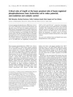
pH studies
The pH–rate profile of wild-type enzyme showed a
bell-shaped curve with pK
a
values of 6.29 ± 0.01 and
8.78 ± 0.09 at the acidic and basic sites, respectively.
The pH–rate profiles for D235A, D257A, and D258A
also showed bell-shaped curves, with two pK
a
values
(Fig. 1). The estimated pK
a
values from the pH profile
studies are summarized in Table 3. The acidic and
basic pK
a
values for D258A were almost identical with
those of the wild-type, and the differences were within
the limits of experimental error. The acidic pK
a
values
for D235A and D257A were also similar to that of the
wild-type, but their basic pK
a
values were increased to
9.10 and 9.23, respectively.
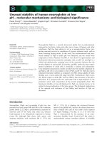
Chemical rescue experiments
Amino-acid residues involved in general acid ⁄ base
mechanisms can be identified using the chemical rescue
method. The abilities of the sodium salts of formate,
acetate, propionate, butanoate, and azide to rescue lost
function of the E234A, D235A, D257A, and D258A
mutants were studied. None of the small acids rescued
the activities of mutants E234A, D235A, or D257A.
The only restoration of activity occurred with the
Table 1. Kinetic parameters for wild-type and mutant pigeon cytosolic NADP
+
-dependent malic enzymes.
K
mNADP
(app)
(l
M)
K
mMal
(mM)
K
dMal
(mM)
K
mMn
(lM)
K
dMn
(lM)
K
mMg
(mM)
K
dMg
(mM)
k
cat
(s
)1
)
Wild-type
(Mn
2+
)
2.07 ± 0.15 0.08 ± 0.01 0.11 ± 0.02 3.78 ± 0.36 5.12 ± 0.87 31.34 ± 1.11
Wild-type
(Mg
2+
)
0.27 ± 0.03 0.27 ± 0.05 0.16 ± 0.02 0.15 ± 0.04 34.83 ± 2.32
E234A 1.80 ± 0.08 13.33 ± 3.26 13.06 ± 2.46 13.96 ± 1.82 13.09 ± 2.87 46.44 ± 5.81
D235A 1.79 ± 0.10 0.10 ± 0.01 0.19 ± 0.02 3.22 ± 0.14 6.55 ± 0.60 (0.04 ± 0.00) · 10
)1
D257A 2.83 ± 0.17 0.10 ± 0.01 0.65 ± 0.10 0.50 ± 0.07 23.36 ± 9.41 1.10 ± 0.04
D258A 2.96 ± 0.07 0.05 ± 0.01 0.15 ± 0.02 3.91 ± 0.24 12.69 ± 2.38 (0.06 ± 0.00) · 10
)1
Table 2. Kinetic parameters of partial reactions for wild-type and mutant malic enzymes.
Reduction reaction Decarboxylation reaction
K
mPyr
(app) (mM) k
cat
(app) (s
)1
) K
mOAA
(app) (mM) k
cat
(app) (s
)1
)
Wild-type 6.05 ± 0.16 0.80 ± 0.01 0.17 ± 0.01 33.41 ± 0.58
D235A 6.11 ± 0.20 (0.10 ± 0.00) · 10
)2
0.91 ± 0.04 0.09 ± 0.00
D257A 1.42 ± 0.04 0.18 ± 0.0.00 0.07 ± 0.01 76.72 ± 2.02
D258A 1.90 ± 0.18 (0.14 ± 0.00) · 10
)2
2.37 ± 0.21 0.22 ± 0.01
Mechanism of malic enzyme S C. Chang et al.
4074 FEBS Journal 273 (2006) 4072–4081 ª 2006 The Authors Journal compilation ª 2006 FEBS
D258A mutant in the presence of azide (Fig. 2A).
Activation of D258A reached a maximum at 100 mm
sodium azide and then declined at higher concentra-
tions. The extent of activation was underestimated
because of the presence of unsaturated l-malate and
Mn
2+
in the assay mixture. To investigate the re-acti-
vation process further, kinetic parameters for mutant
D258A were determined in the presence of sodium
azide (Table 4). Sodium azide had no significant effect
on K
m
values for l-malate and Mn
2+
and on the k
cat
value when wild-type enzyme was used. With D258A,
sodium azide increased the K
m
values for l-malate and
Mn
2+
by 25-fold and 286-fold, respectively, compared
with those observed in the absence of sodium azide.
The k
cat
value for the D258A mutant was 890 times
greater in the presence of the azide ion than in the
absence of the azide ion (Tables 1 and 4). The activity
of the D258A mutant was restored to 42% of that of
the wild-type by sodium azide. To provide further
insight into the catalytic roles of the D258 residue, the
two partial reactions were examined by azide rescue.
Only the pyruvate reduction reaction was rescued by
sodium azide (Fig. 2B). The kinetic studies showed
that the k
cat
value for D258A was identical with that
Table 3. Summary of k
cat
pH data for wild-type and mutant malic
enzymes.
k
cat
(s
)1
)
pK
a1
pK
a2
Wild-type 6.29 ± 0.01 8.78 ± 0.09
D235A 6.29 ± 0.09 9.10 ± 0.11
D257A 6.50 ± 0.04 9.23 ± 0.09
D258A 6.34 ± 0.08 8.72 ± 0.09
Fig. 1. pH–k
cat
profiles for wild-type and mutant pigeon cytosolic
NADP
+
-dependent malic enzyme. The profiles for wild-type (s),
D235A (n), D257A (m), and D258A (d) are shown. Malic enzyme
activity was assayed as described in Experimental procedures.
Points are the experimental data, and traces are the results of a
fit of data for the pH–rate equation log y ¼ log[C ⁄ (1 + H ⁄ K
a1
+
K
a2
⁄ H)].
Fig. 2. Fold of activation of mutant malic enzyme as a function of
the concentration of sodium azide. (A) The mutant malic enzyme
overall oxidative decarboxylation activities of E234A (n), D235A
(d), D257A (h), and D258A (s) were assayed as described in
Experimental procedures. (B) The azide rescue of reduction partial
reaction of wild-type (s) and D258A (d) and decarboxylation activ-
ity of D258A (.).
S C. Chang et al. Mechanism of malic enzyme
FEBS Journal 273 (2006) 4072–4081 ª 2006 The Authors Journal compilation ª 2006 FEBS 4075
of the wild-type in the presence of azide (Table 4). The
results of the chemical rescue studies suggest that
D258 may act as a general base to extract the proton
of the C2 hydroxy of l-malate to facilitate oxaloace-
tate formation.
Discussion
In these studies, site-directed mutagenesis was used to
evaluate the catalytic roles of four highly conserved
acidic residues in the active site of pigeon NADP
+
-
dependent malic enzyme. Steady-state kinetic charac-
terization of the E234A, D235A, D257A, and D258A
mutants suggests that the D257 residue is not directly
involved in enzyme function. E234 is important for the
binding of bivalent metal to the enzyme, and D235
and D258 play critical roles in catalysis.
Our kinetic results for the pigeon D257A mutant
differ from those for the corresponding mutant from
A. suum. The K
m
and k
cat
values and the bell-shaped
pH profile of the pigeon D257A mutant did not differ
significantly from that of the wild-type enzyme. In con-
trast, the corresponding mutant from A. suum, mutant
D294A, had a k
cat
of about 13 000-fold less than that
of the wild-type and exhibited a pH-independent pat-
tern at the basic end of its pH range [13]. The A. suum
mitochondrial enzyme is allosterically activated and
inhibited by fumarate and ATP, respectively [17,18],
whereas the pigeon cytosolic enzyme is not regulated
by any known allosteric effector. The amino-acid
sequences of these two isozymes show 55% identity
and 73% similarity. Therefore, kinetic differences
between pigeon and A. suum mutant enzymes are
probably caused by differences in the microenviron-
ments at their active sites.
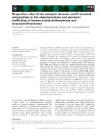
The 3D structure of pigeon malic enzyme showed
that the metal ion was co-ordinated with the carboxy
groups of the E234, D235, and D258 side chains, the
carbonyl group of oxalate (an analogue of enolpyru-
vate), and water to form an octahedral complex [14].
However, our kinetic studies show that only the
E234A mutant has a significant effect on metal bind-
ing. These results are consistent with previous studies
in which the metal-binding ability of E234Q was
decreased 100-fold, whereas D235N had little effect on
the K
m
for Mn
2+
[20]. The unique kinetic properties
of the E234A mutant probably result from the specific
geometrical arrangement of E234. The carboxy groups
of E234 and D235 and the C1 carboxy and C2
hydroxy groups of l-malate are coplanarly chelated
with Mn
2+
D258 and water are located axially above
and beneath this plane, respectively. In this plane,
E234 and D235 are diagonally opposed to the C1
carboxy group of l-malate and the C2 hydroxy group
of l-malate, respectively (Fig. 3). The interaction of
Mn
2+
and the C1 carboxy group of l-malate should
be strengthened by omitting the chelating of the carb-
oxy group of the residue E234 at the opposite direction
in E234A mutant. This trans effect will drive the
Mn
2+
toward l-malate and therefore decrease the
affinity of Mn
2+
for the carboxy groups of D235 and
D258. This may account for the increase in K
m
when
the E234 residue was mutated to alanine. The nominal
change in K
m
values observed with the D235A and
D258A mutant enzymes may reflect the elimination of
an unfavourable repulsive interaction between the
carboxy group and neighbouring negatively charged
ligands. In previous Fe
2+
-ascorbate cleavage and site-
directed mutagenesis studies, we proposed that D258
was involved in metal ion binding [19,20]. However, in
those studies, of the four D258 mutants, only D258E
Table 4. Kinetic parameters of overall and reduction partial reaction for wild-type and D258A mutant malic enzyme in the presence of
50 m
M sodium azide.
Oxidoreduction decarboxylation of malate reaction Reduction of pyruvate reaction
K
mMal
(app) (mM) K
mMn
(app) (lM) k
cat
(app) (s
)1
) K
mPyr
(app) (mM) k
cat
(app) (s
)1
)
Wild-type 0.18 ± 0.01 1.16 ± 0.05 27.80 ± 0.28 0.65 ± 0.04 0.87 ± 0.01
D258A 3.20 ± 0.20 (9.38 ± 0.62) · 10
2
11.76 ± 0.24 5.69 ± 0.74 0.84 ± 0.03
Fig. 3. Proposed mechanism for reduction step of pigeon cytosolic
NADP
+
-dependent malic enzyme. The scheme is not meant to
imply correct geometry or stereochemistry but simply to show the
movement of protons and electrons.
Mechanism of malic enzyme S C. Chang et al.
4076 FEBS Journal 273 (2006) 4072–4081 ª 2006 The Authors Journal compilation ª 2006 FEBS
showed any measurable activity. Its K
m
value for
Mn
2+
increased by 1600-fold. In the present study,
larger amounts of enzyme were used and had a pro-
nounced effect on the k
cat
value but no effect on metal
ion affinity. The previously reported effects of D258E
may have been caused by the extra methylene group,
which would have perturbed the position of Mn
2+
rel-
ative to the other amino-acid residues responsible for
its binding.
A significant decrease in the k
cat
value of the D235A
mutant has not been reported previously. The carboxy
group of D235, Mn
2+
, and the C2 hydroxy group of
l-malate are linear with Mn
2+
at the centre. There-
fore, it is impossible for the D235 residue to act as a
general acid ⁄ base in the catalytic mechanism. Our
chemical rescue and pH–rate profile results also sup-
port this contention, which is based on crystal struc-
ture. It has been proposed that the metal ion acts as a
Lewis acid to stabilize the negatively charged transition
state [21]. In D235A, because the interaction between
the carboxy and Mn
2+
does not occur, the chelating
ability of Mn
2+
for the C2 hydroxy group of l-malate
is increased. This strong electron-withdrawing ability
might propagate through the C2 hydroxy at the
a-position to the C–H bond at the b-position and
make the hydrogen atom partially positive. This effect
might make hydride transfer impossible and inactivate
the enzyme. In the wild-type enzyme, this metal-
induced polarization will not extend to the b-position
and will be limited to the C2 hydroxy group of l-ma-
late. It will increase the acidity of the hydroxy group
and facilitate the transfer of the proton from the
hydroxy group to the general base residue and the
hydride transfer to NADP
+
to complete the oxidore-
duction reaction (Fig. 3). The metal ion will then inter-
act with the carbonyl oxygen of oxaloacetate and
facilitate the decarboxylation reaction to form enol-
pyruvate [21]. Our kinetic data on the D235A mutant
demonstrated a dramatic decrease in k
cat
values for the
overall reaction and both partial reactions, which is in
agreement with a model in which the metal ion partici-
pates in both partial reactions. These results also indi-
cate that both the Lewis acid metal ion and the
general acid ⁄ base residue are important for the cata-
lytic mechanism of malic enzyme.
The D295 (D258 in the pigeon) residue in A. suum
malic enzyme was identified as a general base by kin-
etic and site-directed mutagenesis studies [9]. However,
its role has been questioned because of the inaccessibil-
ity of the carboxyl oxygen to the hydrogen of the hyd-
roxy group of l-malate in human [3] and A. suum
malic enzymes [11]. A similar topology was observed
in pigeon NADP
+
-dependent malic enzymes, in which
the distance between the carboxy oxygen of D258 and
the C2 hydroxy is 3.47 A
˚
. However, our kinetic studies
showed that substitution of alanine for aspartate at the
D258 residue decreased k
cat
values in the overall oxida-
tive decarboxylation reaction and in the pyruvate
reduction partial reaction. Both these enzymatic activ-
ities of D258A mutant could be rescued by sodium
azide. No azide rescue was observed for the decarb-
oxylation partial reaction. These results indicate that
the carboxylic group of D258 is essential for the first
step of the enzymatic reaction in which a general base
is involved. Sodium azide rescue has been widely used
to distinguish nucleophile residues from general bases
in glycosidases, in which azide can act as nucleophile
but not as a proton acceptor [22]. However, the azide
ion was shown to act as an exogenous proton acceptor
in the re-activation of the acid ⁄ base mutants of Ther-
mobacillus xylanilyticus a-l-arabinofuranosidase [23]
and human b-glucuronidase [24]. Therefore, despite the
contradiction between crystal structure and kinetic
studies, we suggest that D258 might still act as a gen-
eral base to accept a proton from the C2 hydroxy
group to form a ketone and facilitate C2 hydride
transfer (Fig. 3).
The pH dependence of k
cat
has been interpreted as
ionization of an enzymatic carboxy group essential for
catalysis. The unexpected bell-shaped pH profile of the
D258A mutant indicated that the acidic pK
a
may
derive from chemical components other than the carb-
oxy group of the D258 residue. The conditions used in
the current studies were not acidic enough to reveal
the pK
a
of the carboxy group of l-malate. Recently,
studies showed that the pK
a
of the deprotonation of
the metal-co-ordinated hydroxy group of isocitrate in
the porcine mitochondrial NADP
+
-dependent isoci-
trate dehydrogenase could be shifted to pH 5 [25].
Therefore, deprotonation of the metal-chelated hyd-
roxy group substrate l-malate may be another reason
for the acidic pK
a
in the pH profile.
There are several possible reasons for the discrep-
ancy between the results of studies of kinetics and
those of studies of crystal structure. Firstly, the D235
and D258 mutants had the most profound effect on
k
cat
values. This suggests that polarization of the C2
hydroxy group and the general acid ⁄ base reaction
co-operatively extract the hydrogen of the C2 hydroxy
group to facilitate hydride transfer. Therefore, the
carboxylic group of D258, a weak base because it is
relatively distal to the hydroxy group of l-malate, may
still be able to act as a general base for the oxidore-
duction reaction. Secondly, an active-site water mole-
cule may exist between the carboxy group of D258
and the hydroxy group of l-malate and serve as a
S C. Chang et al. Mechanism of malic enzyme
FEBS Journal 273 (2006) 4072–4081 ª 2006 The Authors Journal compilation ª 2006 FEBS 4077
proton relay to fulfil the general base role of D258.
Similar active-site water molecules have been observed
in the crystal structure of porcine mitochondrial isoci-
trate dehydrogenase, another oxidoreductive decarbox-
ylated enzyme [26]. In this case, an aspartate residue
and two water molecules form a catalytic triad that is
responsible for the general base mechanism. Finally,
the crystal structures of malic enzyme were solved in
either the E–NADH–malate–Mn
2+
–fumarate penten-
ary complex (human) or the E–NAD(P)H–oxalate–
Mn
2+
tertiary complex (pigeon and Ascarid). Pigeon
and Ascarid malic enzymes show substrate inhibition
in the presence of a high concentration of l-malate
[21,27,28]. It has been suggested that the substrate
inhibition might result from the formation of an
E–malate–NADPH–Mn
2+
aborted complex. Early
kinetic studies showed that oxalate, an analogue of
enolpyruvate, is a dead-end inhibitor for malic enzyme
[29]. Therefore, all the 3D structures of the malic
enzyme examined might represent inactive aborted
enzymatic forms. The inhibition observed in the kinetic
studies may have resulted from inaccessibility between
the carboxy group of D258 and the C2 hydroxy group
of l-malate. Therefore, the carboxy group of D258
may still be close enough to act as a general base to
extract the C2 hydroxy proton of l-malate in the enzy-
matically active complex.
In conclusion, we have described the functional roles
of these conserved carboxylic acid amino-acid residues
using site-directed mutagenesis and steady-state kinet-
ics. We propose the following:
l
E234 is essential for Mn
2+
binding.
l
The carboxy groups of D235 and D258 act
co-operatively.
l
The D235 residue is involved in the polarization of
the hydroxy group of l-malate by chelating the
Mn
2+
ion.
l
The D258 residue acts as a general base to promote
oxaloacetate formation and hydride transfer.
Experimental procedures
Materials
Restriction endonucleases, T4 DNA polymerase, T4 DNA
ligase, and T4 polynucleotide kinase were purchased from
Promega (Madison, WI, USA). Q Sepharose and 2¢,5¢-
ADP–Sepharose were obtained from Amersham (Piscata-
way, NJ, USA). The pET21b expression vector was
purchased from Novagen (Madison, WI, USA). NADP
+
was purchased from Sigma (St Louis, MO, USA). All other
reagents were of molecular biology grade or the highest
grade available.
Cloning of pigeon liver malic enzyme cDNA
The full-length pigeon liver cytosolic malic enzyme cDNA
was cloned into the pET21b vector for expression, as previ-
ously described [30]. The construction was designed in such
a way that no extra nucleotide sequence flanked the 5¢ end
of the ORF of the malic enzyme cDNA. Therefore, the
amino-acid composition and sequence of the recombinant
form were identical with those of the native enzyme. The
plasmid containing malic enzyme cDNA was named
pET21-ME.
Site-directed mutagenesis
Site-directed mutagenesis was carried out according to the
procedures of Zoller & Smith [31] using the M13 origin in
the vector for uracil-containing ssDNA preparation. Other
DNA techniques were performed according to the protocols
of Sambrook et al. [32]. The pET21-ME recombinant
phagemid was amplified in the ung
–
and dut
–
CJ236 E. coli
strain with helper phage R408 for preparation of the uracil-
containing ssDNA template. The uracil-containing template
DNA was annealed with phosphorylated mutagenic oligo-
nucleotides and then extended in vitro and ligated by T4
DNA polymerase and T4 DNA ligase, respectively. The
mutated DNA was screened by transforming into the ung
+
and dut
+
JM109 E. coli strain, and the surviving colonies
were further identified by dideoxy chain-termination
sequencing [33]. The entire cDNA was also sequenced to
exclude any unexpected mutations resulting from in vitro
DNA polymerase extension.
Expression and purification of recombinant
malic enzymes
Expression plasmids for wild-type malic enzyme and mutants
were introduced into the host E. coli BL21(DE3) and grown
in Luria–Bertani medium containing 0.1 mgÆmL
)1
ampicillin
at 37 °CtoanA
660
of 0.5–0.6. Expression was induced
with 1.0 mm isopropyl b-d-thiogalactopyranoside. The
culture was then allowed to grow overnight at 25 °C. The
cells were harvested by centrifugation for 15 min at 5000 g.
Cells were resuspended and sonicated in Tris ⁄ HCl
buffer (25 mm, pH 7.5) containing 2 mm 2-mercaptoethanol.
The recombinant proteins were purified using a Q-Sepharose
column pre-equilibrated with the same buffer. Malic
enzyme was eluted with Tris ⁄ HCl buffer containing 150 mm
NaCl. The fractions containing malic enzyme were
further purified using a 2¢,5¢-ADP–Sepharose column.
The malic enzyme was then eluted by 230 lm NADP
+
.A
Sephadex G-25 gel filtration column was used to remove
NADP
+
. All purified enzymes were subjected to SDS ⁄
PAGE to examine their purity. Protein concentrations were
determined by the Bradford method using BSA as a standard
[34].
Mechanism of malic enzyme S C. Chang et al.
4078 FEBS Journal 273 (2006) 4072–4081 ª 2006 The Authors Journal compilation ª 2006 FEBS
CD measurements
CD measurements were made with a Jasco J-810 spectropo-
larimeter using a 0.1-cm path-length cell and averaging five
repeated scans between 250 and 200 nm. Typically, 30 lg
of the wild-type or mutated NADP
+
-dependent malic
enzyme in Tris ⁄ HCl buffer (25 mm, pH 7.5) containing
2mm 2-mercaptoethanol was used for each measurement.
The spectra were analysed on DICHROWEB (http://
www.cryst.bbk.ac.uk/cdweb/html/home.html) using the
software of CDSSTR [35,36].
Enzyme assay
Malic enzyme activity was assayed as described by Hsu &
Lardy [37]. The reaction mixture contained triethanol-
amine ⁄ HCl buffer (66.7 mm, pH 7.4), l-malate (5 mm),
NADP
+
(0.23 mm), Mn
2+
(4 mm), and an appropriate
amount of enzyme in a total volume of 1 mL. The forma-
tion of NADPH at 25 °C was monitored continuously at
340 nm with a Perkin–Elmer Lambda 3B spectrophoto-
meter. One unit of enzyme activity was defined as the
initial rate of 1 lmol NADPH formed per minute under
the assay conditions. A molar absorption coefficient of
6.22 · 10
3
m
)1
Æcm
)1
for NADPH was used in the cal-
culations. Specific activity was defined as lmol NADPH
formedÆmin
)1
Æ(mg protein)
)1
.
Kinetic analysis
Apparent Michaelis constants for the substrates were
determined by varying one substrate concentration around
its K
m
value while maintaining the other components con-
stant. Initial velocity studies were performed to determine
the Michaelis and dissociation constants for l-malate
and Mn
2+
. For initial velocity studies, the concentrations
of both l-malate and Mn
2+
were varied while that of
NADP
+
was maintained at saturation. The E234A mutant
required a higher concentration of Mn
2+
for initial velo-
city studies than the other mutants. Under these condi-
tions, a brownish Mn–malate complex formed, which
would have interfered with the enzyme assay. Therefore,
Mn
2+
was replaced by Mg
2+
for initial velocity studies of
the E234A mutant. Concentrations of the other compo-
nents were held constant. Data were analysed using the
following equation, which describes a sequential initial
velocity pattern:
t ¼ V
max
AB=ðK
ia
K
b
þ K
a
B þ K
b
A þ ABÞ
in which t and V
max
represent initial and maximum veloci-
ties, A and B represent reactant concentrations, K
a
and K
b
are Michaelis constants for A and B, and K
ia
is the dissoci-
ation constant for A. The linear regression analysis was
carried out with commercial pro fit 6.0 (QuantumSoft,
Uetikon am See, Switzerland).
Partial reaction analysis
The two partial activities of malic enzyme, decarboxylation
and reduction, can be evaluated separately. The decarboxy-
lation activity of malic enzyme was assayed by the method
of Tang & Hsu [38] using oxaloacetate as substrate. The
rate of decarboxylation of oxaloacetate was measured by
monitoring the disappearance of the enolic oxaloacetate
absorbance at 260 nm in the presence of Mn
2+
or Mg
2+
.
Various concentrations of oxaloacetate in 185 mm potas-
sium acetate buffer, pH 4.5, were added to 50 mm EDTA
and incubated at 25 °C for 10 min to reach keto–enol equi-
librium. The oxaloacetate solutions were added to a total
volume of 1 mL containing 4 mm MnCl
2
and 37 mm potas-
sium acetate buffer, pH 4.5 to start the reaction. The rate
of decarboxylation in the presence of enzyme was corrected
by subtracting the spontaneous oxaloacetate decarboxyla-
tion.
Oxidation of l-malate to oxaloacetate cannot be evalu-
ated directly because of interference by the subsequent
decarboxylation. The reversed direction, reduction of a-oxo
acid to a-hydroxy acid, can be analysed using pyruvate and
NADPH as substrates. The reduction partial reaction was
performed as described by Tang & Hsu [39] using pyruvate
as substrate. The rate of reduction of pyruvate to lactate
was measured at 25 °C by monitoring the decrease in
absorbance at 340 mm associated with the oxidation of
NADPH. A typical assay mixture contained 66.7 mm tri-
ethanolamine ⁄ HCl buffer (pH 7.4), 0.23 mm NADPH,
4mm MnCl
2
, 1–50 m m pyruvate (pH 7.4), and an appro-
priate amount of malic enzyme.
pH studies
The pH dependencies of k
cat
for wild-type and mutants
were determined using initial velocity studies and variable
concentrations of l-malate and NADP
+
as a function of
pH over the pH range 5.5–10, which was maintained with
60 mm Bis-Tris propane buffer. The pH values were recor-
ded and showed no significant change before and after the
initial velocity was measured. The pK
a
values were obtained
by fitting the following equation to the data:
log y ¼ log½C=ð1 þ H=K
a1
þ K
a2
=HÞ
where y is the value of the parameter of interest (k
cat
), C is
the pH-independent value of y, H is the hydrogen ion
concentration, and K
a1
and K
a2
are the acid dissociation
constants for functional groups in the enzyme–substrate
complex.
Chemical rescue
The stock solutions of exogenous acids were prepared at
pH 7.4. Various free acids, including formic acid, acetic
acid, butyric acid, or sodium azide, were added to the
S C. Chang et al. Mechanism of malic enzyme
FEBS Journal 273 (2006) 4072–4081 ª 2006 The Authors Journal compilation ª 2006 FEBS 4079
standard reaction mixture to examine their rescue abilities.
To measure the kinetic properties of malic enzyme after
rescue, 50 mm sodium azide was included for all kinetic
studies.
Acknowledgements
This research was supported by a grant from the
National Science Council, China (NSC92-2320-B016-
060 to W.Y.C.). We thank Dr Chi-Ching Hwang
(Kaohsiung Medical University, Taiwan) and Dr
Minghuey Shieh (National Taiwan Normal University,
Taiwan) for helpful discussions.
References
1 Chang GG & Tong L (2003) Structure and function of
malic enzymes, a new class of oxidative decarboxylases.
Biochemistry 42, 12721–12733.
2 Xu Y, Bhargava G, Wu H, Loeber G & Tong L (1999)
Crystal structure of human mitochondrial NAD(P)
+
-dependent malic enzyme: a new class of oxidative
decarboxylases. Struct Fold Des 7, R877–R889.
3 Yang Z, Floyd DL, Loeber G & Tong L (2000) Struc-
ture of a closed form of human malic enzyme and impli-
cations for catalytic mechanism. Nat Struct Biol 7, 251–
257.
4 Kiick DM, Harris BG & Cook PF (1986) Protonation
mechanism and location of rate-determining steps for
the Ascaris suum nicotinamide adenine dinucleotide-
malic enzyme reaction from isotope effects and pH stu-
dies. Biochemistry 25, 227–236.
5 Park SH, Harris BG & Cook PF (1986) pH dependence
of kinetic parameters for oxalacetate decarboxylation
and pyruvate reduction reactions catalyzed by malic
enzyme. Biochemistry 25, 3752–3759.
6 Weiss PM, Gavva SR, Harris BG, Urbauer JL, Cleland
WW & Cook PF (1991) Multiple isotope effects with
alternative dinucleotide substrates as a probe of the
malic enzyme reaction. Biochemistry 30, 5755–5763.
7 Karsten WE & Cook PF (1994) Stepwise versus con-
certed oxidative decarboxylation catalyzed by malic
enzyme: a reinvestigation. Biochemistry 33, 2096–2103.
8 Liu D, Karsten WE & Cook PF (2000) Lysine 199 is
the general acid in the NAD-malic enzyme reaction.
Biochemistry 39, 11955–11960.
9 Karsten WE, Chooback L, Liu D, Hwang CC, Lynch
C & Cook PF (1999) Mapping the active site topo-
graphy of the NAD-malic enzyme via alanine-scanning
site-directed mutagenesis. Biochemistry 38, 10527–
10532.
10 Kuo CC, Tsai LC, Chin TY, Chang GG & Chou WY
(2000) Lysine residues 162 and 340 are involved in the
catalysis and coenzyme binding of NADP(
+
)-dependent
malic enzyme from pigeon. Biochem Biophys Res
Commun 270, 821–825.
11 Tao X, Yang Z & Tong L (2003) Crystal structures of
substrate complexes of malic enzyme and insights into
the catalytic mechanism. Structure (Camb) 11, 1141–
1150.
12 Rao GS, Coleman DE, Karsten WE, Cook PF & Harris
BG (2003) Crystallographic studies on Ascaris suum
NAD-malic enzyme bound to reduced cofactor and
identification of an effector site. J Biol Chem 278,
38051–38058.
13 Karsten WE, Liu D, Rao GS, Harris BG & Cook PF
(2005) A catalytic triad is responsible for acid-base
chemistry in the Ascaris suum NAD-malic enzyme.
Biochemistry 44 , 3626–3635.
14 Yang Z, Zhang H, Hung HC, Kuo CC, Tsai LC, Yuan
HS, Chou WY, Chang GG & Tong L (2002) Structural
studies of the pigeon cytosolic NADP(
+
)-dependent
malic enzyme. Protein Sci 11, 332–341.
15 Chang HC, Chou WY & Chang GG (2002) Effect of
metal binding on the structural stability of pigeon liver
malic enzyme. J Biol Chem 277, 4663–4671.
16 Schimerlik MI, Grimshaw CE & Cleland WW (1977)
Determination of the rate-limiting steps for malic
enzyme by the use of isotope effects and other kinetic
studies. Biochemistry 16, 571–576.
17 Lai CJ, Harris BG & Cook PF (1992) Mechanism of
activation of the NAD-malic enzyme from Ascaris suum
by fumarate. Arch Biochem Biophys 299, 214–219.
18 Landsperger WJ & Harris BG (1976) NAD
+
-malic
enzyme. Regulatory properties of the enzyme from
Ascaris suum. J Biol Chem 251, 3599–3602.
19 Wei CH, Chou WY, Huang SM, Lin CC & Chang GG
(1994) Affinity cleavage at the putative metal-binding
site of pigeon liver malic enzyme by the Fe(
2+
)-ascor-
bate system. Biochemistry 33, 7931–7936.
20 Wei CH, Chou WY & Chang GG (1995) Identification
of Asp258 as the metal coordinate of pigeon liver malic
enzyme by site-specific mutagenesis. Biochemistry 34,
7949–7954.
21 Hsu RY, Mildvan AS, Chang G & Fung C (1976)
Mechanism of malic enzyme from pigeon liver. Mag-
netic resonance and kinetic studies of the role of Mn
2+
.
J Biol Chem 251, 6574–6583.
22 Zechel DL & Withers SG (2001) Dissection of nucleo-
philic and acid-base catalysis in glycosidases. Curr Opin
Chem Biol 5, 643–649.
23 Debeche T, Bliard C, Debeire P & O’Donohue MJ
(2002) Probing the catalytically essential residues of the
alpha-L-arabinofuranosidase from Thermobacillus xylan-
ilyticus. Protein Eng 15, 21–28.
24 Islam MR, Tomatsu S, Shah GN, Grubb JH, Jain S &
Sly WS (1999) Active site residues of human beta-glu-
curonidase. Evidence for Glu (540) as the nucleophile
Mechanism of malic enzyme S C. Chang et al.
4080 FEBS Journal 273 (2006) 4072–4081 ª 2006 The Authors Journal compilation ª 2006 FEBS
and Glu (451) as the acid-base residue. J Biol Chem
274, 23451–23455.
25 Huang YC, Grodsky NB, Kim TK & Colman RF
(2004) Ligands of the Mn
2+
bound to porcine mitoch-
ondrial NADP-dependent isocitrate dehydrogenase, as
assessed by mutagenesis. Biochemistry 43, 2821–2828.
26 Ceccarelli C, Grodsky NB, Ariyaratne N, Colman RF
& Bahnson BJ (2002) Crystal structure of porcine mito-
chondrial NADP
+
-dependent isocitrate dehydrogenase
complexed with Mn
2+
and isocitrate. Insights into the
enzyme mechanism. J Biol Chem 277, 43454–43462.
27 Park SH, Harris BG & Cook PF (1989) Substrate acti-
vation by malate induced by oxalate in the Ascaris suum
NAD-malic enzyme reaction. Biochemistry 28, 6334–
6340.
28 Pry TA & Hsu RY (1980) Equilibrium substrate binding
studies of the malic enzyme of pigeon liver. Equivalence
of nucleotide sites and anticooperativity associated with
the binding of l-malate to the enzyme-manganese(II)-
reduced nicotinamide adenine dinucleotide phosphate
ternary complex. Biochemistry 19, 951–962.
29 Hsu RY (1982) Pigeon liver malic enzyme. Mol Cell
Biochem 43, 3–26.
30 Chou WY, Huang SM, Liu YH & Chang GG (1994)
Cloning and expression of pigeon liver cytosolic
NADP(
+
)-dependent malic enzyme cDNA and some of
its abortive mutants. Arch Biochem Biophys 310, 158–166.
31 Zoller MJ & Smith M (1982) Oligonucleotide-directed
mutagenesis using M13-derived vectors: an efficient and
general procedure for the production of point mutations
in any fragment of DNA. Nucleic Acids Res 10, 6487–
6500.
32 Sambrook J, Fritsch EF & Maniatis T (1989) Molecular
Cloning: a Laboratory Manual. Cold Spring Harbor
Laboratory Press, Cold Spring Harbor, NY.
33 Sanger F, Nicklen S & Coulson AR (1977) DNA
sequencing with chain-terminating inhibitors. Proc Natl
Acad Sci USA 74 , 5463–5467.
34 Bradford MM (1976) A rapid and sensitive method for
the quantitation of microgram quantities of protein util-
izing the principle of protein-dye binding. Anal Biochem
72, 248–254.
35 Whitmore L & Wallace BA (2004) DICHROWEB, an
online server for protein secondary structure analyses
from circular dichroism spectroscopic data. Nucleic
Acids Res 32, W668–W673.
36 Sreerama N & Woody RW (2000) Estimation of protein
secondary structure from circular dichroism spectra:
comparison of CONTIN, SELCON, and CDSSTR
methods with an expanded reference set. Anal Biochem
287, 252–260.
37 Hsu RY & Lardy HA (1967) Pigeon liver malic enzyme.
II. Isolation, crystallization, and some properties. J Biol
Chem 242, 520–526.
38 Tang CL & Hsu RY (1974) Mechanism of pigeon liver
malic enzyme. Modification of sulfhydryl groups by
5,5¢-dithiobis(2-nitrobenzoic acid) and N-ethylmalei-
mide. J Biol Chem 249, 3916–3922.
39 Tang CL & Hsu RY (1973) Reduction of alpha-oxo
carboxylic acids by pigeon liver ‘malic’ enzyme. Biochem
J 135, 287–291.
Supplementary material
The following supplementary material is available
online:
Fig. S1. SDS-PAGE of purified wild-type and
mutant malic enzymes.
Fig. S2. CD spectra of wild-type and mutated
pigeon NADP-malic enzyme.
This material is available as part of the online article
from
S C. Chang et al. Mechanism of malic enzyme
FEBS Journal 273 (2006) 4072–4081 ª 2006 The Authors Journal compilation ª 2006 FEBS 4081