Báo cáo khoa học: Functional analysis of disease-causing mutations in human UDP-galactose 4-epimerase pot
Bạn đang xem bản rút gọn của tài liệu. Xem và tải ngay bản đầy đủ của tài liệu tại đây (314.96 KB, 8 trang )
Functional analysis of disease-causing mutations in human
UDP-galactose 4-epimerase
David J. Timson
School of Biology & Biochemistry, Queen’s University Belfast, Medical Biology Centre, Belfast, UK
UDP-galactose 4-epimerase (GALE; EC 5.1.3.2) cata-
lyses the interconversion of UDP-galactose and UDP-
glucose as part of the Leloir pathway of galactose
catabolism [1]. Mutations in the gene encoding the
human enzyme can lead to a disease known as type III
galactosemia (OMIM 230350), the symptoms of which
can include early-onset cataracts, liver damage, deaf-
ness and mental retardation. Galactosemia can also be
caused by defects in two other Leloir pathway
enzymes, galactose-1-phosphate uridyltransferase (type
I; OMIM 230400) and galactokinase (type II; OMIM
230200) [2–4]. Two forms of epimerase-deficiency
galactosemia are recognized – the more severe, or
generalized, form and the much milder, peripheral
form. In the generalized form little, or no, epimerase
activity can be detected in any tissues, and patients
suffer from restricted growth and mental development,
even when placed on lactose-free diets [5]. As dietary
galactose is the patient’s only source of this sugar for
glycoconjugate biosynthesis, it cannot be withdrawn
completely. In the peripheral form, epimerase activity
is reduced in blood cells, but appears normal in other
tissues. It is not clear why this is so. The symptoms
are milder; indeed some patients may suffer no symp-
toms beyond raised levels of galactose-1-phosphate in
the blood, and no therapy is required [6].
Keywords
galactosemia; GALE; Leloir pathway; SDR
family enzyme; UDP-glucose
Correspondence
D. J. Timson, School of Biology &
Biochemistry, Queen’s University Belfast,
Medical Biology Centre, 97 Lisburn Road,
Belfast, BT9 7BL, UK
Fax: +44 28 90975877
Tel: +44 28 90975875
E-mail:
(Received 14 July 2005, revised 2 September
2005, accepted 17 October 2005)
doi:10.1111/j.1742-4658.2005.05017.x
UDP-galactose 4-epimerase (GALE, EC 5.1.3.2) catalyses the interconver-
sion of UDP-glucose and UDP-galactose. Point mutations in this enzyme
are associated with the genetic disease, type III galactosemia, which exists
in two forms – a milder, or peripheral, form and a more severe, or general-
ized, form. Recombinant wild-type GALE, and nine disease-causing muta-
tions, have all been expressed in, and purified from, Escherichia coli in
soluble, active forms. Two of the mutations (N34S and G319E) display
essentially wild-type kinetics. The remainder (G90E, V94M, D103G,
L183P, K257R, L313M and R335H) are all impaired in turnover number
(k
cat
) and specificity constant (k
cat
⁄ K
m
), with G90E and V94M (which is
associated with the generalized form of galactosemia) being the most affec-
ted. None of the mutations results in a greater than threefold change in the
Michaelis constant (K
m
). Protein–protein crosslinking suggests that none of
the mutants are impaired in homodimer formation. The L183P mutation
suffers from severe proteolytic degradation during expression and purifica-
tion. N34S, G90E and D103G all show increased susceptibility to digestion
in limited proteolysis experiments. Therefore, it is suggested that reduced
catalytic efficiency and increased proteolytic susceptibility of GALE are
causative factors in type III galactosemia. Furthermore, there is an
approximate correlation between the severity of these defects in the protein
structure and function, and the symptoms observed in patients.
Abbreviations
BS
3
, suberic acid bis(3-sulpho-N-hydroxysuccinimide ester); EDC, N-(3-dimethylaminopropyl)-N¢-ethylcarbodiimide; GALE, UDP-galactose
4-epimerase.
6170 FEBS Journal 272 (2005) 6170–6177 ª 2005 The Authors Journal compilation ª 2005 FEBS
GALE is a member of the short-chain dehydrogen-
ases ⁄ reductases family [7,8], and crystal structures
from bacteria, yeast, trypanosomes and humans have
been solved [9–17]. These structures reveal a homodi-
meric enzyme, with each subunit containing one tightly
bound NAD
+
molecule. Structural and kinetic studies
suggest that this cofactor plays a key part in the cata-
lytic mechanism. It is proposed that it transiently oxid-
izes the sugar moiety at carbon-4 and then re-reduces
it in a nonstereospecific manner, permitting inversion
of configuration [18–20]. This reaction is facilitated by
a tyrosine residue (Tyr157 in the human enzyme) act-
ing as an active-site base [16,21].
Nine disease-causing mutations have been identified
so far in human GALE [5,22–26]. Of these, V94M,
which is associated with the generalized form of the
disease, has been studied in greatest detail [27,28].
The other mutations (N34S, G90E, D103G, L183P,
K257R, L313M, G319E and R335H) are associated
with peripheral forms of the disease. In this study, all
nine disease-causing mutations have been expressed in,
and purified from, Escherichia coli, and their steady-
state kinetic parameters, ability to dimerize and
susceptibility to proteolytic digestion have been
compared.
Results
Expression and purification of human GALE
Human GALE was expressed as an N-terminal hexa-
histidine fusion protein in E. coli and purified on
nickel–agarose resin (Fig. 1). Typical yields were
% 10 mg per litre of bacterial culture. The protein is
active and shows saturation kinetics when increasing
amounts of substrate are added (Fig. 2). The kinetic
parameters determined from these data (Table 1) are
similar to those published for human GALE and for
GALEs from other species [21,27,29,30]. There is no
evidence that human GALE is glycosylated in vivo,
and there is no anomalous migration of recombinant
human GALE produced in yeast [27], and thus the
observed activity probably reflects that of the native
enzyme.
With the exception of L183P, all the mutant proteins
could be expressed and purified using similar condi-
tions and procedures. Yields and purity were similar to
those achieved with the wild-type protein. In contrast,
L183P was expressed at much lower levels, and repea-
ted attempts to purify the protein resulted in material
that contained many contaminants of lower molecular
Fig. 1. Expression and purification of human UDP-galactose 4-epimerase (GALE). The hexahistidine-tagged protein was expressed in Escheri-
chia coli HMS174(DE3) cells and purified on nickel agarose. Samples at various stages of the process were analysed by 10% SDS ⁄ PAGE
and stained with Coomassie blue.
D. J. Timson Disease-causing mutations in human GALE
FEBS Journal 272 (2005) 6170–6177 ª 2005 The Authors Journal compilation ª 2005 FEBS 6171
mass. These problems could not be overcome by
expressing the protein at lower temperatures (30 °Cor
22 °C), by using an alternative expression host
{BL21(DE3)[pLysS]} or by including protease inhibi-
tors in the solutions used during purification.
Kinetic analysis of disease-causing mutations
The steady-state kinetic parameters of each of the nine
disease-causing mutant proteins were determined
(Table 1). In general, little change was seen in K
m
(no
change greater than threefold), whereas some mutants
(especially G90E and V94M) showed large changes in
k
cat
and in k
cat
⁄ K
m
. In contrast, two mutants (N34S
and G319E) showed very little change in these para-
meters compared with the wild-type protein.
Dimerization of GALE
Human GALE is known to exist in solution as a
homodimer [1]. One possible explanation for the
in vivo effects of the disease-causing mutations is a fail-
ure to form dimers. However, cross-linking using N-
(3-dimethylaminopropyl)-N¢-ethylcarbodiimide (EDC)
showed that the wild-type protein and all the mutants
were able to form dimers (Fig. 3). In all cases, but
especially with the mutant proteins, some higher
molecular mass species were also observed. Similar
results were seen using suberic acid bis(3-sulpho-N-
hydroxysuccinimide ester) (BS
3
) (data not shown).
Limited proteolysis of GALE
As L183P appears to be highly susceptible to proteo-
lysis during expression and purification, it is possible
that the other disease-causing mutations may also have
increased proteolytic sensitivity compared with the
wild-type protein. Limited proteolysis with thermolysin
(EC 3.4.24.27) showed that while the pattern of frag-
ments produced is not changed by the disease-causing
mutations, the sensitivity to proteolysis is altered, in
some cases (Fig. 4). In particular, N34S, G90E and
D103G are clearly more susceptible to proteolysis than
the wild-type protein. In all cases, the presence of sub-
strate at saturating levels (1 mm) partially protects the
enzyme from proteolysis.
Discussion
Active recombinant human GALE can be produced in
good yields in E. coli. This enabled the creation and
analysis of all nine currently known disease-causing
mutations in this protein. All but one of these mutant
proteins (L183P) could also be produced in an active
and soluble form. This contrasts with human galacto-
kinase where approximately half the disease-causing
mutations were insoluble on expression in E. coli, sug-
gesting that a failure to fold and ⁄ or aggregation of the
protein product could be a major factor in disease
causation [31,32]. This is unlikely to be the case with
most of the mutations in GALE.
Although all the mutant proteins were active, some
of their kinetic properties differed from that of the
wild-type protein and from each other. The main effect
seen was in the turnover, k
cat
, and in the specificity
constant, k
cat
⁄ K
m
. Again, this contrasts with disease-
causing mutations in galactokinase where effects on all
the kinetic parameters were observed [31]. The most
severely affected protein was G90E, which showed
an % 800-fold decrease in k
cat
. This mutation was
Table 1. Kinetic constants of human UDP-galactose 4-epimerase
(GALE) and the consequences of disease-causing mutations. Values
were determined by nonlinear curve fitting, as described in the
Experimental procedures, and are quoted plus ⁄ minus the standard
error.
Protein K
m
(lM) k
cat
(s
)1
) k
cat
⁄ K
m
(LÆmol
)1
Æs
)1
)
Wild type 69 ± 12 36 ± 1.4 5.2 ± 0.72 · 10
5
N34S 82 ± 15 32 ± 1.3 3.9 ± 0.59 · 10
5
G90E 93 ± 24 0.046 ± 0.0028 5.0 ± 0.11 · 10
2
V94M 160 ± 38 1.1 ± 0.088 6.9 ± 1.2 · 10
3
D103G 140 ± 21 5.0 ± 0.23 3.6 ± 0.40 · 10
4
L183P 97 ± 40 11 ± 1.2 1.1 ± 0.35 · 10
5
K257R 66 ± 15 5.1 ± 0.29 7.8 ± 1.5 · 10
4
L313M 35 ± 11 5.8 ± 0.36 1.7 ± 0.46 · 10
5
G319E 78 ± 13 30 ± 1.3 3.9 ± 0.53 · 10
5
R335H 99 ± 12 15 ± 0.48 1.5 ± 0.14 · 10
5
Fig. 2. Steady-state kinetics of recombinant human UDP-galactose
4-epimerase (GALE). Rates (v) were measured in the forward direc-
tion (i.e. the conversion of UDP-galactose to UDP-glucose). Each
point represents the mean of three separate determinations. Error
bars represent the standard deviation of these mean values.
Disease-causing mutations in human GALE D. J. Timson
6172 FEBS Journal 272 (2005) 6170–6177 ª 2005 The Authors Journal compilation ª 2005 FEBS
originally discovered in a patient who was compound
heterozygous for this and another, uncharacterized,
allele [24]. Previous in vivo studies on this mutant
expressed in yeast showed that there was essentially no
GALE activity present in extracts from cells carrying
only this allele [25]. G90 lies close to the bound NAD
+
molecule (Fig. 5) and the increase in size of the side-
chain resulting from mutation to glutamate may well
disrupt the binding or conformation of the cofactor.
The V94M mutation, which is associated with the more
severe generalized form of the disease [5,25], also had an
impaired k
cat
value (% 30-fold reduction compared with
the wild-type protein). This mutation also results in the
biggest change in K
m
(% 2.3-fold). These changes are
consistent with those previously observed with proteins
expressed in yeast [27]. The structure of this mutant has
been solved, showing that a valine to methionine substi-
tution at this position disrupts the packing close to the
sugar-binding site, making it easier for the sugars to
bind nonproductively [28]. This explanation would be
entirely consistent with a reduction in k
cat
.
Three mutations – D103G, K257R and L313M –
are mildly impaired in k
cat
(approx. six- to sevenfold
decreased compared with the wild-type protein). All of
these mutations are associated with the less severe, per-
ipheral form of the disease. D103G was first observed
in a patient homozygous for this mutation [24]. Cell
extracts from yeast expressing only this form of GALE
had % 50% epimerase activity compared with wild-type
protein, and growth on galactose was unaffected [25].
Similar results were found with L313M [25], which was
first isolated from a heterozygous patient [24]. Two
mutations, L183P and R335H, result in small changes
in k
cat
(approx. two- to threefold changes) and two,
N34S and G319E, are essentially unchanged compared
with the wild-type protein (i.e. no change greater than
twofold in any kinetic parameter). Again, these muta-
tions are associated with the peripheral form of the
disease [22,24,26]. K257, L313 and G319 lie within
0.13 nm of each other on one face of the protein.
G319 is on the surface and therefore the protein may
be able to tolerate a larger side-chain at this position
(in contrast to G90). R335 lies far from the other
mutations and from the ligand-binding sites (Fig. 5).
The ability to dimerize does not appear to be
impaired in the mutants. Crosslinking analyses showed
that they are all capable of forming dimers. In all
cases, some higher-order crosslinked products are seen.
This seems to be slightly more pronounced in the case
of the mutants, suggesting that there might be
enhanced aggregation. However, this is not sufficient
to affect the solubility of the proteins.
Decreased protein stability can be a factor in dis-
ease-causation [33]. To test for this, the proteins were
exposed to low concentrations of the protease thermo-
lysin, and the digestion products were observed. The
presence of the mutations did not alter the pattern of
digestion, but did affect the amount of digestion. These
effects were reproducible. The most severe case was
that of L183, which was difficult to obtain in a purified
form. It appeared that this mutation causes the protein
to be degraded in the bacteria following expression
and also during the purification procedure. This sug-
gests that reduced stability may be an important factor
in disease causation in the case of N34S, G90E,
D103G and, particularly, L183P. This is most likely to
manifest itself in increased susceptibility to intracellular
proteases and thus a decreased concentration of active
Fig. 3. Crosslinking of wild-type and disease-causing mutants of UDP-galactose 4-epimerase (GALE). Crosslinking was carried out with N-(3-
dimethylaminopropyl)-N¢-ethylcarbodiimide (EDC), as described in the Experimental procedures. C, control lane containing 25 l
M GALE not
exposed to crosslinker; X, 25 l
M GALE exposed to crosslinker; U, 25 lM GALE exposed to crosslinker in the presence of a saturating
amount (1 m
M) of UDP-galactose; M, molecular mass markers (29, 45, 66, 97, 116 and 205 kDa). All the mutant proteins were exposed to
crosslinker. Bands were identified by reference to the uncrosslinked wild-type protein and by estimation of the molecular mass (Q
UANTITY
ONE software; Bio-Rad, Hercules, CA, USA).
D. J. Timson Disease-causing mutations in human GALE
FEBS Journal 272 (2005) 6170–6177 ª 2005 The Authors Journal compilation ª 2005 FEBS 6173
enzyme present in the cell. Indeed, when L183P was
expressed in yeast, its activity was reduced to 4% of
that observed for the wild-type protein and its abun-
dance to 6% [22]. Residue 183 forms part of a b-sheet
in the interior of the protein (Fig. 5), and mutation
from leucine to proline is highly likely to disrupt this
secondary structure, potentially leading to wider chan-
ges in the overall fold of the enzyme, resulting in
decreased stability and enhanced susceptibility to pro-
teolysis. Enhanced protease sensitivity and reduced
thermal stability has previously been observed for
G90E and D103G [25]. In both cases, the mutation
results in substantial changes to the volume of the
side-chain that are likely to disrupt the local fold.
These two residues, together with N34, are located
within 0.15 nm of each other and all are at, or close
to, the surface of the enzyme. Therefore, it is not sur-
prising that alterations to these residues affect the sen-
sitivity to proteolysis.
In conclusion, there is an approximate correlation
between the severity of symptoms observed in patients
UDPgal
UDPgal
R335
NAD
+
NAD
+
N34
G90
D103
V94
G319
K257
L313
L183
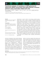
Fig. 5. Structure of human UDP-galactose 4-epimerase (GALE)
showing the positions of the residues altered in the disease-caus-
ing mutations. The image was created from PDB entry 1EK6 [16]
using the program P
YMOL (DeLano Scientific LLC, San Carlos, CA,
USA; ). The lower image is rotated approxi-
mately 90° on a horizontal axis compared with the upper image.
For clarity the residues are highlighted on only one of the two
molecules in the homodimer.
Fig. 4. Limited proteolysis of wild-type and disease-causing
mutants of UDP-galactose 4-epimerase (GALE). Proteolysis was
carried out as described in the Experimental procedures, and then
the products were separated by 15% SDS ⁄ PAGE and stained with
Coomassie blue. C, control lanes containing 50 l
M GALE; P, GALE
digested with 90 n
M thermolysin for 30 min at 37 °C; U, GALE
digested with 90 n
M thermolysin for 30 min at 37 °C in the pres-
ence of 1 m
M UDP-galactose; M, molecular mass markers (29, 45,
66, 97, 116 and 205 kDa).
Disease-causing mutations in human GALE D. J. Timson
6174 FEBS Journal 272 (2005) 6170–6177 ª 2005 The Authors Journal compilation ª 2005 FEBS
with the level of biochemical impairment of the puri-
fied protein. The mutation, V94M, associated with the
generalized form of the disease, has a much reduced
value for k
cat
. A more complete genotype–phenotype
correlation is difficult because of the relatively small
number of patients afflicted with type III galactosemia.
For example, there have been no reports of patients
homozygous for G90E (which has an even lower k
cat
than V94M) or for L183P (which is highly susceptible
to proteolysis). It is reasonable to predict that if such
patients were found, their symptoms would be closer
to the generalized form than the peripheral form of
disease. Some of the mutants have only small changes
in the kinetic parameters. In the case of N34S and
D103G, disease-causation may be explained by a com-
bination of kinetic impairment and increased sensitiv-
ity to proteolysis. In other cases, additional factors
may be important. For example, in the case of K257R
and G319E, it has been suggested that these mutations
are tightly genetically linked to other changes that give
rise to the symptoms (perhaps by affecting the level of
expression of GALE) [34]. That there are two clusters
of disease-causing mutations (N34S ⁄ G90E ⁄ D103G
and K257R ⁄ L313M ⁄ G319E) suggests that these two
parts of the protein are sensitive to changes and that
further mutations may be found in these regions in the
future.
Experimental procedures
Expression and purification of human GALE
Human GALE was expressed in and purified from E. coli,
essentially as described previously for galactose mutarotase
and galactokinase [31,35]. The coding sequence was ampli-
fied by PCR using an IMAGE clone (ID number 3459004)
as a template [36]. The primers were designed in order to
add restriction sites (NcoI at the 5¢ end and EcoRI at the 3¢
end) and to encode a hexa-histidine tag at the N terminus
of the expressed protein. PCR products were cut with these
enzymes and inserted into the corresponding sites in the
expression vector pET-21d (Merck, Nottingham, UK). The
entire GALE coding sequence was verified by DNA sequen-
cing.
The recombinant plasmid was transformed into E. coli
HMS174(DE3) (Merck) for expression. An overnight cul-
ture (volume 5 mL) was diluted into 1 L of Luria–Bertani
(Miller) media supplemented with 100 lgÆmL
)1
ampicillin.
This 1 L culture was incubated, with shaking at 37 °C, for
3–4 h before induction with isopropyl thio-b-d-galactoside
(final concentration, 2 mm). The culture was grown for a
further 2 h and then harvested by centrifugation (4200 g
for 10 min). Cells were resuspended in % 20 mL of 50 mm
Hepes-OH, pH 7.5, containing 150 mm NaCl and 10%
(v ⁄ v) glycerol, and stored at )80 ° C.
Cells were disrupted by sonication on ice (three times:
30 s at 100 W, with 30 s intervals between for cooling) and
cell debris was removed by centrifugation (27 000 g for
20 min at 4 °C). The supernatant was applied to a 1 mL
nickel–agarose (His-select; Sigma, Poole, UK) column and
allowed to pass through by gravity flow. The column was
washed with 20 mL of cold buffer A [50 mm Hepes-OH,
pH 7.5, containing 500 mm NaCl and 10% (v ⁄ v) glycerol]
and the protein was eluted in three, 2 mL washes of cold
buffer B (buffer A supplemented with 250 mm imidazole).
Fractions containing GALE, as judged by SDS ⁄ PAGE,
were dialysed overnight at 4 °C against 50 mm Hepes-OH,
pH 8.0, containing 150 mm NaCl, 1 mm dithiothreitol and
10% (v ⁄ v) glycerol. Protein concentrations were estimated
by the method of Bradford [37] using BSA as a standard.
The protein was stored frozen in small aliquots at ) 80 °C.
Generation of point mutations
Point mutations in the GALE sequence were generated
using the QuikChange method [38]. DNA sequencing was
used to verify each mutation and to ensure that no other
changes had been introduced into the coding sequence. All
mutant proteins were expressed and purified using the same
techniques as for the wild-type protein.
GALE assay
GALE activity was assayed according to the method of Ng
et al. [39], as modified by Wohlers & Fridovich-Keil [27].
The conversion of UDP-galactose to UDP-glucose was cou-
pled to the oxidation of UDP-glucose using the enzyme
UDP-glucose dehydrogenase (Sigma; EC 1.1.1.22). This
results in the reduction of two molecules of NAD
+
per
molecule of UDP-glucose, which can be followed spectro-
photometrically at 340 nm. All assays were carried out at
37 °C in a volume of 1 mL, and each contained 50 mm
Tris ⁄ HCl, pH 8.8, 4 mm NAD
+
and a variable amount of
UDP-galactose (Merck). Reaction mixes were preincubated
at 37 °C for at least 5 min. Then, 0.02 units of UDP-glucose
dehydrogenase (manufacturer’s unit definition) were added
and the absorbance at 340 nm was monitored for 2–3 min.
This is necessary as commercial UDP-galactose contains a
small amount of UDP-glucose [29]. Reactions were initiated
by the addition of GALE (to a final concentration of 2.6 nm
for the wild-type protein and ranging from 2 nm to 330 nm
for the mutants). The absorbance at 340 nm was measured
for a further 7–8 min. Rates (v) were calculated by fitting the
linear part of the A
340
vs. time plot to a straight line and cor-
rected by subtraction of the rate in the absence of GALE.
The kinetic constants K
m
and k
cat
were derived by nonlinear
curve fitting [40] to the Michaelis–Menten equation:
D. J. Timson Disease-causing mutations in human GALE
FEBS Journal 272 (2005) 6170–6177 ª 2005 The Authors Journal compilation ª 2005 FEBS 6175
v=½GALE¼fk
cat
Á½UDP-galactose=ðK
m
þ½UDP-galactoseÞg;
using the program GraphPad Prism 3.0 (GraphPad Soft-
ware, San Diego CA, USA). Specificity constants (k
cat
⁄ K
m
)
were determined directly by fitting to a modified form of
the Michaelis–Menten equation:
v=½GALE¼fk
cat
=K
m
Á½UDP-galactose=
ð1 þ½UDP-galactose=K
m
Þg:
All points were weighted equally and values are quoted
plus ⁄ minus the standard error determined by the program.
Crosslinking of GALE
Wild-type or mutant GALE (25 lm) was incubated for
% 5 min at 37 °C. Cross-linker – either EDC, to a final con-
centration of 70 mm,orBS
3
, to a final concentration of
100 lm – was added and the reaction was allowed to pro-
ceed for 30 min. Reactions were stopped by the addition of
an equal volume of SDS ⁄ PAGE loading buffer [125 mm
Tris ⁄ HCl, pH 6.8, 4% (w ⁄ v) SDS, 20% (v ⁄ v) glycerol, 1%
(w ⁄ v) dithiothreitol, 0.002% (w ⁄ v) bromophenol blue] and
heating at 95 °C for 5 min. Products were analysed by 10%
SDS ⁄ PAGE.
Limited proteolysis of GALE
Wild-type or mutant GALE (50 lm) was incubated for
% 5 min at 37 °C. Thermolysin (Sigma) was added to a final
concentration of 90 nm and digestion allowed to proceed
for 30 min. Reactions were stopped by the addition of an
equal volume of SDS ⁄ PAGE loading buffer and heating
at 95 °C for 5 min. Products were analysed by 15%
SDS ⁄ PAGE.
Acknowledgements
The original GALE-expressing clone was constructed
while I was working in the laboratory of Richard J.
Reece (University of Manchester, UK) to whom I am
grateful for continuing support and advice. This work
was funded by The Royal Society (London, UK).
References
1 Holden HM, Rayment I & Thoden JB (2003) Structure
and function of enzymes of the Leloir pathway for
galactose metabolism. J Biol Chem 278, 43885–43888.
2 Petry KG & Reichardt JK (1998) The fundamental
importance of human galactose metabolism: lessons from
genetics and biochemistry. Trends Genet 14, 98–102.
3 Novelli G & Reichardt JK (2000) Molecular basis of
disorders of human galactose metabolism: past, present,
and future. Mol Genet Metab 71, 62–65.
4 Holden HM, Thoden JB, Timson DJ & Reece RJ
(2004) Galactokinase: structure, function and role in
type II galactosemia. Cell Mol Life Sci 61, 2471–2484.
5 Walter JH, Roberts RE, Besley GT, Wraith JE, Cleary
MA, Holton JB & MacFaul R (1999) Generalised uri-
dine diphosphate galactose-4-epimerase deficiency. Arch
Dis Child 80, 374–376.
6 Gitzelmann R, Steinmann B, Mitchell B & Haigis E
(1977) Uridine diphosphate galactose 4¢-epimerase defi-
ciency. IV. Report of eight cases in three families. Helv
Paediatr Acta 31, 441–452.
7 Kallberg Y, Oppermann U, Jornvall H & Persson B
(2002) Short-chain dehydrogenases ⁄ reductases (SDRs).
Eur J Biochem 269, 4409–4417.
8 Kallberg Y, Oppermann U, Jornvall H & Persson B
(2002) Short-chain dehydrogenase ⁄ reductase (SDR)
relationships: a large family with eight clusters common
to human, animal, and plant genomes. Protein Sci 11,
636–641.
9 Bauer AJ, Rayment I, Frey PA & Holden HM (1991)
The isolation, purification, and preliminary crystallo-
graphic characterization of UDP-galactose-4-epimerase
from Escherichia coli. Proteins 9, 135–142.
10 Bauer AJ, Rayment I, Frey PA & Holden HM (1992)
The molecular structure of UDP-galactose 4-epimerase
from Escherichia coli determined at 2.5 A
˚
resolution.
Proteins 12, 372–381.
11 Thoden JB, Frey PA & Holden HM (1996) High-resolu-
tion X-ray structure of UDP-galactose 4-epimerase com-
plexed with UDP-phenol. Protein Sci 5, 2149–2161.
12 Thoden JB, Frey PA & Holden HM (1996) Crystal
structures of the oxidized and reduced forms of UDP-
galactose 4-epimerase isolated from Escherichia coli.
Biochemistry 35, 2557–2566.
13 Thoden JB, Hegeman AD, Wesenberg G, Chapeau MC,
Frey PA & Holden HM (1997) Structural analysis of
UDP-sugar binding to UDP-galactose 4-epimerase from
Escherichia coli. Biochemistry 36, 6294–6304.
14 Thoden JB & Holden HM (2005) The molecular archi-
tecture of galactose mutarotase ⁄ UDP-galactose 4-epi-
merase from Saccharomyces cerevisiae. J Biol Chem 280,
21900–21907.
15 Thoden JB, Wohlers TM, Fridovich-Keil JL & Holden
HM (2001) Human UDP-galactose 4-epimerase. Accom-
modation of UDP-N-acetylglucosamine within the
active site. J Biol Chem 276, 15131–15136.
16 Thoden JB, Wohlers TM, Fridovich-Keil JL & Holden
HM (2000) Crystallographic evidence for Tyr 157 func-
tioning as the active site base in human UDP-galactose
4-epimerase. Biochemistry 39, 5691–5701.
17 Shaw MP, Bond CS, Roper JR, Gourley DG, Ferguson
MA & Hunter WN (2003) High-resolution crystal struc-
ture of Trypanosoma brucei UDP-galactose 4¢-epimerase:
a potential target for structure-based development of
Disease-causing mutations in human GALE D. J. Timson
6176 FEBS Journal 272 (2005) 6170–6177 ª 2005 The Authors Journal compilation ª 2005 FEBS
novel trypanocides. Mol Biochem Parasitol 126, 173–
180.
18 Maitra US & Ankel H (1971) Uridine diphosphate-4-
keto-glucose, an intermediate in the uridine diphos-
phate-galactose-4-epimerase reaction. Proc Natl Acad
Sci USA 68, 2660–2663.
19 Wong SS & Frey PA (1977) Fluorescence and nucleo-
tide binding properties of Escherichia coli uridine diphos-
phate galactose 4-epimerase: support for a model for
nonstereospedific action. Biochemistry 16, 298–305.
20 Frey PA (1996) The Leloir pathway: a mechanistic
imperative for three enzymes to change the stereo-
chemical configuration of a single carbon in galactose.
FASEB J 10, 461–470.
21 Berger E, Arabshahi A, Wei Y, Schilling JF & Frey PA
(2001) Acid-base catalysis by UDP-galactose 4-epimer-
ase: correlations of kinetically measured acid dissocia-
tion constants with thermodynamic values for tyrosine
149. Biochemistry 40, 6699–6705.
22 Quimby BB, Alano A, Almashanu S, DeSandro AM,
Cowan TM & Fridovich-Keil JL (1997) Characteriza-
tion of two mutations associated with epimerase-defi-
ciency galactosemia, by use of a yeast expression system
for human UDP-galactose-4-epimerase. Am J Hum
Genet 61, 590–598.
23 Alano A, Almashanu S, Chinsky JM, Costeas P, Blitzer
MG, Wulfsberg EA & Cowan TM (1998) Molecular
characterization of a unique patient with epimerase-defi-
ciency galactosaemia. J Inherit Metab Dis 21, 341–350.
24 Maceratesi P, Daude N, Dallapiccola B, Novelli G,
Allen R, Okano Y & Reichardt J (1998) Human UDP-
galactose 4¢ epimerase (GALE) gene and identification
of five missense mutations in patients with epimerase-
deficiency galactosemia. Mol Genet Metab 63, 26–30.
25 Wohlers TM, Christacos NC, Harreman MT & Frido-
vich-Keil JL (1999) Identification and characterization
of a mutation, in the human UDP-galactose-4-epimerase
gene, associated with generalized epimerase-deficiency
galactosemia. Am J Hum Genet 64, 462–470.
26 Henderson JM, Huguenin SM, Cowan TM & Frido-
vich-Keil JL (2001) A PCR-based method for detecting
known mutations in the human UDP galactose-4 ¢-epi-
merase gene associated with epimerase-deficiency galac-
tosemia. Clin Genet 60, 350–355.
27 Wohlers TM & Fridovich-Keil JL (2000) Studies of the
V94M-substituted human UDPgalactose-4-epimerase
enzyme associated with generalized epimerase-deficiency
galactosaemia. J Inherit Metab Dis 23, 713–729.
28 Thoden JB, Wohlers TM, Fridovich-Keil JL & Holden
HM (2001) Molecular basis for severe epimerase
deficiency galactosemia. X-ray structure of the human
V94m-substituted UDP-galactose 4-epimerase. J Biol
Chem 276, 20617–20623.
29 Brahma A & Bhattacharyya D (2004) UDP-galactose 4-
epimerase from Kluyveromyces fragilis: existence of sub-
unit independent functional site. FEBS Lett 577, 27–34.
30 Roper JR & Ferguson MA (2003) Cloning and charac-
terisation of the UDP-glucose 4¢-epimerase of Trypano-
soma cruzi. Mol Biochem Parasitol 132, 47–53.
31 Timson DJ & Reece RJ (2003) Functional analysis of
disease-causing mutations in human galactokinase. Eur
J Biochem 270, 1767–1774.
32 Sangiuolo F, Magnani M, Stambolian D & Novelli G
(2004) Biochemical characterization of two GALK1
mutations in patients with galactokinase deficiency.
Hum Mutat 23, 396.
33 Bullock AN, Henckel J, DeDecker BS, Johnson CM,
Nikolova PV, Proctor MR, Lane DP & Fersht AR
(1997) Thermodynamic stability of wild-type and
mutant p53 core domain. Proc Natl Acad Sci USA 94 ,
14338–14342.
34 Wasilenko J, Lucas ME, Thoden JB, Holden HM &
Fridovich-Keil JL (2005) Functional characterization of
the K257R and G319E-hGALE alleles found in patients
with ostensibly peripheral epimerase deficiency galacto-
semia. Mol Genet Metab 84, 32–38.
35 Timson DJ & Reece RJ (2003) Identification and char-
acterisation of human aldose 1-epimerase. FEBS Lett
543, 21–24.
36 Lennon G, Auffray C, Polymeropoulos M & Soares
MB (1996) The I.M.A.G.E. Consortium: an integrated
molecular analysis of genomes and their expression.
Genomics 33, 151–152.
37 Bradford MM (1976) A rapid and sensitive method for
the quantitation of microgram quantities of protein util-
izing the principle of protein-dye binding. Anal Biochem
72, 248–254.
38 Wang W & Malcolm BA (1999) Two-stage PCR proto-
col allowing introduction of multiple mutations, dele-
tions and insertions using QuikChange Site-Directed
Mutagenesis. Biotechniques 26, 680–682.
39 Ng WG, Donnell GN, Hodgman JE & Bergren WR
(1967) Differences in uridine diphosphate galactose-4-
epimerase between haemolysates of newborns and of
adults. Nature 214, 283–284.
40 Marquardt D (1963) An algorithm for least squares esti-
mation of nonlinear parameters. SIAM J Appl Math 11,
431–441.
D. J. Timson Disease-causing mutations in human GALE
FEBS Journal 272 (2005) 6170–6177 ª 2005 The Authors Journal compilation ª 2005 FEBS 6177