Báo cáo khóa học: Genetic defects in fatty acid b-oxidation and acyl-CoA dehydrogenases Molecular pathogenesis and genotype–phenotype relationships ppt
Bạn đang xem bản rút gọn của tài liệu. Xem và tải ngay bản đầy đủ của tài liệu tại đây (265.63 KB, 13 trang )
MINIREVIEW
Genetic defects in fatty acid b-oxidation and acyl-CoA dehydrogenases
Molecular pathogenesis and genotype–phenotype relationships
Niels Gregersen
1
, Peter Bross
1
and Brage S. Andresen
1,2
1
Research Unit for Molecular Medicine, Aarhus University Hospital and Faculty of Health Sciences and
2
Institute of Human Genetics,
Aarhus University, Aarhus, Denmark
Mitochondrial fatty acid oxidation deficiencies are due to
genetic defects in enzymes of fatty acid b-oxidation and
transport proteins. Genetic defects have been identified in
most of the genes where nearly all types of sequence vari-
ations (mutation types) have been associated with disease. In
this paper, we will discuss the effects of the various types of
sequence variations encountered and review current know-
ledge regarding the genotype–phenotype relationship, espe-
cially in patients with acyl-CoA dehydrogenase deficiencies
where sufficient material exists for a meaningful discussion.
Because mis-sense sequence variations are prevalent in
these diseases, we will discuss the implications of these types
of sequence variations on the processing and folding of
mis-sense variant proteins. As the prevalent mis-sense vari-
ant K304E MCAD protein has been studied intensively, the
investigations on biogenesis, stability and kinetic properties
for this variant enzyme will be discussed in detail and used as
a paradigm for the study of other mis-sense variant proteins.
We conclude that the total effect of mis-sense sequence
variations may comprise an invariable – sequence variation
specific – effect on the catalytic parameters and a conditional
effect, which is dependent on cellular, physiological and
genetic factors other than the sequence variation itself.
Keywords:fattyacidb-oxidation; acyl-CoA dehydrogenase;
VLCAD; MCAD; SCAD; mutation type; protein quality
control system; molecular chaperones; intracellular
proteases; genotype–phenotype.
Introduction
During the last 25 years, the number of known mitochond-
rial fatty acid oxidation defects, as well as the number of
patients with associated disease states, has been increasing
steadily [1,2]. Since the first descriptions of muscle carnitine
palmitoyltransferase (carnitine palmitoyl-CoA transferase
II; CPTII) deficiency [3]; systemic carnitine (carnitine
transporter; CAT) deficiency [4] and nonketotic dicarboxy-
lic aciduria [medium-chain acyl-CoA dehydrogenase
(MCAD) deficiency] in the 1970s [5], defects in many
enzymes and transport proteins involved in the oxidation of
fatty acids have been discovered (Table 1).
The clinical features in patients with different defects, and
among patients with deficiencies of the same transport
protein/enzyme, are very diverse but the most prevalent
symptoms are always related to heart, liver and/or the
neuromuscular systems.
Deficiencies in the transporters and enzymes involved in
the oxidation of long-chain fatty acids are generally severe
and may cause death and severe morbidity early in life. In
contrast, the most common features of disorders of enzymes
involved in the metabolism of medium-chain fatty acids are
episodic hypoglycaemia and liver-associated disturbances
of consciousness, which – if untreated – may lead to coma
and death. These severe, acute life-threatening episodes are
rarely seen in the defects of short-chain fatty acid oxidation,
where the most common symptoms are neuromuscular.
Despite the fact that defects of the long-chain fatty acid
metabolism often cause severe fatal disease, it has become
evident that the whole range of clinical symptoms, from
fatal heart or liver failure to mild muscular disabilities, has
been observed in patients with these diseases. An exception
is in CPTI deficiency, where liver symptoms predominate.
On the other hand, it is unusual to observe heart and liver
pathologies in patients with deficiencies of short-chain fatty
acid metabolism.
Furthermore, in patients with very-long-chain acyl-CoA
dehydrogenase (VLCAD), CPTII and electron transfer
flavoprotein (ETF)/ETF dehydrogenase (ETFDH) defects
Correspondence to N. Gregersen, Research Unit for Molecular
Medicine, Skejby Sygehus, 8200 Aarhus N, Denmark.
Fax: + 45 89496018, Tel.: + 45 89495140, E-mail:
Abbreviations: CPTI (II), carnitine palmitoyl-CoA transferase I (or II);
ETF, electron transfer flavoprotein; ETFDH, ETF dehydrogenase;
Hsp, heat shock protein; HGMD, Human Gene Mutation Database;
MCAD, medium-chain acyl-CoA dehydrogenase; NCBI, National
Centre for Biotechnology Information; PKU, phenylketonuria; PTC,
premature termination codon; SNP, single nucleotide polymorphism;
VLCAD, very-long-chain acyl-CoA dehydrogenase.
Definitions: Sequence variation designates all types of gene sequence
changes, including conventional disease-causing mutations and null-
mutations as well as neutral and susceptibility polymorphisms, as
recommended by The Human Genome Variation Society [den Dun-
nen, J.T. & Antonarakis, S.E. (2001) Hum. Genet. 109, 121–124].
Where not featured in the abbreviations list, enzyme and transport
protein abbreviations are defined in Table 1.
Note: A web site is available at />(Received 17 July 2003, revised 13 October 2003,
accepted 23 October 2003)
Eur. J. Biochem. 271, 470–482 (2004) Ó FEBS 2004 doi:10.1046/j.1432-1033.2003.03949.x
there is a clear correlation between the degree of deficiency
and the clinical phenotype ([1] and references therein). Severe
deficiencies generally result in fatal or severe disabilities,
while milder defects are associated with mainly muscular
symptoms. Such a correlation is not seen in patients with
medium- and short-chain defects. In these diseases mild
defects may not be associated with detectable disease.
The realization of these associations – and lack of
connections – between enzymatic phenotypes and clinical
phenotypes has emerged through careful studies of many
patients over many years. However, the cloning and
elucidation of the genes and genomic structures for nearly
all clinically relevant enzymes and transport proteins of fatty
acid oxidation has stimulated our knowledge considerably,
both with respect to the possibility of specific molecular
genetic diagnostics – which are insensitive to disturbances in
the biochemical and cellular factors – and because this
knowledge has made genotype–phenotype investigations
possible.
In the following we will summarize the current knowledge
regarding the genes that code for clinically relevant trans-
port proteins and the enzymes of mitochondrial fatty acid
oxidation, as available in publicly accessible databases
developed and maintained at the National Center for
Biotechnology Information (NCBI; .
nih.gov/genome/guide/human/).
Despite the fact that the annotated genomic structures
and cDNAs may not be exact, the information is sufficiently
accurate for the purpose of the present discussion and the
databases are an extremely valuable resource with links to
existing original literature. For the discussion concerning
the effects of the various types of sequence variations we
have used the information in the Human Gene Mutation
Database [6] (HGMD, Cardiff,
UK), which remains the most comprehensive database
containing published disease-associated sequence variations
in fatty acid oxidation genes.
Lastly, to give the descriptions of genes and sequence
variations biological significance, we will review the current
knowledge concerning genotype–phenotype relationships in
acyl-CoA dehydrogenase deficiencies, which will illuminate
considerations and ideas that are applicable to the other
fatty acid oxidation deficiencies and many other genetic
disorders.
Genomic structures and disease-associated
sequence variations in genes encoding
enzymes of fatty acid oxidation
The draft sequence of the human genome was published in
2001 [7,8] and the assembly of large contigs and the
annotation of genes makes it possible to find gene and
genome structures for all genes that encode the enzymes
and transport proteins of mitochondrial fatty acid oxida-
tion (except for carnitine/acylcarnitine translocase) in the
NCBI databases (Table 2). The information extracted
includes: chromosome localization; gene length (total
sequence) and the number of exons in the gene and
nucleotides in the coding region of each gene. In addition,
the types of sequence alterations identified in patients with
fatty acid oxidation defects, as extracted from the HGMD
in Cardiff, are also summarized in Table 2. The sequence
variations are categorized into those that probably result
in no enzyme protein (null-mutations) and those for which
the effect is more unpredictable. This is a little different
from the categorization in the database. In Table 2 we
have on one hand counted large deletions, small out-of-
frame deletions/insertions, stop-codon introductions and
Table 1. Transporter proteins and enzymes involved in the mitochondrial saturated fatty acid oxidation. FATP, fatty acid transport protein; CAT,
carnitine transporter; CACT, carnitine/acylcarnitine translocase; CPT I, carnitine palmitoyltransferase I (liver); CPT II, carnitine palmitoyl-
transferase II; ETF/ETFDH, electron transport flavoprotein/electron transport flavoprotein dehydrogenase; VLCAD, very-long-chain acyl-CoA
dehydrogenase; MTP, mitochondrial trifunctional protein (including long-chain enoyl-CoA hydratase, long-chain 3-hydroxyacyl-CoA dehy-
drogenase and long-chain 3-oxoacyl-CoA thiolase); MCAD, medium-chain acyl-CoA dehydrogenase; SCHAD, short-chain 3-hydroxyacyl-CoA
dehydrogenase; SCKAT, short-chain 3-oxoacyl-CoA thiolase; SCAD, short-chain acyl-CoA dehydrogenase.
Year disease discovered Typical organ involvement Recent updates/references
Transporters
FATP (plasma membrane) 1998 [69] Liver [70]
CAT (plasma membrane) 1975 [4] Liver, heart, muscle [32]
CACT (mitochondrial membrane) 1992 [72] Heart, liver, muscle [35]
Enzymes (mitochondrial membrane)
CPT I (liver) 1981 [75] Liver [36]
CPT II 1973 [3] Heart, liver, muscle [36]
ETF/ETFDH 1976 [76] Heart, liver, muscle [30,31]
VLCAD 1993 [77] Heart, liver, muscle [23]
MTP
LCHAD 1989 [78] Liver, heart, muscle [80]
LCKAT 1992 [80] Liver, heart, muscle [80]
Enzymes (mitochondrial matrix)
MCAD 1976 [5] Liver [71]
SCHAD 1991 [53], 2001 [55] Liver [55]
SCKAT 1997 [73] Liver, muscle –
SCAD 1987 [74] Muscle, brain [49]
Ó FEBS 2004 Genetic defects in fatty acid oxidation (Eur. J. Biochem. 271) 471
consensus splice site changes, and on the other hand, mis-
sense variations, small in-frame deletions/insertions and
nonconsensus splice site changes. We will discuss the
various types of sequence variations below. In Fig. 1 the
genes, including information on the HGMD accessible
disease associated gene defects, are depicted for VLCAD,
MCAD and short-chain acyl-CoA dehydrogenase
(SCAD).
Types of sequence variations in fatty acid
oxidation genes
The first level of analysis of the genotype–phenotype
relationship in fatty acid oxidation deficiencies is a discus-
sion of the various types of sequence variations identified
and associated to the disease in patients. As large deletions –
where whole parts of the genes are missing – are rare and
because the description in the database is restricted to the
cDNA level, we do not discuss this type of gene defect
further, but concentrate on the other types.
Small out-of-frame deletions/insertions, including
stop-codon introductions
These have been encountered in nine of the 12 fatty acid
oxidation defects where sequence variations have been
identified in the corresponding genes (Table 2). The change
in reading frame resulting from this type of sequence
variation leads to the introduction of a premature termin-
ation codon (PTC) shortly downstream of the deletion/
insertion. A PTC may also be created by changing an amino
acid codon to a stop-codon. By means of a number of
poorly understood mechanisms, the PTC – if it is present
more than 50 nucleotides upstream of the last intron in the
gene – will be recognized by a RNA surveillance mechanism
[9,10]. This mechanism is mediated by a general mRNA
quality control system, which targets mRNA species
containing PTCs to the so-called nonsense mediated decay
(NMD) pathway. The consequence is that the mRNA is
degraded and no polypeptide is synthesized. If small
amounts of PTC-containing mRNA should escape the
NMD system, it is most probable that the encoded
truncated polypeptide will be rapidly degraded by intracel-
lular proteases, which are part of the protein quality control
system, which will be discussed below. Thus, these types of
sequence variations will, as a rule, result in null-mutations,
characterized by negligible amounts of variant protein
product formed.
Splice site changes
A number of different splice site sequence variations have
been encountered in genes resulting in fatty acid oxidation
deficiencies. Depending on the position in relation to the
intron–exon border the effect may vary. Variations in 100%
conserved AG and GT dinucleotides immediately before
and after an exon may result in exon skipping, intron
retention or activation of cryptic splice sites [11], usually
resulting in a change of reading frame and consequently
degradation of mRNA. In cases where the reading frame is
unchanged, the truncated protein is most probably rapidly
degraded due to misfolding (see below).
Table 2. Chromosomal position, genomic structure length, number of exons, cDNA length, and type of mutations identified in patients. The data for chromosome localization, length of genome structure,
number of exons and nucleotides in coding region are extracted from the publicaly accessible NCBI database, where references to original papers can befound.Theannotationsareinsomecasesnotexactin
details but for the purpose of this review the accuracy is sufficient. In all cases it is necessary to check the sequence oneself before using it as a reference in molecular genetic studies. Sequence variation data are
from the Human Gene Mutation Database ( [6]. del., Deletion; ins., insertion.
Enzyme and
gene
Chromosome
position
Gene
(kbp)
Exon
number
cDNA
(kbp)
Large
del./ins.
Out of frame
del./ins.
Stop-
codons
Consensus
splice changes
Total
null-mutations
Mis-sense
variations
In frame
del./ins.
Nonconsensus
splice changes
Total potential
variable variations
CAT SLC22A5 5q33 25.8 10 1.7 3 3 2 1 7 13 – – 13
CACT SLC25A2 3p21 16.5 [81] 9 0.9 2 2 1 – 6 – – – –
CPTI CPT1A 11q13 60.03 18 2.3 – – – – – 2 – – 2
CPTII CPT2 22q13 17.8 5 2.0 – 2 1 – 3 19 1 – 20
VLCAD ACADVL 17p11 5.3 20 2.0 – 20 7 7 34 41 4 2 47
LCHAD HADHA 2p23 54.0 20 2.3 – 1 3 1 5 4 – 1 5
LCKAT HADHB 2p23 45.5 16 1.4 – 1 – – 1 – 4 – 4
MCAD ACADM 1p31 38.0 12 1.3 1 4 2 – 7 13 2 – 15
SCHAD HADHSC 4q22 45.5 9 0.9 – – – – – 1 [55] – – 1
SCAD ACADS 12q22 13 10 1.2 – – – – – 12 1 – 13
ETF
ETFA 15q23 110.6 12 1.0 1 1 – 2 7 1 – 8
ETFB 19q13 21.2 6 0.8 – – – 1 1 2 – – 2
ETFDH ETFDH 4q32 36.4 13 1.9 – 1 – 1 2 1 – – 1
472 N. Gregersen et al. (Eur. J. Biochem. 271) Ó FEBS 2004
Splice site variations located further from the exons,
as seen in carnitine/acylcarnitine translocase (CACT),
VLCAD and long-chain 3-hydroxy acyl-CoA dehydro-
genase (LCHAD) deficiencies, may or may not result in
complete abolition of the active enzyme [12]. Thus, the effect
may range from severe to mild as discussed for mis-sense
variations below.
Small in-frame deletions/insertions
These sequence defects delete or insert one or more
amino acid codons in the mRNA. Usually sequence
variations of this type have no consequences for the sta-
bility and processing of the mRNA and a truncated or
elongated polypeptide will be produced. This is the case
in several of the fatty acid oxidation deficiencies (Table 2)
but the consequences are difficult to predict. However, as
small insertions or deletions will affect the structural
stability more severely if located in a-helixes or b-sheets
than in structural loops, some idea of the effect can be
predicted if the crystal structure of the protein in question
is known.
In general, the polypeptide is synthesized, but it may have
difficulties in achieving the correct active structure and will
most often be degraded by the protein quality control
system, which is dependent on the nature of the sequence
variation at the protein level and on the cellular conditions,
as discussed below.
Mis-sense sequence variations
About two thirds of all disease-associated sequence varia-
tions in patients with fatty acid oxidation deficiencies are of
the mis-sense type (Table 2), which changes a codon from
one amino acid into another. Usually such sequence
variations result in normal mRNA production and pro-
cessing and normal translation to the corresponding variant
polypeptide. By inspecting the available crystal structures of
wild-type protein it is seen that the vast majority of such
changes are located distant from the active centres. Only a
few seem to be involved in the catalytic mechanism. The rest
perturb folding, resulting in either impaired production of a
correctly folded active enzyme, or in an unstable active
enzyme [13]. Although there have been several attempts, it is
only possible to predict the effect of the mutation from the
nature and position of the altered amino acid [14–16] in a
minority of cases. In certain cases, some rationalization –
mostly post hoc – may be possible. However, the general
conclusion seems to be that predictions on the severity of a
given mis-sense variation are still very uncertain. Despite the
fact that a certain correlation exists between the molecular
interactions in the structured active protein and the
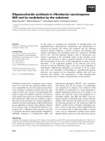
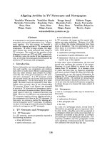
Fig. 1. The gene structures of the ACADVL (VLCAD), ACADM (MCAD) and ACADS (SCAD) genes. The number and approximate size of all
coding regions are shown and the 5¢-UTR (untranslated region) as well as the 3¢-UTR are indicated. The information used for the constructions are:
VLCAD [82,83] and NCBI nucleotide database gi: 3273227; MCAD [84] and NCBI nucleotide database gi: 187432 and SCAD [85] and NCBI
nucleotide database gi: 2995253; 2821943. Sequence variations are designated according to the position relative to the first nucleotide in the start-
codon ATG, and they are taken from the Human Gene Mutation Database (HGMD) in Cardiff ( />Ó FEBS 2004 Genetic defects in fatty acid oxidation (Eur. J. Biochem. 271) 473
interactions that are used during the folding process, the
folding pathway and the molecular forces along this (or
these) path(s) cannot presently be modelled for molecules of
more than 15 kDa [17]. Mis-sense sequence variations may
therefore affect the folding of the enzyme protein severely or
they may only perturb it slightly. The folding process is
monitored by the protein quality control systems, compri-
sing molecular chaperones, assisting the folding, and
intracellular proteases, which eliminate misfolded proteins
[13]. As the efficiency of these systems is dependent on the
cellular conditions, e.g. the temperature and energy level,
and probably also on genetic differences between individ-
uals, the effect of mis-sense sequence variations cannot, in
general, be predicted [18]. As will be discussed in the next
section, experimental evaluation can and should be
performed.
Recently it has been demonstrated that mis-sense sequence
variations, in addition to influencing protein biogenesis, also
may affect the splicing efficiency by interfering with binding
sites for splice modulating factors [19]. Although the effect of
mis-sense variations on splicing has not yet been published in
relation to fatty acid oxidation defects, it has been identified
in relation to isovaleryl CoA dehydrogenase [20] as well as
2-methyl butyryl-CoA dehydrogenase deficiencies [21]. This
fascinating phenomenon is in the process of being charac-
terized in the MCAD and VLCAD genes (K. B. Nielsen,
T. Sinnathamby, T. J. Corydon, L. Cartegni, A. R. Krainer,
O. N. Elpeleg, N. Gregersen, J. Kjems & B. S. Andresen,
unpublished observation).
In conclusion, it is only possible to predict the effect for
one third of the disease associated sequence variations in the
fatty acid oxidation genes, i.e. for large deletions, stop-
codon (PTC) introductions, consensus splice site changes
and small out-of-frame deletions/insertions. These are
sequence variations preventing formation of functional
protein (putative null-mutations). The rest, i.e. in-frame
deletions/insertions, nonconsensus splice site changes and
mis-sense sequence variations, may show an aprioriunpre-
dictable effect, which should be studied experimentally.
With the exception of the few variations directly affecting
the catalytic sites, these types of defects represent sequence
variations with potential variable effects. However, even if
the effect of the sequence variation can be elucidated in vitro,
the in vivo effect may be modulated by cellular and genetic
factors. In spite of these reservations concerning the
predictive value of knowing the disease associated sequence
variations in a given patient, this knowledge has obvious
diagnostic implications, which have been discussed in detail
elsewhere [1,22]. Furthermore, by performing careful
genotype–phenotype studies the relative importance of the
genetic predisposition and possible cellular and metabolic
disturbances, which are often determinants for the
precipitation of the fatty acid oxidation deficiencies, may
be assessed.
Genotype–phenotype relation in fatty acid
oxidation deficiencies
A second level of analysis of the genotype–phenotype
relation in fatty acid oxidation deficiencies is the investiga-
tion of possible associations between the type of sequence
variations and the clinical phenotype. As mentioned in the
Introduction, such associations seem to exist in patients
with certain of the long-chain defects but not – or at least to
a lesser extent – in patients with medium- or short-chain
defects. As our research has focussed on the acyl-CoA
dehydrogenase deficiencies, we will use VLCAD, MCAD
and SCAD deficiencies as examples and try to extrapolate
conclusions drawn from these diseases to the other fatty acid
oxidation defects.
Very-long-chain acyl-CoA dehydrogenase (VLCAD)
deficiency
The clinical spectrum seen in patients with VLCAD
deficiency is a prototype for other long-chain defects. As
discussed in more detail elsewhere [23], it is possible to
distinguish three phenotypes. The first comprises very
young infants who die from cardiac and liver disease within
the first year of life. The second group comprises older
children who do not have cardiac symptoms but show
hypoketotic hypoglycemia and hepatomegaly, symptoms
which are ÔMCAD deficiency-likeÕ (see below). The third
group are composed of adolescents and adults who do not
show cardiac and hepatic symptoms but who suffer from
muscle weakness, which may develop to degenerative
disability [24–27]. A large number of patients with VLCAD
deficiency have been genotyped and Table 3 shows the
distribution of the null-mutation/null-mutation and poten-
tial variable/potential variable genotypes in the three clinical
groups.
The most striking result is that homozygosity for
sequence variations, which lead to mRNA/protein elimin-
ation (null-mutations), is exclusively present in the patient
group with severe symptoms. There is little doubt that this is
a reflection of a severe enzyme deficiency, which is also
reflected in the profile of acyl-carnitines in blood and in
patient cells metabolizing long-chain fatty acids [27]. Not
surprisingly, the severe metabolic block results in profound
energy deficiency and corresponding severe clinical symp-
toms. To what degree the accumulated long-chain fatty
acids and their derivatives, especially the acylcarnitines, may
contribute to the clinical phenotype is not known with
certainty but these species may disturb membrane function,
Table 3. Distribution of VLCAD genotypes among three clinical subtypes of VLCAD deficiency. Data from [23].
Group1
Number of patients with
severe childhood form
Group 2
Number of patients with
mild childhood form
Group 3
Number of patients with
adult form
Null mutation/null mutation 8 0 0
Potential variable genotype/
potential variable genotype
6146
474 N. Gregersen et al. (Eur. J. Biochem. 271) Ó FEBS 2004
including ion-channels, and thereby perhaps, promote
arrhythmia ([28] and references therein).
It is also noteworthy that the sequence variations with
potentially varying effects (potentially variable genotypes)
are distributed to all three groups. This is most probably a
reflection of the fact that the effect of these sequence
variations may be total inactivation of VLCAD function or
it may be mild, leaving sufficient residual activity to avoid
severe energy deficiency. However, the deficiency is suffi-
cient to promote long-term muscle damage [24,25]. Whether
this damage is a result of energy deficiency or the long-term
effect of toxic long-chain fatty acids and their derivatives is
impossible to judge at present.
As far as we can determine, the same situation and
arguments apply to CPTII [29] and to ETF/ETFDH
deficiencies [30,31], where the number of patients and
sequence alterations are sufficiently large to perform a
similar analysis.
In contrast to these diseases, the other diseases related to
long-chain fatty acid metabolism may not show any
significant association between the severity of the defect/
type of sequence variation and the clinical phenotype, i.e.
CAT deficiency [32,33], LCHAD/mitochondrial trifunc-
tional protein (MTP) deficiencies [34], carnitine acylcarni-
tine translocase (CACT) deficiency [35] and CPTI deficiency
[36]. However, despite the fact that the number of patients
as well as the number of known disease-associated sequence
variations is still too small to provide a clear picture of the
genotype–phenotype in these diseases, liver-related patho-
logies are most often encountered. That other pathologies,
such as cardiac dysrhythmia in CPTI and cardiomyopathy
in LCHAD deficiencies, are observed emphasizes the notion
that factors other than the gene defect itself may be decisive,
as is also the case in MCAD deficiency, as discussed below.
Medium-chain acyl-CoA dehydrogenase (MCAD)
deficiency
The situation in MCAD deficiency is different from that in
VLCAD deficiency as a single prevalent sequence variation
(985AfiG), resulting in a mis-sense variant protein
(K304E), is present in homozygous form in 80% of all
patients diagnosed with MCAD deficiency. Eighteen per-
cent of patients are compound heterozygous with 985AfiG
on one allele and a rare disease associated sequence
variation on the other, and only about 2% carry other
(rare) sequence variations on both alleles [37]. Thus, studies
of the clinical impact of different types of sequence
variations are hampered by the fact that nearly all patients
carry one or two copies of the K304E variant MCAD
enzyme. This amino acid change exerts its effect primarily
by compromising the folding [38–40], but the variant
protein is also unstable [41] and the function is impaired
[42]. At least the misfolding and instability are influenced by
cellular and probably also by genetic factors, thus, resulting
in an effect that is totally unpredictable without experimen-
tal approaches, which will be discussed in detail below.
Suffice to say that the 985AfiG sequence variation may
result in varying effects and may blur an analysis similar to
the one described for VLCAD above.
The age of MCAD deficiency at presentation may vary
from birth to middle age. Clinically, the severity ranges from
fatal, through treatable acute symptoms, mild disabilities, to
asymptomatic throughout life. The features are, however,
rather uniform; episodic attacks of hypoketotic hypogly-
caemia accompanied by lethargy and vomiting, that may
develop into hepatic coma and death if not treated by
administration of carbohydrate. Usually, MCAD-deficient
patients do not experience life-threatening heart-related
symptoms, such as arrhythmia [28,43]. This indicates that
the energy deficiency is milder in MCAD than in VLCAD
deficiency or, alternatively, that medium chain-fatty acids
and their derivatives are less toxic to cardiac function than
long-chain fatty acids and their derivatives. However, it is
interesting to note that early investigations of the toxicity of
medium-chain fatty acids, such as octanoic acid, showed
narcotic properties that may contribute significantly to the
lethargy and hepatic coma observed in patients during
periods of metabolic decomposition [44,45].
With respect to the energy deficiency, the milder mani-
festation is probably caused by a combination of the fact
that several cycles of oxidation can proceed before the
pathway is blocked and that some enzyme activity may arise
from long-chain acyl-CoA dehydrogenase (LCAD) and
SCAD because of their overlapping substrate activity with
that of MCAD [46].
Although it is known that physiological factors, i.e.
metabolic stress in connection with fasting and fevers, are
important factors for the expression of the disease, it is still
an open question whether there exist other cellular and
genetic factors that contribute to the susceptibility in some,
but not in all, individuals [18].
Short-chain acyl-CoA dehydrogenase (SCAD) deficiency
SCAD deficiency remains difficult to analyse. Compared to
long-chain and medium-chain defects, there are at least
three peculiarities that are noteworthy with respect to the
relationship between the type of sequence variations and
clinical features in patients. The first is that nearly all
sequence variations identified so far are of the mis-sense
type, which in all cases may result in production of some
variant protein possessing some residual activity. Among
the first 13 disease-associated sequence variations identified
in SCAD-deficient patients, 12 are mis-sense sequence
variations and one is an in-frame deletion of three nucleo-
tides (Table 2). This trend has continued in more than 100
unpublished cases (N. Gregersen, A. Kølvraa, J. Vockley,
D.Matern,I.Tein,R.Ensenauer,C.Vianey-Saban,
M. Kjeldsen, V. S. Winter, C. B. Petersen & S. Kølvraa,
unpublished observations). Although about half of the mis-
sense sequence variations in the SCAD gene are cytidine to
thymine substitutions, which usually arises at CpG dinu-
cleotides, the overrepresentation is still striking and could be
a reflection of negative selection of germ cells with sequence
variations abolishing all enzyme activity. This explanation
has been proposed for the similar phenomenon in fumarate
hydratase deficiency, where the absence of such sequence
variations also is striking [47].
The second point of note is the spectrum of sequence
variations observed in patients with SCAD deficiency. In a
minority of cases, rare inactivating mis-sense sequence
variations are present in homozygous or compound hetero-
zygous form, whereas such variations in many cases are
Ó FEBS 2004 Genetic defects in fatty acid oxidation (Eur. J. Biochem. 271) 475
present in compound heterozygous form together with one
of two common susceptibility mis-sense sequence variations,
625GfiA and 511CfiT. These variations are present in
homozygous or compound heterozygous form in 14% of the
general population [48]. Further, a significant fraction of
patients with SCAD deficiency carry this genotype, in the
absence of any rare inactivating variations [49].
The above situation is apparently different from that
encountered in MCAD deficiency, where a prevalent
sequence variation likewise is present in nearly all patients
but where all individuals carrying the prevalent 985Adefi
Gua on both chromosomes, or in one chromosome with a
rare sequence variation in the other, are at risk of developing
disease. Although preliminary results have shown that the
spectrum of clinical symptoms in patients who are homo-
zygous or compound heterozygous for 625GuafiAde
or/and 511CytfiThy, are indistinguishable from patients
harbouring rare inactivating sequence variation [50], it must
be assumed that only a minority of individuals, who carry
the two susceptibility variations, are at risk of developing
disease. From this, it follows that there must be other
factors, physiological as well as cellular and/or genetic, that
are implicated in the expression of the disease.
This leads to the third point, namely the clinical features
in patients with SCAD deficiency. Only a few patients have
presented with symptoms related to energy deficiency, such
as cardiac symptoms or hypoglycaemia ([49]; N. Gregersen,
A. Kølvraa, J. Vockley, D. Matern, I. Tein, R. Ensenauer,
C.Vianey-Saban,M.Kjeldsen,V.S.Winter,C.B.Petersen
& S. Kølvraa, unpublished observation). The most probable
explanation for this is that SCAD deficiency only blocks the
last cycle of the pathway and that MCAD activity overlaps
with that of SCAD [46], thus, it is probable that near normal
amounts of reducing equivalents are generated. A further
reason is that butyric acid and its derivatives are neither
heart nor liver toxic. On the other hand, butyric acid is
known to exert severe cell toxicity by promotion of cell
differentiation, inhibition of the cell cycle and induction of
apoptosis [51,52]. This may be the reason why the predomi-
nant clinical symptoms are neuromuscular.
Without going into a detailed discussion about this issue,
there are two important remaining questions. First, how is it
possible that two genetic variations, which are implicated in
severe neuromuscular disease, can achieve such high
frequencies in the general population, and second, what
are the genetic/cellular/biochemical mechanisms which
renders some of the individuals carrying the variations in
homozygous or compound heterozygous form at risk of
developing clinically relevant disease. The first question can
not be answered at this time but we will attempt to answer
the second in the next section.
The only other disease affecting the metabolism of short-
chain fatty acids, where sequence variations have been
associated to an enzyme deficiency, is short-chain 3-hydroxy
acyl-CoA dehydrogenase (SCHAD) deficiency. Although
the deficiency has been known in a number of patients for
more than 10 years [53,54] only one patient with disease-
associated sequence variations in both alleles of the SCHAD
gene has been published [55]. The clinical symptoms in this
patient, who was shown to be homozygous for a mis-sense
sequence variation in the SCHAD gene (773CytfiThy;
P258L), are quite different from those found in patients with
SCAD deficiency, and include hyperinsulinism. Only time
will show the degree of clinical and genetic heterogeneity in
this rare disease.
Molecular effect of sequence variations with
an a priori unpredictable effect
A third level of analysis of the genotype–phenotype
correlation in fatty acid oxidation deficiencies may be the
experimental dissection of the molecular effects of sequence
variations with aprioriunpredictable effects.
Traditionally, and long before the genes and protein
structures were elucidated, enzymatic diagnosis of most fatty
acid oxidation deficiencies was possible and, furthermore,
practised in many laboratories. The enzymatic analyses
could correctly determine the residual activity in patient cells
but the question remained whether the enzyme protein was
present with reduced activity, or it was present in diminished
amounts. When antibodies against several of the fatty acid
oxidation enzyme proteins became available, it was possible
to approach this question. It was soon realized that decreased
amounts of protein are the rule rather than the exception.
However, it was only after gene cloning and synthesis of
proteins by recombinant techniques, that it became possible
to resolve the molecular pathogenesis of the increasing
number of identified disease associated sequence variations.
Although it might seem unnecessary to use large resources
to investigate molecular mechanisms, particularly as the
diagnoses can be made by direct enzyme activity measure-
ment in patient cells, there are at least three good reasons for
doing so. The first is to corroborate that an identified
sequence variation is associated to the clinical phenotype
through a functional effect on the variant protein. This is a
very practical and important goal that should be achieved
every time a new sequence variation is encountered.
The second reason is related to the first but extends the
purpose to future diagnostic procedures. The ongoing
genotype–phenotype studies data will alter the content of
sequence variation databases, which, in addition to raw
variation data should also contain information about the
effects on protein and cellular metabolism. For many
diseases, including the fatty acid oxidation deficiencies, it
will thus be possible to replace the laborious and expensive
enzymatic analysis by gene-based in vitro and in silico
methods.
The third reason for elucidating the molecular patho-
genesis, at least for some model variant proteins, is that
knowledge gained from detailed investigation of such
proteins may be generalized to other proteins in other
diseases. As a paradigm – with possible implications for
future treatment of patients – we will discuss the careful
investigation of the biogenesis, stability and function of the
disease-associated K304E MCAD enzyme protein, and the
application of the gained knowledge and methodological
approaches to define the role of the two common suscep-
tibility variations in the SCAD gene.
Molecular effects of the 985AfiG MCAD sequence
variation
Surprisingly, at least at the time of discovery, the amino
acid lysine, which is replaced by glutamic acid by the
476 N. Gregersen et al. (Eur. J. Biochem. 271) Ó FEBS 2004
985AdefiGua sequence variation at position 304 of the
MCAD protein, is located far from the active centre.
However, Western blot analysis of soluble variant protein
after expression in Escherichia coli cells showed diminished
amounts of the K304E MCAD protein compared to the
normal (wild-type) MCAD protein [56]. This result
stimulated further investigations, some of which have
provided the basis for the emerging concept that the
effects of mis-sense sequence variations are not only
dependent on the nature and location of the particular
variation but influenced to varying extents by cellular
factors related to the protein quality control systems
[18,38,41,57].
The studies, which will be summarized below, focused
on one hand on the biogenesis and stability of the
variant K304E MCAD protein, coded from the
985AdefiGua allele, and on the other hand on
the activity and chain-length selectivity of active K304E
MCAD enzyme.
Investigations of biogenesis and stability
In the late 1980s and early 1990s it was realized that the
expression and folding of many cellular proteins, including
mitochondrial proteins, are dependent on assistance by
molecular chaperones [58,59]. As the location was distant
from the active centre, it was hypothesized that the glutamic
acid in position 304 of the K304E MCAD protein distorted
the normal folding. This hypothesis was corroborated by
experiments performed in K. Tanaka’s group [38,39,60] and
in our own [40,41].
Tanaka’s group showed that both wild-type and variant
MCAD proteins were assisted in their intramitochondrial
folding by, first the chaperone heat shock protein 70
(Hsp70) and, subsequently, by chaperonin Hsp60, and
that the K304E protein was retained longer in association
with Hsp60 than was the wild-type protein. These elegant
experiments, which were performed by using rat liver
mitochondria, clearly indicated that MCAD deficiency is
at least in part due to compromised folding of the variant
enzyme protein. In parallel with these studies, we inves-
tigated the effect of co-overexpression of the bacterial
groELS (homologous to human Hsp60/10) in E. coli cells,
which over-expressed K304E or wild-type MCAD protein.
We found that it was possible to increase the yield of
active variant enzyme considerably but further studies also
showed that it was not possible to rescue more than
40–50% of wild-type activity. These experiments clearly
showed that the folding of the variant protein is
compromised.
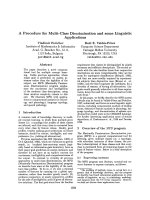
Inspection of the molecular structure surrounding posi-
tion 304 in the mature MCAD protein [41] indicated that
the lysine at position 304 is in close vicinity to two opposite-
charged aspartate residues at positions 300 and 346,
respectively (Fig. 2). Second site mutations at, respectively,
position 300 and 346, indicated that the presence of lysine
at position 304 is important for efficient folding of the
monomer and that the charge interaction between lysine 304
and aspartate 346 is important for tetramer assembly and,
therefore, for the stability of the assembled enzyme protein
[41]. These effects may be decisive for the steady-state level
of variant K304E MCAD but these experiments did not
give any data relating to either enzyme activity or substrate
selectivity.
Investigations of enzyme kinetics
Although the main effect of the 985AdefiGua sequence
variation is most probably due to distortion of folding
and tetramer assembly/stability, small distortions in the
conformation at the active site and substrate binding
pocket could contribute to the pathogenesis of the
985AdefiGua MCAD gene variation. Kieweg and
coworkers [42] addressed this question by determining
the kinetic parameters for purified wild-type and variant
MCAD protein from over-expressing E. coli cells. The
authors showed that V
max
was similar for wild-type and
the variant K304E MCAD proteins (980 vs. 970 lmolÆ
min
)1
), whereas K
m
was 3–4 times higher for the variant
enzyme, indicating a higher saturation concentration for
the optimum substrate octanoyl-CoA compared to the
wild-type enzyme. This may have consequences for the
amounts of available free CoA for other important
cellular processes.
Interestingly, the preferred substrate for K304E variant
MCAD is dodecanoyl-CoA. At this chain length, both V
max
and K
m
are similar for wild-type and variant MCAD
enzyme.
Taken together, these detailed studies on the molecular
pathogenesis of the K304E variant enzyme protein have
illuminated a number of important aspects of the effects of
mis-sense sequence variations in MCAD deficiency in
particular but also in fatty acid oxidation deficiencies in
general – which will be discussed for SCAD deficiency
below – as well as in other genetic diseases, such as
phenylketonuria (PKU) [61,62].
Fig. 2. An enlarged view of the vicinity of K304 of a monomer of porcine
MCAD (PDB accession no. 3MDD or 3MDE). Helices H and I are
shown in ribbons and side-chain atoms of K304, D346, Q342, D300
and the main chain carbonyl atoms of Q342 are shown as solid balls.
The side chain of R383 of the neighbouring monomer is represented by
open ball-and-stick. Distances between polar atoms in A
˚
are shown
with dotted lines. Reproduced with permission from Journal of
Biological Chemistry [41].
Ó FEBS 2004 Genetic defects in fatty acid oxidation (Eur. J. Biochem. 271) 477
The SCAD enigma
As mentioned earlier in this review, the genetic defect in
most patients with SCAD deficiency is not due to rare
inactivating sequence variations but rather to the presence of
one of two (or both) susceptibility gene variations, which are
present in 14% of the general population in configurations
also seen in patients with enzymatically proven SCAD
deficiency [49,63]. The goal is to delineate the nature of these
variations, which may help to explain why only certain
individuals carrying these variations develop clinically
relevant disease.
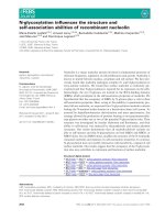
The structure of SCAD from rat has been elucidated [64].
From an inspection of the positions of the two variations,
G185S and R147W, it is not obvious how amino acid
changes at these positions could be pathogenic (Fig. 3).
Both positions are at the outer surface of the monomeric
structure. In agreement with this location and the fact that
severe defects on enzyme function would not be compatible
with the high frequency in the general population, the
kinetic disturbances were not found to be serious. Purified
R147W protein had kinetic properties similar to the wild-
type, and the kinetic efficiency of G185S protein was about
50% compared to the wild-type enzyme [65]. This probably
reflects the change from glycine to serine distorting the
conformation and exact positions of other amino acids
involved in the enzyme mechanism. These results – at least
concerning the G185S variant enzyme – underscore the
predisposing nature and indicate that other factors must be
involved.
Early biogenesis and stability studies showed that wild-
type SCAD is more dependent on the chaperonin system
Hsp60/10 (GroELS in E. coli) than MCAD [66]. While
wild-type MCAD does not need additional assistance by the
chaperonins in E. coli at 31 °C to achieve the active
conformation [40], the yield of functional wild-type SCAD
is increased eightfold by co-overexpression of GroELS at
thesametemperature.Inthesametypeofexperiment,
G185S variant SCAD showed about 30% of wild-type
activity without co-overexpression of GroELS but achieved
wild-type activity after co-overexpression of GroELS. This
indicated a greater dependence on chaperonin assistance for
the variant protein than for the wild-type but also that when
the folding capacity is sufficiently high, the biogenesis of the
variant enzyme is as effective as the wild-type enzyme.
Due to the ineffective folding behaviour of SCAD
compared to MCAD in E. coli cells, we looked into
eukaryotic expression that has been shown to be intrinsic-
ally more effective than bacterial expression for a number of
MCAD mutant proteins [67]. By varying the culture
temperature it was possible to detect differences in biogen-
esis between wild-type and the two variant proteins, G185S
and R147W SCAD. At physiological temperature, 37 °C,
the relative SCAD activities in extracts from transfected
COS-7 cells for G185S and R147W were 136 and 45%,
respectively. At 41 °C the relative activities were, respect-
ively, 58 and 13% for G185S and R147W SCAD, while they
were 183 and 85% at 26 °C [49]. These results support the
notion that the variant proteins in their biogenesis at
physiological temperatures may achieve sufficient activity to
sustain normal fatty acid oxidation but that both variant
proteins at higher temperatures, as experienced during
fevers, may result in insufficient amounts and activity and
thus the development of SCAD deficiency. This conclusion
is supported by further in vitro studies, where the biogenesis
of the two variant SCAD enzyme proteins was shown to be
delayed and compromised, especially at higher temperatures
[68]. Together with the fact that the stability of the active
G185S SCAD protein is decreased compared to that of
wild-type SCAD [66], these studies further contribute to the
notion that especially the G185S SCAD protein may be
disease-associated.
Whether other perturbations of the cellular homeostasis
in addition to high temperatures, such as alterations of
redox state, ATP depletion and pH changes, may show
differential effects on the biogenesis and/or stability of the
two variant proteins are pressing questions. If this is the
case, a number of conditions encountered in other metabolic
and endocrine diseases may result in Ôfunctional SCAD
deficiencyÕ and add to the clinical features of these other
diseases.
With the present knowledge levels we still do not know
how many of the 14% of the general population are at risk
of developing – perhaps in a mild and unrecognized form –
SCAD deficiency. We only know that a small fraction
develop clinically relevant disease [50], and we know that
this is possible by a combination of high fatty acid oxidation
activity and high temperature, which may result in accu-
mulation of cytotoxic butyric acid.
The challenge is to define further the cellular conditions
under which the deficiency occurs and to delineate whether
there exist inter-individual genetic differences in susceptibi-
lity to develop clinical disease.
Generalization and future aspects
Many elements of the above discussion can be generalized
to defects in other fatty acid oxidation enzymes and to
variant proteins present in other genetic diseases. To our
knowledge only a few other metabolic diseases have been
investigated in the same detail as MCAD and SCAD
deficiencies, and with PKU as a prominent example [61,62].
Fig. 3. Schematic overview of monomeric SCAD with the positions of
the 12 published mutations. The figure is based on the coordinates for
rat SCAD (PDB acc. no. 1JQI). SCAD protein is shown as a solid
ribbon and the Ca atoms at variant residues are represented as balls.
FAD and butyryl-CoA are shown as sticks.
478 N. Gregersen et al. (Eur. J. Biochem. 271) Ó FEBS 2004
In the future it may be important, in special cases, to do
similar experiments but the real challenge for disease-related
research, in relation to the biological significance of varying
effects of disease-causing and disease associated susceptibi-
lity variations, is to define the cellular conditions and
perturbations as well as genetic factors that may modulate
their effect. This challenge is still unappreciated but with the
identification of single nucleotide polymorphisms (SNPs),
which may associate to clinical features in complex diseases,
there will be a need for biochemical and cellular approaches
to delineate the functional significance of putative disease-
associated SNPs.
Furthermore, another challenge, which has not been
addressed in this review, is to describe and characterize
sequence variations that influence splicing by modulation of
the binding of splicing factors [19]. Doing this will also – as
has been seen for the mis-sense sequence variations – open
avenues to new questions about the plasticity of the cellular
response to gene variations, and give new insights in
biological mechanisms, which may be the target for
intervention by conventional treatments or future gene
therapeutic treatment.
Acknowledgements
The molecular genetic analyses of the VLCAD, MCAD and SCAD
genes have been performed by medical laboratory technologists
Vibeke Winter, Inga Knudsen, Margrethe Kjeldsen and Lisbeth
Schrøder. The investigations of our own group referred to in this
review have been supported by The Danish Medical Research
Council; Danish Human Genome Centre; Karen Elise Jensen
Foundation; Aarhus County Research Initiative; Institute of Experi-
mental Clinical Research, Aarhus University; Institute of Human
Genetics, Aarhus University and Aarhus University Hospital. We
thank colleagues from all over the world for providing genetic and
cell material for the studies and certain of them for inspiring
discussions concerning genotype–phenotype interactions in especially
the acyl-CoA dehydrogenase deficiencies.
References
1. Gregersen, N., Andresen, B.S., Corydon, M.J., Corydon, T.J.,
Olsen, R.K., Bolund, L. & Bross, P. (2001) Mutation analysis in
mitochondrial fatty acid oxidation defects exemplified by acyl-
CoA dehydrogenase deficiencies, with special focus on genotype–
phenotype relationship. Hum. Mutat. 18, 169–189.
2. Vockley, J. & Whiteman, D.A. (2002) Defects of mitochondrial
beta-oxidation: a growing group of disorders. Neuromuscul.
Disord. 12, 235–246.
3. DiMauro, S. & DiMauro, P.M. (1973) Muscle carnitine palmi-
tyltransferase deficiency and myoglobinuria. Science 182, 929–931.
4. Karpati,G.,Carpenter,S.,Engel,A.G.,Watters,G.,Allen,J.,
Rothman, S., Klassen, G. & Mamer, O.A. (1975) The syndrome of
systemic carnitine deficiency. Clinical, morphologic, biochemical,
and pathophysiologic features. Neurology 25, 16–24.
5. Gregersen, N., Lauritzen, R. & Rasmussen, K. (1976) Sub-
erylglycine excretion in the urine from a patient with dicarboxylic
aciduria. Clin. Chim. Acta 70, 417–425.
6. Krawczak,M.,Ball,E.V.,Fenton,I.,Stenson,P.D.,Abeysinghe,
S., Thomas, N. & Cooper, D.N. (2000) Human gene mutation
database – a biomedical information and research resource. Hum.
Mutat. 15, 45–51.
7. Lander, E.S., Linton, L.M., Birren, B., Nusbaum, C., Zody, M.C.,
Baldwin,J.,Devon,K.,Dewar,K.,Doyle,M.,FitzHugh,W.,
et al. (2001) Initial sequencing and analysis of the human genome.
Nature 409, 860–921.
8. Venter,J.C.,Adams,M.D.,Myers,E.W.,Li,P.W.,Mural,R.J.,
Sutton,G.G.,Smith,H.O.,Yandell,M.,Evans,C.A.,Holt,R.A.,
et al. (2001) The sequence of the human genome. Science 291,
1304–1351.
9. Culbertson, M.R. (1999) RNA surveillance. Unforeseen conse-
quences for gene expression, inherited genetic disorders and
cancer. Trends Genet. 15, 74–80.
10. Frischmeyer, P.A. & Dietz, H.C. (1999) Nonsense-mediated
mRNA decay in health and disease. Hum. Mol. Genet. 8, 1893–
1900.
11. Cooper, D.N. & Krawczak, M. (1993) Human Gene Mutation.
Bios. Scientific Publishers Ltd, Oxford, UK.
12. Nissim-Rafinia, M. & Kerem, B. (2002) Splicing regulation as a
potential genetic modifier. Trends Genet. 18, 123–127.
13. Bross, P., Corydon, T.J., Andresen, B.S., Jørgensen, M.M.,
Bolund, L. & Gregersen, N. (1999) Protein misfolding and
degradation in genetic disease. Hum. Mutat. 14, 186–198.
14. Wang, Z. & Moult, J. (2001) SNPs, protein structure, and disease.
Hum. Mutat. 17, 263–270.
15. Sunyaev, S., Ramensky, V., Koch, I., Lathe, W. III, Kondrashov,
A.S. & Bork, P. (2001) Prediction of deleterious human alleles.
Hum. Mol. Genet. 10, 591–597.
16.Terp,B.N.,Cooper,D.N.,Christensen,I.T.,Jorgensen,F.S.,
Bross, P., Gregersen, N. & Krawczak, M. (2002) Assessing the
relative importance of the biophysical properties of amino acid
substitutions associated with human genetic disease. Hum. Mutat.
20, 98–109.
17. Fersht, A.R. & Daggett, V. (2002) Protein folding and unfolding
at atomic resolution. Cell 108, 573–582.
18. Gregersen, N., Bross, P., Andresen, B.S., Pedersen, C.B., Cory-
don, T.J. & Bolund, L. (2001) The role of chaperone-assisted
folding and quality control in inborn errors of metabolism: protein
folding disorders. J. Inherit. Metab. Dis. 24, 189–212.
19. Cartegni, L., Chew, S.L. & Krainer, A.R. (2002) Listening to
silence and understanding nonsense: exonic mutations that affect
splicing. Nat. Rev. Genet. 3, 285–298.
20. Vockley,J.,Rogan,P.K.,Anderson,B.D.,Willard,J.,Seelan,
R.S., Smith, D.I. & Liu, W. (2000) Exon skipping in IVD RNA
processing in isovaleric acidemia caused by point mutations in the
coding region of the IVD gene. Am.J.Hum.Genet.66, 356–367.
21. Andresen,B.S.,Christensen,E.,Corydon,T.J.,Bross,P.,Pilg-
aard,B.,Wanders,R.J.,Ruiter,J.P.,Simonsen,H.,Winter,V.,
Knudsen,I.,Schroeder,L.D.,Gregersen,N.&Skovby,F.(2000)
Isolated 2-methylbutyrylglycinuria caused by short/branched-
chain acyl-CoA dehydrogenase deficiency: identification of a new
enzyme defect, resolution of its molecular basis, and evidence for
distinct acyl-CoA dehydrogenases in isoleucine and valine meta-
bolism. Am.J.Hum.Genet.67, 1095–1103.
22. Gregersen, N., Andresen, B.S. & Bross, P. (2000) Prevalent
mutations in fatty acid oxidation disorders: diagnostic considera-
tions. Eur. J. Pediatr. 159, S213–S218.
23. Andresen, B.S., Olpin, S., Poorthuis, B.J., Scholte, H.R., Vianey-
Saban, C., Wanders, R., Ijlst, L., Morris, A., Pourfarzam, M.,
Bartlett, K., Baumgartner, E.R., deKlerk, J.B., Schrøder, L.D.,
Corydon, T.J., Lund, H., Winter, V., Bross, P., Bolund, L. &
Gregersen, N. (1999) Clear correlation of genotype with disease
phenotype in very- long-chain acyl-CoA dehydrogenase defi-
ciency. Am.J.Hum.Genet.64, 479–494.
24. Smelt, A.H., Poorthuis, B.J., Onkenhout, W., Scholte, H.R.,
Andresen,B.S.,vanDuinen,S.G.,Gregersen,N.&Wintzen,A.R.
(1998) Very long chain acyl-coenzyme A dehydrogenase deficiency
with adult onset. Ann. Neurol. 43, 540–544.
25. Scholte,H.R.,VanCoster,R.N.,deJonge,P.C.,Poorthuis,B.J.,
Jeneson, J.A., Andresen, B.S., Gregersen, N., de Klerk, J.B. &
Ó FEBS 2004 Genetic defects in fatty acid oxidation (Eur. J. Biochem. 271) 479
Busch, H.F. (1999) Myopathy in very-long-chain acyl-CoA
dehydrogenase deficiency: clinical and biochemical differences
with the fatal cardiac phenotype. Neuromuscul. Disord. 9, 313–319.
26. Takusa,Y.,Fukao,T.,Kimura,M.,Uchiyama,A.,Abo,W.,
Tsuboi,Y.,Hirose,S.,Fujioka,H.,Kondo,N.&Yamaguchi,S.
(2002) Identification and characterization of temperature-sensitive
mild mutations in three japanese patients with nonsevere forms of
very-long-chain acyl-CoA dehydrogenase deficiency. Mol. Genet.
Metab. 75, 227–234.
27. Vianey-Saban, C., Divry, P., Brivet, M., Nada, M., Zabot, M.T.,
Mathieu, M. & Roe, C. (1998) Mitochondrial very-long-chain
acyl-coenzyme A dehydrogenase deficiency: clinical characteristics
and diagnostic considerations in 30 patients. Clin. Chim. Acta 269,
43–62.
28. Bonnet, D., Martin, D., Pascale, D.L., Villain, E., Jouvet, P.,
Rabier, D., Brivet, M. & Saudubray, J.M. (1999) Arrhythmias and
conduction defects as presenting symptoms of fatty acid oxidation
disorders in children. Circulation 100, 2248–2253.
29. Brivet, M., Boutron, A., Slama, A., Costa, C., Thuillier, L.,
Demaugre, F., Rabier, D., Saudubray, J.M. & Bonnefont, J.P.
(1999) Defects in activation and transport of fatty acids. J. Inherit.
Metab. Dis. 22, 428–441.
30. Frerman, F.E. & Goodman, S.I. (2001) Defects of electron
transfer flavoprotein and electron transfer flavoprotein-ubiqui-
none oxidoreductase: Glutaric aciduria type II. In The Metabolic
and Molecular Basis of Inherited Disease (Scriver, C.R., Beaudet,
A.L. & Valle, D., eds), pp. 2357–2365. McGraw-Hill, New York.
31. Olsen, R.K., Andresen, B.S., Christensen, E., Bross, P., Skovby, F.
& Gregersen, N. (2003) Clear relationship between ETF/ETFDH
genotype and phenotype in patients with multiple acyl-CoA
dehydrogenation deficiency. Hum. Mutat. 22, 12–23.
32. Wang, Y., Korman, S.H., Ye, J., Gargus, J.J., Gutman, A.,
Taroni, F., Garavaglia, B. & Longo, N. (2001) Phenotype and
genotype variation in primary carnitine deficiency. Genet. Med. 3,
387–392.
33. Wang, Y., Taroni, F., Garavaglia, B. & Longo, N. (2000) Func-
tional analysis of mutations in the OCTN2 transporter causing
primary carnitine deficiency: lack of genotype-phenotype corre-
lation. Hum. Mutat. 16, 401–407.
34. Tyni, T. & Pihko, H. (1999) Long-chain 3-hydroxyacyl-CoA
dehydrogenase deficiency. Acta Paediatr. 88, 237–245.
35. Hsu, B.Y., Iacobazzi, V., Wang, Z., Harvie, H., Chalmers, R.A.,
Saudubray,J.M.,Palmieri,F.,Ganguly,A.&Stanley,C.A.(2001)
Aberrant mRNA splicing associated with coding region mutations
in children with carnitine-acylcarnitine translocase deficiency.
Mol. Genet. Metab. 74, 248–255.
36. Bonnefont, J.P., Demaugre, F., Prip-Buus, C., Saudubray, J.M.,
Brivet, M., Abadi, N. & Thuillier, L. (1999) Carnitine palmitoyl-
transferase deficiencies. Mol. Genet. Metab. 68, 424–440.
37. Coates,P.M.,Chen,Y.T.,Curtis,D.,Gregersen,N.,Kelly,D.P.,
Matsubara, Y. & Yokota, I. (1992) Mutations causing medium-
chain acyl-CoA dehydrogenase deficiency: a collective compilation
of the data from 172 patients. Prog. Clin. Biol. Res. 375, 499–506.
38. Saijo, T., Welch, W.J. & Tanaka, K. (1994) Intramitochondrial
folding and assembly of medium-chain acyl-CoA dehydrogenase
(MCAD) – Demonstration of impaired transfer of K304E-variant
MCAD from Its complex with Hsp60 to the native tetramer.
J. Biol. Chem. 269, 4401–4408.
39. Saijo, T. & Tanaka, K. (1995) Isoalloxazine ring of FAD is
required for the formation of the core in the Hsp60-assisted
folding of medium chain Acyl-CoA dehydrogenase subunit into
the assembly competent conformation in mitochondria. J. Biol.
Chem. 270, 1899–1907.
40. Bross, P., Andresen, B.S., Winter, V., Krautle, F., Jensen, T.G.,
Nandy, A., Kølvraa, S., Ghisla, S., Bolund, L. & Gregersen, N.
(1993) Co-overexpression of bacterial GroESL chaperonins partly
overcomes non-productive folding and tetramer assembly of
E. coli-expressed human medium-chain acyl-CoA dehydrogenase
(MCAD) carrying the prevalent disease-causing K304E mutation.
Biochim. Biophys. Acta 1182, 264–274.
41. 81–89.Bross, P., Jespersen, C., Jensen, T.G., Andresen, B.S.,
Kristensen, M.J., Winter, V., Nandy, A., Krautle, F., Ghisla, S.,
Bolund, L., Kim, J.J.P. & Gregersen, N. (1995) Effects of two
mutations detected in medium chain acyl-CoA dehydrogenase
(MCAD)-deficient patients on folding, oligomer assembly, and
stability of MCAD enzyme. J. Biol. Chem. 270, 10284–10290.
42. Kieweg,V.,Krautle,F.G.,Nandy,A.,Engst,S.,Vock,P.,Abdel-
Ghany, A.G., Bross, P., Gregersen, N., Rasched, I., Strauss, A. &
Ghisla, S. (1997) Biochemical characterization of purified, human
recombinant Lys304fiGlu medium-chain acyl-CoA dehydro-
genase containing the common disease-causing mutation and
comparison with the normal enzyme. Eur. J. Biochem. 246, 548–
556.
43. Roe, C.R. & Ding, J.H. (2001) Mitochondrial fatty acid oxidation
disorders. In The Metabolic and Molecular Bases of Inherited
Disease (Scriver,C.R.,Beaudet,A.L.,Valle,D.&Sly,W.S.,eds),
pp. 2297–2326. McGraw-Hill, New York.
44. Trauner, D.A. & Huttenlocher, P.R. (1978) Short chain fatty acid-
induced central hyperventilation in rabbits. Neurology 28, 940–
944.
45. Gregersen, N. (1985) The acyl-CoA dehydrogenation deficiencies:
recent advances in the enzymic characterization and understand-
ing of the metabolic and pathophysiological disturbances in
patients with acyl-CoA dehydrogenation deficiencies. Scand. J.
Clin. Laboratory Invest. 45, 1–60.
46. Eaton, S., Bartlett, K. & Pourfarzam, M. (1996) Mammalian
mitochondrial beta-oxidation. Biochem. J. 320, 345–357.
47. Tomlinson,I.P.,Alam,N.A.,Rowan,A.J.,Barclay,E.,Jaeger,
E.E.,Kelsell,D.,Leigh,I.,Gorman,P.,Lamlum,H.,Rahman,S.,
et al. (2002) Germline mutations in FH predispose to dominantly
inherited uterine fibroids, skin leiomyomata and papillary renal
cell cancer. Nat. Genet 30, 406–410.
48. Gregersen, N., Winter, V.S., Corydon, M.J., Corydon, T.J.,
Rinaldo,P.,Ribes,A.,Martinez,G.,Bennett,M.J.,Vianey-
Saban,C.,Bhala,A.,Hale,D.E.,Lehnert,W.,Kmoch,S.,Roig,
M., Riudor, E., Eiberg, H., Andresen, B.S., Bross, P., Bolund,
L.A. & Kølvraa, S. (1998) Identification of four new mutations in
the short-chain acyl-CoA dehydrogenase (SCAD) gene in two
patients: one of the variant alleles, 511CfiT,ispresentatan
unexpectedly high frequency in the general population, as was the
case for 625GfiA, together conferring susceptibility to ethylma-
lonic aciduria. Hum. Mol. Genet. 7, 619–627.
49. Corydon, M.J., Vockley, J., Rinaldo, P., Rhead, W.J., Kjeldsen,
M.,Winter,V.,Riggs,C.,Babovic-Vuksanovic,D.,Smeitink,J.,
DeJong,J.,Levy,H.,Clive,S.A.,Roe,C.,Matern,D.,Dasouki,
M. & Gregersen, N. (2001) Role of common gene variations in the
molecular pathogenesis of short-chain acyl-CoA dehydrogenase
deficiency. Pediatr. Res. 49, 18–23.
50. Gregersen, N., Vockley, J., Matern, D., Rinaldo, P., Vianey-
Saban, C., Andresen, B.S., Bross, P., Kjeldsen, M., Winter,
V.S., Ensenauer, R., Kølvraa, A. & Kølvraa, S. (2002) Ethyl-
malonic aciduria/SCAD deficiency: there are correlations between
genotype and metabolic and enzymatic phenotype, but not
between genotype and clinical phenotype. J. Inher. Metab. Dis.
25,65.
51. Barnard, J.A. & Warwick, G. (1993) Butyrate rapidly induces
growth inhibition and differentiation in HT-29 cells. Cell Growth
Differ. 4, 495–501.
52. Giuliano, M., Lauricella, M., Calvaruso, G., Carabillo, M.,
Emanuele, S., Vento, R. & Tesoriere, G. (1999) The apoptotic
effects and synergistic interaction of sodium butyrate and MG132
in human retinoblastoma Y79 cells. Cancer Res. 59, 5586–5595.
480 N. Gregersen et al. (Eur. J. Biochem. 271) Ó FEBS 2004
53. Tein,I.,DeVivo,D.C.,Hale,D.E.,Clarke,J.T.,Zinman,H.,
Laxer, R., Shore, A. & DiMauro, S. (1991) Short-chain
L
-3-
hydroxyacyl-CoA dehydrogenase deficiency in muscle: a new
cause for recurrent myoglobinuria and encephalopathy. Ann.
Neurol. 30, 415–419.
54. Bennett, M.J., Rinaldo, P. & Strauss, A.W. (2000) Inborn errors
of mitochondrial fatty acid oxidation. Crit.Rev.Clin.Lab.Sci.37,
1–44.
55.Clayton,P.T.,Eaton,S.,Aynsley-Green,A.,Edginton,M.,
Hussain,K.,Krywawych,S.,Datta,V.,Malingre,H.E.,Berger,
R. & van den Berg, I.E. (2001) Hyperinsulinism in short-chain
L
-3-hydroxyacyl-CoA dehydrogenase deficiency reveals the
importance of beta-oxidation in insulin secretion. J. Clin. Invest.
108, 457–465.
56. Gregersen, N., Andresen, B.S., Bross, P., Winter, V., Ru
¨
diger, N.,
Engst, S., Christensen, E., Kelly, D., Strauss, A.W., Kølvraa, S.,
Bolund, L. & Ghisla, S. (1991) Molecular characterization of
medium-chain acyl-CoA dehydrogenase (MCAD) deficiency:
identification of a lys329 to glu mutation in the MCAD gene, and
expression of inactive mutant enzyme protein in E. coli. Hum.
Genet. 86, 545–551.
57. Bross, P., Andresen, B.S. & Gregersen, N. (1998) Impaired folding
and subunit assembly as disease mechanism: the example of
medium-chain acyl-CoA dehydrogenase deficiency. Prog. Nucleic
Acid Res. Mol. Biol. 58, 301–337.
58. Ostermann,J.,Horwich,A.L.,Neupert,W.&Hartl,F.U.(1989)
Protein folding in mitochondria requires complex formation with
hsp60 and ATP hydrolysis. Nature 341, 125–130.
59. Horwich, A.L., Neupert, W. & Hartl, F.U. (1990) Protein-
catalysed protein folding. Trends Biotechnol. 8, 126–131.
60. Yokota, I., Saijo, T., Vockley, J. & Tanaka, K. (1992) Impaired
tetramer assembly of variant medium-chain acyl-coenzyme A
dehydrogenase with a glutamate or aspartate substitution for
lysine 304 causing instability of the protein. J. Biol. Chem. 267,
26004–26010.
61. Gamez, A., Perez, B., Ugarte, M. & Desviat, L.R. (2000)
Expression analysis of phenylketonuria mutations. Effect on
folding and stability of the phenylalanine hydroxylase protein.
J. Biol. Chem. 275, 29737–29742.
62.Waters,P.J.,Parniak,M.A.,Akerman,B.R.&Scriver,C.R.
(2000) Characterization of phenylketonuria missense substitu-
tions, distant from the phenylalanine hydroxylase active site,
illustrates a paradigm for mechanism and potential modulation of
phenotype. Mol. Genet. Metab. 69, 101–110.
63. Matern,D.,Hart,P.,Murtha,A.P.,Vockley,J.,Gregersen,N.,
Millington, D.S. & Treem, W.R. (2001) Acute fatty liver of
pregnancy associated with short-chain acyl-coenzyme A dehydro-
genase deficiency. J. Pediatr. 138, 585–588.
64. Battaile,K.P.,Molin-Case,J.,Paschke,R.,Wang,M.,Bennett,
D., Vockley, J. & Kim, J.J. (2002) Crystal structure of rat short
chain acyl-CoA dehydrogenase complexed with acetoacetyl-CoA:
comparison with other acyl-CoA dehydrogenases. J. Biol. Chem.
277, 12200–12207.
65. Nguyen, T.V., Riggs, C., Babovic-Vuksanovic, D., Kim, Y.S.,
Carpenter,J.F.,Burghardt,T.P.,Gregersen,N.&Vockley,J.
(2002) Purification and characterization of two polymorphic
variants of short chain acyl-CoA dehydrogenase reveal reduction
of catalytic activity and stability of the gly185ser enzyme.
Biochemistry 41, 11126–11133.
66. Corydon,M.J.,Gregersen,N.,Lehnert,W.,Ribes,A.,Rinaldo,
P.,Kmoch,S.,Christensen,E.,Kristensen,T.J.,Andresen,B.S.,
Bross,P.,Winter,V.,Martinez,G.,Neve,S.,Jensen,T.G.,Bol-
und, L. & Kølvraa, S. (1996) Ethylmalonic aciduria is associated
with an amino acid variant of short-chain acyl-coenzyme A
dehydrogenase. Pediatr. Res. 39, 1059–1966.
67. Jensen, T.G., Bross, P., Andresen, B.S., Lund, T.B., Kristensen,
T.J.,Jensen,U.B.,Winther,V.,Kølvraa,S.,Gregersen,N.&
Bolund, L. (1995) Comparison between medium-chain acyl-CoA
dehydrogenase mutant proteins overexpressed in bacterial and
mammalian cells. Hum. Mutat. 6, 226–231.
68. Pedersen,C.B.,Bross,P.,Winter,V.S.,Corydon,T.J.,Bolund,L.,
Bartlett, K., Vockley, J. & Gregersen, N. (2003) Misfolding,
degradation and aggregation of variant proteins – the molecular
pathogenesis of short chain acyl-CoA dehydrogenase (SCAD)
deficiency. J. Biol. Chem. 278, 47449–47458.
69. Odaib, A.A., Shneider, B.L., Bennett, M.J., Pober, B.R.,
Reyes-Mugica, M., Friedman, A.L., Suchy, F.J. & Rinaldo,
P. (1998) A defect in the transport of long-chain fatty acids
associated with acute liver failure. N.Engl.J.Med.339,
1752–1757.
70. Rinaldo, P., Matern, D. & Bennett, M.J. (2002) Fatty acid oxi-
dation disorders. Annu. Rev. Physiol. 64, 477–502.
71. Andresen, B.S., Dobrowolski, S.F., O’Reilly, L., Muenzer, J.,
McCandless,S.E.,Frazier,D.M.,Udvari,S.,Bross,P.,Knudsen,
I.,Banas,R.,Chace,D.H.,Engel,P.&Gregersen,N.(2001)
Medium-chain acyl-CoA dehydrogenase (mcad) mutations iden-
tified by ms/ms-based prospective screening of newborns differ
from those observed in patients with clinical symptoms: identifi-
cation and characterization of a new, prevalent mutation that
results in mild MCAD deficiency. Am. J. Hum. Genet. 68, 1408–
1418.
72. Stanley,C.A.,Hale,D.E.,Berry,G.T.,Deleeuw,S.,Boxer,J.&
Bonnefont, J.P. (1992) Brief report: a deficiency of carnitine-
acylcarnitine translocase in the inner mitochondrial membrane.
N.Engl.J.Med.327, 19–23.
73. Kamijo,T.,Indo,Y.,Souri,M.,Aoyama,T.,Hara,T.,Yama-
moto,S.,Ushikubo,S.,Rinaldo,P.,Matsuda,I.,Komiyama,A.
& Hashimoto, T. (1997) Medium chain 3-ketoacyl-coenzyme A
thiolase deficiency: a new disorder of mitochondrial fatty acid
beta-oxidation. Pediatr. Res. 42, 569–576.
74. Amendt, B.A., Greene, C., Sweetman, L., Cloherty, J., Shih, V.,
Moon, A., Teel, L. & Rhead, W.J. (1987) Short-chain acyl-
coenzyme A dehydrogenase deficiency: clinical and biochemical
studies in two patients. J. Clin. Invest. 79, 1303–1309.
75. Bougneres, P.F., Saudubray, J.M., Marsac, C., Bernard, O.,
Odievre, M. & Girard, J. (1981) Fasting hypoglycemia resulting
from hepatic carnitine palmitoyl transferase deficiency. J. Pediatr.
98, 742–746.
76. Przyrembel, H., Wendel, U., Becker, K., Bremer, H.J., Bruinvis,
L., Ketting, D. & Wadman, S.K. (1976) Glutaric aciduria type II:
report on a previously undescribed metabolic disorder. Clin. Chim.
Acta 66, 227–239.
77. Bertrand, C., Largilliere, C., Zabot, M.T., Mathieu, M. & Vianey-
Saban, C. (1993) Very long chain acyl-CoA dehydrogenase defi-
ciency: identification of a new inborn error of mitochondrial fatty
acid oxidation in fibroblasts. Biochim. Biophys. Acta 1180,
327–329.
78. Wanders,R.J.,Duran,M.,Ijlst,L.,deJager,J.P.,vanGennip,
A.H., Jakobs, C., Dorland, L. & van Sprang, F.J. (1989) Sudden
infant death and long-chain 3-hydroxyacyl-CoA dehydrogenase.
Lancet 2, 52–53.
79. Jackson, S., Kler, R.S., Bartlett, K., Briggs, H., Bindoff, L.A.,
Pourfarzam,M.,Gardnermedwin,D.&Turnbull,D.M.(1992)
Combined enzyme defect of mitochondrial fatty acid oxidation.
J. Clin. Invest. 90, 1219–1225.
80. Hintz, S.R., Matern, D., Strauss, A., Bennett, M.J., Hoyme, H.E.,
Schelley, S., Kobori, J., Colby, C., Lehman, N.L. & Enns, G.M.
(2002) Early neonatal diagnosis of long-chain 3-hydroxyacyl
coenzyme a dehydrogenase and mitochondrial trifunctional pro-
tein deficiencies. Mol. Genet. Metab. 75, 120–127.
Ó FEBS 2004 Genetic defects in fatty acid oxidation (Eur. J. Biochem. 271) 481
81.Iacobazzi,V.,Naglieri,M.A.,Stanley,C.A.,Wanders,R.J.&
Palmieri, F. (1998) The structure and organization of the human
carnitine/acylcarnitine translocase (CACT1) gene2. Biochem.
Biophys. Res. Commun. 252, 770–774.
82. Strauss, A.W., Powell, C.K., Hale, D.E., Anderson, M.M., Ahuja,
A., Brackett, J.C. & Sims, H.F. (1995) Molecular basis of human
mitochondrial very-long-chain acyl-CoA dehydrogenase defi-
ciency causing cardiomyopathy and sudden death in childhood.
Proc.NatlAcad.Sci.USA92, 10496–10500.
83. Orii,K.O.,Aoyama,T.,Souri,M.,Orii,K.E.,Kondo,N.,Orii,T.
& Hashimoto, T. (1995) Genomic DNA organization of human
mitochondrial very-long- chain acyl-CoA dehydrogenase and
mutation analysis. Biochem. Biophys. Res. Commun. 217, 987–992.
84. Zhang,Z.F.,Kelly,D.P.,Kim,J.J.,Zhou,Y.Q.,Ogden,M.L.,
Whelan, A.J. & Strauss, A.W. (1992) Structural organization and
regulatory regions of the human medium-chain acyl-CoA dehy-
drogenase gene. Biochem. 31, 81–89.
85. Corydon, M.J., Andresen, B.S., Bross, P., Kjeldsen, M.,
Andreasen, P.H., Eiberg, H., Kølvraa, S. & Gregersen, N. (1997)
Structural organization of the human short-chain acyl-CoA
dehydrogenase gene. Mamm. Genome 8, 922–926.
482 N. Gregersen et al. (Eur. J. Biochem. 271) Ó FEBS 2004