Báo cáo khóa học: Different associational and conformational behaviors between the second and third repeat fragments in the tau microtubule-binding domain pdf
Bạn đang xem bản rút gọn của tài liệu. Xem và tải ngay bản đầy đủ của tài liệu tại đây (403.36 KB, 8 trang )
Different associational and conformational behaviors between
the second and third repeat fragments in the tau
microtubule-binding domain
Katsuhiko Minoura
1
, Tian-Ming Yao
1
, Koji Tomoo
1
, Miho Sumida
2
, Masahiro Sasaki
2
, Taizo Taniguchi
2,3
and Toshimasa Ishida
1
1
Osaka University of Pharmaceutical Sciences, Takatsuki, Osaka, Japan;
2
Behavioral and Medical Sciences Research Consortium,
Akashi, Hyogo, Japan;
3
Biosignal Research Center, Kobe University, Kobe, Japan
The third repeat fragment (R3) in the four-repeat micro-
tubule-binding domain of the water-soluble tau protein has
been considered to play an essential role in the protein’s
filamentous assembly. To clarify the associational and con-
formational features that differentiate R3 from the second
repeat, R2, the heparin-induced assembly profiles of these
peptide fragments were monitored by the thioflavin fluor-
escence method and electron microscopy. The trifluoro-
ethanol-induced reversible conformational change from a
random structure to an a-helical structure, in an aqueous
solution, was monitored by CD measurement, and the
structure of R2 in trifluoroethanol solution was analyzed by
a combination of two-dimensional
1
H-NMR measurements
and molecular modeling calculations to facilitate compar-
ison with the structure of R3. The speed of R3 assembly was
remarkably faster than that of R2, in spite of their similar
amino acid sequences. The averaged NMR conformers of
R2 exhibited the whole-spanning a-helical structure. Similar
features observed in R2 and R3 conformers in trifluoro-
ethanol were that the Leu10–Leu20/Lys20 sequence takes a
helical structure with the amphipathic-like distribution of the
respective side-chains, whereas the C-terminal moieties are
both flexible. In contrast, a notable difference was observed
at the N-terminal Val1–Lys6 sequence, namely, a helical
conformation for R2 and an extended conformation for R3.
These conformational behaviors would be associated with
the different self-aggregation speeds and seeding reactions
between R2 and R3.
Keywords: tau protein; microtubule-binding domain; repea-
ted fragment; self-assembly; amphipathic structure.
Aggregation of the microtubule-associated tau protein is a
significant event in neurodegradation [1], because the water-
soluble tau protein self-aggregates into a water-insoluble
structure known as the paired helical filament (PHF),
which is a major component of the pathological lesion in
Alzheimer’s and other diseases [2]. These aggregates are
neurotoxic, as they destroy the cell interior and lead to the
development of neuropathological diseases. Therefore, the
inhibition of PHF formation may be effective in preventing
such a pathological progression.
Although data on the physicochemical behaviors of the
tau protein, associated with self-assembly, have increased
in recent years, the underlying mechanism at the mole-
cular level remains to be clarified, because the water-soluble
tau protein is flexible and takes a random conformation
under physiological conditions. It has been reported that the
three- or four-repeat microtubule-binding domain (MBD),
each repeat consisting of 31 or 32 amino acid residues,
located in the C-terminal half (Fig. 1), assumes the core
structure of PHF [3] and promotes tau assembly in vitro [4].
Therefore, it is important to examine the structural features
of the MBD, in order to understand the mechanism
underlying PHF formation. In particular, we have focused
on clarifying the self-associational and conformational
features of the repeat fragments in MBD. Little such data
has previously been reported, despite its usefulness in
determining the contribution of each repeat fragment to
PHF formation.
Recently, we have determined the structure of the third
repeat of MBD (R3 in Fig. 1), in water and trifluoroethanol
(TFE) solutions, by using the
1
H-NMR method [5,6], and
clarified the extended-like structure of the N-terminal
VQIVYK sequence and the helical structure of the
Leu10–Leu20 sequence, with an amphipathic distribution
of the corresponding side-chains, in TFE. This conforma-
tional behavior may be associated with the filament
formation of MBD, because the VQIVYK local sequence
of R3 was reported to play an important role in the assembly
of the tau protein into PHF, which is responsible for
Alzheimer’s lesion [7]. On the other hand, it has been
Correspondence to K. Minoura, Research Center, Osaka University of
Pharmaceutical Sciences, 4-20-1 Nasahara, Takatsuki,
Osaka 569-1094, Japan. Fax/Tel.: + 81 726 90 1039,
E-mail: or T M. Yao, Department of
Physical Chemistry, Osaka University of Pharmaceutical Sciences,
4-20-1 Nasahara, Takatsuki, Osaka 569-1094, Japan.
Fax/Tel.: + 81 726 90 1068, E-mail:
Abbreviations: MBD, microtubule-binding domain; PHF, paired
helical filament; TFE, trifluoroethanol; ThS, thioflavin S.
(Received 29 August 2003, revised 12 November 2003,
accepted 5 December 2003)
Eur. J. Biochem. 271, 545–552 (2004) Ó FEBS 2004 doi:10.1046/j.1432-1033.2003.03956.x
reported that the R2-included and -deleted tau proteins
demonstrate a notable difference in their microtubule-
binding ability and PHF formation [8–10]; the four-repeat
and three-repeat isoforms of the tau MBD are a direct
result of the presence or absence of R2, respectively [11].
Therefore, in order to clarify the difference in the associ-
ational and conformational behaviors between R3 and R2
repeat fragments, we investigated their assembly profiles by
the thioflavin S (ThS) fluorescence method and electron
microscopy. Moreover, the TFE-induced conformation of
R2 was analyzed by
1
H-NMR spectroscopy and was
compared with that of R3. It is important to investigate
to what extent their averaged conformations differ in
solution (even in a nonphysiological solution), because the
reason why R2 does not play as crucial a role as R3 in PHF
formation, in spite of their nearly identical N-terminal
sequences and the fact that both possess one Cys residue
each (Fig. 1) is, as yet, unclarified.
Materials and methods
Peptide
R1, R2, R3 and R4 peptides, corresponding to the first
(244–274), second (275–305), third (306–336) and fourth
(337–378) repeat fragments of the full-length human tau
protein, respectively, were synthesized in the form of
lyophilized powders (including trifluoroacetic acid as a
counter ion). These peptides were characterized by MS and
were purified to > 95.0%, as assessed by reverse-phase
HPLC.
Electron microscopy
Each repeat peptide (15 l
M
)wasmixedwith3.8l
M
heparin
in 50 m
M
Tris/HCl (pH 7.5). The solution was then
incubated at 37 °C for 24 h. For negative-staining electron
microscopy, 600-mesh copper grids were used. A drop of
peptide solution and a drop of 2% uranyl acetate were
placed on the grids. After 2 min, excess fluid was removed
from the grids. Negative-staining electron microscopy was
performed using an electron microscope (Hitachi H-600)
operated at 75 kV.
Monitoring of aggregation of the MBD repeat fragment
by ThS fluorescence
Each repeat peptide was adjusted to a concentration of
15 l
M
using 50 m
M
Tris/HCl (pH 7.5) containing 10 l
M
ThS dye. Aggregation was induced by adding heparin (final
concentration 3.8 l
M
) to the solution, which was then
mixed with a pipette prior to measuring the fluorescence.
The time-scanning of fluorescence was carried out on a
JASCO FP-6500 instrument using a 2-mm quartz cell, in
which the temperature was maintained at 37 °Cbya
circulating water bath. The kinetics of MBD aggregation
was analyzed by recording the time-dependent curve of the
fluorescence intensity, with excitation at 440 nm and
emission at 490 nm. Background fluorescence of the sample
was reduced as required.
CD measurement
The sample solution was adjusted to 40 l
M
in water, TFE,
and these mixed solvents, where the pH value was adjusted
by adding HCl or NaOH. All measurements at 25 °Cwere
conducted using a JASCO J-820 spectrometer in a cuvette
with a 2-mm path length. For each experiment under N
2
gas flow, measurement from 190 to 260 nm was repeated
eight times and the results were summed. Then, the molar
ellipticity was determined after normalizing the sample
concentration. The same experiment was performed at least
three times using newly prepared samples; their averaged
values are presented below, in the Results and discussion.
Data were expressed in terms of a mean residue ellipticity (h)
in units of deg cm
2
Ædmol
)1
.
NMR measurement
The method used to determine the structure of R2 was the
same as that used for R3 [5,6]. The peptide (2 m
M
)was
dissolved in TFE-d
2
,andits
1
H-NMR spectra were
recorded using a Varian unity INOVA500 spectrometer
equipped with a variable temperature-control unit.
1
H
chemical shifts were referenced to 0 p.p.m. for 3-(trimethyl-
silyl) propionic acid at 298 K. Owing to the low solubility at
pH values of > 5.0, the pH was adjusted to 3.9 by adding
HCl or NaOH. In order to trace direct single- and multiple-
relayed through-bond connectivities, successively, TOCSY
spectra were recorded at mixing times of 40 and 100 ms.
The NOESY spectra were also measured at mixing times of
100, 200, and 300 ms. Assuming the same correlation time
for all the protons, the offset dependence of the NOESY
cross-peaks was used for the estimation of proton–proton
distance. The NOE intensities were classified into three
groups (strong, medium and weak). The vicinal coupling
constants obtained from DQF-COSY measurements were
used to estimate the possible torsion angles:
3
J
HNCaH
¼ 1:9 À 1:4cosh þ 6:4cos
2
h ð1Þ
where / ¼ h-60° for the / torsion angle around the C¢
i-1
-N
i
-
Ca
i
-C¢
i
bond sequence [12].
Fig. 1. Schematic representation of the four-repeat microtubule-binding
domain (MBD) moiety in the entire human tau protein (A) and the amino
acid sequence of each repeat (B). The regions from the first to the fourth
repeat fragments in MBD (A) are named R1 to R4, respectively. The
numbering of the amino acid residues in (A) refers to the longest
isoform of human tau protein (441 residues).
546 K. Minoura et al. (Eur. J. Biochem. 271) Ó FEBS 2004
Conformational calculations
Three dimensional structures that fulfil the NOE distance
and J torsion angle constraints of intramolecular proton
pairs were constructed by dynamic simulated annealing
calculations [13] using the
CNS
program [14]. After rand-
omizing the peptide into extended strands, corresponding to
each disjointed molecular entity, the initial structures were
constructed by referring to the data structures and statistical
analysis of the average property. The constructed structure
was then annealed for 15 ps at 50 000 K and cooled to
300 K (at a rate of 250 K/step) for 10 ps, and the minimi-
zation of more than 5000 steps was continued. The
constraints for distances and torsion angles were used as
the harmonic potential function. As the input data for
distance constraint, the proton–proton pairs were classified
into three distance groups according to the NOE intensi-
ties: strong (1.8–3.0 A
˚
), medium (1.8–4.0 A
˚
)andweak
(1.8–5.0 A
˚
). The torsional constraint was applied to the
torsion / angle, i.e. )120 ± 40° for
3
J
HNCaH
>8Hz,
)75 ± 25° for
3
J
HNCaH
<6Hz,and)100 ± 60° for the
others. The root mean square deviation analyses of energy-
minimized structures were carried out using the
MOLMOL
program [15].
Results and discussion
Different behaviors among four repeat fragments
for filament formation
It has been reported that thioflavin dyes, such as ThS, can be
used to quantify the filament formation in solution in real
time [16]. We used this assay to monitor the filamentous
assembly of each repeat MBD peptide, the aggregation of
which was induced by adding heparin. The aggregation
kinetics was then derived from the time dependence of
fluorescence intensity. As shown in Fig. 2A, the fluorescence
intensity of R3 reached a maximum within 50 min
(t
½
¼%15 min). However, the presence of dithiothreitol
caused a significant decrease in its fluorescence intensity,
indicating that the intermolecular disulfide bond formation
between the Cys residues of neighboring R3 is a major step
for initiating filament formation. In contrast, the increase in
ThS intensity was very slow in R2 (Fig. 2B), indicating that
the aggregation mechanism of R2 is different from that of
R3. As the ThS intensity of R2 also decreased in the presence
of dithiothreitol, the filament of R2 may be formed through
disulfide bonds. However, R2 showed a considerably
different profile from R3, although both peptides contain
one Cys residue and possess similar amino acid sequences
(Fig. 1B). In order to consider the biological/structural
implication of this difference, the effect of seeding for
filament formation was investigated. As shown in Fig. 2C,D,
a notable difference was observed. The R2 peptide showed
an R2-dependent seeding effect, whereas R3 was only
Fig. 2. Heparin-induced in vitro aggregation profiles. Heparin-induced
in vitro aggregation profiles of R3 with and without 1 m
M
dithio-
threitol (A), of R2 with and without 1 m
M
dithiothreitol (B), and of
seeded R3 (C) and R2 (D), as functions of reaction time, monitored
based on thioflavin S (ThS) fluorescence intensity.
Ó FEBS 2004 Different features of repeat fragments in tau MBD (Eur. J. Biochem. 271) 547
slightly affected by any seeding. From these results, it would
be reasonable to consider that the R3 peptide aggregates
easily, without the help of any template. In contrast, the R2
peptide does not aggregate easily, and its filament formation
progresses via a nucleation step, in which the template of
homogeneous aggregates is required.
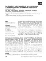
Figure 3 shows the electron micrographs of R2 and R3
filaments. Both R2 and R3 peptides exhibited thin and
straight filaments. However, a notable difference was
observed in their shapes, that is, the R2 filaments were
considerably longer and wider than the R3 filaments.
Compared with R2 fibrils, those of R3 showed a nonphys-
iological morphology, probably as a result of the high speed
of assembly, because the R3 peptide, when mixed with a
diluted heparin concentration (< % 1 l
M
) formed biologic-
ally relevant fibrils similar to those of R2.
The filament formation of MBD was therefore thought
to start through the aggregation of R3 and/or R2
peptide, because neither R1 nor R4 peptides showed a
lack of ThS fluorescence intensity and filament forma-
tion, as judged by EM, under the same experimental
conditions.
Fig. 3. Electron micrographs of R3 (A) and R2 (B). Samples were negatively stained with 2% uranyl acetate.
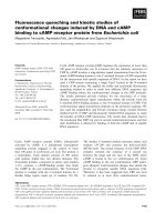
Fig. 4. CD spectra of R2 and R3. CD spectra of R2 (A) and R3 (B) at different ratios of water/trifluoroethanol (TFE) mixture at pH 4.3.
548 K. Minoura et al. (Eur. J. Biochem. 271) Ó FEBS 2004
TFE-induced conformational change
In order to determine, in greater detail, the reason for the
above-mentioned difference, it is important to investigate
the difference in flexibility and conformational features
between R2 and R3 peptides. To estimate their flexibilities,
the CD spectra at pH 4.3 were measured at different ratios
of the water/TFE mixture (Fig. 4); approximately the same
profiles were also observed at pH 7.0. The conformations of
R2 and R3 peptides showed a similar solvent-dependent
behavior, although their ellipticities were considerably
different. Whereas their CD spectra in water predominantly
showed a random conformation characterized by a negative
peak at % 197 nm, the spectra in TFE indicated an a-helical
structure characterized by two negative peaks at % 209 nm
and 222 nm. The conformational transitions started at
% 20% TFE and the a-helical structure content showed a
direct increase in proportion to the TFE concentration. The
conformations were reversibly transformed to the random
structure upon addition of water to the TFE solution and
were scarcely affected by the pH change. No notable time
lag was observed between the reversible conformational
transitions, indicating that the helical conformations of R2
and R3 peptides are both sufficiently flexible to change their
structures, depending on the hydrophobic and hydrophilic
balance of the solvent. On the other hand, the calculation
from the CD ellipticity [17] indicated a meaningful differ-
ence in the a-helical structure content between R2 and
R3 peptides, i.e. R2 ¼ 9.7% and R3 ¼ 7.6% at 0% TFE,
R2 ¼ 12.2% and R3 ¼ 8% at 10% TFE, R2 ¼ 17.6%
and R3 ¼ 12.1% at 20% TFE, R2 ¼ 21.7% and R3 ¼
17.8% at 30% TFE, R2 ¼ 28.2% and R3 ¼ 20.6% at 50%
TFE, and R2 ¼ 53.6% and R3 ¼ 34.2% at 100% TFE.
This shows that it is much easier to induce a conformational
change of R2 than of R3, and also indicates that the
transition energy of R2 is less than that of R3.
Conformation of R2 in TFE solution
The structure of R2 in the TFE solution was analyzed by
both
1
H-NMR spectroscopy and molecular modeling
calculations; we have previously determined the TFE-
induced conformation of R3 by using the same method
[5,6]. Proton peak assignments were performed using a
combination of (a) connectivity information via scalar
coupling in phase-sensitive TOCSY experiments and (b)
sequential NOE networks along the peptide backbone
protons. The diagram of short-, medium- and long-range
proton–proton connectivity along the peptide backbone,
observed by NOESY, is shown in Fig. 5. The orientation
around the Val26–Pro27 x bond was determined to be trans
from the strong NOE of the CaH(Val26)–CdH (Pro27)
proton pair. The NOESY cross-peak pattern among
neighboring protons suggested an a-helical structure of
the Val1–His25 sequence. Using 374 NOE constraints for
proton–proton distances, and 25 J
HNCaH
constraints for /
torsion angles, 100 possible conformers were constructed by
a dynamic simulated annealing calculation. The statistics of
the 20 most stable conformers are summarized in Table 1,
and their superposition on the backbone structure is shown
in Fig. 6. The constructed conformers exhibited a-helical
structures of the Ile3–His25 sequence, whereas the
C-terminal Gly-Gly-Gly-Ser sequence was flexible and did
Fig. 5. Diagram of NOE connectivity between neighboring [d
aN(i, i+1)
,d
NN(i, i+1)
,d
aN(i, i+3)
and d
aN(i, i+3)
]protons.The strength of the observed NOE
is represented by the thickness of respective bars.
Table 1. Structural statistics of 20 stable structures of the R2 domain.
Structural feature Value
Number of structures 20
Number of constraints:
Total number of NOEs 374
Intraresidue NOEs 211
Sequential NOEs 106
Inter-residue NOEs 57
Dihedral angles 25
Average values (esd)
RMS deviation (N, Ca,C¢)(A
˚
) 0.68 (25)
a
RMS deviation from NOE (A
˚
) 0.069 (2)
NOE violations > 0.10 (A
˚
) 11.0 (8)
Energy (kcal/mol)
Overall 315 (6)
NOE 133 (7)
Angle 90 (3)
Bond 25 (1)
Improper 9.0 (8)
van der Waals 58 (4)
a
Calculated from residues 3–20.
Ó FEBS 2004 Different features of repeat fragments in tau MBD (Eur. J. Biochem. 271) 549
not show any definite 3D structure. As the CD spectrum
in the TFE solution suggested an % 50% content of the
a-helical structure, it can be predicted that the R2 peptide
is in equilibrium between equimolar amounts of random
and helical conformers; Fig. 6 corresponds to an ensemble
of the latter conformers.
Conformational comparison between R2 and R3
in TFE solution
As the NMR data reflect an ensemble of various dynamic
conformers, the conformational comparison between R2
and R3 peptides is possible only in terms of the structural
features observed commonly in various NMR-constructed
conformers of each peptide, and the common features of R2
and R3 backbone conformers are schematically shown in
Fig. 7A,B respectively. A similar conformational feature of
R2 and R3 peptides can be described as follows. The Leu10–
Lys20 sequence of R2 forms an a-helical structure, and the
helix wheel drawing of this sequence shows an amphipathic
distribution of the respective amino acid residues (Fig. 8A),
where the hydrophobic residues, Leu10 and Val13, and the
hydrophilic residues, Ser11, Ser15, and Ser19, are arranged
on the both sides of the helix axis, respectively, and the polar
residues, Asn12, Gln14, Lys16, and Lys20, are located at the
interface between both sides. Similar features can also be
observed in the conformation of R3 (Fig. 8B).
The remarkable conformational discrepancy between
the two peptides can be characterized as follows. The
N-terminal Val1–Lys6 sequence of R2 takes a typical
a-helical structure, while that of R3 shows an extended-like
conformation; this structure has also been observed in
aqueous solution [6] and would not be a result of the
presence of the Pro7 residue in R3, because this residue
Fig. 6. Stereoscopic superposition of the most
stable 20 conformers of R2. Each conformer is
projected in order to superimpose on the Ile3–
Lys20 sequence. The upper and lower sides of
conformers correspond to N- and C-terminal
regions, respectively.
Fig. 7. Average backbone conformations. Comparison of averaged
backbone conformations commonly observed in various NMR con-
formers of R2 (A) and R3 (B). The N- and C-terminal regions cor-
respond to the upper and lower sides, respectively.
550 K. Minoura et al. (Eur. J. Biochem. 271) Ó FEBS 2004
takes a trans orientationwithregardtothex torsion angle
bothinwaterandTFE.
This conformational result indicates the following, con-
cerning the difference in time profile between R2 and R3
filament formations (Fig. 2), although the experimental
conditions are different. The extended-like structure of the
N-terminal VQIVYK sequence in R3 is important for
facilitating aggregation without requiring any template for
forming an ordered filament structure, whereas the helical
structure of the N-terminal VQIINK sequence of R2 is
flexible and changes easily into the conformation required
in a particular environmental condition, as judged from the
very slow aggregation and the rapid template-dependent
filament formation.
Concerning the conformation–filament formation rela-
tionship of the MBD, the present work proposes the fol-
lowing possibility, namely, an association through
the helical structures of R2 and R3 repeats and/or the
b-structure-mediated association of the N-terminal
VQIVYK sequence in R3. At present, a unified scheme
has not yet been established concerning the mechanism of
PHF formation, although it has been proposed [18]. The
present TFE-induced helical structures of R2 and R3
cannot be directly associated with the PHF aggregation of
tau MBD under physiological conditions. However, the
speed of three- or four-repeat MBD assembly could be R3-
dependent, because a lack of the R3 domain leads to a
considerable slow down of the assembly process. Also, the
physiological morphology of the MBD filament formation
absorbs almost the entire effect of the nonphysiological one
of the R3 filament. Therefore, it would be reasonable to
consider that the relationship proposed above is likely to
occur in the PHF formation of tau protein. This is also
suggested from the association through the helical structures
with an amphipathic character, which is enthalpy advanta-
geous [6], and the importance of the extended-like VQIVYK
sequence as a core structure for the PHF formation of tau
protein has been proposed [19].
In conclusion, the present study has clarified, for the first
time, the notable difference between the second and third
repeat fragments in the tau MBD in terms of (a) self- and
seeded-aggregations and (b) the conformation induced by
TFE solution. Knowledge of these different conformational
behaviors will be helpful in future investigations undertaken
to clarify the mechanism underlying the MBD assembly of
the tau protein.
Acknowledgements
This work was supported by Grants-in-Aid for Scientific Research
from the Ministry of Education, Culture, Sports, Science and
Technology of Japan, by JSPS Postdoctoral Fellowship for Foreign
Researchers (T M. Y), and by The Science Research Promotion
Fund of The Promotion and Mutual Aid Corporation for Private
Schools of Japan.
References
1. Lee, V.M., Goedert, M. & Trojanowski, J.Q. (2001) Neuro-
degenerative tauopathies. Annu. Rev. Neurosci. 24, 1121–1159.
2. Friedhoff, P., Von Bergen, M., Mandelkow, E M. & Mandelkow,
E. (2000) Structure of tau protein and assembly into paired helical
filaments. Biochim. Biophys. Acta 1502, 122–132.
3. Priedhoff, F., Von Bergen, M., Mandelkow, E M. & Mandelkow,
E. (1998) A nucleated assembly mechanism of Alzheimer paired
helical filaments. Proc. Natl Acad. Sci. USA 95, 15712–15717.
4. Wille,H.,Drewes,G.,Biernat,J.,Mandelkow,E M.&Man-
delkow, E. (1992) Alzheimer-like paired helical filaments and
antiparallel dimers formed from microtubule-associated protein
tau in vitro. J. Cell Biol. 118, 573–584.
5. Minoura, K., Tomoo, K., Ishida, T., Hasegawa, H., Sasaki, M. &
Taniguchi, T. (2002) Amphipathic helical behavior of the third
repeat fragment in the tau microtubule-binding domain, studied
by
1
H-NMR spectroscopy. Biochem. Biophys. Res. Commun. 294,
210–214.
6. Minoura, K., Tomoo, K., Ishida, T., Hasegawa, H., Sasaki, M. &
Taniguchi, T. (2003) Solvent-dependent conformation of the third
repeat fragment in microtubule-binding domain of tau protein,
analyzed by
1
H-NMR spectroscopy and molecular modeling cal-
culation. Bull. Chem. Soc. Jpn 76, 1617–1624.
7. Von Bergen, M., Friedhoff, P., Biernat, J., Heberle, J., Mandel-
kow, E M. & Mandelkow, E. (2000) Assembly of tau protein into
Alzheimer paired helical filaments depends on a local sequence
motif (306) VQIVYK (311) forming beta structure. Proc. Natl
Acad. Sci. USA 97, 5129–5134.
8. Schweers, O., Mandelkow, E M., Biernat, J. & Mandelkow, E.
(1995) Oxidation of cysteine-322 in the repeat domain of micro-
tubule-associated protein tau controls the in vivo assembly of
paired helical filament. Proc.NatlAcad.Sci.USA92, 8463–8467.
9. Bhattacharya, K., Rank, K.B., Evans, D.B. & Sharma, S.K.
(2001) Role of cysteine-291 and cysteine-322 in the polymerization
of human tau into Altzheimer-like filaments. Biochem. Biophys.
Res. Commun. 285, 20–26.
10. Goode, B.L., Chau, M., Denis, P.E. & Feinstein, S.C. (2000)
Structural and functional differences between 3-repeat and
4-repeat tau isoforms. J. Biol. Chem. 275, 38182–38189.
Fig. 8. Helical wheel drawings. Helical wheel
drawings of the Leu10–Leu20 sequences of the
most stable conformers of R2 (A) and R3 (B),
viewed from the N-terminal side.
Ó FEBS 2004 Different features of repeat fragments in tau MBD (Eur. J. Biochem. 271) 551
11. Goedert, M. & Spillantini, M.G. (2000) Tau mutations in fron-
totemporal dementia FTDP-17 and their relevance for Alzhei-
mer’s disease. Biochim. Biophys. Acta 1502, 110–121.
12. Bystrov, V.F. (1976) Spin-spin coupling and the conformational
states of peptide systems. Prog. Nucl. Magn. Reson. Spectrosc. 10,
41–81.
13. Nilges, M., Clore, G.M. & Gronenborn, A.M. (1988) Determi-
nation of three-dimensional structures of proteins from inter-
proton distance data by hybrid distance geometry-dynamical
simulated annealing calculations. FEBS Lett. 229, 317–324.
14. Brunger, A.T., Adams, P.D., Clore, G.M., DeLano, W.L., Gros,
P., Grosse-Kunstleve, R.W., Jiang, J.S., Kuszewski, J., Nilges, M.,
Pannu,N.S.,Read,R.J.,Rice,L.M.,Simonson,T.&Warren,
G.L. (1998) Crystallography and NMR system: a new software
suited for macromolecular structure determination. Acta Crys-
tallogr. D54, 905–921.
15. Koradi, R., Billeter, M. & Wuthrich, K. (1996)
MOLMOL
:apro-
gram for display and analysis of macromolecular structures.
J. Mol. Graphics 14, 51–55.
16. Friedhoff, P., Schneider, A.E.M., Davies, P. & Mandelkow, E.
(1998) Rapid assembly of Alzheimer-like paired helical filaments
from microtubule-associated protein tau monitored by fluores-
cence in solution. Biochemistry 37, 10223–10230.
17. Chen, Y.H., Yang, J.T. & Martinez, H.M. (1972) Determination
of the secondary structures of proteins by circular dichroism and
optical rotatory dispersion. Biochemistry 11, 4120–4131.
18. Barghorn, S. & Mandelkow, E. (2002) Toward a unified scheme
for the aggregation of tau into Alzheimer paired helical filaments.
Biochemistry 41, 14885–14896.
19. Von Bergen, M., Barghorn, S., Li, L., Marx, A., Biernat, J.,
Mandelkow, E M. & Mandelkow, E. (2001) Mutations of tau
protein in frontotemporal dementia promote aggregation of
paired helical filaments by enhancing local b-structure. J. Biol.
Chem. 276, 48165–48174.
552 K. Minoura et al. (Eur. J. Biochem. 271) Ó FEBS 2004