Báo cáo khóa học: Three cyclin-dependent kinases preferentially phosphorylate different parts of the C-terminal domain of the large subunit of RNA polymerase II potx
Bạn đang xem bản rút gọn của tài liệu. Xem và tải ngay bản đầy đủ của tài liệu tại đây (352.33 KB, 11 trang )
Three cyclin-dependent kinases preferentially phosphorylate different
parts of the C-terminal domain of the large subunit of RNA
polymerase II
Reena Pinhero, Peter Liaw, Kimberly Bertens and Krassimir Yankulov
Department of Molecular Biology and Genetics, University of Guelph, Ontario, Canada
The C-terminal domain (CTD) of the largest subunit of
RNA polymerase II plays critical roles in the initiation,
elongation and processing of primary transcripts. These
activities are at least partially regulated by the phosphory-
lation of the CTD by three cyclin-dependent protein kinases
(CDKs), namely CDK7, CDK8 and CDK9. In this study,
we systematically compared the phosphorylation of differ-
ent recombinant CTD substrates by recombinant CDK7/
CycH/MAT1, CDK8/CycC and CDK9/CycT1 kinases. We
showed that CDK7, CDK8 and CDK9 produce different
patterns of phosphorylation of the CTD. CDK7/CycH/
MAT1 generates mostly hyperphosphorylated full-length
and truncated CTD peptides, while CDK8/CycC and
CDK9/CycT1 generate predominantly hypophosphoryl-
ated peptides. Total activity towards different parts of the
CTD also differs between the three kinases; however, these
differences did not correlate with their ability to hyper-
phosphorylate the substrates. The last 10 repeats of the CTD
can act as a suppressor of the activity of the kinases in the
context of longer peptides. Our results indicate that the three
kinases possess different biochemical properties that could
reflect their actions in vivo.
Keywords: carboxy-terminal domain; cyclin-dependent kin-
ase; phosphorylation; RNA pol II.
The C-terminus of the largest subunit of the eukaryotic
RNA polymerase II consists of multiple repeats of a
YSPTSPS consensus heptapeptide sequence [1,2]. This part
of the polypeptide is referred to as CTD (C-terminal
domain). In higher eukaryotes, the CTD consists of 52
heptapeptide repeats [1–3]. The N-terminal portion of the
CTD contains mainly perfect YSPTSPS repeats; however,
the repeats in the C-terminal portion significantly deviate
from the consensus [2–5], probably reflecting a more
specialized function of this part of the polypeptide. It has
been demonstrated that the N-terminal half of the CTD
supports RNA synthesis and capping of the primary
transcript [6–8], whereas the C-terminal half supports
splicing and 3¢ processing of the transcripts [6]. The
importance of the C-terminal ISPDDSDEEN sequence of
the CTD in the regulation of transcript processing has also
been shown [9]. The CTD is phosphorylated at multiple
sites, which leads to the production of two forms of RNA
polymerase II in vivo: a hypophosphorylated form called
IIa, and a hyperphosphorylated form called IIo [1,2,4,5].
It is well established that phosphorylation of the CTD
regulates the transition of RNA polymerase II from
initiation to elongation, the capping of primary transcripts
and the efficiency of pol II elongation [1,2,10]. CTD
phosphorylation has also been implicated in the cotran-
scriptional splicing and polyadenylation of nascent tran-
scripts [1,2,10]. However, little is known about how the
phosphorylation of different parts of the CTD contributes
to these functions.
At least three protein kinases are involved directly in the
phosphorylation of the CTD and in the regulation of
different stages of mRNA synthesis [1,2]. Cyclin dependent
kinase (CDK)7, in conjunction with cyclin (Cyc)H and
MAT1, forms a tripartite complex known as CAK (CDK-
activating kinase); however, a less abundant bipartite form
(CDK7/CycH) has also been observed [11]. At the same
time, CDK7/CycH/MAT1 has been identified as a compo-
nent of the general pol II transcription factor, TFIIH [1,2],
and of large protein complexes containing RNA polymerase
II and general pol II transcription factors that are referred
to as pol II holoenzyme complexes [12]. Another protein
kinase, CDK8/CycC, has also been found in the pol II
holoenzyme [13] and in other MED/SRB containing
complexes such as TRAP/SMCC and NAT [14–17].
TRAP/SMCC and NAT both phosphorylate the CTD
and repress activated, but not basal, transcription [17].
Another study indicates that NAT and TRAP/SMCC
phosphorylate CycH of the TFIIH complex via its CDK8
kinase activity and inhibit TFIIH protein kinase activity
[18]. Studies in Saccharomyces cerevisiae suggestthatthe
Correspondence to K. Yankulov, Department of Molecular Biology
and Genetics, University of Guelph, Guelph, Ontario, Canada,
N1G 2W1. Fax: + 519 837 4120, Tel.: + 519 824 4120 ext. 56466,
E-mail:
Abbreviations: CAK, CDK activating kinase; CDK, cyclin dependent
kinase; CTD, C-terminal domain; Cyc, cyclin; MED, mediator;
GST, glutathione S-transferase; MBP, myelin basic protein;
MOI, multiplicity of infection; NAT, negative regulator of activated
transcription; P-TEFb, positive transcriptional elongation factor b;
SMCC, SRB/MED containing complex; SRB, suppressor of RNA
polymerase B; TRAP, thyroid hormone receptor associated
protein complex.
(Received 18 November 2003, revised 9 January 2004,
accepted 19 January 2004)
Eur. J. Biochem. 271, 1004–1014 (2004) Ó FEBS 2004 doi:10.1111/j.1432-1033.2004.04002.x
CDK8 homolog, Srb10p, phosphorylates the CTD prior to
the formation of an initiation complex at promoters, which
results in the repression of pol II transcription [19]. The third
CTD kinase, CDK9, in complex with one of several
homologous CycT molecules, has been initially identified as
P-TEFb (positive transcription elongation factor-b) [20,21].
Independently, CDK9/CycT1 has been isolated as the HIV-
tat-associated kinase, TAK. It has been reported that the
P-TEFb kinase activity operates in a CAK-independent
manner [22]. Unlike CDK7/CycH/MAT1 and CDK8/
CycC, which are recruited to promoters prior to transcrip-
tion initiation, P-TEFb is recruited to the elongating
polymerase at a later stage of the transcription reaction
[23–26]. The in vitro effects of P-TEFb on elongation cannot
be replaced by TFIIH, thus suggesting that these complexes
perform non-redundant functions [24].
Several studies have attempted to directly compare the
phosphorylation of the CTD or synthetic CTD deriva-
tives by CDK7/CycH/MAT1, CDK8/CycC and CDK9/
CycT1 [25,27–31]. CDK7/CycH/MAT1 and CDK8/CycC
preferentially phosphorylate the S5 residue in the
YSPTSPS repeat [25,27–29,31]. CDK7/CycH/MAT1 might
also phosphorylate some S2 residues in the less conserved
C-terminal portion of the CTD [32]. CDK9/CycT1 seems
to preferentially phosphorylate the S2 residue of the
YSPTSPS repeat on longer CTD substrates [21,25];
however, it can also phosphorylate S5 on short peptide
substrates [28,31]. In addition, CDK9/CycT1 can shift its
preference from S2 to S5 in the presence of the HIV-tat
protein [25].
There is a greater uncertainty as to how these kinases
phosphorylate full-length CTD and parts derived from it.
One study demonstrates that the C-terminal portion of the
CTD is phosphorylated more efficiently by CDK7/CycH/
MAT1 than by CDK8/CycC [27]. This effect is attributed
to the frequent presence of K in position 7 of the heptad
repeats in the C-terminal part of the CTD. Indeed, synthetic
(YSPTSPK)
4
peptides are preferentially phosphorylated by
CDK7/CycH/MAT1 as compared to CDK8/CycC [27].
Another study, using immunoprecipitated CDK7, CDK8
and CDK9, indicates that the three kinases phosphorylate
equally well the N terminus of the CTD (repeats 1–29), but
only CDK7 is able to produce the hyperphosphorylated IIo
form of this substrate [28]. The C terminus (repeats 30–52) is
efficiently phosphorylated by CDK7 only, but the hyper-
phosphorylated IIo form was not produced [28]. The
authors conclude that the hyperphosphorylation, and the
production of the IIo form of pol II and the CTD, is a result
of phosphorylation of the first half of the CTD [28]. On full-
length CTD, the immunoprecipitated CDK7 has a much
higher activity relative to CDK8 and CDK9. Surprisingly,
both CDK7 and CDK9 produced the full-length hyper-
phosphorylated IIo form [28].
In this study, we systematically compared the phosphory-
lation pattern of recombinant CTD substrates by recom-
binant CDK7/CycH/MAT1, CDK8/CycC and CDK9/
CycT1 kinases. We showed that the three kinases do not
dramatically differ in their activity towards the CTD in vitro;
however, they displayed different abilities to hyperphospho-
rylate CTD substrates. Only CDK7/CycH/MAT1 was able
to efficiently hyperphosphorylate the full-length CTD and
produce the IIo form of this substrate. The N- and
C-terminal portions of the CTD were differentially phos-
phorylated by CDK7/CycH/MAT1, CDK8/CycC and
CDK9/CycT1. Finally, we showed data suggesting that
certain CTD repeats in the context of larger polypeptides
can suppress the activities of these kinases.
Materials and methods
Expression vectors
The baculoviruses for the expression of CDK7, MAT1,
CycC and His
6
-CDK9/CycT1 were as described previously
[21,33,34]. The baculovirus for the expression of His-tagged
CycH was produced by subcloning the human CycH into
pBlueBac (Invitrogen) and transfecting Sf9 cells according
to the instructions of the manufacturer. The baculovirus
containing His
6
-CDK8 was produced by subcloning the
human CDK8 into pFASTBACHta and using the BAC-
to-BAC recombination system (Life Technologies). The
plasmids for the expression of glutathione S-transferase
(GST)-CTD(1–52), GST-CTD(1–15, S5>A) and GST-
CDK2 were as described previously [35]. Plasmids for the
expression of GST-CTD(1–15), GST-CTD(1–25), GST-
CTD(27–39), GST-CTD(27–42) and GST-CTD(27–52)
were as described previously [6]. The plasmid for the
expression of GST-CTD(42–52) was prepared by subclon-
ing a PCR fragment, encompassing repeats 42–52, into
pGEX2T (Amersham). GST-CTD(1–52), GST-CTD(27–
52) and GST-CTD(42–52) also contained the C-terminus
ISPDDSDEEN peptide that is positioned next to the 52
heptad repeat in vertebrate RPB1.
Expression and purification of recombinant kinases
Recombinant kinases were expressed by infecting 0.5–1 L of
Sf9 cells (1.5–2 · 10
6
cells per mL) with combinations of
individual baculoviruses at a multiplicity of infection
(MOI) of 5 for 48 h. The cells were harvested by centri-
fugation (275 g,5min)at4°C and lysed in lysis buffer
[10 m
M
Tris/HCl, pH 7.5, 10 m
M
NaCl, 2 m
M
2-merca-
ptoethanol, 0.5 m
M
EDTA, 10 m
M
2-glycerophosphate,
0.5 m
M
sodium vanadate, 2 m
M
NaF, 2 lgÆmL
)1
leupeptin,
2 lgÆmL
)1
aprotonin, 2 lgÆmL
)1
pepstatin, 0.2% (v/v)
Nonidet P-40, 50 lgÆmL
)1
phenylmethanesulfonyl fluoride]
by 10 strokes with the Dounce homogenizer. The proteins
were extracted by adding NaCl to a final concentration of
0.4
M
and then rocking for 30 min. The extract was clarified
by spinning (75 000 g,30min)at4°C in an SW50.1 rotor
(Beckman) and mixed with 1 mL of Ni
2+
nitrilotriacetic
acid–agarose beads (Qiagen) that had been equilibrated
with 10 m
M
Tris/HCl, pH 7.6, containing 0.5
M
NaCl,
5m
M
imidazole, 50 lgÆmL
)1
phenylmethanesulfonyl fluor-
ide, and 10% (v/v) glycerol. The beads were washed in
the equilibration buffer and transferred to a column.
Proteins were eluted in batch by buffers containing
15–400 m
M
imidazole, 10 m
M
Tris/HCl, pH 7.6, 0.1
M
NaCl, 50 lgÆmL
)1
phenylmethanesulfonyl fluoride and
10% (v/v) glycerol. The fractions containing the recombin-
ant protein kinases were pooled and the buffer was
exchangedinPD10columns(Bio-Rad)to25m
M
sodium
Hepes, pH 7.6, 0.1 m
M
EDTA, 1 m
M
dithiothreitol, 5%
(v/v) glycerol. The proteins were then loaded onto a 5 mL
Ó FEBS 2004 Phosphorylation of pol II C-terminal domain (Eur. J. Biochem. 271) 1005
Econo-Pac Mono S cartridge (Bio-Rad) and eluted with a
linear 0.08–0.5
M
NaCl gradient in 25 m
M
sodium Hepes,
pH 7.6, 0.1 m
M
EDTA, 1 m
M
dithiothreitol, 5% (v/v)
glycerol. The fractions containing recombinant protein
kinases were identified by SDS/PAGE followed by silver
staining, pooled and stored at )80 °C. The identity of the
recombinant proteins was confirmed by Western blot with
antibodies against CDK7, CycH, MAT1, CDK8, CycC
and CDK9.
Expression and purification of recombinant substrates
All GST-CTD fusion proteins and GST-CDK2 were
expressed in BL21 cells using 0.5 m
M
isopropyl thio-b-
D
-galactoside (IPTG) for 3 h at 30 °C. Cells were lysed by
sonication in TEN buffer (20 m
M
Tris/HCl, pH 7.5, 5 m
M
EDTA, 200 m
M
NaCl, 1 lgÆmL
)1
aprotonin, 1 lgÆmL
)1
leupeptin, 1 lgÆmL
)1
pepstatin, 2 m
M
benzamidine and
1m
M
phenylmethanesulfonyl fluoride). Triton-X-100 was
added to 1% (v/v) and the extract was rocked for 20 min at
4 °C and then spun at 12 100 g in a JA20 rotor (Beckman)
at 4 °C. The supernatant was loaded onto glutathione–
sepharose 4B beads (Amersham). The bound proteins were
eluted with 15 m
M
glutathione, 50 m
M
KCl, 20 m
M
Tris/
HCl, pH 8.0, 15% (v/v) glycerol, and stored at )80 °C.
Highly purified myelin basic protein (MBP) from bovine
brain was a gift from G. Harauz (University of Guelph).
Kinase assay
Kinase reactions were performed in a 20 lL volume
containing 50 m
M
KCl, 20 m
M
Tris/HCl, pH 8.0, 7 m
M
MgCl
2
,5m
M
2-glycerophosphate, 100 lgÆmL
)1
BSA,
10 l
M
ATP, 2 lCi (7.4 · 10
4
Bq) [
32
P]ATP[cP] (ICN),
40 lgÆmL
)1
recombinant substrate and 100–400 ngÆmL
)1
purified kinase, or the same volume of control fractions
from uninfected Sf9 cells. The kinase reactions were
incubated for 30 min at 30 °C, stopped by the addition of
SDS/PAGE loading buffer, and analyzed on SDS/PAGE
gels and by autoradiography. The separation of substrates
from kinases after the kinase reaction was carried out as
follows. The kinase reaction was stopped by adding 200 lL
of ice-cold STOP buffer [10 m
M
sodium EDTA, pH 8,
50 m
M
KCl, 0.2% (v/v) Nonidet P-40] and incubated with
20 lL of glutathione–sepharose 4B beads. The suspension
was rocked for 20 min, the beads were washed three times
in STOP buffer containing 200 m
M
NaCl, and the bound
proteins were eluted by boiling in SDS/PAGE loading
buffer.
Quantification of levels of phosphorylation
Levels of phosphorylation were measured by scanning
exposed films on a Kodak DS 440CF image station using
the
KODAK
1
D
image analysis software. Relative signals
along each lane in the gels were evaluated by using the grid
option of the data analysis software. Quantification was
conducted only with subsaturated films and only if the
grids did not show saturation (flat) signals. Signals in each
segment of the grid were corrected in Microsoft
EXCEL
by
subtracting the corresponding signals from the identical
segment in the grid from a sample without a substrate.
Intensity curves were prepared in Microsoft
EXCEL
.Total
phosphorylation of each substrate was calculated as the
sum of signals in all segments corresponding to the
substrate bands. Relative phosphorylation of individual
substrates was calculated by measuring the signals from
different substrates on the same X-ray film and normal-
izing them to a postulated value of 1 for the intensity of the
phosphorylation of the GST-CTD(1–52) substrate. Aver-
age relative phosphorylation was calculated from these
values.
The incorporation of ATP in GST-CTD(1–52) and MBP
(pmols of ATP min
)1
Æmg
)1
of protein) was determined
according to the previously published procedure [36].
Results
Expression, purification and characterization
of recombinant CDK7/CycH/MAT1, CDK8/CycC
and CDK9/CycT1
Earlier studies have provided important information on the
substrate preferences of CDK7/CycH/MAT1, CDK8/CycC
and CDK9/CycT1. However, a comprehensive description
of their properties is far from complete. We therefore
attempted a more systematic comparison of the activities of
these kinases towards different substrates. To minimize
variations resulting from different sources of material or
purification procedures, we prepared the three recombinant
kinases following the same expression/purification scheme.
Briefly, CycH, CDK8 and CDK9 were cloned in baculo-
virus vectors as N-terminally 6-Histidine tagged proteins.
CDK7, MAT1, CycC and CycT1 were expressed as
untagged proteins. Sf9 cells were infected with combina-
tions of CDK7/His
6
-CycH/MAT1, His
6
-CDK8/CycC and
His
6
-CDK9/CycT1 baculoviruses. The kinases were subse-
quently purified by immobilized metal-affinity chromato-
graphy (IMAC) using Ni
2+
nitrilotriacetic acid–agarose
and then by ion-exchange chromatography on MonoS
beads. This procedure purified the three kinases to near-
homogeneity, as determined by silver staining (Fig. 1A).
The identities of the CDK7, CycH, MAT1, CDK8, CycC,
and CDK9 bands in Fig. 1A were confirmed by Western
blot (data not shown). All of these preparations displayed
strong kinase activities towards the GST-CTD(1–52) and
MBP (Figs 1B, 2 and 3). Typically, different preparations
of CDK7/CycH/MAT1 and CDK9/CycT1 transferred
between 0.3 and 1.6 nmols of ATP min
)1
Æmg
)1
of protein
with both substrates. The CDK8/CycC preparations
showed somewhat lower specific activities, of 0.09–0.12
nmols of ATP min
)1
Æmg
)1
of protein. Importantly, when
the Ni
2+
nitrilotriacetic acid–agarose/MonoS fractions that
correspond to the fractions with kinase complexes were
isolated from uninfected cells, none showed detectable
kinase activity towards these substrates (results not shown).
We concluded that most, if not all, of the CTD- and MBP-
kinase activity in these preparations belonged to the
expressed kinases.
Next, we tested whether the three kinases would show
substrate specificities that were reported by other groups.
Kinase reactions were performed with MBP, GST-
CDK2(K33>R) and GST-CTD15(S5>A). MBP is a
common non-physiological kinase substrate that is rich in
1006 R. Pinhero et al. (Eur. J. Biochem. 271) Ó FEBS 2004
serine/threonine (17%) and lysine/arginine residues (19%).
GST-CDK2(K33>R) is a catalytically inactive CDK2
molecule [37]. CDK2 is believed to be a physiological sub-
strate of CDK7/CycH/MAT1 [11]. GST-CTD15(S5>A)
contains 15 synthetic consensus YSPTAPS repeats [37].
As expected, all three kinases showed significant activity
towards the generic MBP substrate (Fig. 1B, lanes 2, 6 and
10), transferring between 0.0001 and 0.001 pmols of ATP
per pmol of MBP per min (data not shown). Only CDK7/
CycH/MAT1 phosphorylated the GST-CDK2
(K33>R) substrate (Fig. 1B, lane 3). In agreement with
previous studies [21,25,38], only CDK9/CycT1 phosphor-
ylated the GST-CTD15(S5>A) substrate (Fig. 1B, lane
12), thus stressing the specificity of CDK7/CycH/MAT1
and CDK8/CycC for S5 of the YSPTSPS consensus and the
preference of CDK9/CycT1 for S2. None of the substrates
was phosphorylated in the absence of a kinase (Fig. 1B,
lanes 13–16). We also noticed phosphorylated bands in
the CDK8/CycC and CDK9/CycT1 samples that had the
mobility of CDK8 (Fig. 1B, lanes 5–8) or CDK9, respect-
ively (Fig. 1B, lanes 9–12). These bands probably represen-
ted the autophosphorylation of CDK8 and CDK9 that was
reported previously [21,27,38]. In summary, we established
that our recombinant kinases had properties that were
similar or identical to the ones reported in previous
studies for their native counterparts. We concluded
that further comparison of the recombinant kinases was
justified.
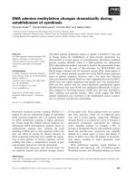
Fig. 1. Characteristics of the recombinant CDK7/CycH/MAT1, CDK8/CycC and CDK9/CycT1 kinases. (A) Samples from pooled Mono S
chromatography fractions containing the three kinases were separated by SDS/PAGE (10% gel) and silver stained. The position of each individual
recombinant polypeptide is shown on the left. The CycH/CDK7 band corresponds to a doublet of CDK7 and His
6
-CycH ( 40 kDa). MAT1 is
36 kDa. The CDK8 corresponds to a molecular mass of 53 kDa and CycC 36 kDa; CDK9 is 43 kDa and CycT1, 81 kDa. (B) Phos-
phorylation of the myelin basic protein (MBP), glutathione S-transferase (GST)-CDK2 and GST-C-terminal domain (CTD)(S5>A)
15
substrates
by CDK7/CycH/MAT1, CDK8/CycC and CDK9/CycT1. Kinase reactions were performed with the combinations of kinase and substrate as
indicated above each lane. The mobility of the substrate polypeptides are indicated on the left. The mobility of 60 and 20 kDa molecular mass
markers are indicated on the right.
Ó FEBS 2004 Phosphorylation of pol II C-terminal domain (Eur. J. Biochem. 271) 1007
CDK7/CycH/MAT1 hyperphosphorylates CTD
Next, we normalized the activity of the three kinases using
MBP and compared their activity towards the full-length
CTD substrate [GST-CTD(1–52)] (Fig. 2A). In these and
all subsequent reactions, we used at least a 100-fold molar
excess of CTD substrates vs. kinase. We did not notice
major differences in the preference of the three kinases
Fig. 2. CDK7/CycH/MAT1, but not CDK8/CycC and CDK9/CycT1
produce a hyperphosphorylated GST-CTD. (A) Kinase reactions were
performed with the combinations of kinase and substrate, as indicated
above each lane and as described in the Materials and methods. (B)
Kinase reactions were performed with a fixed amount (800 ng) of
GST-CTD(1–52) and serial 1 : 3 dilutions of the kinases, as indicated
above each panel of lanes. The mobility of the hypophosphorylated
GST-CTD(1–52)-IIa and the hyperphosphorylated GST-CTD(1–52)-
IIo bands is indicated on the left. (C) Kinase reactions were performed
with GST-CTD(1–52) and the kinases as indicated above each lane.
The samples in the input lanes were loaded without further manipu-
lations. The samples in the GST pull-down lanes were incubated with
GSH-Separose 4B and the bound proteins were eluted from the
washed beads. The mobility of the hypophosphorylated GST-CTD(1–
52)-IIa and the hyperphosphorylated GST-CTD(1–52)-IIo bands are
indicated on the left. The mobility of the 90 kDa molecular mass
markersisindicatedontheright.
Fig. 3. Differential phosphorylation of parts of the C-terminal domain
(CTD) by CDK7/CycH/MAT1, CDK8/CycC and CDK9/CycT1. (A)
Kinase reactions were performed with the combinations of kinase and
substrate, as indicated above each lane. The positions of the unphos-
phorylated substrate polypeptides (IIa) were derived from Coomassie
stained gels without any kinase added (data not shown) and are
marked by the asterisk in each lane. The bars above each lane represent
relative levels of phosphorylation of the substrates. The signals of
phosphorylation of the GST-CTD(1–52) were equalized between the
three different kinases (lanes 2, 9, 16) and the signals of phosphory-
lation of the truncated CTD substrates were plotted relative to GST-
CTD(1–52). The figure is representative of at least three independent
kinase assays with each substrate/kinase combination. (B) The average
ratios of phosphorylation of individual substrates relative to the GST-
CTD(1–52) substrate were calculated and plotted. The bars represent
at least three independent parallel experiments with each substrate and
the three kinases.
1008 R. Pinhero et al. (Eur. J. Biochem. 271) Ó FEBS 2004
towards MBP or GST-CTD(1–52). Therefore, in contrast
to a previous report [28], we do not support the idea that
there was a markedly higher CTD activity in CDK7/CycH/
MAT1 as compared to CDK8/CycC and CDK9/CycT1
(Fig. 2A, lanes 2, 5, 8). However, there was a substantial
difference in the mobility of the phosphorylated GST-
CTD(1–52) species that were generated by the three kinases.
Whereas CDK8/CycC and CDK9/CycT1 produced mostly
the higher mobility (hypophosphorylated) IIa form, CDK7/
CycH/MAT1 produced equal amounts of both the higher-
mobility IIa and lower-mobility (hyperphosphorylated) IIo
forms (Fig. 2A, compare lanes 2, 5 and 8). Most of the
GST-CTD(1–52) retained the mobility of the unphosphory-
lated/hypophosphorylated band, as determined by Coo-
massie staining of the gels after the kinase reactions (data
not shown). In addition, in the reactions with GST-CTD(1–
52), the three kinases transferred between 0.002 and 0.008
pmols of ATP per pmol of GST-CTD(1–52) per min (data
not shown). Thus, assuming only one phosphorylation per
CTD molecule, a maximum of 6–20% of the GST-CTD
molecules could be phosphorylated over the course of the
reaction. It is therefore unlikely that the observed generation
of the IIo band was a consequence of limiting substrate
leading to high levels of phosphorylation. Nevertheless, to
further test the possibility of limiting substrate, we titrated
the kinases, thus reducing the kinase/substrate ratios. As
indicated in Fig. 2B, titration of the kinases over a 24-fold
range did not significantly alter the pattern of phosphory-
lation of the GST-CTD(1–52) substrate. Similarly, extend-
ing the incubation time of the kinase reactions did not
produce a different pattern of phosphorylation of the GST-
CTD (data not shown). Hence, the differential pattern of
CTD phosphorylation does not appear to be solely a
function of the level of kinase activity.
A possible cause for the differential mobility of the GST-
CTD substrate phosphorylated by the three kinases could
be the contamination of the kinase reactions with the
peptidy-prolyl isomerase, Pin1/Ess1 [39–42]. Pin1/Ess1 is
a known modifier of the CTD structure that has been
implicated in pol II transcription and RNA processing
[39–42]. To test the possibility of Pin1/Ess1 involvement, we
performed reactions with GST-CTD and the three kinases
in the presence of the Pin1/Ess1 inhibitor, juglone [41,42].
Because we found no effect of juglone at concentrations up
to 30 l
M
(data not shown), we believe it unlikely that the
effects observed occurred as a result of Pin/Ess1 contami-
nation.
In all preparations of CDK8/CycC and CDK9/CycT1 we
noticed the appearance of phosphorylated bands with
similar mobility to the GST-CTD(1–52)-IIo band (Fig. 2B,
lanes 5–12). These bands could potentially obscure the
detection of the GST-CTD(1–52)-IIo form in the kinase
reactions with CDK8/CycC and CDK9/CycT1. In order to
circumvent this potential problem, we pulled out the GST-
CTD(1–52) substrate molecules after completion of the
kinase reactions and analyzed them separately. Briefly,
kinase reactions were performed as usual and terminated by
the addition of EDTA. Glutathione–sepharose 4B beads
(Amersham) were used to pull out GST-CTD(1–52) and
elute it in SDS sample-loading buffer. In Fig. 2C (lanes
10–12), we clearly show that under conditions of non-
limiting substrate, only CDK7/CycH/MAT1 could produce
substantial amounts of the hyperphosphorylated GST-
CTD(1–52) IIo form.
CDK7/CycH/MAT1, CDK8/CycC and CDK9/CycT1 have
preferences towards different parts of the CTD
In a set of subsequent experiments, we analyzed the activity
of the three kinases towards different parts of the CTD.
CTD heptad repeats 1–15, 1–25, 27–39, 27–42, 27–52 and
42–52 (see Fig. 6) were expressed as GST fusion proteins.
Kinase assays were performed exactly as with the full length
GST-CTD(1–52) substrate. In these analyses, we first
determined the relative levels of phosphorylation (IIa + IIo
signals) of each of these substrates by the three kinases.
The CTD repeats 27–39, 27–42 and 27–52 were definitely
much better substrates for CDK7/CycH/MAT1 than
repeats 1–15 and 1–25 (Fig. 3A, compare lanes 3 and 4
with lanes 5, 6 and 7). The best substrate for CDK7/CycH/
MAT1 appeared to be repeats 27–42 (Fig. 3A, lane 6).
Noticeably, repeats 1–25 and 27–52 were less favored
substrates than the shorter substrates represented by repeats
1–15 and 27–42, respectively (Fig. 3A, compare lanes 3 and
4 with lanes 6 and 7). CDK8/CycC phosphorylated repeats
1–25, 27–39 and 27–42 well (Fig. 3A, lanes 11–13), repeats
1–15 less well (Fig. 3A, lane 10) and repeats 27–52 very
poorly (Fig. 3A, lane 14). CDK9/CycT1 followed a pattern
of total (IIo + IIa) phosphorylation that was similar to
that of CDK8/CycC. However, the phosphorylation of
repeats 27–52 was comparable to that of repeats 1–15
(Fig. 3A, lanes 15–21).
A comparison between the phosphorylation of individual
CTD substrates by the three kinases is shown in Fig. 3B.
CDK7/CycH/MAT1, CDK8/CycC and CDK9/CycT1
phosphorylated repeats 1–15 and 27–39 at comparable
levels. CDK7/CycH/MAT1 consistently showed slightly
higher activity; however, the difference in total phosphory-
lation (IIo + IIa) of these substrates in several independent
experiments was not greater than twofold (Fig. 3B, graphs
a, c). Repeats 1–25 were phosphorylated well by CDK8/
CycC and CDK9/CycT1 and only moderately by CDK7/
CycH/MAT1 (Fig. 3B, graph b). In contrast, repeats 27–42
and 27–52 were much better phosphorylated by CDK7/
CycH/MAT1 than CDK8/CycC and CDK9/CycT1
(Fig. 3B, graphs d, e). In the case of repeats 27–42, these
differences were a result of the remarkably higher activity of
CDK7/CycH/MAT1, while, in the case of repeats 27–52,
the differences were caused by the modest-to-poor activity
of CDK8/CycC and CDK9/CycT1 (Fig. 3A). In summary,
we showed that the three kinases did not phosphorylate
different parts of the CTD equally. Importantly, we outlined
regions that enhance or suppress the activity of each kinase.
Generation of the IIo form by different
parts of the CTD
We noticed substantial variations in the generation of the
hyperphosphorylated IIo form of different CTD substrates
by the three kinases. We decided to assess these variations
by calculating the percentage of the signal in the IIo
substrate bands. In order to do so, we measured the
intensity of the radioactive signal along each lane of the gel
and then subtracted the corresponding signals from the
Ó FEBS 2004 Phosphorylation of pol II C-terminal domain (Eur. J. Biochem. 271) 1009
lanes with samples that contained no substrate. After
preparing a graph of the intensity of the signal from the
substrates only, we calculated the percentage of signal in the
IIo and IIa bands. Thus, assuming that the lower mobility
bands corresponded to the hyperphosphorylated forms of
the substrate, we evaluated the levels of production of
hyperphosphorylated GST-CTD substrates by each kinase.
In order to obtain measurable signals for all substrates and a
comprehensive picture of the generation of the IIo form
along the CTD, we performed kinase assays with the GST-
CTD(1–15) and GST-CTD(27–52) substrates with higher
amounts of the CDK8/CycC and CDK9/CycT1 (Fig. 4A).
The results from these experiments and the calculations
are presented in Fig. 4B. As in the case of the full length
CTD (repeats 1–52), CDK7/CycH/MAT1 was very efficient
in generating slowly migrating bands with all but the
CTD(1–25) substrate (Fig. 3A, lanes 2–7, and Fig. 4B). We
estimated that > 50% of the signal in these reactions was
derived from the IIo-band of the substrates (Fig. 4B). In
sharp contrast, CDK8/CycC did not generate considerable
signals in the IIo band with repeats 1–25, 27–39 and 27–42,
despite the similar levels of phosphorylation with CDK7/
CycH/MAT1 (Fig. 3A, lanes 11–13 and Fig. 4B). CDK9/
CycT1 produced a slightly higher percentage of signal from
the IIo bands in these three substrates, but still the pattern of
phosphorylation was similar to that observed with CDK8/
CycC (Fig. 3A, lanes 18–20 and Fig. 4B). Surprisingly,
CDK8/CycC and CDK9/CycT1 generated an ample per-
centage of signal in the IIo form of the CTD(1–15) and
CTD(27–52) substrates, while total phosphorylation
(IIo + IIa) was lower as compared to the other substrates
(Fig. 3A, lanes 10, 14, 17, 21 and Fig. 4B). In the case of
CDK9/CycT1, the GST-CTD(27–52) substrate generated
42% signal in the IIo band, which is comparable to that
Fig. 4. Generation of hyperphosphorylated
GST-CTD substrates. (A) Kinase reactions
were performed with the combinations of
kinase and substrate, as indicated above each
lane. The positions of the unphosphorylated
substrate polypeptides (IIa) were derived from
Coomassie stained gels without any kinase
added (data not shown) and are marked by the
asterisk in each lane. In order to obtain
measurable signals, the kinase activity in lanes
4–9 was increased threefold as compared to
the experiments in Fig. 3. (B) The intensity of
the phosphorylation signal was determined
along each lane in the gels of Fig. 3A. The
signals from lanes 2–7, lanes 9–13 and lanes
16–21 of the gels in Fig. 3A were plotted after
subtracting the signals from lanes 1, 8 and 15,
respectively. The graph representing the
phosphorylation of GST-CTD(27–52) by
CDK8/CycC was derived from lane 6 of (A),
after subtracting the signal from lane 4. The
asterisk indicates the position of the unphos-
phorylated/hypophosphorylated (IIa) sub-
strate. The percentage of the signal in the IIo
band is shown above each peak.
1010 R. Pinhero et al. (Eur. J. Biochem. 271) Ó FEBS 2004
produced by CDK7/CycH/MAT1 (Fig. 4B). In conclusion,
we observed differential ability of the three kinases to
hyperphosphorylate different parts of the CTD. This ability
did not necessarily correlate to the levels of total phos-
phorylation of these parts.
We also need to mention the mobility of the IIo forms of
the different substrates. The IIo form of the N-terminal 1–15
repeats was only slightly retarded relative to the position of
the unphosphorylated polypeptide (Fig. 3A, lanes 3, 10, 17,
Fig. 4A). In comparison, the IIo forms of the C-terminal
repeats 27–39, 27–42 and 27–52 were dramatically retarded,
independently of the kinase that produced them (Figs 3A
and 4A). The magnitude of mobility shift was not dependent
on the number of repeats. For example, both CTD(1–15)
and CTD(27–42) contain 15 heptad repeats, but their
mobility shift was substantially different (Fig. 3A).
Phosphorylation of GST-CTD(42–52)
CDK8/CycC and CDK9/CycT1 phosphorylated GST-
CTD(27–52) very weakly as compared to GST-CTD(27–
42) (Fig. 3A). These observations suggested that repeats
42–52 are a poor substrate for these kinases and that they
contributed to the overall decrease of the phosphorylation
of GST-CTD(27–52). We tested this possibility by perform-
ing kinase assays with a GST-CTD(42–52) substrate. In
Fig. 5, we show that all three kinases poorly phosphorylated
repeats 42–52 as compared to the full length CTD (Fig. 5,
compare lanes 2, 6 and 10 to 4, 8 and 12, respectively).
CDK7/CycH/MAT1 phosphorylated repeats 42–52 better
than CDK8/CycC and CDK9/CycT1 (Fig. 5, lanes 4, 8,
12), but the overall signal was low. Thus, we obtained a
separate set of data, which indicated that repeats 42–52 are a
poor substrate for the three kinases and that they can
influence the phosphorylation of the C-terminal portion of
the CTD.
Discussion
In this study we performed a systematic comparison of the
activity of CDK7/CycH/MAT1, CDK8/CycC and CDK9/
CycT1 towards recombinant CTD substrates. We expressed
and purified all three recombinant kinases from insect cells
following the same purification strategy. Our CTD sub-
strates were portions of the natural mouse CTD. We
worked under the conditions of non-limiting substrates and
evaluated the relative activities of the kinases and the levels
of production of hypophosphorylated (IIa) and hyperphos-
phorylated (IIo) forms for each of the substrates. This
approach unveiled important differences between the three
kinases that were not noticed in earlier studies [25,27–29,31].
First, we demonstrated that the three recombinant
kinases transferred approximately equal amounts of phos-
phoryl groups to the full-length CTD substrate, yet they
clearly produced different amounts of the hyperphosphory-
lated IIo form (Fig. 2A). CDK7/CycH/MAT1 generated
approximately equal amounts of the hyperphosphorylated
IIo and hypophosphorylated IIa forms, whereas CDK8/
CycC and CDK9/CycT1 produced predominantly the
hypophosphorylated IIa form (Fig. 2). Titration of the
kinases (Fig. 2b) and time-course experiments (data not
shown) indicated that these specific patterns of phosphory-
lation were independent of the kinase/substrate ratio. Our
observations strongly suggest that the three kinases act by
different mechanisms on the pol II CTD substrate.
Because the CTD contains multiple serine residues on the
same polypeptide, it can be phosphorylated in two modes.
In the disruptive mode, the kinase–CTD complex will
uncouple after the transfer of a phosphoryl group to the
CTD, then the kinase will form a complex with another
CTD molecule and phosphorylate it. Under the conditions
of not-limiting substrate, a hypophosphorylated CTD (IIa)
will be predominantly produced. If the kinase forms a
complex with the CTD substrate and phosphorylates
multiple S residues, then it acts by a processive mechanism.
Under the conditions of not-limiting substrate, a hyper-
phosphorylated (IIo) form of the CTD will be produced.
Another way of producing a hyperphosphorylated IIo form
would be if (after the initial phosphorylation) the phos-
phorylated molecules become higher affinity substrates,
leading to a multiple phosphorylation in a disruptive mode.
Our results suggest that the CTD is phosphorylated in the
disruptive mode by CDK8/CycC and CDK9/CycT1.
However, CDK7/CycH/MAT1 operates by both processive
and disruptive modes. Previous studies have shown that
CDK7/CycH/MAT1 acts by the disruptive mechanism on
short (YSPTSPS)
2
substrates [43] and that longer CTD
substrates are much better phosphorylated [27]. Taken
together, these and our observations suggest that the
processive mechanism by CDK7/CycH/MAT1 needs more
than two YSPTSPS repeats.
Second, we demonstrated that different parts of the CTD
are differentially phosphorylated by CDK7/CycH/MAT1.
We showed that this kinase phosphorylates GST-CTD(27–
Fig. 5. Phosphorylation of GST-CTD(42–52). Kinase reactions were
performed with the combinations of kinase and substrate, as indicated
above each lane. The position of the unphosphorylated GST-CTD(42–
52) was derived from Coomassie stained gels without any kinase added
(not shown) and is marked by asterisks.
Ó FEBS 2004 Phosphorylation of pol II C-terminal domain (Eur. J. Biochem. 271) 1011
42) significantly better than GST-CTD(27–39) (Fig. 3A).
This minor extension of three heptad repeats creates a
cluster of YSPTSPK repeats that leads to very high levels
of phosphorylation of the rest of the molecule by CDK7/
CycH/MAT1 (Fig. 3A). Our observation is agreement
with results of a previous study, which showed that a
(YSPTSPK)
4
peptide is a better substrate for CDK7/CycH/
MAT1 than (YSPTSPS)
4
[27]. We therefore propose that
the region encompassing repeats 38–42 is the major site of
CTD phosphorylation by CDK7/CycH/MAT1. At the
same time, we showed that CDK7/CycH/MAT1 weakly
phosphorylates GST-CTD(1–25) relative to the shorter
GST-CTD(1–15) substrate (Fig. 3A). This difference bet-
ween the two substrates applied only to CDK7/CycH/
MAT1. For CDK8/CycC and CDK9/CycT1, the better of
the two substrates was GST-CTD(1–25) (Fig. 3A). Repeats
16–25 contained two YSPTSPN repeats (Fig. 6). It has been
shown that (YSPTSPN)
4
peptides are a less favored
substrate of CDK7/CycH/MAT1 than (YSPTSPS)
4
[27].
We therefore propose that these repeats act as a suppressor
of CDK7/CycH/MAT1 in the context of longer CTD
substrates.
Third, the last 10 C-terminal repeats (42–52) are a very
poor substrate for all three kinases (Fig. 5). The presence of
these repeats in the GST-CTD(27–52) substrate has a
significantly negative effect on the activity of CDK8/CycC
and CDK9/CycT1 and a moderately negative effect on the
activity of CDK7/CycH/MAT1 (Fig. 3A). These results are
consistent with the idea that repeats 42–52 could act as a
kinase suppressor in the context of the full-length CTD. The
moderate effect on the activity of CDK7/CycH/MAT1
(Fig. 3A) could be attributed to the potent positive influence
of the YSPTSPK repeats in 37–42. It is noteworthy that the
42–52 domain contains YSPTSPK repeats that alternate
with an equal number of YSPTSPT repeats (see Fig. 6). In
addition, both GST-CTD(27–52) and GST-CTD(42–52)
contain the C-terminal ISPDDSDEEN sequence that is
missing from GST-CTD(27–42). The importance of this
peculiar alternating of the seventh amino acid in the
C-terminal repeats and the ISPDDSDEEN sequence
remains to be established.
Fourth, we showed that the production of the hyper-
phosphorylated CTD substrates is not necessarily a result
of their total phosphorylation. For example, total phos-
phorylation of GST-CTD(27–39) was approximately equal
between the three kinases (Fig. 3B, column c). However,
CDK7/CycH/MAT1 generated 62% of the signal in the
IIo form, while CDK8/CycC and CDK9/CycT1 generated
2% and 5%, respectively (Fig. 4B, column d). At the
same time, on the longer GST-CTD(27–52) substrate,
CDK9/CycT1 produced 42% in the IIo form, yet total
phosphorylation was very low (Figs 3A and 4B). The
structure of the CTD provides little explanation for the
basis of these differences. Nonetheless, it is clear that
the ability of the kinases to produce hyperphosphorylated
substrates is not related to their overall activity towards
them.
On a minor note, we noticed clear differences in the extent
of retardation of the IIo band in SDS/PAGE between the
C-terminal and the N-terminal parts of the CTD (Figs 3A,
4A and 5). The first 15 CTD repeats only slightly change
their mobility upon hyperphosphorylation, independently
of the phosphorylating kinase (Figs 3A and 4A), while the
other CTD substrates display a dramatic retardation
(Figs 3A, 4A and 5). We therefore suggest that the
phosphorylation of the C-terminus of the CTD is respon-
sible for the generation of the IIo form of pol II in vivo.An
earlier study had reached the opposite conclusion [28]. This
discrepancy might stem from the different substrates used.
Furthermore, we used recombinant kinases, while the other
group used immunoprecipitated CDK7 that might contain
other kinase activities.
Some of our conclusions and observations do not
completely agree with separate pieces of evidence reported
by other groups. Some of the differences can be explained
by the fact that these studies used short synthetic CTD
heptad peptides [25,27–29,31], while we used longer regions
of the natural mouse CTD. For example, synthetic peptides
were used to address the preference towards S2 or S5 and
the effect of the seventh amino acid in YSPTSPS [27–29,31],
but these substrates might have a limited use in assessing the
preferences in the context of the natural CTD. Indeed,
CDK9/CycT1 seems to phosphorylate well S5 of the CTD
heptad consensus on short synthetic peptides [28,31], but it
definitely prefers S2 on full length CTD [25]. In addition, in
some of these studies, high (1 : 3) or unknown enzyme/
substrate ratios were used, thus posing the risk of masking
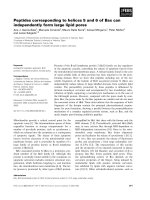
Fig. 6. A model depicting the possible action of the three kinases on the
C-terminal domain (CTD). The amino acid sequence of the mouse
CTD is shown at the bottom. In the diagram, the heptad repeats, with
N at position 7 of the YSPTSPS consensus, are shown as solid rec-
tangles. Heptad repeats with K at position 7 are shown as halftone
rectangles. Heptad repeats with T at position 7 are shown as striated
rectangles. Bent arrows indicate processive phosphorylation. Straight
arrows indicate disruptive phosphorylation. The three kinases seem to
employ the processive mode of phosphorylation at the N-terminus of
the CTD. The YSPTSPN repeats between the 20th and 30th repeats
act as a suppressor of CDK7/CycH/MAT1 and could possibly prevent
the spreading of the phosphorylation by this kinase into the N-ter-
minal portion. The C-terminal subdomain is phosphorylated by the
processive CDK7/CycH/MAT1 kinase via the cluster of YSPTSPKs
around the 40th repeat. At the same time, CDK9/CycT1 is a weak
processive kinase in this region.
1012 R. Pinhero et al. (Eur. J. Biochem. 271) Ó FEBS 2004
the differences in the kinase activity because of limiting
substrates.
The data in this report are summarized in the model
presented in Fig. 6. All three kinases phosphorylate equally
well the N-terminal repeats of the CTD. Even though to
a different extent, all three kinases seem to employ the
processive mode of phosphorylation in this region. The
YSPTSPN repeats between the 20th and 30th repeats act
as a suppressor of CDK7/CycH/MAT1 and may prevent
the spreading of phosphorylation by this kinase into the
C-terminal portion. Thus, the CTD seems to be separated
into two subdomains. The C-terminal subdomain is mainly
phosphorylated by the processive CDK7/CycH/MAT1
kinase via a focal point in the cluster of YSPTSPKs around
the 40th repeat. At the same time, CDK9/CycT1 is a weak
processive kinase in this region. As indicated previously
[44,45], partially phosphorylated CTD is a better substrate
for CDK9/CycT1 than unphosphorylated CTD. It is
therefore possible that the CDK9/CycT1 activity could
significantly increase upon initial phosphorylation of the
CTD by another kinase. It is also possible that the potent
phosphorylation of repeats 38–42 by CDK7/CycH/MAT1
could spread partially into the last 10 repeats, thus triggering
higher levels of processive activity by CDK9/CycT1. Such
an idea is in agreement with the concept that TFIIH, which
contains CDK7/CycH/MAT1, acts early in the transcrip-
tion process [2,46]. P-TEFb, which contains CDK9/CycT1,
acts after TFIIH [2,46].
In vivo, the function of the CTD is influenced by other
modifications, including other phosphorylations, glycosyla-
tion and proline isomerization [1,2,5]. All these modifica-
tions and the corresponding enzymes could have additional
effects on the substrate specificities of CDK7/CycH/MAT1,
CDK8/CycC and CDK9/CycT1. These effects are beyond
the scope of the current study.
The proposed model indicates a probable pattern of
phosphorylation of the CTD by CDK7/CycH/MAT1,
CDK8/CycC and CDK9/CycT1. The physiological signifi-
cance of certain potential sites of phosphorylation has
already been investigated [1,2,10]. However, future studies
are needed to link the described effects to the phosphory-
lation of these sites.
Acknowledgements
We would like to thank D. Morgan, E. Lees and D. Price for providing
baculoviruses and vectors for the expression of the recombinant
kinases; N. Fong and D. Bentley for vectors for the expression of
recombinant CTD substrates; C. Hill, J. Haines and G. Harauz for
MBP; and L. Holland and R. Dziak for comments and advice. This
study was supported by grants to K. Y. from the Natural Sciences and
Engineering Research Council of Canada (NSERC no. 217548) and the
Ontario Genomics Institute (OGI no. 043567). K. B. was supported by
an NSERC studentship.
References
1. Palancade, B. & Bensaude, O. (2003) Investigating RNA poly-
merase II carboxyl-terminal domain (CTD) phosphorylation. Eur.
J. Biochem. 270, 3859–3870.
2. Kobor, M.S. & Greenblatt, J. (2002) Regulation of transcription
elongation by phosphorylation. Biochim. Biophys. Acta 1577,261–
275.
3. Dahmus, M.E. (1996) Reversible phosphorylation of the C-ter-
minal domain of RNA polymerase II. J. Biol. Chem. 271, 19009–
19012.
4. Dahmus, M.E. (1994) The role of multisite phosphorylation in the
regulation of RNA-polymerase-II activity. Prog. Nucleic Acid Res.
Mol. Biol. 48, 143–179.
5. Oelgeschlager, T. (2002) Regulation of RNA polymerase II
activity by CTD phosphorylation and cell cycle control. J. Cell.
Physiol. 190, 160–169.
6. Fong, N. & Bentley, D.L. (2001) Capping, splicing, and 3¢ pro-
cessing are independently stimulated by RNA polymerase II:
different functions for different segments of the CTD. Genes Dev.
15, 1783–1795.
7. Meininghaus, M., Chapman, R.D., Horndasch, M. & Eick, D.
(2000) Conditional expression of RNA polymerase II in mam-
malian cells. Deletion of the carboxyl-terminal domain of the large
subunit affects early steps in transcription. J. Biol. Chem. 275,
24375–24382.
8. Meininghaus, M. & Eick, D. (1999) Requirement of the carboxy-
terminal domain of RNA polymerase II for the transcriptional
activation of chromosomal c-fos and hsp70A genes. FEBS Lett.
446, 173–176.
9. Fong, N., Bird, G., Vigneron, M. & Bentley, D.L. (2003) A 10
residue motif at the C-terminus of the RNA pol II CTD is required
for transcription, splicing and 3¢ end processing. EMBO J. 22,
4274–4282.
10. Bentley, D. (2002) The mRNA assembly line: transcription and
processing machines in the same factory. Curr. Opin. Cell Biol. 14,
336–342.
11. Nigg, E.A. (1996) Cyclin-dependent kinase-7 – at the cross-roads
of transcription, DNA-repair and cell-cycle control. Curr. Opin.
Cell Biol. 8, 312–317.
12. Lee, T.I. & Young, R.A. (2000) Transcription of eukaryotic pro-
tein-coding genes. Annu. Rev. Genet. 34, 77–137.
13. Liao, S.M., Zhang, J., Jeffery, D.A., Koleske, A.J., Thompson,
C.M., Chao, D.M., Viljoen, M., van Vuuren, H.J. &Young, R.A.
(1995) A kinase-cyclin pair in the RNA polymerase II holo-
enzyme. Nature 374, 193–196.
14. Rachez, C., Lemon, B.D., Suldan, Z., Bromleigh, V., Gamble, M.,
Naar, A.M., Erdjument-Bromage, H., Tempst, P. & Freedman,
L.P. (1999) Ligand-dependent transcription activation by nuclear
receptors requires the DRIP complex. Nature 398, 824–828.
15. Malik, S., Gu, W., Wu, W., Qin, J. & Roeder, R.G. (2000) The
USA-derived transcriptional coactivator PC2 is a submodule of
TRAP/SMCC and acts synergistically with other PCs. Mol. Cell 5,
753–760.
16. Wang, G., Cantin, G.T., Stevens, J.L. & Berk, A.J. (2001) Char-
acterization of mediator complexes from HeLa cell nuclear
extract. Mol. Cell. Biol. 21, 4604–4613.
17. Sun, X., Zhang, Y., Cho, H., Rickert, P., Lees, E., Lane, W. &
Reinberg, D. (1998) NAT, a human complex containing Srb
polypeptides that functions as a negative regulator of activated
transcription. Mol. Cell 2, 213–222.
18. Akoulitchev, S., Chuikov, S. & Reinberg, D. (2000) TFIIH is
negatively regulated by cdk8-containing mediator complexes.
Nature 407, 102–106.
19. Hengartner, C.J., Myer, V.E., Liao, S.M., Wilson, C.J., Koh, S.S.
& Young, R.A. (1998) Temporal regulation of RNA polymerase
II by Srb10 and Kin28 cyclin-dependent kinases. Mol. Cell 2,
43–53.
20. Marshal, N. & Price, D. (1995) Purification of P-ETFb, a tran-
scription factor required for transition into productive elongation.
J. Biol. Chem. 270, 12335–12338.
21. Peng, J., Zhu, Y., Milton, J.T. & Price, D.H. (1998) Identification
of multiple cyclin subunits of human P-TEFb. Genes Dev. 12,
755–762.
Ó FEBS 2004 Phosphorylation of pol II C-terminal domain (Eur. J. Biochem. 271) 1013
22. Kim, J.B. & Sharp, P.A. (2001) Positive transcription elongation
factor B phosphorylates hSPT5 and RNA polymerase II carboxyl-
terminal domain independently of cyclin-dependent kinase-acti-
vating kinase. J. Biol. Chem. 276, 12317–12323.
23. Marshall, N.F. & Price, D.H. (1992) Control of formation of two
distinct classes of RNA polymerase II elongation complexes. Mol.
Cell. Biol. 12, 2078–2090.
24. Ping, Y.H. & Rana, T.M. (1999) Tat-associated kinase (P-TEFb):
a component of transcription preinitiation and elongation com-
plexes. J. Biol. Chem. 274, 7399–7404.
25. Zhou, M., Halanski, M.A., Radonovich, M.F., Kashanchi, F.,
Peng, J., Price, D.H. & Brady, J.N. (2000) Tat modifies the activity
of CDK9 to phosphorylate serine 5 of the RNA polymerase II
carboxyl-terminal domain during human immunodeficiency virus
type 1 transcription. Mol. Cell. Biol. 20, 5077–5086.
26. Orphanides, G., Lagrange, T. & Reinberg, D. (1996) The general
transcription factors of RNA polymerase II. Genes Dev. 10, 2657–
2683.
27. Rickert, P., Corden, J.L. & Lees, E. (1999) Cyclin C/CDK8 and
cyclin H/CDK7/p36 are biochemically distinct CTD kinases.
Oncogene 18, 1093–1102.
28. Ramanathan, Y., Rajpara, S.M., Reza, S.M., Lees, E., Shuman,
S., Mathews, M.B. & Pe’ery, T. (2001) Three RNA polymerase II
carboxyl-terminal domain kinases display distinct substrate
preferences. J. Biol. Chem. 276, 10913–10920.
29. Trigon, S., Serizawa, H., Conaway, J.W., Conaway, R.C., Jack-
son, S.P. & Morange, M. (1998) Characterization of the residues
phosphorylated in vitro by different C-terminal domain kinases.
J. Biol. Chem. 273, 6769–6775.
30. Marshall, N.F. & Price, D.H. (1995) Purification of P-TEFb, a
transcription factor required for the transition into productive
elongation. J. Biol. Chem. 270, 12335–12338.
31. Ramanathan, Y., Reza, S.M., Young, T.M., Mathews, M.B. &
Pe’ery, T. (1999) Human and rodent transcription elongation
factor P-TEFb: interactions with human immunodeficiency virus
type 1 tat and carboxy-terminal domain substrate. J. Virol. 73,
5448–5458.
32. Dubois, M.F., Vincent, M., Vigneron, M., Adamczewski, J., Egly,
J.M. & Bensaude, O. (1997) Heat-shock inactivation of the
TFIIH-associated kinase and change in the phosphorylation sites
on the C-terminal domain of RNA polymerase II. Nucleic Acids
Res. 25, 694–700.
33. Fisher,R.,Jin,P.,Chamberlin,H.&Morgan,D.(1995)Alter-
native mechanisms of CAK assembly require an assembly factor
or an activating kinase. Cell 83, 47–58.
34. Rickert, P.S.W., Shanahan, F., Cho, H. & Lees, E. (1996) Cyclin
C/CDK8 is a novel CTD kinase associated with RNA polymerase
II. Oncogene 12, 2631–2640.
35. Yankulov, K.Y. & Bentley, D.L. (1997) Regulation of CDK7
substrate specificity by MAT1 and TFIIH. EMBO J. 16, 1638–
1646.
36. Kikkawa, U., Minakuchi, R., Takai, Y. & Nishizuka, Y. (1983)
Calcium-activated, phospholipid-dependent protein kinase (pro-
tein kinase C) from rat brain. Methods Enzymol. 99, 288–298.
37. Poon, R.Y., Yamashita, K., Adamczewski, J.P., Hunt, T. &
Shuttleworth, J. (1993) The cdc2-related protein p40
MO15
is the
catalytic subunit of a protein kinase that can activate p33
cdk2
and
p34
cdc2
. EMBO J. 12, 3123–3132.
38. Peng, J.M.N. & Price, D.H. (1988) Identification of a cyclin sub-
unit required for the function of Drosophila P-TEFb. J. Biol.
Chem. 273, 13855–13860.
39. Xu, Y.X., Hirose, Y., Zhou, X.Z., Lu, K.P. & Manley, J.L. (2003)
Pin1 modulates the structure and function of human RNA poly-
merase II. Genes Dev. 17, 2765–2776.
40. Lavoie, S.B., Albert, A.L., Handa, H., Vincent, M. & Bensaude,
O. (2001) The peptidyl-prolyl isomerase Pin1 interacts with hSpt5
phosphorylated by Cdk9. J. Mol. Biol. 312, 675–685.
41. Chao, S.H., Greenleaf, A.L. & Price, D.H. (2001) Juglone, an
inhibitor of the peptidyl-prolyl isomerase Pin1, also directly blocks
transcription. Nucleic Acids Res. 29, 767–773.
42. Morris, D.P., Phatnani, H.P. & Greenleaf, A.L. (1999) Phospho-
carboxyl-terminal domain binding and the role of a prolyl iso-
merase in pre-mRNA-3¢-end formation. J. Biol. Chem. 274,
31583–31587.
43. Larochelle, S., Chen, J., Knights, R., Pandur, J., Morcillo, P.,
Erdjument-Bromage, H., Tempst, P., Suter, B. & Fisher, R.P.
(2001) T-loop phosphorylation stabilizes the CDK7-cyclin
H-MAT1 complex in vivo and regulates its CTD kinase activity.
EMBO J. 20, 3749–3759.
44. Marshall,N.F.,Peng,J.,Xie,Z.&Price,D.H.(1996)Controlof
RNA polymerase II elongation potential by a novel carboxyl-
terminal domain kinase. J. Biol. Chem. 271, 27176–27183.
45. Parada, C.A. & Roeder, R.G. (1996) Enhanced processivity of
RNA polymerase II triggered by Tat-induced phosphorylation of
its carboxy-terminal domain. Nature 384, 375–378.
46. Yankulov, K. & Bentley, D. (1998) Transcriptional control: Tat
cofactors and transcriptional elongation. Curr. Biol. 8, R447–
R449.
1014 R. Pinhero et al. (Eur. J. Biochem. 271) Ó FEBS 2004