Báo cáo khoa học: Human salivary a-amylase Trp58 situated at subsite )2 is critical for enzyme activity potx
Bạn đang xem bản rút gọn của tài liệu. Xem và tải ngay bản đầy đủ của tài liệu tại đây (1.02 MB, 13 trang )
Human salivary a-amylase Trp58 situated at subsite )2 is critical
for enzyme activity
Narayanan Ramasubbu
1
, Chandran Ragunath
1
, Prasunkumar J. Mishra
1
, Leonard M. Thomas
2
,
Gyo¨ ngyi Gye
´
ma
´
nt
3
and Lili Kandra
3
1
Department of Oral Biology, University of Medicine and Dentistry of New Jersey, Newark, NJ, USA;
2
Howard Hughes Medical
Institute, Division of Biology, California Institute of Technology, Pasadena, CA, USA;
3
Department of Biochemistry,
Faculty of Sciences, University of Debrecen, Hungary
The nonreducing end of the substrate-binding site of human
salivary a-amylase contains two residues Trp58 and Trp59,
which belong to b2–a2 loop of the catalytic (b/a)
8
barrel.
While Trp59 stacks onto the substrate, the exact role of
Trp58 is unknown. To investigate its role in enzyme activity
the residue Trp58 was mutated to Ala, Leu or Tyr. Kinetic
analysis of the wild-type and mutant enzymes was carried
out with starch and oligosaccharides as substrates. All three
mutants exhibited a reduction in specific activity (150–180-
fold lower than the wild type) with starch as substrate. With
oligosaccharides as substrates, a reduction in k
cat
,anincrease
in K
m
and distinct differences in the cleavage pattern were
observed for the mutants W58A and W58L compared with
the wild type. Glucose was the smallest product generated
by these two mutants in the hydrolysis oligosaccharides;
in contrast, wild-type enzyme generated maltose as the
smallest product. The production of glucose by W58L was
confirmed from both reducing and nonreducing ends of
CNP-labeled oligosaccharide substrates. The mutant W58L
exhibited lower binding affinity at subsites )2, )3and+2
and showed an increase in transglycosylation activity com-
pared with the wild type. The lowered affinity at subsites
)2and)3 due to the mutation was also inferred from the
electron density at these subsites in the structure of W58A in
complex with acarbose-derived pseudooligosaccharide.
Collectively, these results suggest that the residue Trp58
plays a critical role in substrate binding and hydrolytic
activity of human salivary a-amylase.
Keywords: salivary a-amylase; site-directed mutagenesis;
subsite engineering; oligosaccharide hydrolysis; crystal
structure.
a-Amylases (a-1,4-
D
-glucan glucanohydrolases, EC 3.2.1.1)
are endoglucanases, widely distributed in all three domains
of life (Bacteria, Archaea and Eucarya), and catalyze
reactions such as hydrolysis and transglycosylation of
polysaccharides [1,2]. These enzymes, belonging to the
glycoside hydrolase family 13 [3], possess very low overall
sequence similarity among the various members; nonethe-
less, in four small regions around the active site, the
members exhibit a strong sequence similarity [4–6] and
harbor the (b/a)
8
barrel topology [7]. This small number of
conserved but critical short regions whose residues are lined
up along the surface of a deep cleft carries out substrate
binding and catalysis in a-amylases [2].
In humans, a-amylase is present in both salivary and
pancreatic secretions; the overall primary sequences of the
pancreatic and salivary a-amylases are highly homologous,
and exhibit a high level of structural similarity [8,9]. Human
salivary a-amylase (HSAmy) is monomeric, calcium binding
protein with a single polypeptide chain of 496 amino acids
[9]. The structure of HSAmy consists of three domains:
domain A (residues 1–99, 170–404), domain B (residues
100–169) and domain C (residues 405–496). The domain A
adopts a (b/a)
8
barrel structure bearing three catalytic
residues Asp197, Glu233 and Asp300. The domain B occurs
as an excursion from domain A and contains one calcium-
binding site. Domain C forms an all b-structure and
seems to be an independent domain with as yet unknown
function [9].
The active site of HSAmy and mammalian a-amylases
is well established and is present in domain A as a deep
V-shaped cleft [8–13]. The active site of HSAmy is divided
into glycone binding sites ()4, )3, )2, and )1) and aglycone
binding sites (+1, +2 and +3) [13]. These consecutive sites
(whose nomenclature follows the currently accepted nomen-
clature [14]), have been suggested to interact with substrate
glucosyl residues with cleavage occurring between subsites
)1 and +1 [9,15,16]. Enzymatic subsite mapping has been
used to characterize the number of recognized substrate
residues and the individual subsite binding affinity for
HSAmy [17]. Using this method, the high and low affinity
subsites, which control the productive binding modes in
HSAmy has been determined. Among these, the subsite )2
of the glycone binding site and +2 of the aglycone binding
sites possessed the highest affinity [17].
Correspondence to Narayanan Ramasubbu, Department of Oral
Biology, C-634, MSB, UMDNJ, 185 South Orange Ave, Newark,
NJ 07103 USA. Fax: + 1 973 9720705, Tel.: + 1 973 9720704,
E-mail:
Abbreviations: CNP, 2-chloro-4-nitrophenyl; G2, maltose;
G3, maltotriose; G4, maltotetraose; G5, maltopentaose; G6, malto-
hexaose; G7, maltoheptaose; HSAmy, human salivary a-amylase;
MPD, 2-methyl-2,4-pentanediol; PNP, p-nitrophenyl.
Enzyme: a-amylase (a-1,4-
D
-glucan glucanohydrolase) (EC 3.2.1.1).
(Received 9 March 2004, accepted 23 April 2004)
Eur. J. Biochem. 271, 2517–2529 (2004) Ó FEBS 2004 doi:10.1111/j.1432-1033.2004.04182.x
Subsite mapping of the substrate-binding site has also
been reported based on the crystal structure of HSAmy in
complex with a pseudohexasaccharide inhibitor derived
from acarbose [13]. This structure has provided the detailed
stacking and hydrogen bond interactions occurring at
subsites )4 through +2. The glucose moieties occupying
the subsites )1 through )4 are each involved in a number
of interactions with the protein atoms. While subsite )1
interacts with domain A (Arg195, Asp197, Glu233, His299
and Asp300) and domain B residues (His101 and Leu165),
the subsite )4 interacts only with domain B residues
(Asn105, Asp147 and Ser163). These residues are dispersed
in the loops following the strands b2 through b7. In contrast,
subsites )2and)3 interact with residues Trp58, Trp59 and
Gln63 (contained in a loop connecting b2anda2) and
His305 (in mobile loop 304–310). Although the residues
Trp58 and Trp59 are present in a number of a-amylases of
the Eukarya family, there are a few enzymes with Ala at
position 58 and a Tyr at 59 ([18]; follow the links Multi-
alignments and then Eukaryota at mica.
urv.es/pujadas/AAMY/AAMY_01/).
The two aromatic residues, Trp58 and Trp59 interact
with the bound substrate to different extent [8–13]. The
residue Trp59 is involved in a stacking with the 4-amino-4,
6-dideoxy glucose and the glucose moiety at subsites )3and
)2, respectively, and a hydrogen bond interaction to the
glucose moiety at subsite )2. In contrast, Trp58 has no such
stacking interaction with sugar moieties either at subsite )3
or at )2. Interestingly, the residue Trp58 is juxtaposed in
such a way that it interacts with many protein atoms both
in unliganded and in complex structures of HSAmy and
other mammalian a-amylases [8–13]. For instance, hydro-
phobic interactions with residues Trp59, His299 and His305
and a hydrogen bond interaction with Asp356 are dominant
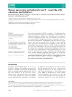
around Trp58 (Fig. 1). As Trp58 is located in the vicinity of
subsite )2 and this subsite had the highest binding affinity
among the glycone subsites [17], we investigated the role
of Trp58 in the activity of HSAmy. For this,
mutants Trp58 fi Ala (W58A), Trp58 fi Leu (W58L)
and Trp58 fi Tyr (W58Y) were generated and their kinetic
properties were compared with wild type using starch and
oligosaccharides (both labeled and unlabeled) as substrates.
The crystal structure analysis of uncomplexed W58L
and acarbose-soaked W58A mutant enzymes were also
determined to analyze the structural differences, if any that
might be used to explain the kinetic behavior of the mutants.
Materials and methods
General procedures
All buffer reagents and other chemicals were obtained from
Sigma Chemical Co. The acarbose was a generous gift from
Bayer. The expression and purification of the recombinant
proteins was carried out as previously described [19]. All
oligonucleotides used in this study were synthetic products
purchased from Integrated DNA Technologies; the oligo-
nucleotide sequences used in this study are given below.
Sequencing was performed at the DNA Sequencing
Resource Center at the Rockefeller University, New York.
Bacterial strain, media and plasmids
Bac-To-Bac Baculovirus Expression System was used to
generate recombinant and mutant proteins using procedures
outlined previously [19,20]. The following forward pri-
mers (5¢-CCTTTCAGACCTXXXTGGGAAAGATAC-
3¢, where XXX ¼ GCG, CTG, and TAC, respectively, for
W58A, W58L and W58Y) and the corresponding reverse
oligonucleotide primers used to create the mutants studied
in this paper. For W59A and W59L, the forward primer
was designed based on W58 mutation except for
the position change (5¢-CCTTTCAGACCTTGGXXXGA
AAGATAC-3¢,whereXXX ¼ GCG, CTG, respectively,
for W59A, and W59L). All primers were used in vector
pFASTBAC1 (Invitrogen) into which HSAmy gene was
cloned [19]. The mutations were verified by nucleotide
sequencing of the HSAmy cDNA using appropriate primer.
The plasmid pFASTBAC1 with mutant HSAmy was used
to transform into MAX EFFICIENCY DH10BAC
TM
Fig. 1. Conformational space occupied by Trp58 in wild-type HSAmy. The Trp58 site of the wild-type HSAmy crystallized with acarbose showing
the interactions involving the Trp residue (PDB Code 1mfv). Note that the side chain of Trp58 enters into a hydrogen bond with the main chain of
Asp356. Note that Asp300 is one of the three catalytic residues. All other contacts are of hydrophobic nature. The distances are given in Angstroms.
All structural figures were drawn using
SETOR
[48].
2518 N. Ramasubbu et al. (Eur. J. Biochem. 271) Ó FEBS 2004
(Invitrogen) cells that contained baculovirus genomic DNA
(bacmid) as well as a helper plasmid. Transformed cells were
plated on Kanamycin, Gentamycin, Tetracycline, Bluo-gal
and IPTG-containing plates. A single white colony was
cultured overnight and the high molecular recombinant
bacmid DNA was isolated and transfected into Sf9 cells
using CELLFECTIN Reagent
TM
(Invitrogen). After 72 h
of incubation at 28 °C in SF900II serum-free medium
(Invitrogen), recombinant baculovirus was harvested from
the medium. Viral stocks were amplified by re-infection
into suspension culture of Sf9 cells at 28 °C with continuous
shaking at a speed of 140 r.p.m.
Protein expression and purification
The proteins were isolated from Sf9 cell culture grown in
1 L of the medium by following protocol previously
established for native HSAmy [19] after observing 100%
cell death. Briefly, cell debris was removed from a 5-day
postinfected medium, adjusted to pH 8.0 with NaOH. After
centrifugation, the supernatant was further clarified by
passing through a 0.45 l
M
filter (Corning Inc.) and the low
molecular weight proteins were removed by ultrafiltration
(Amicon Inc.) using a 30 kDa cut-off spiral cartridge. The
medium was lyophilized and resuspended in 100 m
M
Tris/
HCl, pH. 8.0. Following dialysis against a buffer (5 m
M
Tris/HCl, pH 8.0) containing 2 m
M
CaCl
2
, and centrifuga-
tion, the supernatant was applied to a 3 · 13 cm DEAE-52
cellulose column (Whatman). Bound materials were eluted
from the column as previously described [19]. Fractions
containing recombinant protein were pooled based on
SDS/PAGE [21] and Western blotting and dialyzed against
cold deionized water using Spectra/Por2 (MWCO of
12–14 000 Da; Spectrum Medical Industries, Inc.) and
lyophilized. At this stage, the enriched enzymes were
subjected to a BioGel P60 size exclusion chromatography
following a procedure described previously [22]. After
pooling the fractions containing the desired mutant enzymes
based on Western blotting, enzymes with greater than 99%
purity were obtained at approximately 5 mgÆL
)1
of the
culture medium.
The mass and purity of the enzymes were confirmed by
mass spectral analysis using Perspective Biosystems, a DE
Pro MALDI-TOF instrument equipped with a laser at
337 nm and operated with a positive or negative detection
with 6 kV acceleration potential. Samples were analyzed in
delayed extraction linear mode, calibrated externally with
bovine serum albumin (Sigma Chemical Co.). All spectra
were the result of averaging 200 shots.
Enzyme activity assays
Dinitrosalicylic acid assay was used for measuring the
starch-hydrolyzing activity of HSAmy and mutants at
25 °C for 3 min in 20 m
M
phosphate buffer (pH 6.9)
containing 6 m
M
NaCl using 1% soluble starch as substrate
[23]. Kinetic measurements were carried out using
4-nitrophenyl-a-
D
-maltoheptaoside (G
7
-PNP; Boehringer
Mannheim) and p-nitrophenyl-a-
D
-maltopentaoside (G5-
PNP; Sigma) in a coupled assay with 20 UÆmL
)1
of yeast
a-glucosidase (Boehringer Mannheim). Kinetic parameters
were calculated using the initial velocities (v) obtained from
seven substrate concentrations [S] in the range of 0.078–
5m
M
. The concentration of the wild type was 2 n
M
and
concentration of the mutants W58A, W58Y and W58L was
20 n
M
as determined from molar absorbance at 280 nm
(26.1 for HSAmy) and/or BCA protein assay (Pierce). A
typical reaction was carried out in 100 m
M
HEPES buffer
(pH 7.1) containing 50 m
M
NaCl and 10 m
M
CaCl
2
at
30 °C. All experiments were carried out in triplicate and the
average value is reported.
Hydrolysis of maltooligosaccharides
Assays measuring the products of oligosaccharide hydro-
lysis were carried out using a Varian HPLC (ProStar)
system equipped with a single port manual injector and a
refractive index detector (model number 350). The product
distribution of the hydrolysis of oligosaccharide substrates
by the wild-type and mutant enzymes was determined by
HPLC analyses at a single substrate concentration (0.5 m
M
)
at room temperature. In these experiments, the secondary
attacks on products were avoided by analyzing the reaction
at time points wherein the conversion was < 20%. The
hydrolysates were analyzed using an analytical Dextropak
column (100 · 8 mm) to which a Novapak C18 Guard Pak
precolumn module was attached (Waters). Water was used
as an eluent. Integration of the HPLC profiles was carried
out using Varian Star software (Version 5.51). The
a-anomers of the oligosaccharides (maltotriose through
maltoheptaose) were identified from the retention times of
the products obtained by the hydrolysis of amylose. In a
typical run, a total reaction volume (200 lL) consisted of
either an enzyme concentration of 60 n
M
(HSAmy) or
500 n
M
(W58A, W58Y and W58L), oligosaccharide at
0.5 m
M
(G3 through G7) in water. The reaction mixture
(20 lL) was injected in to the HPLC system after a specific
interval (1–15 min). The product profile was analyzed based
on retention times of standards run under similar conditions
without the addition of the enzyme. The retention times of
the oligosaccharides were also compared using a mixture of
G3 through G7 at 0.5 m
M
each separated using the same
HPLC system. The amount of each product formed was
determined using the area under each peak and converting it
in to molar concentration using values obtained previously
for the standards. These measured data were used to
calculate the action pattern of various HSAmy enzymes for
a given substrate.
Hydrolysis of maltooligosaccharide glycosides
Oligosaccharides labeled with 2-chloro-4-nitrophenyl moi-
ety (CNP) were synthesized from b-cylcodextrin [24].
Incubations of the various CNP-labeled oligosaccharides
in 25 m
M
glycerophosphate buffer (pH 7.0) containing
5m
M
Ca(OAc)
2
and 50 m
M
NaCl were carried out at 37 °C
for 30, 40 and 60 min for W58L. The reactions were
initiated by the addition of enzyme (final concentration of
1.85 n
M
HSA and 18.8 n
M
for the mutant W58L) to the
solution containing 1.0 m
M
of substrate. Samples (20 lL)
were taken at various time intervals and injected into the
chromatographic column. The products were separated on
a Spherisorb ODS2 5 lm column (250 · 4.0 mm) with
acetonitrile–water (13 : 87) as the mobile phase and at a
Ó FEBS 2004 Trp58 mutants at subsite )2 of human salivary a-amylase (Eur. J. Biochem. 271) 2519
flow rate of 1 mLÆmin
)1
at 40 °C using a Hewlett-Packard
1090 Series II liquid chromatograph equipped with a diode
array detector and an automatic sampler. As noted above,
care was taken to exclude the secondary attack on the
products by obtaining the product ratios from the early
stages of hydrolysis wherein the conversion was always
< 10%. The effluent was monitored for CNP-glycosides at
302 nm and the products of the hydrolysis were identified
by using relevant standards and analyzed using ChemSta-
tion software suite. The measured hydrolysis data were used
to calculate the catalytic efficiencies of the enzymes.
Structure determinations
Crystals of the mutants W58L, W58A were grown using
conditions previously described [9,25]. All crystallization
experiments were conducted at room temperature. A
protein concentration of 16 mgÆmL
)1
in 10 m
M
Tris/HCl
(pH 9.0) containing 5 m
M
CaCl
2
was used. The reservoir
solution contained 40% 2-methyl-2,4-pentanediol (MPD)
and the hanging drops consisted of 2 lLofproteinand
2 lL of reservoir solution. Diffraction quality crystals
appeared over a period of one to 4 weeks. To obtain the
complexes with acarbose, these crystals were soaked with
acarbose (1 m
M
final concentration) in 40% MPD for 24 h
and used for data collection. Diffraction data were collected
on a Mar Research imaging plate area detector system
(W58L) or on a Rigaku R-AXIS IV + image plate area
detector (W58A) using Cu K
a
radiation (1.5418 A
˚
) gener-
ated from a Rigaku RU200 rotating anode generator
operating at 50 kV and 100 mA. The crystals were mounted
on loops (Hampton Research) and flash frozen to )170 °C
in liquid nitrogen. One hundred frames were measured with
a1° oscillation to give 98–100% complete data to 2.0 A
˚
(W58L) or 2.1 A
˚
(W58A). The data frames were exposed
for 10 min each. Intensity data were integrated, scaled and
reduced to structure factor amplitudes using HKL suite of
programs [26]. Data collection statistics are given in Table 1.
The unit cell parameters were found to be isomorphous with
those of the wild-type HSAmy [19].
The refinement of these solutions was carried out using
the CNS package [27] wherein cycles of rigid body
refinement, simulated annealing, positional and thermal B
factor refinements were carried out. Bulk solvent corrections
were incorporated in the refinement protocols. A test set
consisting of 5% of reflections was used to monitor the R
free
behaviour. Manual model rebuilding was carried out using
TOM
-
FRODO
[28] and
O
[29]. The complete polypeptide
chains of the mutants were examined with Fo-Fc, 2Fo-Fc
and omit maps. During this process, the mutant enzymes
W58L and W58A clearly showed absence of side chain
density for Trp. The residues were changed to reflect the
respective mutations and for the remainder of the refine-
ment this enzyme was treated as such.
At this stage, clear-cut continuous density corresponding
to the oligosaccharide ligand was observed in the active site
region of only the W58A crystal soaked in acarbose at
subsites )1, +1 and +2. However, no oligosaccharide
atoms were included in the refinement until the refinement
of the protein reached convergence. The identity of the
sugar moieties (either 5-hydroxymethylchonduritol or
4-amino-4,6-dideoxy-a-
D
-glucose or glucose) was deduced
from the presence or absence density for the hydroxyl group
of the side chain at position C5 in the ring [13]. Additionally,
difference density maps calculated by giving zero occupancy
to the O6 atoms were used to assist in the identifications.
The refinements were continued by the inclusion of the
sugar atoms. Further examination of the density maps
revealed no additional binding sites in the complex.
The final rounds of refinement were carried out using
maximum likelihood method as implemented in REF-
MAC-5 of the CCP4 package [30]. Solvent molecules were
added using the arp/warp procedure [31] in the CCP4
package. The validity of the water molecules were assessed
on the basis of the presence of a peak at least 3 r in the
difference map, at least one hydrogen bond to a protein
atom (N or O) or if the water molecules were part of a chain
connecting protein atoms, and refinement of thermal factor
less than 50 A
˚
2
. Manual fitting was interspaced between
refinements when necessary. The programs
PROCHECK
[32],
CCP
4and
CNS
were used for model analysis of the final
refined structures. The coordinates and structure factors
have been deposited with the Protein Data Bank [PDB
codes are 1jxj (W58L) and 1nm9 (W58A complex)].
Table 1. Summary of diffraction data collection values and structure refinement statistics. NA, not applicable.
Parameters W58L W58A–acarbose complex
Space group P2
1
2
1
2
1
P2
1
2
1
2
1
Cell dimensions: a,b,c (A
˚
) 52.3 · 75.2 · 135.0 51.9 · 74.0 · 134.5
Resolution range (A
˚
) 65.9–2.0 42.6–2.1
Total/unique number of reflections 11 613/36 285 188 351/31 104
Completeness (%): overall/last shell
a
98.3/95.0 99.7/99.7
Mean I/rI: overall/last shell 16.2/3.0 19.1/9.4
Multiplicity 3.8 6.0
R
merge
(%) (overall/last shell)
a
6.2/28.1 6.9/23.1
Number of protein/solvent/other atoms 3926/322/0 3926/293/212
Number of reflections used 34 452 29 481
B factor (A
˚
2
): protein/solvent/other 17/23/NA 23/29/29
R/R
free
(%)
b
16.6/19.8 15.5/19.3
r.m.s. deviations: bonds (A
˚
)/angles (°) 0.015/1.6 0.009/1.1
a
Last shell: 2.07/2.0 A
˚
; 2.15–2.10 A
˚
.
b
Reflections in the test set (number/%): 1795/5.0; 1564/5.0.
2520 N. Ramasubbu et al. (Eur. J. Biochem. 271) Ó FEBS 2004
Results
Kinetics studies of mutants
Replacements at position 58 were based on decreasing bulk
(Ala and Leu) or partial retention of aromatic character
(Tyr). All mutants gave as a single band in SDS/PAGE after
final purification and no isozyme corresponding to the
glycosylated a-amylase ( 62 kDa) was observed in either
SDS/PAGE or through mass spectral analysis [19]. The
effect of the mutations on the hydrolysis of starch was
examined by comparing the specific activities for starch
hydrolysis (Table 2). For the mutants W58A, W58L or
W58Y, the specific activity is 150–180-fold lower compared
with the wild-type enzyme. For smaller oligosaccharides
such as a p-nitrophenyl derivative of maltopentaoside (G5-
PNP) and maltoheptaoside (G7-PNP), the k
cat
values were
lower significantly. Interestingly, although the K
m
values for
the mutants were similar to the wild type for the substrate
G7-PNP, the corresponding K
m
values for the substrate G5-
PNP were higher. Thus, for the G5 substrate, there is an
increase (5-fold) in the K
m
and a decrease in k
cat
(30–500-
fold) compared with HSAmy. The k
cat
/K
m
valueforthetwo
mutants W58L and W58A, which have no aromatic ring, is
less than W58Y but similar to that obtained for the D300N
mutant of human pancreatic a-amylase [33]. The k
cat
/K
m
for
the W58Y mutant, albeit lower than the wild type, is 10-fold
higher than either W58L or W58A suggesting that an
aromatic residue at this position might be necessary. In
sharp contrast, the values for the position 59 mutants were
only approximately twofold lower compared with the wild
typeforstarchaswellasG7-PNPassubstrates.Clearly,the
mutation of Trp58 affects the ground state binding of the
substrate and enzyme activity.
Hydrolysis of maltooligosaccharides
Product distributions were determined by HPLC for the
wild type as well as all three mutants with several
oligosaccharides. A typical chromatogram using G4 is
shown in Fig. 2. The substrates were assayed at 0.5 m
M
,
which was used in the standard assay and in previous studies
[13,20]. For each of the mutants, the sites of cleavage for a
given oligosaccharide and the ratio of the products formed
were determined as described previously [20]. The hydrolysis
of each oligosaccharide at a single concentration was
monitored by means of HPLC with an aid of Dextropak
column. The Dextropak column is able to separate the two
anomers of maltooligosaccharides containing three or more
glucose units. The retention times for the a-andb-anomers
of these oligosaccharides were deduced by first determining
the retention time for the a-anomer using HPLC as
described earlier [20]. Briefly, amylose was used as substrate
under similar conditions and the products were separated by
HPLC. The products of hydrolysis of maltooligosaccharides
(G3-G7) were all composed of only a-anomers as HSAmy,
like other a-amylases, is a retaining enzyme. The retention
times of the a-anomers of G3 through G7 thus obtained
were used to identify the b-anomer and its retention time in
the hydrolysis experiment using oligosaccharide substrates.
These values were used then in the analysis of the action
pattern of the HSAmy enzymes. This approach allowed us
to determine the site of cleavage in the various productive
binding modes [12]. The results obtained from this analysis
are shown in Fig. 3 in which the arrows indicate the site
of cleavage and the numbers reflect the percent cleavage
attained at each point.
The analyses were carried out at time points wherein the
substrate consumption was less than 20%. Maltotriose is a
smaller substrate, which is very weakly cleaved by the wild-
type enzyme and W58Y whereas both W58L and W58A
cleaved it into glucose and maltose. The production of
glucose, which was observed in the hydrolysis of higher
oligosaccharides as well, was a characteristic of the mutants
W58A and W58L (Fig. 2A). The amount of glucose
produced was dependent upon the nature of the mutant.
Thus, W58A produced more glucose than W58L, which in
turn produced more than W58Y. In contrast, the wild
type did not produce any detectable glucose for any of the
substrates. Comparison of the productive binding modes
for the wild type with the mutants revealed that the number
of cleavage modes in the mutants is higher than the wild
type for all substrates (Fig. 3). The presence of the aromatic
side chain in the mutant W58Y, results in binding modes
closely resembling the wild type albeit with significantly
lowered k
cat
/K
m
values (Table 2). In contrast, the other two
Table 2. Parameters for the hydrolysis of starch and oligosaccharides. All assays were performed at pH 7.1. Average kinetic errors in kinetic
parameters: specific activity (± 2–5%) for HSAmy and 15–20% for the mutants; K
m
(± 7–10%) and k
cat
(± 5–7%). N.D., not determined.
Enzyme
Hydrolysis of
x-fold decrease in k
cat
/K
m
for G5-PNP substrate
compared with wild-type
enzyme
Starch
specific activity
(UÆmg of protein
)1
)
Heptasaccharide (G7-PNP) Pentasaccharide (G5-PNP)
k
cat
(s
)1
)
K
m
(m
M
)
k
cat
/K
m
(s
)1
Æm
M
)1
)
k
cat
(s
)1
)
K
m
(m
M
)
k
cat
/K
m
(s
)1
Æm
M
)
HSAmy 66 212 175 0.27 648 131 0.35 372 1
W58A 350 0.43 0.39 1.1 0.60 1.70 0.353 1.09 · 10
3
W58L 356 0.43 0.20 2.1 0.26 1.55 0.168 2.31 · 10
3
W58Y 434 0.80 0.31 2.6 4.1 1.63 2.515 0.15 · 10
3
W59A 38 100 111 0.26 427 N.D. N.D. N.D.
W59L 34 415 75 0.16 468 N.D. N.D. N.D.
H305A
a
5016 12 0.28 43 N.D. N.D. N.D. –
a
[37].
Ó FEBS 2004 Trp58 mutants at subsite )2 of human salivary a-amylase (Eur. J. Biochem. 271) 2521
mutants, W58L and W58A, lacking the aromatic residue
exhibit apparently altered binding modes.
Hydrolysis of maltooliogosaccharide glycosides
The production of glucose in the hydrolysis by mutants
might have occurred in two different binding modes, where
the subsites from )4 to +1 were occupied or where the
subsites from )1 to +3 were occupied. If the mutation of
the residue Trp58 affected binding at glycone subsites, use of
labeled substrates might provide additional insights into the
binding modes in these mutants. For this purpose, we used
the mutant W58L and CNP-labeled substrates to determine
unambiguously the exact glycosidic linkage being cleaved
and the cleavage frequency. A sample chromatogram is
given in Fig. 2B and the distribution of the products were
calculated for a given substrate and summarized in Table 3.
The product distribution for the W58L mutant is
different from that of the wild-type HSAmy. When CNP-
G3 was used as the substrate productive binding modes in
which )1 alone is occupied (leading to CNP-G2) or when
)2and)1 are occupied (leading to CNP-G1) occurs with
equal frequency. Three different binding modes are
observed in the hydrolysis of G4 for both wild type and
W58L but with an increase in the production of glucose
from the nonreducing side only in W58L. As seen with the
unlabeled oligosaccharides (Fig. 3), W58L generates: (a)
more products from CNP-labeled oligosaccharides and (b)
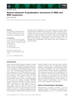
Fig. 2. HPLC analysis of the products of the reactions of the HSAmy and W5L enzymes with G4 oligosaccharide and CNP-G4. (A) HPLC analysis of
the products of the reactions of the HSAmy enzymes with G4 oligosaccharide. Note that oligosaccharides G3 or higher give rise to two peaks
corresponding to the a-anomer (early eluting) or the b-anomer (late eluting). The products were identified using standards and amylose as described
in the Experimental section. Note that glucose is generated by the mutant W58A as well as W58L. (B) HPLC analysis of the products of the reaction
of the W58L enzyme with CNP-G4. As a result of increased transglycosylation activity of this enzyme significant amounts of CNP-G5, CNP-G6
and CNP-G7 are produced. The wild-type enzyme did not show such activity. Note the generation of CNP-G3, which suggests that in this cleavage
mode, W58L generates glucose from the nonreducing end.
2522 N. Ramasubbu et al. (Eur. J. Biochem. 271) Ó FEBS 2004
more CNP-G or CNP-G2 than the wild type. This suggests
that higher population of the productive binding modes, in
which subsite )1 alone or )2and)1 at the nonreducing
ends are occupied, occurs in the mutant W58L than in wild
type. Also, the binding at the subsites )3and)4 might be
affected by the mutation.
The relative rate of formation of each product from the
hydrolysis of a series of oligomeric substrates has been used
to estimate the subsite-binding energy in HSAmy and its
mutants [17]. Using this method the binding affinities for the
four glycone and three aglycone binding sites in the mutant
W58L, with the exception of the two subsites adjacent to the
catalytic site, were calculated using a procedure suggested
by Allen and Thoma [34]. The binding energies for the
subsites )3, )2 and +2 are substantially lower compared
with that of HSAmy (Fig. 3B).
As Trp58 is situated at subsite )2 with the highest affinity
among the glycone binding sites, its mutation affects the
cleavage propensity of individual bonds in maltooligosac-
charides (Fig. 3A; Table 3). Reducing the bulk of the side
chain at position 58 appears to suppress the binding beyond
subsite )2. A reduction of the binding affinities in neigh-
boring subsites is expected for multivalent ligands that bind
in a cooperative manner. Thus, if one binding site shows
reduced affinity, the neighboring binding sites will too,
because the binding sites are not independent of each other.
Because of this reduction in the binding affinity at these
subsites, there is an increase in the productive binding mode
in which glucose from the nonreducing end is generated;
however, this is accompanied by a severe loss in activity
(Table 2).
Structural studies of W58L and W58A
Substitution of a Leu residue for Trp58 causes little
perturbation in the structure (Fig. 4A). The conformation
of the main chain and the orientation of the side chains of
the active site residues are very close to those of the
counterparts in the wild-type enzyme. A notable exception is
the region 304–310, the mobile surface loop, which adopts a
completely different conformation (Fig. 4B). This adapta-
tion of a loop conformation is in accordance with the
absence of substrate in the active site as has been shown
previously in several wild-type a-amylases including
HSAmy [8–10]. However, W58L had clear electron density,
except for His305, for the entire loop (Fig. 4C) unlike wild-
type a-amylase structures that exhibited only weak density
in this region [8–10]. The substitution of the bulkier Trp
with a shorter nonaromatic side chain leads to more open
space in the vicinity of the Leu58 site. However, this void in
the mutant is unoccupied with any water molecules.
Another interesting feature about the W58L structure is
the absence of a hydrogen bond between the catalytic
Asp300 carbonyl oxygen and the nitrogen atom of His305.
Interestingly, this interaction between these two residues has
been suggested to mediate the information flow during
substrate binding and catalysis [13]. The conformation
adopted by the loop structure in W58L is another snapshot
for different possible conformations that can be adopted by
the mobile loop when there is no substrate present.
In the crystal structure of the W58A mutant (Fig. 5A) no
significant deviations in the active site architecture were
Fig. 3. Kinetic analysis of a-amylase enzymes. (A) Comparison of
action pattern in HSAmy and W58 mutants. The enzymes are given in
the order from the top: HSAmy, W58Y, W58L and W58A. The
arrows indicate cleavage positions and the numbers reflect the percent
cleavage observed at each point. Note that the mutants W58A and
W58L, which do not possess an aromatic residue at position 58, have
distinctly different cleavage pattern than either HSAmy or W58Y. (B)
Subsite maps for HSAmy (solid bar) and W58L (open bar). The arrow
indicates the scissile bond. The reducing end of maltooligomers is
situated at the right hand of the subsite map. Negative energy values
indicate binding between the enzyme and aligned glucopyranosyl res-
idues, while positive values indicate repulsion.
Ó FEBS 2004 Trp58 mutants at subsite )2 of human salivary a-amylase (Eur. J. Biochem. 271) 2523
observed when compared with either W58L or HSAmy
except for an altered orientation of the His305 side chain
compared with HSAmy/acarbose complex (v
2
171°,W58A/
acarbose vs. v
2
)114°, HSAmy/acarbose complex). This
altered conformation alone could not account for the
significant reduction in the k
cat
as a mutation of His305 to
Ala reduced the k
cat
by only 15-fold ([35]; Table 2). While
His305 is known to shift its position in the liganded
structures to interact with the bound oligosaccharide
[11,13], in this structure (W58A complex) part of the loop
(residues 305, 306 and 307) is not well defined. This
suggested that the presence of a well-occupied sugar moiety
at subsite )2 might be required for interaction mobilizing
the entire mobile loop.
The other notable feature of the W58A/acarbose complex
that is different from the wild-type/acarbose enzyme is the
way acarbose was modified in the crystal. Unlike the wild-
type/acarbose complex, wherein acarbose was modified into
a hexasaccharide (PDB code 1mfv), only a pseudotrisac-
charide (acarvosine-glucose) was fully occupied in the
complex W58A/acarbose (Fig. 5B). The trisaccharide is
part of the acarbose (acarvosine-glucose) but lacks the
reducing end glucose unit. The three sugar rings of the
bound ligand occupy subsites )1, +1 and +2 in a manner
nearly identical to the same trisaccharide component in the
wild-type enzyme [13]. The subsites corresponding to the
nonreducing end, subsites )4, )3and)2 are not fully
occupied. The size and shape of the difference density can be
interpreted by fitting the acarvosine-glucose moiety, which
is produced by hydrolytic cleavage of acarbose by the
enzyme in the crystal. Alternatively, the observed map could
be due to a longer saccharide formed through a transgly-
cosylation reaction but exhibiting significant positional
disorder at these sites. Modeling sugar units at )4, )3and
)2 subsites resulted in an increase in R
free
as well as very
high thermal parameters for the sugar atoms (<B>> 60
A
˚
2
vs. <B> of 35 A
˚
2
for the subsite )1, +1 and +2
atoms). Refinement with partial occupancy for the atoms at
these subsites also did not improve the model. Therefore,
only water molecules, which satisfied the criteria given in the
Materials and methods section, were modeled into the
disordered density.
The structural analyses reveal that the inhibitor binding
at subsites )1, +1 and +2 has little impact on the
interactions and orientation of the catalytic groups in the
active site. The complex W58A/acarbose displayed a
secondary sugar-binding site on its surface centered on the
residues Trp284 (and Tyr276). This binding site has been
previously observed in the complex structures of wild-type
HSAmy and the mutant D306 lacking the mobile loop
residues 306–310 [13]. Smaller oligosaccharides have been
observed to occupy similar surface sites in several
a-amylases including porcine pancreatic a-amylase [36]
and barley a-amylase [37].
Discussion
Enzymatic properties of Trp58 mutants
The major effect of the mutation appears to be the loss of
the catalytic efficiency as illustrated by the decrease in the
k
cat
and an increase in K
m
for smaller oligosaccharides. This
suggested that the transition state stabilization is hampered
by the removal of the bulky side chain in the middle of the
binding pocket and that interactions around subsites )2and
)3, which control the substrate binding, might be affected.
This is partially supported by results from the subsite
binding affinity using CNP derivatives. The ability of the
mutants W58A and W58L to bind the substrates such as
G5 and G6 in several binding modes, suggests that there is
flexibility of binding around these subsites. The crystal
structure of the W58A complex provides some evidence for
the flexibility in the binding. In this structure, clear electron
density was visible only for subsites )1, +1 and +2
(Fig. 5B). The difference density at the other sites was too
weak to fit additional saccharide residues. The very poor
electron density observed at subsites )4, )3and)2 suggests
that glucose units at these subsites might be highly
positionally disordered. The positional disorder around
subsites )2and)3 has been suggested as a possible reason
for the absence of binding at subsites in the crystal structure
of acarbose-soaked human pancreatic a-amylase mutant
D300N [12].
It is known that a-amylases, can display transglycosy-
lation activity in the crystal in which the cleavage
products are rearranged to form an extended oligosac-
charide species. Several recent crystallographic studies
strongly support that transglycosylation activity occurs in
the crystals of a-amylases [38–40]. During a transglyco-
sylation reaction, the glycosyl covalent intermediate is
Table 3. Action pattern of HSAmy and the W58L mutant.
Substrate Enzyme
Products of hydrolysis (area % of CNP-glycosides)
CNP-G
1
CNP-G
2
CNP-G
3
CNP-G
4
CNP-G
5
CNP-G
6
CNP-G
3
W58L 47 53
HSAmy – –
CNP-G
4
W58L 13 74 13
HSAmy 10 85 5
CNP-G
5
W58L2612710
HSAmy 2 86 12
CNP-G
6
W58L2353028 5
HSAmy 51 44 5
CNP-G
7
W58L1133338132
HSAmy 18 50 30 2
2524 N. Ramasubbu et al. (Eur. J. Biochem. 271) Ó FEBS 2004
attacked by an oligosaccharide moiety instead of water to
lead to a longer sugar chain. We have recently shown
that mutants of HSAmy can be used in synthetic
chemistry for producing oligosaccharides by transglyco-
sylation [41]. Interestingly, while the Trp58 mutant
exhibits such an activity in solution (Fig. 2B), evidence
for such a reaction in the crystal is not very clear due to
positional disorder exhibited by the bound pseudooligo-
saccharide. Thus, although additional density is present
at subsites )4, )3and)2, only a trisaccharide moiety
was modeled in to the active site. It is also possible that
the added acarbose might have been cleaved to generate
a trisaccharide, which accumulates over the soaking
period. Due to the flexibility existing in the binding
pocket, the concentration of the extended pseudooligo-
saccharide is less and hence very low density is observed
for such a higher oligosaccharide in the crystal. Partial
support for this comes from the length of the bound
pseudooligosaccharide observed at the secondary binding
site in the W58A complex. This site also shows clear
evidence for only a trisaccharide. Thus, in the W58A
mutant, the void generated by the absence of Trp at
position 58 might be lead to flexibility in the binding of
longer oligosaccharides even when they are present.
Interestingly, the mutants W58L and W58A cleave G3
while HSAmy does not as the number of nonproductive
binding modes is reduced in these mutants.
Conformational freedom at subsites )2 and )3
in W58A mutant
In the study of barley a-amylase, it was shown recently
that Met53 (equivalent to Gln63 at subsite )2in
HSAmy) was required for wild-type kinetic properties
such as affinity [42]. Inadequate binding at subsite )2
caused distortions at the subsite )1. In the mutants
studied here, such a distortion at subsite )1 may not
occur as subsite )1 is fully occupied. The interactions
around this subsite agree well with interactions observed
around subsite )1 in wild-type complex [13]. However,
local rearrangement of some side chain residues around
Trp58 does occur, most notably in His305 and Lys352.
Two water molecules bridging Asp356 and subsite )2
glucose moiety are also absent (Fig. 6). It should be
pointed out, however, that at the present resolution
(2.1 A
˚
), the mobile loop His305 side chain is not well
resolved. This might be taken to be suggestive of the
inability of the loop to become ordered upon saccharide
binding, a characteristic feature in a-amylases containing
such a mobile loop, as critical subsites )2and)3 are not
occupied. Nonetheless, from the structural and kinetic
data obtained in this study for the W58A/L/Y mutation,
it is clear that the residue Trp58 plays a critical role in
the proper binding of the substrates and thus, for
maintenance of the optimal catalytic activity of HSAmy.
The role of Trp58 in enzyme activity
Several conserved residues, dispersed throughout the
sequence, are juxtaposed around the active site of
a-amylases, some of which have been shown to be
important in the enzyme activity [12,13,20,33,35,43,44].
The potential role of many of these residues in the hydrolytic
activity can be easily surmised from the available crystal
structures of a-amylases in complex with acarbose-derived
Fig. 4. Structure analysis of the HSAmy mutants. (A) Stereodrawing of
the 2Fo-Fc omit maps corresponding to residues 58 and 59 in the
mutant W58L. (B) Superposition of the mobile loop of the active site
region in the HSAmy enzymes HSAmy/acarbose complex (thick lines;
PDB Code 1mfv) and W58L (thin lines). Note in the absence of a
bound oligosaccharide, the residue His305, which is part of a mobile
loop, occupies a different space in W58L and lacks the hydrogen bond
between the His305 nitrogen atom and the Asp300 carbonyl oxygen
atom. (C) Stereodiagram of the 2Fo-Fc omit map corresponding to the
mobile loop (residues 304–310). Unlike the wild-type structure (PDB
Code 1smd), this region of the structure is well defined. In (A) and (C),
the electron density map has been contoured at 1 r and the final
refined coordinates of the corresponding oligosaccharide residues are
overlaid.
Ó FEBS 2004 Trp58 mutants at subsite )2 of human salivary a-amylase (Eur. J. Biochem. 271) 2525
pseudooligosaccharides. The residue Trp58 occurs in a loop
region following the b2strandof(b/a)
8
-barrel fold that has
been suggested to be important from the evolutionary point
of view in a-amylase and several other (b/a)
8
-barrel enzymes
[45]. In spite of this importance, the sequence similarity
around b2-a2 loop region is very thin ([18]; http://www.
quimica.urv.es/pujadas/AAMY/AAMY_01/; follow the
link Multialignments). The length of this loop varies in size
in different a-amylases and contains one invariant residue
Tyr62 that provides a stacking interaction at subsite )1. An
examination of the reported a-amylase structures contain-
ing acarbose-derived pseudooligosaccharides revealed that
noncontiguous residues occupy the space occupied by
Trp58-Trp59 in HSAmy. For example, in TAKA-amylase
[46], residues Arg344 and His80 are located at the positions
occupied by Trp58 and Trp59, respectively). Thus, the
stacking interaction provided by the Trp59 in a-amylases
appears to be compensated by His80 in TAKA-amylase.
However, as a result of the variations in the sequence,
HSAmy and TAKA-amylases bind pseudooligosaccharides
in two orientations (Fig. 7) [13,46]. The conformational
freedom of the substrate, if any, in TAKA-amylase is
restricted probably due to the orientation of two peptide
segments TTAYG(72–76) and GDNTV(167–171) around
subsite )3. Modeling studies showed that the residues Trp58
(and Trp59) of HSAmy will encounter severe steric inter-
actions with the pseudooligosaccharide if the sugar units
occupying subsite )2/)3 adopt alternate conformations as
observed in TAKA-amylase (Fig. 7). Interestingly, the size
of the substrate-binding pocket around the glycone subsites
in a-amylase enzymes that possess Trp58Trp59 segment
appears to be larger. Why mammalian a-amylases and some
other bacterial a-amylases possess a larger substrate-binding
pocket is unclear at present. Nonetheless, these amylases
with Trp58-Trp59 segment also possess a loop segment
GHGGA (residues 304–310 in HSAmy and residues 268–
Fig. 5. Difference electron density maps (omit maps) in the mutants W58L and W58A/acarbose complex. (A) Stereodrawing of the 2Fo-Fc omit maps
corresponding to residues 58 and 59 in the mutant W58L. (B) Stereodrawing of the 2Fo-Fc omitting density maps corresponding to the bound
oligosaccharide in W58A. This complex is made up of a trisaccharide and is named according to subsite location. The electron density map has been
contoured at 1 r and the final refined coordinates of the corresponding oligosaccharide residues are overlaid.
2526 N. Ramasubbu et al. (Eur. J. Biochem. 271) Ó FEBS 2004
272 in psychrophilic a-amylase from Altermonas halopanctis
[47]) containing a His residue that becomes ordered upon
sugar binding [11,13]. Interestingly, Trp58 interacts with
His305 of the mobile loop in HSAmy through a hydro-
phobic interaction (Fig. 1).
Recently, we showed that the mobile loop (residues 304–
310) might be involved in the HSAmy enzyme activity [13].
It is tempting to suggest that the two regions might act in
concert to assist the enzyme during the hydrolytic reaction.
Thus, the residue Trp58 might be involved in one or more of
the following scenarios. First, it might help orient the sugar
chain in the active site by forcing a conformation of the
oligosaccharide to adopt a sharp V-shaped turn. This
enables Trp59 to form a floor onto which subsite )3/)2
glucose moieties stack against [11]. Note that the indole side
chain of Trp58 protrudes into the binding pocket (Fig. 6).
Fig. 6. Stereodiagram depicting the changes in the interactions around position 58 in W58A–acarbose complex (thin lines) as compared with the wild-
type enzyme complex (thick lines). The mutation has resulted in minor changes in the side conformation of residues Lys352 and His305. Clear
density for the side chain of His305 was not observed in the difference density maps (see text).
Fig. 7. Comparison of the conformation of the bound pseudooligosaccharide in human and fungal a-amylases. Superposition of the glycone subsites in
HSAmy (thick lines; PDB Code 1mfv) and TAKA-amylase (thin lines; PDB Code 7taa) along with the bound pseudooligosaccharide. These two
structures were superposed by fitting the spatial location of the three catalytic residues (equivalent to Asp197, Glu233 and Asp300 in HSAmy). The
two bound pseudooligosaccharides (HSAmy vs. TAKA-amylase) traverse two different paths beyond the subsite )2 as shown by the dotted
spheres. Note that Trp58 and Trp59 of HSAmy would encounter severe steric interaction if the bound oligosaccharide in HSAmy adopted a
conformation as in TAKA-amylase.
Ó FEBS 2004 Trp58 mutants at subsite )2 of human salivary a-amylase (Eur. J. Biochem. 271) 2527
Second, it might assist the imidazole side chain of His305 to
be properly juxtaposed to interact with the subsite )2
glucose moiety and also assist in ordering the water
structure around these subsites (Fig. 6). Thus, only in the
presence of Trp58, the mobile loop along with His305
repositions to anchor glucose at )2 (and possibly )3)
susbites at the binding pocket. In this regard, Trp58 might
be involved in the pathway in which information flows
between substrate and the catalytic site through His305 [13].
Third, the proximity of the Trp side chain to the catalytic
carboxyl group Asp300 suggests that some distortion of the
negative electrostatic potential might be occurring at the
catalytic site. To test this, atomic charges on the carboxyl
group atoms of Asp300 were evaluated by semiempirical
methods (MOPAC with AM1 as provided in the SYBYL
software, Tripos Inc.). The atomic charges on the atoms
around position 58 in HSAmy, W58L and W58A were
calculated and compared (data not shown). This prelimin-
ary analysis suggested that the overall negative charge on
the two oxygen atoms of the carboxylate group was higher
in the wild type than in the mutants. The positive charge on
the carbon atom of the carboxylate group remained the
same in all three structures. Such distortion of negative
electrostatic potential has been suggested to be partially
responsible for the very low activity when Met53 located at
subsite )2 was replaced either with Tyr or Trp in barley
a-amylase [42]. Whether such an effect occurs in HSAmy is
beyond the scope of this report. Nonetheless, it should be
pointed out that mutation of Asp300 to Asn in human
pancreatic a-amylase, wherein the charge polarization could
not occur, also leads to the reduction in k
cat
/K
m
values
similar to that obtained for W58A [12,33].
Thus, in HSAmy, and possibly other a-amylases
containing Trp58 and the mobile loop, Trp58 is required
for wild-type kinetic properties especially in the affinity
and in the hydrolysis of maltooligosaccharides. Replace-
ment of this residue with residues containing smaller side
chain resulted in the decrease of enzyme activity. Such a
replacement also misguided the substrate binding in the
glycone subsites )2, )3and)4toanextentthat
interactions around these subsites might be affected.
Additionally, Trp58 might help the catalytic residue
Asp300 to maintain a higher negative electrostatic poten-
tial around the catalytic subsites )1 and +1. The residue
Trp58 and the mobile loop residues, especially His305,
might act in concert in substrate binding and help to
anchor it during hydrolysis.
Acknowledgements
We thank Dr Hong Li of the Biochemistry and Molecular Biology
Department, UMDNJ for the mass spectral data. Part of this work is
based upon the use of the X-ray diffraction facilities in the University of
California at San Diego and California Institute of Technology. This
project was supported by the USPHS Grant DE12585 (NR) and grants
T032005 and M041829 from the Hungarian Scientific Research Fund
(KL).
References
1. Yamamoto, T. (1995) Enzyme Chemistry and Molecular Biology of
Amylases and Related Enzymes. CRC Press Inc, Boca Raton, FL.
2. Janecek, S. (2000) Structural features and evolutionary relation-
ships in the a-amylase family. In Glycoenzymes (Ohnishi, M.,
Hayashi, T., Ishima, S. & Kuriki, T., eds), pp. 19–54. The Japanese
Scientific Societies Press, Tokyo, Japan.
3. Svensson, B. (1994) Protein engineering in the alpha-amylase
family: catalytic mechanism, substrate specificity, and stability.
Plant Mol. Biol. 25, 141–157.
4. Svensson, B. (1988) Regional distant sequence homology between
amylases, a-glucosidases and transglucanosylases. FEBS Lett.
230, 72–76.
5. MacGregor, E.A. & Svensson, B. (1989) A super-secondary
structure predicted to be common to several a-1,4-
D
-glucan-clea-
ving enzymes. Biochem. J. 259, 145–152.
6. Janecek, S. (1994) Sequence similarities and evolutionary rela-
tionships of microbial, plant and animal a-amylases. Eur. J. Bio-
chem. 224, 519–224.
7. Farber, G.K. & Petsko, G.A. (1990) The evolution of alpha/beta
barrel enzymes. Trends Biochem. Sci. 15, 228–234.
8. Brayer, G.D., Luo, Y. & Withers, S.G. (1995) The structure of
human pancreatic alpha-amylase at 1.8 Angstrom resolution and
comparisons with related enzymes. Protein Sci. 4, 1730–1742.
9. Ramasubbu, N., Paloth, V., Luo, Y., Brayer, G.D. & Levine, M.J.
(1996) Structure of human salivary a-amylase at 1.6 Angstrom
resolution: implications for its role in the oral cavity. Acta Crys-
tallogr. D52, 435–446.
10. Qian, M., Haser, R. & Payan, F. (1993) Structure and molecular
model refinement of pig pancreatic a-amylase at 2.1 Angstrom
resolution. J. Mol. Biol. 231, 785–799.
11. Qian, M., Haser, R., Buisson, G., Duee, E. & Payan, F. (1994)
The active center of a mammalian alpha-amylase: structure of the
complex of a pancreatic a-amylase with a carbohydrate inhib-
itor refined to 2.2-Angstrom resolution. Biochemistry 33, 6284–
6294.
12. Brayer,G.D.,Sidhu,G.,Maurus,R.,Rydberg,E.H.,Braun,C.,
Wang, Y., Nguyen, N.T., Overall, C.M. & Withers, S.G. (2000)
Subsite mapping of the human pancreatic alpha-amylase active
site through structural, kinetic, and mutagenesis techniques. Bio-
chemistry 39, 4778–4791.
13. Ramasubbu, N., Ragunath, C. & Mishra, P.J. (2003) Probing the
role of a mobile loop in substrate binding and enzyme activity of
human salivary amylase. J. Mol. Biol. 325, 1061–1076.
14. Davies, G.J., Wilson, K.S. & Henrissat, B. (1997) Nomenclature
for sugar-binding subsites in glycosyl hydrolases. Biochem. J. 321,
557–559.
15. Nagamine, Y., Omichi, K. & Ikenaka, T. (1988) Investigation of
the active site of human salivary alpha-amylase from the modes of
action on modified maltooligosaccharides. J. Biochem. (Tokyo)
104, 409–415.
16. Omichi, K., Hase, S. & Ikenaka, T. (1991) Examination of agly-
cone-binding site of human salivary alpha-amylase by means of
transglycosylation reactions. J. Biochem. (Tokyo) 109, 410–415.
17. Kandra, L., Gyemant, G., Remenyik, J., Ragunath, C. &
Ramasubbu, N. (2003) Subsite mapping of human salivary alpha-
amylase and the mutant Y151M. FEBS Lett. 544, 194–198.
18. Pujadas, G. & Palau, J. (2001) Evolution of alpha-amylases:
architectural features and key residues in the stabilization of the
(beta/alpha) (8) scaffold. Mol. Biol. Evol. 18, 38–54.
19. Ragunath, C., Sundar, K. & Ramasubbu, N. (2002) Expression,
characterization, and biochemical properties of recombinant
human salivary amylase. Protein Exp. Purif. 24, 202–211.
20. Mishra, P.J., Ragunath, C. & Ramasubbu, N. (2002) The
mechanism of salivary amylase hydrolysis: role of residues at
subsite S2¢. Biochem. Biophys. Res. Commun. 292, 468–473.
21. Laemmli, U.K. (1970) Cleavage of structural proteins during the
assembly of the head of bacteriophage T4. Nature 227, 680–685.
2528 N. Ramasubbu et al. (Eur. J. Biochem. 271) Ó FEBS 2004
22. Scannapieco, F.A., Bergey, E.J., Reddy, M.S. & Levine, M.J.
(1989) Characterization of salivary alpha-amylase binding to
Streptococcus sanguis. Infect. Immun. 57, 2853–2863.
23. Bernfeld, P. (1955) Amylases, a and b. Methods Enzymol. 1,
149–159.
24. Farkas, E., Janossy, L., Harangi, J., Kandra, L. & Liptak, A.
(1997) Synthesis of chromogenic substrates of alpha-amylases on a
cyclodextrin basis. Carbohydr. Res. 303, 407–415.
25. Ramasubbu, N., Bhandary, K.K., Scannapieco, F.A. & Levine,
M.J. (1991) Crystallization and preliminary X-ray diffraction
studies of human salivary alpha-amylase. Proteins 11, 230–232.
26. Otwinowski, Z. & Minor, W. (1997) Processing of X-ray crystal-
lographic data in oscillation mode. Methods Enzymol. 276,307–
326.
27. Brunger, A.T., Adams, P.D., Clore, G.M., DeLano, W.L., Gros,
P., Grosse-Kunstleve, R.W., Jiang, J.S., Kuszewski, J., Nilges, M.,
Pannu,N.S.,Read,R.J.,Rice,L.M.,Simonson,T.&Warren,
G.L. (1998) Crystallography & NMR system: a new software suite
for macromolecular structure determination. Acta Cryst. D54,
905–921.
28. Jones, T.A. (1985) Interactive computer graphics: FRODO.
Methods Enzymol. 115, 157–171.
29. Jones, T.A., Zou, J.Y., Cowan, S.W. & Kjeldgaard, M. (1991)
Improved methods for building protein models in electron density
maps and the location of errors in these models. Acta Cryst. A47,
110–119.
30. CCP4. (1994) The CCP4 Suite: programs for protein crystallo-
graphy. Acta Cryst. D50, 760–763.
31. Lamzin, V.S. & Wilson, K.S. (1993) Automated refinement of
protein models. Acta Cryst. D49, 129–149.
32. Laskowski, R.A., MacArthur, M.W., Moss, D.S. & Thornton,
J.M. (1993)
PROCHECK
: a program to check the stereochemical
quality of protein structures. J. Appl. Cryst. 26, 283–291.
33. Rydberg, E.H., Li, C., Maurus, R., Overall, C.M., Brayer, G.D. &
Withers, S.G. (2002) Mechanistic analyses of catalysis in human
pancreatic alpha-amylase: detailed kinetic and structural studies of
mutants of three conserved carboxylic acids. Biochemistry 41,
4492–4502.
34. Allen, J.D. & Thoma, J.A. (1976) Subsite mapping of enzymes.
Application of the depolymerase computer model to two alpha-
amylases. Biochem. J. 159, 121–132.
35. Tseng, C.C., Miyamoto, M., Ramalingam, K., Hemavathy, K.C.,
Levine, M.J. & Ramasubbu, N. (1999) The roles of histidine
residues at the starch-binding site in streptococcal-binding activ-
ities of human salivary amylase. Arch. Oral Biol. 44, 119–127.
36. Qian, M., Haser, R. & Payan, F. (1995) Carbohydrate binding
sites in a pancreatic alpha-amylase-substrate complex, derived
from X-ray structure analysis at 2.1 Angstrom resolution. Protein
Sci. 4, 747–755.
37. Kadziola, A., Sogaard, M., Svensson, B. & Haser, R. (1998)
Molecular structure of a barley alpha-amylase-inhibitor complex:
implications for starch binding and catalysis. J. Mol. Biol. 278,
205–217.
38. Qian,M.,Nahoum,V.,Bonicel,J.,Bischoff,H.,Henrissat,B.&
Payan, F. (2001) Enzyme-catalyzed condensation reaction in a
mammalian alpha-amylase: high-resolution structural analysis of
an enzyme-inhibitor complex. Biochemistry 40, 7700–7709.
39. Aghajari, N., Roth, M. & Haser, R. (2002) Crystallographic evi-
dence of a transglycosylation reaction: ternary complexes of a
psychrophilic alpha-amylase. Biochemistry 41, 4273–4280.
40. Ramasubbu, N., Ragunath, C. & Mishra, P.J. (2003) Probing the
role of a mobile loop in substrate binding and enzyme activity of
human salivary amylase. J. Mol. Biol. 325, 1061–1076.
41. Remenyik, J., Ragunath, C., Ramasubbu, N., Gyemant, G.,
Liptak, A. & Kandra, L. (2003) Introducing transglycosylation
activity into human salivary alpha-amylase (HSA). Org. Lett. 5,
4895–4898.
42. Mori, H., Bak-Jensen, K.S. & Svensson, B. (2002) Barley alpha-
amylase Met53 situated at the high-affinity subsite-2 belongs to a
substrate binding motif in the beta fi alpha loop 2 of the catalytic
(beta/alpha) 8-barrel and is critical for activity and substrate spe-
cificity. Eur. J. Biochem. 269, 5377–5390.
43. Ishikawa, K., Matsui, I., Honda, K. & Nakatani, H. (1992) Multi-
functional roles of a histidine residue in human pancreatic alpha-
amylase. Biochem. Biophys. Res. Commun. 183, 286–291.
44. Rydberg, E.H., Sidhu, G., Vo, H.C., Hewitt, J., Cote, H.C.,
Wang, Y., Numao, S., MacGillivray, R.T., Overall, C.M., Brayer,
G.D. & Withers, S.G. (1999) Cloning, mutagenesis, and structural
analysis of human pancreatic alpha-amylase expressed in Pichia
pastoris. Protein Sci. 8, 635–643.
45. Janecek, S. (1996) Invariant glycines and prolines flanking in loops
the strand beta 2 of various (alpha/beta) 8-barrel enzymes: a
hidden homology? Protein Sci. 5, 1136–1143.
46. Brzozowski, A.M. & Davies, G.J. (1997) Structure of the Asper-
gillus oryzae alpha-amylase complexed with the inhibitor acarbose
at 2.0 Angstrom resolution. Biochemistry 36, 10837–10845.
47. Aghajari, N., Feller, G., Gerday, C. & Haser, R. (1998) Crystal
structures of the psychrophilic alpha-amylase from Alteromonas
haloplanctis in its native form and complexed with an inhibitor.
Protein Sci. 7, 564–572.
48. Evans, S.V. (1993)
SETOR
: hardware-lighted three-dimensional
solid model representations of macromolecules. J. Mol. Graph. 11,
127–128.
Ó FEBS 2004 Trp58 mutants at subsite )2 of human salivary a-amylase (Eur. J. Biochem. 271) 2529