Báo cáo khoa học: Surface exposed amino acid differences between mesophilic and thermophilic phosphoribosyl diphosphate synthase ppt
Bạn đang xem bản rút gọn của tài liệu. Xem và tải ngay bản đầy đủ của tài liệu tại đây (412.81 KB, 8 trang )
Surface exposed amino acid differences between mesophilic and
thermophilic phosphoribosyl diphosphate synthase
Bjarne Hove-Jensen
1
and James N. McGuire
2
1
Department of Biological Chemistry and
2
Center for Enzyme Research, Institute of Molecular Biology, University of Copenhagen,
Denmark
The amino acid sequence of 5-phospho-a-
D
-ribosyl
1-diphosphate synthase from the thermophile Bacillus
caldolyticus is 81% identical to the amino acid sequence of
5-phospho-a-
D
-ribosyl 1-diphosphate synthase from the
mesophile Bacillus subtilis. Nevertheless the enzyme from the
two organisms possesses very different thermal properties.
The B. caldolyticus enzyme has optimal activity at 60–65 °C
and a half-life of 26 min at 65 °C, compared to values of
46 °Cand60sat65°C, respectively, for the B. subtilis
enzyme. Chemical cross-linking shows that both enzymes
are hexamers. V
max
is determined as 440 lmolÆmin
)1
Æmg
protein
)1
and K
m
values for ATP and ribose 5-phosphate
are determined as 310 and 530 l
M
, respectively, for the
B. caldolyticus enzyme. The enzyme requires 50 m
M
P
i
as
well as free Mg
2+
for maximal activity. Manganese ion
substitutes for Mg
2+
, but only at 30% of the activity
obtained with Mg
2+
. ADP and GDP inhibit the B. caldo-
lyticus enzyme in a cooperative fashion with Hill coefficients
of 2.9 for ADP and 2.6 for GDP. K
i
values are determined as
113 and 490 l
M
for ADP and GDP, respectively. At low
concentrations ADP inhibition is linearly competitive with
respect to ATP. A p redicted structure of t he B. caldolyticus
enzyme based on homology m odelling with the structu re of
B. subtilis 5-phospho-a-
D
-ribosyl 1-diphosphate synthase
shows 92% of the amino acid differences to be on solvent
exposed surfaces in the hexameric structure.
Keywords: kinetics; mesophile; nucleotide metabolism;
PRPP; thermophile.
The compound 5-phospho-a-
D
-ribosyl 1-diphosphate
(PRibPP) is a central intermediate in the de novo and salvage
biosynthesis of pyrimidine, purine and pyridine nucleotides
as well as in the biosynthesis of the amino acids histidine
and tryptophan [1,2]. In addition, methanopterin, a folate
analogue involved in C1 metabolism of methanogenic
archaea, is synthesized with PRibPP as an inte rmediate [3].
PRib PP is the s ubstrate for a number of phosphoribosyl-
transferases which catalyse the phosphoribosylation of a
variety of nucleobases to the corresponding ribonucleoside
monophosphates, i.e. the formation of N-glycosidic bonds.
In methanopterin biosynthesis, a carbon–carbon bond is
formed to C1 of the phosphoribosyl moiety of PRibPP [3,4].
Bacterial s pecies like Bacillus subtilis and Escherichia coli
contain 10 enzymes, which utilize PRibPP as a substrate [5].
The s ynthesis of PRibPP is catalysed by PRibPP synthase,
which transfers the b,c-diphosphoryl group of ATP to ribose
5-phosphate (Rib5P) to produce PRibPP and 5¢-AMP [6,7]
(Scheme 1 ). The r eaction proceeds by a ttack of the b-
phosphate by O-1 of Rib5P [7,8]. PRibPP synthase from
E. coli [9,10], Salmonella enterica serovar Typhimurium
[11,12] and B. subtilis [13] requires two Mg
2+
per subunit
and a P
i
concentration of 50 m
M
. S. enterica and E. coli
PRib PP synthases bind A TP (as MgÆATP) before Rib5 P.
The E. coli enzyme furthermore b inds free Mg
2+
before
binding MgÆATP in the catalytic cycle [14]. Regulation of the
activity of PRibPP synthase is achieved primarily thro ugh
the inhibition by ADP or GDP. It has been shown that ADP
inhibits the enzyme by binding to the allosteric site in
competition with P
i
as well as by competing w ith ATP for the
active site [9,15,16]. GDP also inhibits PRibPP synthases
from Gram-negative b acteria and mammals, but to a lesser
extent and by binding at the allosteric site [13,17]. PRibPP
synthase is active as a homomultimer with oligomerization
states ranging from hexamer to higher st ates of aggregation
depending on the detection method and the source of
organism [18]. In the present work we describe the charac-
terization of PRibPP synthase, which is encoded by the
prs gene, from the thermophile Bacillus caldolyticus and
compare i t with t he enzyme from the mesophile B. subtilis.
Experimental procedures
Materials
Ribonucleotides were obtained from Pharmacia (Uppsala,
Sweden), Sigma (St. Louis, MO, USA) or Roche (Mann-
Correspondence to B. Hove-Jensen, Department of Biological Chem-
istry, Institute of Molecular Biology, University of Copenhagen, 83H
Sølvgade, DK-1307 Copenhagen K, Denmark. Fax: +45 3532 2040,
Tel.: +45 3532 2027, E- m ail: hov
Abbreviations: PRibPP, 5-phospho-a-
D
-ribosyl 1-diphosphate;
Rib5P, ribose 5-phosphate.
Enzyme: 5 -phospho-a-
D
-ribosyl 1-di phosphate synthase or A TP:
D
-ribose-5-phosphate p yrophosphotransferase ( EC 2. 7.6.1).
Note: A department website i s available at
Note: Dedicated to the memory of the late Professor Agnete M unch-
Petersen, a fine colleagu e and a great mentor.
(Received 4 August 2004, rev ised 17 September 2004,
accepted 4 October 2004)
Eur. J. Biochem. 271, 4526–4533 (2004) Ó FEBS 2004 doi:10.1111/j.1432-1033.2004.04412.x
heim, Germany). Antibiotics, isopropyl thio-b-
D
-galactoside
and EGTA were obtained from Sigma. Restriction endo-
nucleases were obtained from Promega (Madison, WI,
USA). Oligodeoxyribonucleotides were purchased from
DNA Technology (A
˚
rhus, Denmark) or Hobolth DNA
Syntese (Hillerød, Denmark). FPLC was performed using a
Bio-Rad Bio Logic system with UV detection at 280 nm.
Polyethyleneimine-cellulose coated TLC sheets were from
Baker-flex (J. T. Baker, Phillipsburg, NJ, USA).
Cloning and expression of the
B. caldolyticus prs
gene
The prs gene was s ynthesized by PCR w ith pHO219 DNA
[19] as the template, the oligodeoxyribonucleotides 5¢-AA
GAAA
GAATTC-TAGCGGAGGTCTATCATG-3¢ and
5¢-ATGTTT
AAGCTTA-TTAGTCGAACAGGACGCT-3¢
as primers and DNA polymerase f rom Pyrococcus furiosus
in the presence of the four deoxyribonucleoside triphos-
phates. The nucleotides preceding hyphens indicate non-
complementary extensions and recognition sites for the
restriction endonuclease Eco RI and Hin dIII are underlined.
Standard procedures were used for thermocycling in a T rio-
Thermoblock (Biometra, Go
¨
ttingen, Germany). T he PCR
product w as digested by Eco RI and HindIII, a nd ligated to
EcoRI an d Hin dIII digested DNA of the expression vector
pUHE23-2 [H. Bujard, University of Heidelberg, Germany,
personal communication]. The nucleotide sequence of the
insert of the r esulting plasmid, pJM1, was determined in an
Abi Prism Genetic Analyser (model 310) with the Bigdye
Terminator Cycle S equencing Ready Reaction Kit as
recommended by the supplier (PE Applied Biosystems,
Foster City, CA, USA).
Purification of recombinant
P
Rib
PP
synthase
of
B. caldolyticus
and
B. subtilis
The plasmid pJM1 was t ransformed i nto the PRibPP-
less E. coli strain HO1986 (Dprs-4::Kan
R
araC
am
araD
D(lac)U169 trp
am
mal
am
rpsL relA thi deoD gsk-3 udp supF
ÔFÕ
R
/F lacI
q
zzf::Tn10), which contains no endogenous
PRib PP synthase activity. HO1986 is a deriva tive of strain
HO1088 [20] and was kindly p rovided b y B . N . K rath (t his
institute). It is resistant to an unspecified nonlambdoid, non-
P type bacteriophage. Cultures of s train HO1986/pJM1 were
grown at 37 °C to an attenuance at 436 nm of 1.2–1.5
( 3 · 10
11
cellsÆL
)1
), measured in an Eppendorf 6121
spectrophotometer. At t his time isopropyl thio-b-
D
-galacto-
side was added to a final concentration of 50 l
M
,and
incubation continued for 16 h. Unless otherwise s tated the
following steps were performed at 4 °C. Cells were harvested
by centrifugation at 20 000 g for 2 0 min. Collected cells were
resuspended in five volumes of 50 m
M
potassium phosphate
buffer (pH 7.5), and sonicated f or 20 min (60 s bursts with
60 s pauses) followed by centrifugation at 20 000 g for
15 min. The supernatant fluid was 40 % saturated with
ammonium sulphate. The precipitate was removed by
centrifugation, and the supernatant fluid was 60% saturated
with ammonium sulphate. The precipitate, collected by
centrifugation, was redissolved in 50 m
M
potassium phos-
phate buffer (pH 7.5) in half the original volume and
dialyzed for 16 h against 2 L of 50 m
M
potassium phosphate
buffer (pH 8.2). The dialysed enzyme preparation was
applied to a Dyematrex Gel Green A column (Millipore,
Bedford, MA, USA), and washed with five volumes of
50 m
M
potassium phosphate buffer (pH 8.2). Protein was
eluted by u sing a linear gradient o ver s ix column volumes
from 50 m
M
potassium phosphate buffer (pH 8.2) to 50 m
M
potassium phosphate, 300 m
M
potassium chloride (pH 8.2).
PRib PP synthase activity eluted as two major peaks, which
were pooled, dialyzed against 50 m
M
potassium phosphate
buffer (pH 8.2), reapplied to the same column, and eluted
under the same conditions as before. The larger of two
activity peaks (fraction A) was further dialysed against
50 m
M
potassium phosphate buffer (pH 8.2) eluted isocrat-
ically through a Pharmacia Superose 12 10/30 gel filtration
column using an FPLC instrument at room temperature.
PRib PP synthase activity eluted as three or four peaks. The
largest was chosen for further study. The final enzyme
fraction was greater than 95% pure as determined by SDS/
PAGE and staining i n Coomassie Brilliant Blue. T he enzyme
was s tored i n 5 0% glycerol in aliquots at )80 °C.
Recombinant B. subtilis PRibPP synthase was isolated
from cells overexpressing the prs gene essentially as
described previously [21] with a modification of the final
anion exchange step as follows. The enzyme, dissolved in
50 m
M
potassium phosphate buffer (pH 7.5) was a pp lied to
a 20 mL anion exchange Hiload Q-Sepharose column
(Pharmacia), previously equilibrated with t he same buffer.
PRib PP synthase was e luted b y applying a s alt g radient of
0% Salt Buffer [50 m
M
potassium phosphate buffer
(pH 7 .5)] to 100% Salt Buffer [1
M
sodium chloride in
50 m
M
potassium phosphate buffer (pH 7.5)] at a rate of
2mLÆmin
)1
over 60 min. The gradient w as an initial linear
increase from 0 t o 20% Salt Buffer, f ollowed by a hold for
40 mL and an increase to 35% Salt Buffer over approxi-
mately 120 mL and finally a raise to 100% Salt Buffer.
PRib PP synthase eluted at a sodium chloride concentration
of approximately 0.30
M
. The fractions with highest purity
evaluatedbyassayofPRibPP synthase activity and by
SDS/PAGE were pooled and dialyzed against 50 m
M
potassium phosphate buffer (pH 7.5). The enzyme was
stored refrigerated [22].
Protein content was determined by the bicinchoninic acid
procedure (Pierce Chemical Company, Rockford, IL, USA)
as described previously with BSA as the standard [23].
MALDI-TOF mass spectrometry analysis was performed
by the School of Chemical Sciences Mass Spectrometry
Center, University of Illinois, Urbana-Champaign, IL,
Scheme 1. Reaction catal y sed by PRib PP.
Ó FEBS 2004 Bacillus caldolyticus PRibPP synthase (Eur. J. Biochem. 271) 4527
USA. Amino acid sequencing by automated Edman
degradation was performed by the Department of Protein
Chemistry, Institute of Molecular Biology, University of
Copenhagen, Denmark.
Assay of
P
Rib
PP
synthase activity
The standard reaction buffer consisted o f 50 m
M
Tris/HCl,
50 m
M
potassium phosphate, 2.0 m
M
EGTA (pH 8.5,
adjusted at 65 °C). The standard reaction contained
2.0 m
M
(10 G BqÆmol
)1
)[
32
P]ATP[cP] (prepared a s des-
cribed previously [24]), 5.0 m
M
Rib5P,5.0m
M
magnesium
chloride. Unless otherwise indicated the Mg
2+
concentra-
tion w as 3 .0 m
M
in excess of the r ibonucleoside t riphos-
phate concentration. In analyses of inhibition by ADP and
in determination of K
m
for ATP and R ib5P, a buffer
without EGTA was used. For the P
i
or sulphate dependence
analysis, the en zyme was d iluted in 5 0 m
M
Tris/HCl buffer
(pH 8 .2) containing BSA (2 gÆL
)1
) without prior dialysis.
The reaction bu ffer for these studies was 50 m
M
Tris/HCl
(pH 8 .5, adjusted at 65 °C). In all cases, the assay buffer
with ATP, Rib5P and magnesium chloride present was
prewarmed for 2 m in at the desired temperature and
reaction initiated by the addition of enzyme. The enzyme
had b een previously diluted in 50 m
M
potassium phosphate
buffer (pH 8.5, adjusted at 20 °C) containing BSA
(2 mg ÆmL
)1
) and prewarmed for 2 min at 20 °C. Reaction
was performed for 3 min at three different enzyme dilutions.
The reaction was terminated by mixing the s ample ( 10 lL)
with 0.33
M
formic acid (5 lL) and applying the 1 5 lLtoa
polyethyleneimine-cellulose coated TLC sheet. The chro-
matogram was developed i n 0.85
M
potassium phosphate,
which had been previously titrated to pH 3.4 with 0.85
M
phosphoric acid. The radioactive content in individual spots
wasdeterminedinaPackardInstant Imager (model 2024).
B. subtilis PRibPP synthase activity was assayed by
the same p rocedure. Enzyme activity is expressed as
lmolÆmin
)1
Æmg protein
)1
.
Kinetic analysis
Results of initial velocity determinations, which were
averages of at least three d eterminations, were fitted t o the
following equations using the program
ULTRAFIT
(version
3.0.5, Biosoft, Cambridge, UK). E quation 1 is the Micha-
elis–Menten equation for hyperbolic substrate saturation
kinetics, w hereas Eqn 2 is the rate equation for a sequential
mechanism. For competitive and noncompetitive inhibition
the initial velocities were fitted to Eqn 3 a nd 4, respectively
[25]. Equation 5 was used to estimate the H ill coefficient in
inhibition studies.
v ¼
V
app
S
K
m
þ S
ð1Þ
v¼
V
max
½ATP½Rib5P
K
ATP
½Rib5Pþ K
Rib5P
½ATPþ K
iATP
K
Rib5P
þ½ATP½Rib5P
ð2Þ
v ¼
V
app
S
K
m
1 þ
I
K
is
þ S
ð3Þ
v ¼
V
app
S
K
m
1 þ
I
K
is
þ S1þ
I
K
ii
ð4Þ
v ¼
V
max
1 þ
I
K
i
n
ð5Þ
where v is the initial v elocity, V
app
is the a pparent maximal
velocity, K
m
is the a pparent Michaelis–Menten c onstant for
the varied substrate S, V
max
is the maximal velocity, K
ATP
and K
Rib5P
are the Michaelis–Menten constants for ATP
and R ib5P, respectively. K
iATP
is the dissociation constant
for ATP, K
is
and K
ii
are inhibitor constants f or the inhibitor
I obtained from t he effect on slopes a nd intercept, respect-
ively, K
i
is the inhibitor constant for the substrate S, and n is
the Hill coefficient.
Chemical cross-linking
Cross-linking was performed with bis(sulphosuccinimidyl)
suberate (Pierce) at a concentration of 1.8 m
M
in 20 m
M
potassium phosphate buffer (pH 8.3) with a protein
concentration range of 91–910 lgÆmL
)1
(equivalent to
3–30 l
M
PRibPP sy nthase subunit). The reaction (10 lL)
was incubated at room temp erature f or 30 min f ollowed
by quenching with an equal volume of 100 m
M
Tris/HCl
(pH 8 .5). Samples were analysed by SDS/PAGE (10%
acrylamide).
Molecular modelling
Molecular modelling was based on the coordinates of the
crystal form of B. subtilis PRib PP synthase with sulphate
present [26]. An unresolved loop, RPKPNVAEVM(199–
208), w as added t o this s tructure using
HOMOLOGY
software
(Biosym/Msi, San Diego, C A, USA) and minimized using
the manufacturer’s suggested settings. The resulting struc-
ture was u sed as a template to build a m odel of B. caldo-
lyticus P RibPP synthase by using the program
HOMOLOGY
.
The residues that deviated from the B. subtilis sequence
were minimized to remove any gross errors. The whole
structure w as subjected to r epeated rounds of minimization
and molecular dynamics using the
DISCOVER
module
(Biosym/Msi) again using the manufacturer’s suggested
settings. The final root-mean-square deviation between the
two backbones was 0.005. Analysis of the structure with
PROSTAT
in
HOMOLOGY
and
VERIFY
3-
D
[27] revealed only
two problem areas. The first was the loop RQDRKAR-
SRN(99–108), which had some non-ideal torsion angles, but
they arose from the analogous loop in the original structure.
The other problem was the constructed loop (amino acids
residues 197–206), which i s flexible anyway , so s mall errors
were of little consequence. Graphics were made by using the
program
INSIGHT
(Biosym/Msi).
Results
Purification and characterization
B. caldolyticus PRibPP synthase was purified to homo-
geneity by ammonium sulphate precipitation, triazyl dye
4528 B. Hove-Jensen and J. N. McGuire (Eur. J. Biochem. 271) Ó FEBS 2004
chromatography and gel filtration. An approximate subunit
mass was determined by MALDI-TOF mass spectrometry
as 34 496.8 Da and agreed within 1% deviation with the
value, 34 296 Da, calculated f rom the deduced amino a cid
sequence. N-terminal sequencing r evealed the sequence Ser-
Asp-Xaa-Gln-His-Gln-Leu-Lys-Leu-Phe, which is in agree-
ment with the deduced amino acid sequence and shows t hat
the initial methionine has been removed. Comparison of the
nucleotide sequences of the insert o f p JM1 a nd the original
insert of pHO219 (GenBank and EBI Data Bank accession
number X83708) revealed three discrepancies. Lys289 and
Arg294 were found to be glutamic acid and alanine,
respectively. The c odon for Val292 was found to be GUG
and not GUC as published originally [19].
Temperature and pH dependency
Temperature d ependency of t he enzymatic activity of
B. caldolyticus PRibPP synthase was determined in the
range 40–75 °C using the standard reaction buffer. A bell
shaped profile was obtained w ith maximal activity at 60 °C
(data not shown). In all of the experiments reported here,
the reactions we re initiated with enz yme that had been
prewarmed at room temperature. Initiating the r eaction
with Rib5P gave an optimum at 60–65 °C. This suggests
that the presence of the substrate ATP prior to initiating
the reaction may stabilize the enz yme. The optimal
temperature a ppeared to vary between 60 and 6 5 °C
among enzyme preparations. For comparison the tem-
perature dependency o f t he enzymatic activity o f B. subtilis
PRib PP synthase was determined as well and revealed an
optimal temperature of 46 °C. The s tability of the two
enzymes at 65 °C was determined. A dramatic difference
was observed. The half-life of the B. caldoly ticus enzyme
was 2 6 min, w hereas that of the B. subtilis enzyme was 60 s
(data not shown).
The optimal pH of B. caldolyticus P RibPP synthase was
8.25–8.75 when the activity was assayed at 6 5 °C. The
activity dropped to 8 0% of maximal a t p H 9.5 and t o o nly
about 25% at pH 6.5 compared to the activity at pH 8.50.
At least in part this reduction in enzyme activ ity at higher
pH may be caused by the formation of magnesium–
phosphate complexes, and, thus, cause a depletion of
Mg
2+
. An i dentical pH optimum was obtained w ith
B. subtilis PRibPP synthase when activity was assayed at
37 °C.
P
i
and metal ion requirements
In the a bsence of added P
i
, which corresponds to a minimal
P
i
concentration of 12.5 l
M
intheassay,theenzymewas
weakly ac tiv e (4 .8% of maximum). As the P
i
concentration
was raised, the enzyme gained activity and re ached a
maximum at 50 m
M
, whereas it was slowly reduced to 58%
at 120 m
M
and 17% at 200 m
M
. The enzyme could use
sulphate ion in p lace of P
i
but on ly at about 30% of
maximal activity at a concentration of 0.50
M
.At50m
M
,
the optimal concentration for P
i
, sulphate was hardly
activating (5% of maximal activity), whereas 1
M
sulphate
was strongly inhibitory (5% of maximal activity). The
enzyme clearly preferred Mg
2+
as the metal ion, but could
use Mn
2+
,Zn
2+
,Cd
2+
or Cu
2+
. The activity in the
presence of Mn
2+
was about 30% of the activity determined
inthepresenceofMg
2+
, while the activity in the presence
of Zn
2+
,Cd
2+
or Cu
2+
was only 5–10% of the activity
determined in the presence of Mg
2+
.Itislikelythattwo
Mg
2+
were bound per subunit, one in complex with ATP
and one bound at the active site, because activity increased
as the Mg
2+
concentration w as raised above the ribo-
nucleoside triphosphate concentration. No activity was
observed in the presence of Ca
2+
,Fe
2+
,Co
2+
or Ni
2+
.
Kinetic analysis
It was necessary to use an excess of Mg
2+
over ATP, similar
to what has been observed for other PRibPP synthases.
Even under these conditions ATP exerted substrate inhibi-
tion at concentrations above 1 m
M
. However, results of
initial v elocity v s. the concentration of A TP or Rib5P were
found to follow Michaelis–Menten kinetics a t ATP con-
centrations below 0.8 m
M
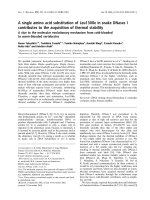
. I n double reciprocal plots of the
data, intersecting lines indicated that the reaction followed a
sequential mechanism (Fig. 1). The data were fitted to
Eqn 2 and the following values were obtained: K
ATP
310 ± 110 l
M
, K
Rib5P
530 ± 140 l
M
and V
max
440 ± 69 lmolÆmin
)1
Æmg protein
)1
.
Assay of enzyme activity in the presence of a variety
of nucleotides showed that 5¢-AMP, GTP, 5¢-GMP and
CTP, each at a concentration of 5.0 m
M
, had little or no
Fig. 1. Reaction mechanism o f PRibPP synthase and determination o f
kinetic constants. Activity was determined as described in Experimental
procedures. The magnesium ch loride c oncentration was 3.0 m
M
over
the ATP concentratio n. 1/v is expressed as lmol
)1
ÆminÆmg protein.
Double reciprocal plots of initial velocity vs. Rib5P at five concen-
trations of ATP are shown. The concentration of Rib5P was varied
from 0.2 to 0.8 m
M
in the presence of different concentrations of ATP:
e,0.1m
M
; n,0.2m
M
; h,0.4m
M
; ·,0.6m
M
;ors,0.8m
M
.Lines
represent fitting of the data t o Eqn 2.
Ó FEBS 2004 Bacillus caldolyticus PRibPP synthase (Eur. J. Biochem. 271) 4529
effect on the enzyme activity, as activity varied from 92
to 109% of the activity obtained in the absence of these
nucleotides. The activity in the presence of 5.0 m
M
UTP
was only 20% of that in the absence of UTP, indicating
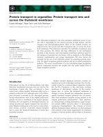
significant inhibition. Only ADP and GDP showed
significant inhibition at physiologically relevant concen-
trations, less than 1% residual activity in the presence of
1m
M
ADP or 5 m
M
GDP. As e xpected from these
results, GDP was a less efficient inhibitor (Fig. 2).
Inhibition by ADP as well as by GDP was strongly
cooperative, with Hill coefficients for ADP and GDP
determined as 2.9 ± 0.1 and 2.6 ± 0.1, respectively. The
apparent K
i
values determined under these assay condi-
tions (3.0 m
M
ATP) were 113 ± 1 l
M
for ADP and
490 ± 9 l
M
for GDP.
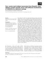
Inhibition with ADP at various ATP c oncentrations was
analysed. In the inhibitor concentration range employed
here, 0.06–0.18 m
M
, ADP was a linear competitive inhibitor
of ATP saturation (Fig. 3). Analysis of the data with respect
to noncompetitive inhibition (Eqn 4) failed t o give a
satisfying fit.
Quaternary structure
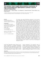
Chemical cross-linking of PRibPP synthase followed by
SDS/PAGE revealed two major bands of M
r
220 000 and
100 000 (Fig. 4). The monomer behaved as a 36 000 M
r
polypeptide. This result indicates the formation of hexa-
mers and trimers. In addition some higher order oligomers
were seen. I nterestingly, n o o r very little dimer was
observed. Higher order oligomers o f B. caldolyticus
PRib PP synthase were consistently seen by gel fi ltration,
and they possessed significant a ctivity but not as high as
the hexamer (data not shown). Identical results, i.e.
Fig. 4. The quaternary structure of PRibPP synthase. Cross-linking
was performed as described in Experimental procedures. Lanes 1 and 7
contain M
r
standards (Bio-Rad): I, M
r
208 000; II, M
r
115 000; III, M
r
79 500; IV, M
r
49 500; V, M
r
34 800. Lane 2 contains untreated
enzyme (0.9 lg app lied in gel). Lanes 3–6 contain cross-linked enzyme.
The amount of protein loaded in e ach lane of t he ge l: lane 3, 4.5 lg
applied in gel; l ane 4, 2 .3 lg; lane 5, 1.1 lg; lane 6, 0.5 lg.
Fig. 3. Inhibition of B. caldolyticus PRibPP synthase activity by ADP.
Activity was determined as described in Experimental procedures. The
magnesium chloride c once ntration exce eded total nucleotid e con cen-
tration by 3.0 m
M
.1/v is expressed as lmol
)1
ÆminÆmg protein. Double
reciprocal plots of initial velocity vs. ATP at six concentrations of ADP
are shown. The concentration of ATP was varied from 0.05 to
0.80 m
M
in the presence of different concentrations of ADP: ,,0m
M
;
s,0.06m
M
; h,0.09m
M
; n,0.12m
M
; e,0.15m
M
,or· ,0.18m
M
.
Lines represent fitting o f the data to Eqn 3.
Fig. 2. Inhibition by ADP and GDP of B. caldo lyticus PRibPP syn-
thase activity. A ctivity was determined as described in E xperimental
procedures wit h ATP a nd Rib 5P c onc entrations o f 3.0 and 5.0 m
M
,
respectively, a nd Mg
2+
exceeding the total ribon ucleotid e co ncentra-
tion by 3.0 m
M
. The specific activity of the enzyme was 400 lmolÆ
min
)1
Æmg protein
)1
(determined at 65 °C). Ribonucleoside diphos-
phate varied from 0 to 5 m
M
. Curves represent fitting of the entire data
sets to Eq n 5 . h,ADP;s,GDP.
4530 B. Hove-Jensen and J. N. McGuire (Eur. J. Biochem. 271) Ó FEBS 2004
chemical cross-linking products with M
r
of 220 000 and
100 000 were obtained with B. subtilis P RibPP synthase
as well.
Model structure
An alignment of B. caldolyticus and B. subtilis PRibPP
synthases is shown in Fig. 5. The amino acid sequences of
the two polypeptides are 81% identical. The crystal
structure of B. subtilis PRibPP synthase has been solved
with two ADP molecules per monomer, one bound at the
active site and one bound in an allosteric cleft. The
structure has also been solved with sulphate bound in the
allosteric cleft and in place of the phosphate group of
Rib5P in the a ctive site [26]. A model based on the
sulphate structure was constructed using a homology-
based method (Fig. 6). All of t he amino acids of the
active sites as well as those of the monomer–monomer
contact surfaces were identical in the two proteins. The
only exceptions were Leu70 and Lys199, which are
isoleucine and a rginine, respec tively, i n t he B. subtilis
enzyme. In addition, all of the amino acids involved in
allosteric regulation by ADP were conserved [28]. Inter-
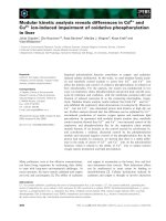
estingly, of the 59 altered amino acid residues, 54
(i.e. 92%) were solvent exposed in the hexameric struc-
ture. The five buried residues of B. caldolyticus PRibPP
synthase were as follows, with the corresponding amino
acid of B. subtilis PRibPP synthase given in parenthesis:
Ile43 (Val), Val56 (Cys), Leu70 (Ile), Asn108 (Glu) and
Val115 (Phe). Consistent with the surface lo cation of the
altered amino acids were hydrophobicity surface maps of
monomers from the two Bacillus PRibPP synthases.
These revealed a n increase in polar surface area in the
B. caldolyticus enzyme compared to that of B. subtilis
(data not shown).
Discussion
It is apparent that the thermophilic version of the Bacillus
enzyme possesses the sam e basic s tructure as its m esophilic
relative and that both enzymes function by the same
mechanism. In particular all of t he residues identified as
important in c atalysis a nd allosteric regulation as well as in
monomer–monomer contact of the B. subtilis PRibPP
synthase were retained in the B. caldolyt icus enzyme with
the t wo exceptions of conservative replacements mentioned
above [22,26,28–30]. T hus, the mechanism of catalysis and
regulation appe ars to b e s imilar for the two enzymes. The
two enzymes differed primarily in their thermal properties.
The origin of this d ifference is at present unknown. In
general, the number of individual amino a cids varied little
among the two enzymes. Exceptions were asparagine,
alanine, glycine a nd methionine. Analysis of the number o f
asparagine and glutamine residues revealed a bias against
these thermolabile amino acids. Both enzymes contained
10 glutamine residues. B. subtilis PRibPP synthase con-
tained 17 asparagines c ompared to 11 of the B. caldolyticus
enzyme. Curiously, however, four of these 17 asparagines of
the B. subtilis enzyme were replaced by glutamines in the
B. caldolyticus enzyme. T hus, the A sn + G ln content may
Fig. 5. Alignment of B. ca ldolyticus and B. subtilis PRibPP synthase amino acid sequences. Bc, B. caldolyticus; Bs, B. subtilis. b-Sheets are shown as
yellow letters, a- helices as blue letters. Residues that are different among the two sequenc es, are shown as red letters in the B. caldolyticus sequence.
Fig. 6. Model structure of hexameric B. c aldo lyticus PRibPP synthase.
One dimer is shown with grey shading, a second dimer with green and
purple shading and a third dimer with blue a nd yellow shading. Red
atoms i ndicate amino acids that differ among B. c aldolyticus and
B. subt ilis PRibPP s ynthases (detailed i n Fig. 5).
Ó FEBS 2004 Bacillus caldolyticus PRibPP synthase (Eur. J. Biochem. 271) 4531
be of significance for the enhanced thermostability of
B. caldolyticus PRibPP synthase, similar to what has been
shown for ce rtain enzymes from hyperthermophilic organ-
isms [31]. Furthermore, the B. caldolyticus enzyme con-
tained 33 alanines compared to 28 in the B. subtilis enzyme
as well as one additional change to alanine. The amino acids
of the B. subtilis enzyme at positions corresponding to these
six alanines were serine, glutamate, valine, lysine and two
glycines. It is possible therefore th at these alanines contribute
compactness to t he thermophilic enzyme. T he glycine
content of the B. caldolyticus enzyme was three less than
that of the B. subtilis enzyme. In the former enzyme the
corresponding amino acids were cysteine, alanine and serine.
Therefore, it is possible that the thermophilic enzyme is more
rigid in structure than the mesophilic enzyme. Finally, t he
B. caldolyticus enzyme contains four more methionines than
the B. subtilis enzyme, corresponding to proline, valine,
isoleucine and glutamine in the latter e nzyme. The signifi-
cance of this difference, if any, remains unknown. It is
possible that subtle changes along the primary structure
together contribute to the increased thermostability [32].
Altogether the modelling of B. caldolyticus P RibPP syn-
thase indicated that the altered amino acids were primarily
located o n t he surface of t he hexameric protein.
Apart f rom t he thermal properties, the two enzymes also
differ widely in their regulation. We determined K
i
values
for ADP and GDP, in the presence of 3.0 m
M
ATP and
5.0 m
M
Rib5P, as 113 and 490 l
M
, respectively, for the
B. caldolyticus enzyme. In comparison, the concentration of
ADP and GDP resulting in 50% inhibition, and determined
at identical s ubstrate c oncentrations as before, w ere g reater
than 1 m
M
and greater than 5 m
M
, respectively, for the
B. subtilis enzyme [12]. Similarly, UTP inhibited the
B. caldolyticus to a higher extent, 20% residual activity,
than the B. subtilis enzyme, 80% residual activity. Again,
determined under identical assay conditions, other kinetic
values differed by approximately two-fold or less. A
summary of the properties of t he two enzymes is given in
Table 1.
Acknowledgements
We are grateful to M. Willem oe
¨
s for discussions and for carefully
reading the manu script, to B . N . Krath for providin g strain H O1986
and for assistance with analysis of kinetic data. We wish to thank T. D.
Hansen for excellent technic al assistance. Financ ial support w as
obtained from the Danish Natural Science R esearch Council.
References
1. Hove-Jensen, B. (1988) Mutation in the phosphoribosylpyro-
phosphate synthetase gene (prs) th at results in simultaneous
requirements for purine and pyrimidine nucleosides, nicotinamide
nucleotide, histidine and tryptophan in Escherichia coli. J. Bacte-
riol. 170 , 1148–1152.
2. Hove-Jensen, B. (1989) Phosphoribosylpyroph osphate (PRPP)-
less mutants of Escherichia coli. Mol. Microbiol. 3, 1487–
1492.
3. White, R.H. (1996) Biosynthesis of methanopterin. B ioc h emi stry
35, 3 447–3456.
4. Scott, J.W. & Rasche, M .E. (2002 ) P urification, o verproduction,
and partial characterization of b-RFAP synthase, a key enzyme in
the m ethanopterin bio synthesis p athway. J. Bacteriol. 184 , 4442–
4448.
5. Jensen, K.F. (1983) Metabolism of 5-phosphoribosyl 1-pyro-
phosphate (PRPP) in Escherichia coli and Salmonella typhimu-
rium.InMetabolism of Nucleotides, Nucleosides and Nucleobases in
Microorganisms (Munch-Petersen, A., ed.), pp. 1–25. Academic
Press, Lo ndon.
6. Kornberg, A., Lieberman, I. & Simms, E.S. (1955) Enzymatic
synthesis and properties of 5- phospho ribosylpyrophosph ate.
J. Bi ol. Chem. 215, 389–402.
7. Khorana, H.G., Fernandes, J.F. & Kornberg, A. (1958) Pyr-
ophosphorylation of ribo se 5-phosphat e in the en zymatic synt h-
esis of 5-phosphorylribose 1-pyrophosphate. J. Biol. Chem. 230,
941–948.
8. Miller, G.A. Jr, Rosenzweig, S. & Switzer, R.L. (1975) Oxygen-18
studies of the mechanism of pyrophosphoryl group transfer cata-
lyzed by phosphoribosylpyropho sphate synthetase. Arch. Bio-
chem. B iophys. 171, 732–736.
9. Hove-Jensen, B., Harlow, K.W., King, C.J. & Switzer, R.L. (1986)
Phosphoribosylpyrophosphate synthetase of Escherichia coli.
Properties of the purified enzyme and primary structure of the prs
gene. J. Biol. Chem. 261, 6 765–6771.
10. Willemoe
¨
s, M. & Hove-Jensen, B . (1997) Binding of divalent
magnesium by Escherichia coli phosphoribosyl diphosphate syn-
thetase. Biochemistry 36, 507 8–5083.
11. Switzer, R.L. (1969) Regulation and mechanism of phospho-
ribosylpyrophosphate synthetase. I. Purification and properties of
the enzyme from Salmonella typhimurium. J. Biol. Chem. 244,
2854–2863.
12. Switzer, R.L. (1971) Regulation and mechanism of phospho-
ribosylpyrophosphate s ynthetase. III. Kinetic studies of the reac-
tion me chanism. J. Biol. Chem. 246 , 2447–2458.
13. Arnvig, K., Hove-Jensen, B. & Switzer, R.L. (1990) Purification
and properties of phosphoribosyl-diphosphat e synthetase from
Bacillus subtilis. Eur. J. Biochem. 192, 195–200.
14. Willemoe
¨
s, M., Hove-Jensen, B . & La rsen, S. (2000) Steady s tate
kinetic m odel for the bin ding of substrates an d allosteric effectors
to Escherichia coli ph osphoribosyl-diphosphate synthase. J. Biol.
Chem. 275 , 35408–35412.
15. Switzer, R.L. & Sogin, D.C. (1973) Regulation and mechanism of
phosphoribosylpyrophosphate synthetase. V. Inhibition by end
product and regu lation by adenosine d iphosp hate. J. Biol . Chem.
248, 1 063–1073.
16. Gibson, K.J., Schubert, K.R. & Switzer, R.L. (1982) Binding of
the substrates and the allosteric inhibitor adenosine 5¢-diphos-
Table 1. Comparison of properties of Bacillus PRibPP synthases. Values for pH optimum, K
m
, V
max
and K
i
of the B. caldolyticus enzyme we re
determined at 65 °C. Values for K
m
, V
max
and K
i
of the B. subtilis enzyme were determined at 37 °C[13].
Source of PRibPP
synthase
No. of amino
acids
Optimal K
m
V
max
(lmolÆ
min
)1
Æmg protein
)1
) K
ADP
i
(m
M
)
Predominant
oligomer
Temp. (°C) pH ATP (m
M
) Rib5P (m
M
)
B. caldolyticus 314 60–65 8.5 0.31 0.53 440 0.113 Hexamer
B. subtilis 316 46 8.5 0.66 0.48 250 > 1 Hexamer
4532 B. Hove-Jensen and J. N. McGuire (Eur. J. Biochem. 271) Ó FEBS 2004
phate t o phosphoribo sylpyropho sphate synthet ase fro m Salm on-
ella typhimurium . J. Bi ol. Chem. 257, 2391–2396.
17. Sonoda, T., Ishiharu, T., Ishijima, S., Kita, K., Ahmad, I. &
Tatibana, M. (1998) Rat liver pho sphoribosylpyro phosphate
synthetase is activated by free Mg
2+
in a manner that overcomes
its inhibition by nucleotides. Biochem. Biophys. A cta 1387, 32–40.
18. Becker, M.A. (2001) Phosphoribosylpyrophosphate synthetase
and the regulation of ph osph oribosylpyrop hosphate p rodu ction in
human c ells. Prog. Nucle ic Acid R es. Mol. Biol. 69, 115–148.
19. Krath, B.N. & Hove-Jensen, B. (1996) Bacillus caldolyticus prs
gene encoding phospho ribosyl-diphosph ate synthase. Gene 176,
73–79.
20. Krath, B.N. & Hove-Jensen, B. (2001) Class II recombinant
phosphoribosyl diphosphate synthase from spinach. Phosphate-
independence and diphosphoryl donor s pecificity. J. Biol. Chem.
276, 17851–17856.
21. Bentsen, A K., Larsen, T.A., Kadziola, A., Larsen, S. & Harlow,
K.W. (1996) Overexpression of Bacillus subtilis phospho-
ribosylpyrophosphate synthetase and crystallization and pre-
liminary x-ray characterization of t he free enzyme and its
substrate-effector complex. Proteins 24 , 238 –246.
22. Bentsen, A K. (1999) Structure–function relationships in Bacillus
subtilis PRPP synthetase. PhD Thesis, University of Copenhagen,
Denmark.
23. Smith, P.K., Krohn, R.I., Hermanson, G.T., Mallia, A.K.,
Gartner, F.H., Provenzano, M.D., Fujimoto, E.K., Goeke, N.M.,
Olson, B .J. & Klenk, D.C. (1985) Measurement of protein using
bicinchoninic acid. Anal . Biochem. 150 , 76–85.
24. Jensen, K.F., Houlberg, U. & Nygaard, P. (1979) Thin-layer
chromatographic methods to isolate
32
P-labeled 5-phospho -
ribosyl-a-1-pyrophosphate (PRPP): Determination of cellular
PRPP pools and assay of PRPP synthetase activity. Anal. Bio-
chem. 98, 2 54–263.
25. Cleland, W.W. (1963) The kinetics of e nzyme-catalyzed reactions
with two or more s ubstrates or products. II. Inhibition: N o men-
clature an d theory. Bio chim. Biophys. A cta 67, 1 73–187.
26. Eriksen, T.A., Kadziola, A., Bentsen, A K., Harlow, K.W. &
Larsen, S. (2000) Structural basis for the function of Bacillus
subtilis phosphoribosyl-pyrophosphate synthetase. Nat. Struct.
Biol. 7, 303–308.
27. Luthy, R., Bowie, J.U. & Eisenberg, D. (1992) Assessment of
protein models with t hree -dimensional profi les. Nature 356, 83–85.
28. Nygaard, F.B. (2001) The molecular mechanism of catalysis and
allosteric regulation in the phosphoribosyldiphosphate synthase
from Bacillus subtilis. PhD Thesis, University of Copenhagen,
Denmark.
29. Eriksen, T.A., Kadziola, A. & Larsen, S. (2002) Binding of cations
in Bacillus subtilis phosphoribosyldiphosphate synthetase an d
their role in c atalysis. Protein Sci. 11, 271–279.
30. Hilden, I., Hove-Jensen, B. & Harlow, K .W. (1 995) Ina ctivation
of Escherichia coli phosphoribosylpyrophosphate synthetase by
the 2¢,3¢-dialdehyde derivative of ATP. Identification of active site
lysines. J. Biol. Chem. 270, 20730–20736.
31. Vieille, C., Epting, K.L., Kelly, R.M. & Zeikus, J.G. (2001)
Bivalent cations and amino-acid composition contribute to the
thermostability of Bacillus licheniformes xylose isomerase. Eur. J.
Biochem. 26 8, 6291–6301.
32. Petsko, G.A. (2001) St ructural basis of thermostability in hyper-
thermophilic proteins, or ÔThe re is m ore than one way to skin a
catÕ. Methods En zymol. 334, 469– 478.
Ó FEBS 2004 Bacillus caldolyticus PRibPP synthase (Eur. J. Biochem. 271) 4533