Báo cáo khoa học: Structural determination of the polar glycoglycerolipids from thermophilic bacteria Meiothermus taiwanensis pdf
Bạn đang xem bản rút gọn của tài liệu. Xem và tải ngay bản đầy đủ của tài liệu tại đây (235.85 KB, 7 trang )
Structural determination of the polar glycoglycerolipids from
thermophilic bacteria
Meiothermus taiwanensis
Feng-Ling Yang
1
, Chun-Ping Lu
1
, Chien-Sheng Chen
2
, Mao-Yen Chen
3
, Hung-Liang Hsiao
4
, Yeu Su
4
,
San-San Tsay
3
, Wei Zou
5
and Shih-Hsiung Wu
1
1
Institute of Biological Chemistry, Academia Sinica, Taipei, Taiwan;
2
Department of Chemistry and
3
Department of Life Science
and Institute of Plant Biology, National Taiwan University, Taipei, Taiwan;
4
Institute of Pharmacology, College of Life Science,
National Yang-Ming University, Shih-Pai, Taipei, Taiwan;
5
Institute for Biological Sciences, National Research Council of Canada,
Ottawa, Ontario, Canada
The polar glycolipids were isolated from the thermophilic
bacteria Meiothermus taiwanensis ATCC BAA-400 by eth-
anol extraction and purified by Se phadex LH-20 and s ilica
gel column chromatography. The fatty acid composition of
O-acyl groups in the glycolipids was obtained b y gas chro-
matography mass spectroscopy analysis on their methyl
esters derived from m ethanolysis and was made mainly of
C
15:0
(34.0%) and C
17:0
(42.3%) fatty acids, with the majority
as branched fatty acids (over 80% ). R emoval of O-acyl
groups under mild basic conditions provided two glycolipids,
which differ only in N-acyl substitution on a h exosamine.
Electrospray mass spectroscopy analysis revealed that one
has a C
17:0
N-acyl group and the other hydroxy C
17:0
in a
ratio of about 1 : 3.5. Furthermore, complete de-lipidation
with strong base followed by selective N-acetylation re sulted
in a homogeneous tetraglycosyl glycerol. T he linkages and
configurations of the carbohydrate moiety were then eluci-
dated by MS and various NMR a nalyses. Thus, the major
glycolipid from M. taiwanensis A TCC BAA-400 w as
determined with the following structure: a-Galp(1-6)-b-
Galp(1-6)-b-GalNAcyl(1,2)-a-Glc(1,1)-Gro diester, where
N-acyl is C
17:0
or hydroxy C
17:0
fatty acid and the glycerol
esters were mainly iso- and a nteisobranched C
15:0
and C
17:0
.
Keywords: glycolipid; Meiothermus taiwanensis;MS;NMR;
thermophilic b acteria.
The thermophilic bacteria such as Aquifex pyrophilus,
Thermodesulfotobacterium commune, Thermus scotoductus,
Thermomicrobium roseum and Thermodesulfatator indicus
contain unique polar lipids as major membrane components
[1–8]. Those lipids are essential for the thermal stability a nd
biological functions of the bacteria i n extreme environments
[9–11]. The polar lipids found in Thermus aquaticus,
Thermus filiformis, Thermus scotoductus,andThermus oshi-
mai were mostly phospholipids and glycolipids [12], and the
glycolipids from Thermus species examined thus far usually
contain t hree hexoses, one N-hexosamine, and one glycerol
[7,10,12–15]. Although the sequences of those carbohydrate
moieties have been studied by chemical and mass spectro-
scopic analysis, no complete structure is a vailable as yet due
to the lack of information on the linkages and configura-
tions of the carbohydrate moiety. We h ave been working on
a n ewly discovered species of thermophilic bacteria, Meio-
thermus taiwanensis, recently isolated from the Wu-rai hot
spring in Taiwan [16], as part of our program to investigate
the immunomodulation activity of the glycolipids and the
structure–activity relationship. In this study, we determined
the structure of a major glycolipid from the thermophilic
bacteria M. taiwanensis ATCC BAA-400. The fatty acids
were examined by gas chromatography mass spectroscopy
(GC-MS) analysis on their methyl esters derived from
methanolysis, whereas, t he structure of t he carbohydrate
moiety was elucidated by MS/MS and NMR spectroscopic
analyses. To the best of our knowledge this is the first
complete glycolipid structure from thermophilic bacteria.
Materials and methods
Isolation and purification of the glycolipids
M. taiwanensis ATCC BAA-400 (Wu-rai hot spring, Tai-
wan) was grown aerobically in Thermus modified medium
[14,16] at 55 °C and harvested until the late exponential
phase ( D
660
¼ 1.6). A suspension of wet b acteria in absolute
ethanol (1 : 10, w/v, Riedel-de-Hae
¨
n, Germany) was shaken
at room temperature for 2 h . After centrifugation, the
supernatant was collected, concentrated, and purified
through a Sephadex LH-20 column (Am ersham Pharmacia,
80 · 1.1 cm) eluted with methanol. T he glycolipid s
Correspondence to S H. Wu, Institute o f Bi ological Chemistry,
Academia Sinica, Taipei 115, Taiwan.
E-mail:
Abbreviations: HMBC, heteronuclear multiple quantum coherence;
HSQC, heteronuclear single quantum coherence; NOESY, nuclear
Overhauser effect spectroscopy; ROESY, rotational frame nuclear
Overhauser effect spectroscopy; TOCSY, total correlation spectros-
copy; HPAEC-PAD, high performance anion exchange chromato-
graphy with pulsed amperometric detection; GC-MS, gas
chromatography mass spectroscopy; ES-MS, electrospray mass
spectroscopy; CE-MS, capillary electrophoresis mass spectroscopy;
MALDI, matrix-assisted laser desorption ionization; FAMEs, fatty
acid methyl esters; TMS, trimethylsilylated.
(Received 30 April 2004, revised 2 1 September 2004,
accepted 4 October 2004)
Eur. J. Biochem. 271, 4545–4551 (2004) Ó FEBS 2004 doi:10.1111/j.1432-1033.2004.04415.x
obtained above were further purified on a silica gel G-60
(Merck, Darmstadt, Germany) chromatography eluted by a
chloroform/methanol gradient from 20 : 1 to 3 : 1. The
carbohydrate-containing fractions were detected by TLC
stained with a molybdate solution [0.02
M
ammonium
cerium sulfate dihydrate/ammonium molydate tetrahydrate
in aqueous 10% (w/v) H
2
SO
4
] a nd collected. T he glycolipids
were still heterogeneous accor ding to the MS analysis due to
the variations in lipids, and soluble in neither water nor
chloroform.
Chemical modification
De-O-acylation. Glycolipids from silica gel purification
weretreatedwith1%(w/v)NaOMe/MeOHatroom
temperature for 5 h. The mixture was neutralized by the
addition of Dowex 50 (H
+
) resin (Acros, NJ, USA) and the
filtrate was concentrated. Purification by silica g el G-60
chromatography (MeOH/CHCl
3
, 1 : 3) gave de-O-acylated
glycolipids.
Per-acetylated glycosyl glycerol. The glycolipids were
treatedwith2
M
NaOH at 100 °C for 8 h to remove both
O- and N -acyl groups; neutralization of the reaction mixture
by acetic anhydride resulted in partial N-acetylation. The
precipitates were removed by centrifugation (3000 g,
15 min, room temperature), and the supernatant containing
sugar w as co llected and lyophilized. The above sample was
then treated with Ac
2
O/pyridine (1 : 2) at room tempera-
ture for 1 h. The reaction was quenched b y the addition of
MeOH, an d the mixture was concentrated to a residue.
Purification by silica gel G-60 chromatography (EtOAc/
hexanes, 2 : 1) gave the per-acetylated glycosyl glycerol.
N-Acetyl glycosyl glycerol. De-O-acetylation was per-
formed on per-acetylated glycosyl glycerol by treatment
with 0.01
M
NaOMe/MeOH at room temperature for 3 h .
The solution was neutralized by the addition of Dowex 5 0
(H
+
) resin and concentrated to a residue. A solution of the
above sample in water was passed t hrough a Sephadex G-10
column using w ater as eluent. The fractions were collected
and lyophilized to give N-acetyl glycosyl glycerol.
Composition and linkage analyses
The fatty acid composition of the O-acyl groups in the
glycolipid was determined b y comparing the r etention times
of FAMEs (fatty a cid methyl esters) from glycolipids to the
standards in GC-MS analysis. The methyl esters were
prepared by treatment of the glycolipids with 0.5
M
HCl/
MeOH at 80 °C for 1 h. Solvent was removed under a
nitrogen stream, and the residue was partitioned between
CHCl
3
and H
2
O. FAMEs in o rganic phase were analyzed
by GC-MS. The f atty acid composition o f the N-acyl group
in the glycolipid was determined by t he MS analysis of
de-O-acylated glycolipid.
The sugar composition analysis was determined by either
GC-MS or high performance anion e xchange chromato-
graphy with pulsed amperometric detection (HPAEC-
PAD) (Dionex, CA, USA). The GC-MS analyses of
glycolipid or N-acetyl glycosyl glycerol were perfo rmed by
methanolysis with 0.5
M
methanolic/HCl at 80 °Cfor16h,
re-N-acetylation with pyridine/ace tic anhydride (in low
temperature with equivalent quantity of acetic a nhydride),
and trimethylsilylation with Sylon HTP (HMDS/TMCS/
pyridine, 3 : 1 : 9) trimethylsilylation reagent (Supelco, PA,
USA). The final trimethylsilylated (TMS) derivatives were
kept in n-hexane for GC-MS analysis. For the HPAEC-
PAD analysis, N-acetyl glycosyl glycerol was subjected to
acidic hydrolysis (2
M
trifluoroacetic acid at 100 °Cfor5h)
to release monosaccahrides, which were then analyzed by
HPAEC-PAD.
For the carbohydrate linkage analysis, the Hakomori
methylation analysis [ 17] w as carried out. The glycolipid or
N-acetyl glycosyl glycerol was per-O-methylated with
methyl iodide and dimethylsulfoxide anion in dimethylsulf-
oxide, and then hydrolyzed by 2
M
trifluoroacetic acid at
100 °C for 5 h. The solvent was evaporated by compressed
air, the residue was r educed with 0.25
M
NaBD
4
in 1
M
NH
4
OH for 40 min. The reaction was quenched with 20%
HOAc and coevaporated with MeOH. The residue was then
per-acetylated with Ac
2
O/pyridine (1 : 1, v/v) overnight,
dried with toluene, and finally analyzed by GC-MS.
Analytical methods
GC-MS was carried out on a H ewlett Packard Gas
Chromatography HP6890 connected to an HP5973 Mass
Selective Detector. The HP-5MS fused silica capillary
column (30 m · 0.25 mm i.d., Hewlett Packard) at 60 °C
was used. The programs for analyses of TMS and FAMEs
weresetupat60°C for 1 m in, increasing t o 140 °Cat
25 °CÆmin
)1
, to 200 °Cat5°CÆmin
)1
, and finally to 300 °C
at 10 °CÆmin
)1
. For partial m ethylated a ditol a cetate s
derivatives, the oven was programmed at 60 °Cfor1min
before increasing to 290 °Cat8°CÆmin
)1
, and finally to
300 °Catarateof10°CÆmin
)1
. Peaks were analyzed by
GC-MS and compared with the database. Also, t he arabitol
derivative was used as an internal standard.
HPAEC-PAD analysis was used to determine the sugar
composition. The hydrolysates f rom N-acetyl glycosyl gly-
cerol were analyzed by HPAEC-PAD in a DX-500 BioLC
system, which included a GP40 gradient pump, an ED40
electrochemical detector (PAD detection) with a working
gold electrode, an LC30 column oven, and an AS3500
autosampler. The Dionex Eluant Degas Module was
employed to purge and pressurize the eluants with helium.
The monosaccharides were separated on Carbopac PA10
analytical column (4 · 250 mm) with Carbopac PA10
Guard (4 · 50 mm) column, flowing at a rate of 1 mLÆmin
)1
at 30 °C, and detected by following pulse potentials and
durations: E
1
¼ 0.05 V (0.4 ms); E
2
¼ 0.75 V (0.2 ms); and
E
3
¼ )0.15 V (0.4 ms). The integration was recorded from
0.2 to 0.4 ms during the E
1
application.
NMR analysis
NMR analytic conditions for carbohydrate analysis were
carried out based on approaches reported previously
[18,19]. NMR spectra were recorded in D
2
O(0.6mL)with
a Varian I NOVA-500 spectrometer at 298 K with s tandard
pulse sequences provided by Varian. Chemical shifts
1
Hand
13
C were given in p.p.m. relative to HDO (4.75 p.p.m.) and
external methanol-d
4
(49.15 p.p.m.), respectively. 1D total
4546 F L. Yang et al.(Eur. J. Biochem. 271) Ó FEBS 2004
correlation spectroscopy (TOCSY) spectra were recorded
with mixing times (20 ms, 100 ms, and 180 ms) which
allowed the assign ment of the proton s H-1 t o H-4 for Gal
and GalNAc, and H-1 to H-6 f or Glc. Four anomeric
protons were selected in respective 1D TOCSY experiments.
2D heteronuclear multiple quantum coherence ( gradient
HMBC) and heteronuclear single quantum coherence
(gradient HSQC) spectra were performed with H -C coup-
ling constants at both 8 Hz/140 Hz and 5 Hz/150 Hz.
Rotational frame n uclear Overhauser effect spectroscopy
(ROESY) spectrum was obtained w ith m ixing t ime 2 00 ms
2D nuclear Overhauser effect spectroscopy (NOESY)
spectra were obtained with m ixing t ime 300 ms and 500 ms.
Mass analysis
MALDI mass spectroscopy. Glycolipids from silica g el
purification were dissolved in CH
3
OH and analyzed by a
MALDI-TOF mass s pectrometer (MALDI
TM
;Micromass,
Manchester, UK). Mass spectra were acquired for the mass
range of 600–2000 Da under a pulsed nitrogen laser of
wavelength 337 nm. Cyano-4- hydroxycinnamic acid was
used as matrix.
CE-MS and MS/MS. A crystal Model 310 CE instrument
(ATI Unicam, Boston, MA, USA) was coupled to an API
3000 mass spectrometer (MDS/Sciex, Concord, ON,
Canada) via a microionspray interface. A sheath solution
(isopropanol/methanol, 2 : 1) was delivered at a fl ow rate of
1 lLÆmin
)1
to a low dead volume t ee (250 lmi.d.,
Chromatographic Specialities, Brockville, ON, Canada).
All a queous solutions were filtered through a 0.45-lmfilter
(Millipore, Bedford, MA, USA) b efore u se. A n electrospray
stainless steel needle (27 gauge) w as butted against the low
dead volume tee a nd enabled the delivery of the sheath
solution to the end of the capillary column. The separation
was obtained on about 90 cm length bare fused-silica
capillary using 10 m
M
ammonium acetate/ammonium
hydroxide in deionized water, pH 9.0, containing 5% (v/v)
methanol. A voltage of 20 kV was t ypically applied at the
injection. The outlet of the capillary was tapered to 15 lm
i.d. using a laser puller (Sutter Instruments, Novato, CA,
USA). Mass spectra were acquired with dwell times of
3.0 ms per step of 1 m/z unit in full-mass scan mode. For
capillary electrophoresis mass spectroscopy (CE-ESMS)
experiments, about 30 nL sample was introduced using
4.35 PSI for 0.1 min. The MS/MS data were acquired w ith
dwell times of 3.0 ms per step of 1 m/ z unit. Fragment ions
formed by collision a ctivation of selected precursor ions
with nitrogen in the R F-only quadrupole collision ce ll, were
analyzed by scanning the third quadru pole.
Results and Discussion
Sugar/fatty acid compositions and sugar linkage analysis
The f atty acid composition of the O-acylated groups linked
on glycerol part of the glycolipid was determined b y GC-MS
analysis on FAMEs derived from glycolipid by methanolysis
in 0.5
M
HCl/MeOH. Quantitative analysis indicated that
the glycolipid contains mainly isobranched (61.7%) and
Table 1. The O-acylated fatty acids present in the glycoglycerolipids
from Meiothermus taiwanensis ATCC BAA-400.
Fatty acids Composition (%)
Straight chain
15:0 3.5
16:0 2.4
17:0 6.6
Isobranched
14:0 1.3
15:0 22.8
16:0 12.3
17:0 23.7
18:0 1.6
Anteisobranched
15:0 7.7
17:0 12.0
Unsaturated
17:1 1.0
Unknown 4.1
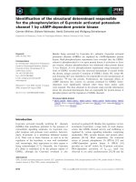
Fig. 1. MS Sp ec tra of native and de-O-acety-
lated glycolipids. (A) MALDI-TOF MS (+ev)
of native glycolipids from Meiothermus tai-
wanensis ATCC BAA-400. A c luster of peaks
was observed due to the f atty acid heterogen-
eity. The peak at m/z 1491 (M + Na
+
)rep-
resents a glycolipid with three hexoses, one
hexosamine, on e glyce rol, and th ree fatty ac ids
(two C
17:0
and one C
15:0
lipids), and the g ly-
colipid a t m/z 1507 (M + Na
+
)containsone
C
17:0
, one hydroxy-C
17:0
and one C
15:0
.(B)ES-
MS (+ev) spectra of de-O-actylated glyco-
lipids, m/z 993 (M
1
+H
+
) and 1009
(M
2
+H
+
)andm/z 1026 (M
2
+NH
4
+
).
(C) MS/MS analysis of peak 1026 (in B) and
(D) MS/MS analysis of peak 993 (in B).
Ó FEBS 2004 Glycolipids from thermophilic bacteria (Eur. J. Biochem. 271) 4547
anteisobranched (19.7%) fatty acids. Over 80% of fatty acids
were C
15:0
and C
17:0
(Table 1). The fatty a cid composition o f
the N-acylated group will be discussed later.
Compositional analysis of sugar was independently
performed using two methods. One was based on
HPAEC-PAD analysis on the acid hydrolyzates of the
N-acetyl glycosyl glycerol. Glucose, galactose, and galacto-
samine were found to be in a ratio of 1 : 2 : 1. The other
followed a standard methanolysis/trimethyl-silylation pro-
cedure, by which we analyzed the TMS methylated sugar
alditol ace tates by GC-MS a nd c ompared with t he standard
profiles for quantitative and qualitative measurement.
In addition, to confirm the sugar composition, the s ugar
linkage analysis als o indicated that the glycolipid contains
one terminal galactopyranose (t-Gal-1-), one 1,6-linked
galactopyranose (-6-Gal-1-), one 1,6-linked galactopyrano-
samine (-6-GalNAc-1-), and one 1,2-linked glucopyranose
(-2-Glc-1-). All sugar residues in the glycolipids are pyra-
noses.
N-Amide and sugar sequence
MALDI-TOF mass spectrosco pic analysis of the glycolipids
showed a cluster of peaks at m/z (+ev) 1433, 1449, 1463,
1477, 1491, and 1507 with mass differences of 14 and 16
(Fig. 1 A), which probably resulted from t he heterogeneity
of fatty acids. On the other hand, the E S-MS spectrum
(Fig. 1 B) obtained from the de-O-acylated glycolipid (see
above) was simpler, s howing major peaks at m/z (+ev) 993,
1009 and 1026. In fact m/z 1009 a nd 1026 were derived from
the same molecule but only differently ionized as they
provided identical fragmentation in MS/MS experiments.
One (m/z 1009) represents (M + H
+
), and the other (m/z
1026) probably added an ammonium ion (M + NH
4
+
)
from the buffer used in MS analysis (Fig. 1B). A compar-
ison of the d aughter ions from MS/MS a nalysis on m/z 1026
and 993 revealed a difference of m/z 16 on all major
fragments as indicated in Fig. 1C,D. Two ions, m/ z 414 and
430, from N-acyl hexosamine are indicative that the
hexosamine was acylated by two major fatty acids, C
17:0
(m/z 414) and hydroxy (presumably 3-hydroxy) C
17:0
(m/z
430). The ratio of the C
17:0
and hydroxy C
17:0
is approxi-
mately 1 : 3.5 according to relative peak heights in MS
spectrum. Small amounts of other N-acyl lipids in glycolipid
were also detected, e.g. C
16:0
(m/z 979), C
22:0
(m/z 1080), and
hydroxy C
22:0
(m/z 1096) (Fig. 1B). The lack of adequate
detection of N-acyl lipids was due to the relative stability
of the amide bond under methanolysis conditions. The
significant amount of hydro xy fatty acids presented in this
glycolipid as amide linked to galactosamine is similar t o
those of T. filiformis and T. aquaticus [15].
N-Acetyl glycosyl glycerol was obt ained b y the total
deacylation, full acetyla tion and de-O-acetylation of the
glycolipid (see above). T he ES-MS spectrum of N-acetyl
Fig. 2. Mass spectra of t he N-ace tyl glycolipid s. (A) ES-MS spectrum
(–ev)oftheN-acetyl tetraglycosyl glycerol derived from the major
glycolipids o f Meiothermus taiwanensis ATCC BAA-400. Both O - and
N-acyl groups were removed a nd the am ino group was acetyl ated. (B)
MS/MS spectra (+ev) revealed the sugar sequence of the tetraglycosyl
glycerol.
Fig. 3. The 500 MHz spectra o f
1
HNMRand
1D TOCSY o f the N-acetyl tetraglycosyl gly-
cerol. Four anomeric p roton s were irradiated
in respective 1D TOCSY experimen ts.
Chemical shifts of the anomeric protons were
assigned as following: t-a-Gal at d 4.96
(J
1,2
¼ 3.3 Hz), 1 ,6-b-Gal at 4.44
(J
1,2
¼ 7.8 Hz), 1 ,6-b-GalNAc at d 4.58
(J
1,2
¼ 8.5 Hz), a nd 1,2-a-Glc at 5.15
(J
1,2
¼ 3.5 Hz) p.p.m.
4548 F L. Yang et al.(Eur. J. Biochem. 271) Ó FEBS 2004
glycosyl glycerol (Fig. 2A) showed a major peak at m/z
(–ev) 780.0 with minor peaks at 618.0 (- Hex) and 456.0
(-2Hex). MS/MS a nalysis on t he major ion, m/z (+ev) 782,
provided more d etailed information on the sequence o f the
carbohydrate moiety (Fig. 2B). Because the breakup of the
HexNAc glycosidic bond often produces a relatively stable
positive-charged oxazoline-like fragment, the hig h intensit y
peaks at m/z 204, 366 and 528 were indicative that those
fragments contain HexNAc at t he reducing end. On the
other hand, the observation of m/z 620 (M-Hex) and 458
(M-2Hex, HexNAc-Hex-Gro) as daughter ions suggested
Hex-Hex at the nonreducing end. Further ES-MS/MS
analysis of th ese daughter i ons (m/z 620 and 4 58) w as
performed a nd the results were consistent with the following
tetraglycosyl glycerol sequence: Hex-Hex-HexNAc-Hex-
Gro. This sequence is similar to the ones previously
reported with s ome strains of thermophilic eubacterial
genus T. aquaticus and T. filiformis [15].
Glycosyl linkage and anomeric configuration
With the solid results of the sugar composition and
sequence, the linkages and configurations of glycosidic
bonds would be investigated by NMR to determine the
complete structure of the glycolipid. A clean
1
H-NMR
spectrum of the tetraglycosyl glycerol is shown in Fig. 3.
Four H-1 anomeric proto n signals were observed a s
expected and their configurations could be identified by
their coupling constants. 1D-TOCSY experiments further
indicated that they represent the anomeric protons of a-Glc
(5.15 p .p.m., J
1,2
¼ 3.5 Hz), a-Gal (4.96 p.p.m., J
1,2
¼
3.3 Hz), b-GalNAc (4.58 p.p.m., J
1,2
¼ 8.5 Hz) and b-Gal
(4.44 p .p.m., J
1,2
¼ 7.8 Hz), respectively [19]. Based on
1D-TOCSY spectra, the chemical shifts of Glc residue from
H-1 to H -6 and t hose of Gal and GalNAc residues from H-1
to H-4 were able t o b e a ssigned ( Fig. 3). The H-5 protons of
b-Gal and b-GalNAcwereassignedbasedonNOE
interaction to H-1 by NOESY or ROESY experiments
(data not shown).
13
C chemical shifts were obtained from
HSQC experiment and both
1
Hand
13
C chemical shifts are
summarized in Table 2 . Six methylene carbons (-CH
2
-O-)
were detected as negative peaks in the HSQC experiment.
Table 2. NMR data of the tetraglycosyl glycerol derived from the major
glycolipid from Meiothermus taiwanensis ATCC BAA-400. In p.p.m.
from the HSQC spectrum obtained in D2O at 25 °C.
Residue Atom d
H
d
C
A
a-Gal(1fi}
1 4.96 98.5
2 3.82 68.5
3 3.83 69.6
4 3.96 69.4
5 3.96 71.2
6 3.72 61.3
B
6)-b-
D
-Galp(1fi}
1 4.44 103.5
2 3.51 70.9
3 3.64 72.9
4 3.96 68.9
5 3.87 73.1
6 3.87, 3.71 66.5
C
6)-b-GalNAc(1fi}
1 4.58 103.4
2 3.90 52.8
3 3.72 71.1
4 3.93 68.0
5 3.84 73.8
6 4.02, 3.87 69.5
NAc 2.01 22.4
D
2)-a-Glc(1fi}
1 5.15 98.5
2 3.59 80.9
3 3.75 71.8
4 3.38 70.1
5 3.65 71.9
6 3.82, 3.72 60.7
E
1)-Glycerol
1 3.53, 3.74 69.2
2 3.94 70.5
3 3.58, 3.68 62.8
Fig. 4. 2D gH SQC (red) and gHMBC (blue)
spectra of the N-acetyl tetraglycosyl glycerol
were used to assign the glycosyl linkages and
configurations.
Ó FEBS 2004 Glycolipids from thermophilic bacteria (Eur. J. Biochem. 271) 4549
Three of them (d
C
66.5, 69.2, and 69.5) were in residues in
which a glycosyl substituent was present at O
6
,andtheother
three (d
C
60.7, 61.3, and 62.8) were in residues in which an
unsubstituted h ydroxyl group was present at O
6
.The
interglycosidic linkages were determined b ased on the
HMBC (Fig. 4, Table 3) and NOE interactions (Table 3),
the terminal Gal (A) wa s a-(1-6)-linkedtoGal(B)because
of the N OE and HMBC correlations observed between H-1
ofGal(A)andH-6andC-6ofGal(B).Similarly,theGal
(B) was assigned to be b-(1-6)-linked to GalNAc (C), w hich
was then b-(1-2)-linked to Glc (D) based on both NOE and
HMBC correlations. Finally, the Glc (D) at the reducing
end was then a-(1-1)-linkedtoglycerol(E).
Based on all the information obtained from sugar and
fatty acid composition analyses and MS and NMR
experiments, we are able to report the major g lycolipid
from thermop hilic b acteria M. taiwanensis ATCC BAA-
400 having the following structure: a-Galp(1-6)-b-
Galp(1-6)-b-GalNAcyl(1-2)-a-Glc(1-1)Gro diester, where,
the N-acyl lipids were mainly C
17:0
and hydroxy C
17:0
fatty
acids, and t he glycerol diester was mainly made of branched
C
15:0
and C
17:0
fatty acids.
The monosacchrides in the major glycoglycerolipids of
Meiothermus ruber, M. silvanus, M. chliarophilus,and
M. ce rbereus are two or three g lucoses, one galactose, either
one galactosamine or one glucosamine, and glycerol [15]. I n
M. taiwanensis ATCC BAA-400 glycoglycerolipids, there
are two galactoses, one glucose, one galactosamine, a nd one
glycerol, which is different from other Meiothermus, but
similar to its relative genus Thermus spp. 3-Hydroxy fatty
acids linked to N-acyl galactosamine is specific, which is
quite different from Thermus glycolipids [11]. This study is
the first to determine the full structure of glycoglycerolipid
in thermophilic bacteria Meiothermus spp. The structural
information will be very useful for further investigations of
the mechanisms of glycoglycerolipid biosynthesis in vivo,
and even for chemical synthesis in vitro and their physio-
logical roles.
Acknowledgements
The authors thank the National S cience Council, Taiwan for support.
MS and NMR spectra were performed i n the Institute for Biological
Sciences, National Research Coun cil of Canada. We are also grateful to
DrJianjunLiofNRCforES-MS/MSanalysis.
References
1. Langworthy , T.A. & Pond, J.L. ( 1986) Membranes and lipids of
thermophiles. In Thermophiles: general, molecular and applied
microbiology. (Brock, T.D., ed.), pp. 107–135. John Wiley and
Sons, New York, NY.
2. Pond,J.L.,Langworthy,T.A.&Holzer,G.(1986)Long-chain
diols: a new class of membrane lipids from a thermophilic bac-
terium. Science 231, 1134–1136.
3. Jahnke, L.L., Eder, W., Huber, R., Hope, J.M., Hinrichs, K.U.,
Hayes,J.M.,DesMarais,D.J.,Cady,S.L.&Summons,R.E.
(2001) Signature lipids and stable carbon isotope analyses of
Octopus Spring hyperthermophilic communities compared with
those of Aquifi cales rep resen tatives. Appl. Environ. Microbiol. 67,
5179–5189.
4. Huber,R.,Wilharm,T.,Huber,D.,Trincone,A.,Burggraf,S.,
Koenig, H., Ra chel, R., R ockinger, I., F ricke, H. & Stetter, K O.
(1992) Aquifex pyrophilus gen. nov., sp. no v., represen ts a novel
group of marine hyperthermophilic hydrogen-oxidizing b acteria.
Syst. Appl. Microbiol. 15, 340–351.
5. Langworthy, T.A., Holzer, G., Zeikus, J.G. & Tornaben e, T.G.
(1983) Iso- and anteiso-branched glycerol diethers of the ther-
mophilic anaerobe Thermodesulfotobacterium commune. Syst.
Appl. Microbiol. 4, 1–17.
6. Huber, R. & Stetter, K O. (1992) The order Thermotogales.In
The Prokaryotes.(Balows,A.,Tru
¨
per, H.G., D workin, M., Har-
der, W. & Schleifer, K.H., eds), pp. 3809–3815. Springer-Verlag,
Berlin, Germany.
7. Wait, R., Carreto, L., Nobre, M.F., Ferreira, A.M. & da Costa,
M.S. (1997) Characterization of novel long-chain 1 ,2-diols in
Thermus species a nd demonstration that Thermus strains contain
both glycero l-linked and diol-linked glycolip ids. J.Bacteriol. 179,
6154–6162.
8. Moussard, H., L’Haridon, S., Tindall, B.J., Banta, A., Schumann,
P., Stackebrandt, E., Reysenbach, A.L. & Jeanthon, C. (2004)
Thermodesulfatator indicus gen. nov., sp. nov., a novel thermo-
philic chemolithoaut otrophic sulfate-redu cing bacterium isolate d
from the Cen tral Ind ian Ridge. In t. J. Syst. E vol. Microbiol. 54,
227–233.
9. Ray, P.H., White, D.C. & Brock, T.D. (1971) Effect of growth
temperature on the lipid compos ition of Thermus aquaticus.
J. Bacteriol. 108, 227–235.
10. Pask-Hug hes, R.A. & Shaw, N. (1982) Glycolipids from some
extreme thermoph ilic bacte ria b elongin g to the genus Thermus.
J. Bacteriol. 149, 54–58.
11. Ferreira, A.M., Wait, R., Nobre, M.F. & da Costa, M.S. (1999)
Characterization of glycolipids from Meiothermus spp. Micro-
biology 145, 1191–1199.
12. Donato, M .M., Seleiro, E.A. & da Costa, M.S. (1990) Polar l ipid
and f atty acid com position of strains of the genus Thermus. Sys.
Appl. Microbiol. 13 , 234–239.
13. Ferraz, A.S., Carreto, L., Tenreiro, S., Nobre, M.F. & da Costa,
M.S. (1994) Polar lipids and fatty acid composition of Thermus
strains from New Zealand. Antonie Van Leeuwenhoek 66, 357–363.
14. Williams, R.A.D. & da Cost a, M.S. (1992) The genus Thermus
and related microorganisms. In The Prokaryotes 2nd edn.
(Balows, A., Trueper, H.G., Dworkin, M., Harder, W. & Schlei-
fer, K.H., e ds.), pp. 3745–3753. S pringer, New York, NY.
15. Carreto, L., Wait, R., Nobre, M.F. & da Costa, M.S. (1996)
Determination of the structure of a novel glycolipid f rom Thermus
aquaticus 15004 and d emonstration that hydroxy fatty acids are
amide linked to glycolipids in Thermus spp. J. Bacteriol. 178,
6479–6486.
16. Chen, M.Y., Lin, G.H., Lin, Y.T. & Tsay, S .S. (2002) Meiother-
mus taiwanensis sp. nov., a novel filamentous, t hermophilic species
isolated in Taiwan. Int. J. Sys. Evol. Microbiol. 52, 1647–1654.
Table 3. HMBC and NOE correlations observed in the tetraglycosyl
glycerol.
Residue
From
proton
NOE to
protons
HMBC to
carbons
a-Gal1- A1 A2, B6a, 6b A2, A5, B6
(A)
-6-b-Gal1- B1 B2, B5, C6a, 6b C6
(B)
-6-b-GalNAc-1- C1 C2, D2, D3 D2
(C)
-2-a-Gal1- D1 D2, E1a,1b D3, D5, E1
(D)
OCH
2
CH(OH)CH
2
OH E1 E2, E3
(E) E2 E1, E3
4550 F L. Yang et al.(Eur. J. Biochem. 271) Ó FEBS 2004
17. Waeghe, T.J., Darvill, A.G., McNeil,M.&Albersheim,P.(1983)
Determination, b y met hy lation a nalysis, of the glycosyl-linkage
compositions of m icrogram qu antities o f complex carbohydrates.
Carbohydr. R es. 123, 281–304.
18. Kogan, G. & Uhrin, D. (2000) Current NMR methods in the
structural elucidation of polysaccharides. In New Advances in
Analytical Chemistry (Rahman, A., ed.), pp. 73–134. Harwood
Academic Press, Amsterdam.
19. Du us, J., Gotfredsen, C.H. & Bock, K. (2000) Carbohydrate
structural determination by NMR spectroscopy: modern m etho ds
and limitations. Chem. Rev. 100, 4589–4614.
Ó FEBS 2004 Glycolipids from thermophilic bacteria (Eur. J. Biochem. 271) 4551