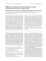
Báo cáo khoa học: Structure of the core oligosaccharide of a rough-type lipopolysaccharide of Pseudomonas syringae pv. phaseolicola docx
Bạn đang xem bản rút gọn của tài liệu. Xem và tải ngay bản đầy đủ của tài liệu tại đây (240.12 KB, 10 trang )
Structure of the core oligosaccharide of a rough-type
lipopolysaccharide of
Pseudomonas syringae
pv.
phaseolicola
Evelina L. Zdorovenko
1,2
, Evgeny Vinogradov
1,
*, Galina M. Zdorovenko
3
, Buko Lindner
2
,
Olga V. Bystrova
1,2
, Alexander S. Shashkov
1
, Klaus Rudolph
4
, Ulrich Za¨ hringer
2
and Yuriy A. Knirel
1,2
1
N. D. Zelinsky Institute of Organic Chemistry, Russian Academy of Sciences Moscow, Russia;
2
Research Center Borstel,
Leibniz Center for Medicine and Biosciences, Borstel, Germany;
3
D.K. Zabolotny Institute of Microbiology and Virology,
National Academy of Sciences of Ukraine, Kiev, Ukraine;
4
Institute for Plant Pathology and Plant Defence, Georg August University,
Go
¨
ttingen, Germany
The core structure of the lipopolysaccharide (LPS) isolated
from a rough strain of the phytopathogenic bacter ium
Pseudomonas syringae pv. phaseolicola, GSPB 711, was
investigated by sugar and methylation analyses, Fourier
transform ion-cyclotron r esonance ESI M S, and one- and
two-dimensional
1
H-,
13
C- and
31
P-NMR spectroscopy.
Strong alkaline deacylation of the LPS r esulted in two
core-lipid A backbone undecasaccharide pentakisphos-
phates in the ratio 2.5 : 1, which corresponded to outer
core glycoforms 1 and 2 terminated with either
L
-rham-
nose or 3-deoxy-
D
-manno-oct-2-ulosonic acid (Kdo), res-
pectively. Mild acid degradation of the LPS gave the
major glycoform 1 c ore octasaccharide and a minor trun-
cated glycoform 2 core heptasaccharide, which resulted
from the cleavage of the terminal Kdo residues. The inner
core of P. syringae is distinguished by a high degree of
phosphorylation of
L
-glycero-
D
-manno-heptose residues
with phosphate, diphosphate and ethanolamine diphos-
phate groups. The glycoform 1 core is structurally similar
but not identical to one of the core glycoforms of the
human pathogenic bacterium Pseudomonas aeruginosa.
The outer core composition and structure may be useful
as a chemotaxonomic marker for the P. syringae group of
bacteria, whereas a more conserved inner core structure
appears t o be r epresentative for the whole genus Pseudo-
monas.
Keywords: core oligosaccharide; glycoform; lipopolysac-
charide s tructure; phytopathogen; Pseudomonas syringae.
The bacteria Pseudomonas syringae cause serious diseases
in most cultivated plants and are widespread in nature as
epiphytes. More than 50 pathovars o f P. syringae and related
species have been described based on the distinctive patho-
genicity of the strains to one or more host plants [1]. The
P. syringae group is characterized by a high degree of het-
erogeneity also in respect to gen omic features. Recently, type
strains of v arious P. syringae pathovars have been delineated
into nine genomospecies [2]. However, the taxonomic status
of the pathovars a nd genomospecies remains uncertain.
The lipopolysaccharide (LPS) is the m ajor component of
the out er membrane of Gram-negative b acteria, which plays
an important role in interaction of bacteria with their hosts.
LPS i s c omposed of lipid A, a c ore oligosaccharide, and an
O-polysaccharide (O-antigen) built up of oligosaccharide
repeats. The structures of the O-polysaccharides of all
known serologically distinguishable smooth strains of
P. syringae have been determined [3–12]. Aiming at solving
the problems of r ecognition, taxonomy and classification o f
P. syringae strains, we established, for the first time, t he full
structure of the core region of the L PS from a rough strain
of P. syringae pv. phaseolicola GSPB 711. According to
published composition [ 11,13–16] and serological [17,18]
data, t his core structure is shared by most P. syringae strain s
tested.
Materials and methods
Bacterium, growth and isolation of the
lipopolysaccharide
P. syringae pv. pha seolicola rough strain GSPB 711
was received f rom t he Go
¨
ttingen Collection o f P lant
Pathogenic Bacteria (Germany) were grown on Potato
agar at 22 °C for 24 h, washed with physiological saline,
separated by centrifugatio n, washed with acetone and d ried.
LPS was isolated from dry bacterial cells by the method
of Galanos [19] and purified by ultracentrifugation
(105 000 g, 4 h). The supernatant was dialyzed against
distilled water and lyophilized.
Correspondence to E. L. Zdorovenko, N. D. Zelinsky Institute of
Organic Chemistry, Leninsky Prospekt 47, 119991, Moscow,
GSP-1, Russia. Fax: +7095 1355328, Tel.: + 7095 9383613,
E-mail:
Abbreviations: Cm, carbamoyl; CSD, capillary skimmer dissociation;
6dHex, 6-deoxyhexose; Etn, ethanolamine; FT-ICR, Fourier trans-
form ion-cyclotron resonance; Hep,
L
-glycero-
D
-manno-heptose; Hex,
hexose; HexN, hexosamine; HPAEC, high-performance anion-
exchange chromatography; Kdo, 3-deoxy-
D
-manno-oct-2-ulosonic
acid; LPS, lipopolysaccharide; OS, oligosaccharide.
*Present address: Institute for Biological S ciences, National Research
Council, 100 S ussex Drive, O ttawa, ON, Canada K1A 0R6.
(Received 2 9 June 2004, revised 30 September 2004,
accepted 27 October 2004)
Eur. J. Biochem. 271, 4968–4977 (2004) Ó FEBS 2004 doi:10.1111/j.1432-1033.2004.04467.x
Alkaline degradation of the lipopolysaccharide
The LPS (110 mg) was treated with anhydrous hydrazine
(4 mL) for 1 h at 37 °C, then 16 h at 20 °C. Hydrazine was
flushed out in a stream of air at 3 0–33 °C, the residue washed
with cold ac etone at 4 °C, dried i n v acuum, disso lved in 4
M
NaOH (8 mL) supplemented with a small amount of
NaBH
4
, and then heated at 100 °C for 4 h. After cooling
to 4 °C, the solution was acidified to pH 5.5 w ith concen-
trated HCl, extracted twice with dichloromethane, and the
aqueous solution desalted by gel-permeation chromatogra-
phy on a column (60 · 2.5 cm) of Sephadex G-50 (Amer-
sham Biosciences, Uppsala, Swe den) in pyridinium a cetate
buffer (4 mL pyridine and 10 mL HOAc in 1 L water,
pH 4.5) at 30 mLÆh
)1
. Elution was monitored with a
differential refractometer (Knauer, Berlin, Germany). The
isolated oligosaccharide mixture (OS
NaOH
) (35 mg ) was
fractionated by h igh-performance anion-exchange chroma-
tography (HPAEC) on a semipreparative CarboPac PA1
column (250 · 9 mm; Dionex, Sunnyvale, CA, USA) using
a linear gradient of 0.02–0.6
M
NaOAc in 0 .1
M
NaOH at a
flow rate of 2 mLÆmin
)1
for 100 min and 2-mL fractions were
collected and analyzed by HPAEC using pulsed ampero-
metric detection (Dionex) on an analytical CarboPac PA1
column (250 · 4.6 mm) using the same eluent at 1 mLÆmin
)1
for 3 0 min. Desalting on a column ( 40 · 2.6 cm) of Sepha-
dex G-50 afforded two major oligosaccharides, OS
NaOH
-I
and O S
NaOH
-II (7.2 a nd 3.6 mg, respectively), having
retention times 11.7 a nd 18.0 min in analytical HPAEC.
Mild-acid degradation of the lipopolysaccharide
The LPS was d issolved in aqueous 1 % HOAc and heated for
1.5 h at 100 °C. The p recipitate was r emoved by centrifuga-
tion ( 12 000 g, 20 min), and the supernatant fractionated by
gel-permeation chromatography on a column (40 · 2.6 cm)
of Sephad ex G-50 as described above to give a mixture of
phosphorylated oligosaccharides (OS
HOAc
).
Chemical analysis
For neutral sugar analysis, the oligosaccharides (0.5 mg
each) were hydrolyzed with 2
M
CF
3
CO
2
H(120°C, 2 h),
monosaccharides were conventionally converted into the
alditol acetates and analyzed by GLC on a Hewlett-Packard
HP 5890 Series II chromatograph (Palo Alto, CA, USA)
equipped with a 30-m fused-silica S PB-5 column (Supelco,
Bellefoute, PA, USA) using a temperature gradient of
150 °C(3min)fi 320 °Cat5°CÆmin
)1
. After hydrolysis of
the oligosaccharides (40 lgeach)with4
M
HCl (80 lL,
100 °C, 16 h), amino components were analyzed as p he-
nylthiocarbamoyl derivatives b y HPLC o n a reversed-phase
Pico-Tag column (150 · 3.9 mm) using buffers for Pico-
Tag amino acid analysis of protein hydrolysates (Waters,
Milford, MA, USA) at 42 °C and a flow rate 1 mLÆmin
)1
for 10 min; monitoring was performed with a dual k
absorbance detector (Waters) at 254 nm.
Methylation analysis
OS
NaOH
-I and OS
NaOH
-II (1 mg each) were dephosphoryl-
ated with aqueous 48% HF (25 lL) at 4 °C f or 16 h, the
solution was diluted with water and lyophilized, the
products were N-acetylated with Ac
2
O (100 lL) in aqueous
saturated N aHCO
3
at 20 °C for 1 h at stirring, reduced with
NaBH
4
and d esalted b y gel-permeation c hromatography on
Sephadex G-15. Methylatio n was performed by the proce-
dure of Ciucanu and Kerek [20] with CH
3
I(0.3mL)in
dimethylsulfoxide (0.5 mL) in the p resence of solid NaOH
(stirring for 20 min before and 2 h after a dding CH
3
I), the
reaction mixture was diluted with water, the methylated
compounds were extracted with chloroform, h ydrolyzed
with 3
M
CF
3
CO
2
H (100 °C, 2 h), reduced with NaBD
4
,
acetylated and analyzed by GLC MS on a HP Ultra 1
column (25 m · 0.3 mm) using a Varian Saturn 2000
instrument (Palo A lto, CA, USA) equipped with an ion-
trap MS detector.
Electrospray ionization mass spectrometry (ESI MS)
High-resolution electrospray ionization Fourier t ransform
ion-cyclotron resonance mass spectrometry (ESI FT-ICR
MS) was performed in the negative ion mode using an
ApexII-instrument (Bruker Daltonics, Billerica, USA)
equipped with a 7 T actively shielded magnet and an
Apollo electrospray ion source. Mass spectra were a cquired
using standard experimental sequ ences as provided by the
manufacturer. Samples were dissolved at a concentration of
10 ngÆlL
)1
in a 50 : 50 : 0.001 (v/v/v) 2 -propanol, water,
and triethylamine mixture and sprayed at a flow rate of
2 lLÆmin
)1
. Capillary entrance voltage was set to 3.8 kV,
and dry gas temperature to 150 °C. Capillary skimmer
dissociation (CSD) was induced by increasing the capillary
exit voltage from )100 to )350 V.
NMR spectroscopy
NMR spectra were obtained on a Varian Inova 500, Bruker
DRX-500 and DRX-600 spectrometers (Karlsruhe,
Germany) in 99.96% D
2
Oat25or50°C and pD 3, 6 or
9 (uncorrected), respectively, using internal acetone (d
H
2.225, d
C
31.45) or external aqueous 85% H
3
PO
4
(d
P
0.0) as
reference. Prior to the measurements, the samples w ere
lyophilized twice from D
2
O. Bruker software
XWINNMR
2.6
was used to acquire and process the data. M ixing times of
120 and 100 ms were used i n TOCSY and 250 and 225 ms
in ROESY experiments at 500 and 600 MHz, respectively.
Results and Discussion
Oligosaccharides derived b y strong alkaline degaradation of
the LPS [21] were used to determine the structure of the
core-lipid A c arbohydrate b ackbone of the P. syringae LPS.
The LPS was O-deacetylated by mild hydrazinolysis and
then N-deacylated under strong alkaline conditions (4
M
NaOH, 100 °C, 4 h). After desalting, the resultant mixture
of oligosaccharides (OS
NaOH
) was fractionated b y HPAEC
on CarboPak PA1 at super-high pH to give the major and
minor products (OS
NaOH
-I and O S
NaOH
-II, respectively).
The charge deconvoluted ESI FT-ICR mass spectrum
of OS
NaOH
showed an abundant molecular ion with the
molecular mass 2356.55 Da as well as less intense peaks
(Fig. 1 ). The measured molecular masses of two ions,
2356.55 an d 2430.57 Da, were i n agreement with those
Ó FEBS 2004 Core oligosaccharide of Pseudomonas syringae (Eur. J. Biochem. 271) 4969
calculated for undecasaccharide pentakisphosphates h aving
the following composition: 6dHex
1
Hex
2
Hep
2
Kdo
2
HexN
4
P
5
and Hex
2
Hep
2
Kdo
3
HexN
4
P
5
(OS
NaOH
-I and OS
NaOH
-II,
respectively), where 6dHex stands f or a 6-deoxyhexose, H ex
for a hexose, Hep for a heptose, HexN for a hexosamine, and
Kdo for 3-deoxy-
D
-manno-oct-2-ulosonic acid. These com-
pounds differ in one of the constituent monosaccharides,
which is either a 6dHex residue or the third Kdo residue.
Accordingly, the
1
H-NMR spectra of OS
NaOH
-I and
OS
NaOH
-II isolated b y HPAEC showed signals for two and
three K do residues, respectively. This finding is in agreement
with a significantly higher retention time of OS
NaOH
-II in
HPAEC as compared with OS
NaOH
-I due to the presence
of an additional negatively c harged Kdo residue.
As depicted in Fig. 1, the other minor mass peaks
belonged to (a) OS
NaOH
-I bearing a 3-hydroxydodecanoyl
group (Dm/z 198), which resulted from incomplete
N-deacylation of lipid A, and (b) to fragment ions due
to losses of Kdo (Dm/z )220), bisphosphorylated diglu-
cosamine lipid A backbone (Dm/z )500), and decarboxy-
lation (Dm/z )44).
The
1
H- and
13
C-NMR spectra of OS
NaOH
-I and OS
NaOH
-
II at two different temperature and pD conditions were
assigned using t wo-dimensional COSY, TOCSY and
1
H,
13
C
HSQC experiments (Table 1). Spin systems for a ll constitu-
ent monosaccharides, including rhamnose (Rha), Glc,
L
-glycero-
D
-manno-heptose (Hep), GlcN, GalN and Kdo,
were identified by
3
J coupling constants a nd using published
data for stru cturally similar oligo saccharides derived from
the Pseudomonas aeruginosa LPS [ 22,23]. The configurations
of the glycosidic linkages were determined based on J
1,2
coupling constant values for Glc, GlcN and G alN (3–3.5 and
7–8 Hz for a-andb-linked monosaccharides, respectively)
and by typical
1
H- and
13
C-NMR chemical shifts for Rha,
Hep and Kdo [24]. The anomeric configurations of Rha and
Hep were confirmed by the presence of H-1,H-2 and no
H-1,H-3 or H-1,H-5 cross-peaks in the two-dimensional
ROESY spectra of the oligosaccharides.
Linkage and sequence analysis of OS
NaOH
-I and
OS
NaOH
-II was performed using a two-dimensional
ROESY experiment. This revealed a lipid A carbohydrate
backbone of a GlcN
II
fiGl cN
I
disaccharide and an inner
core region composed of two Hep and two Kdo residues
(Hep
I
,Hep
II
,Kdo
I
and Kdo
II
). The ROESY correlation
pattern was essentially identical to t hat reported earlier for
the inner core of the other Pseudomonas LPS studied
[22,23,25]. In particular, a correlation of Kdo
II
H6 with
Kdo
I
H3eq at d 3.98/2.26 showed the presence of a n a2fi4-
linkage between these residues, and a correlation of Hep
I
H1 with Kdo
I
H5 and H7 at d 5.39/4.27 and 5.39/3.87,
respectively, is characteristic for an a1fi5-linka ge [25].
The following correlations in the ROESY spectrum of
OS
NaOH
-I were observed between the anomeric protons of
the outer core monosaccharides and the protons at the
linkage carbons of the neighboring monosaccharide resi-
dues: GalN H1/Hep
I
H3 at d 5.50/4.09; Glc
I
H1/GalN H3
at d 4.69/4.25; Glc
II
H1/GalN H4 at d 4.97/4.35; GlcN
III
H1/Glc
I
H2 at d 4.57/3.31; Rha H 1/Glc
II
H6a,6b at d 4.77/
3.79 and 4.77/3.91. These data were in ag reement with
methylation analysis data (see below) and
13
C-NMR
chemical shift data showing downfield displacements of
the signals for the corresponding linkage carbons (Table 2)
as compared with their positions in the nonsubstituted
monosaccharides [26].
In the
31
P-NMR s pectrum of OS
NaOH
-I, fi ve signals for
phosphate groups were present at d 2 .58, 2.72 , 4.29, 4.47 and
4.95 (at pD 6). A t wo-dimensional
1
H,
31
P-HMQC experi-
ment with OS
NaOH
-I revealed a pattern essentially identical
to that of Pseudomonas aeruginosa core-lipid A backbone
oligosaccharide pentakisphosphate [22,23] and defined the
positions of the phosphate groups at GlcN
I
O1, GlcN
II
O4,
Hep
I
O2 and O4 and Hep
II
O6. These data together
demonstrated that OS
NaOH
-I has the structure shown in
Fig. 2.
Similar studies, including ROESY and
1
H,
31
P-HMQC
experiments, demonstrated that OS
NaOH
-II has the same
structure except for that the terminal Rha residue in the
outer core region is replaced with a terminal Kdo residue
(Kdo
III
). The chemical shift for H3eq in Kdo
III
was similar
to that in a-Kdo
II
and published values for a-linked Kdo
[27] (d 2.17 vs. 2.06–2.13) and significantly different from
published data for b-linked Kdo [27] (d 2.37–2.47), thus
indicating the a-configuration of Kdo
III
.
An additional
1
H,
13
C-HMBC experiment confirmed the
linkage pattern and the sugar sequence in OS
NaOH
-II but
failed t o r eveal correlation for Kdo
III
C2 to a proton at the
linkage carbon of the neighbouring sugar. Substitution
with a keto sugar is known to cause a small downfield
displacement of t he linkage carbon signal ( a-effect of
glycosylation), and no displacement was observed in the
13
C-NMR s pectrum o f OS
NaOH
-II f or the C6 s ignal of Glc
II
,
which is a putative linkage carbon for Kdo
III
(Table 2).
However, the attachment of Kdo
III
at position 6 of Glc
II
could be demonstrated by a significant upfield b-effect of
glycosylation on the C5 signal from d 73.2 in nonsubstituted
a-Glc [26] to d 71.9 in Glc
II
as well as by displacements of
the H4-H6 signals from d 3.42, 3.84, 3.84, respectively, in
nonsubstuted Glc [28] to d 3.66, 4.03, 3.43, respectively, in
Glc
II
as a r esult of the anisotropy of the carboxyl carbon of
Kdo
III
. The data obtained suggested that OS
NaOH
-II h as the
structure shown in Fig. 2.
The s tructures o f t he alkaline degradation products were
further confirmed by methylation analysis after dephospho-
rylaton, N-acetylation and borohydride reduction. The
Fig. 1. Charge de convoluted negative io n ESI FT-ICR mass sp ectrum of
OS
NaOH
obtained by stron g alkaline degradation of the LPS. 3HOC12:0
stands for the 3-hydroxydodecanoyl group.
4970 E. L. Zdorovenko et al. (Eur. J. Biochem. 271) Ó FEBS 2004
analysis of OS
NaOH
-I revealed terminal Rha, 2-substituted
and 6-substituted Glc, 3- substituted Hep, 6 -substituted
2-acetamido-2-deoxyglucitol (GlcNAc-ol; from GlcN-P of
lipid A), terminal GlcNAc and 3,4-disubstituted GalNAc in
the ratios 0 .67 : 1: 1.67 : 0.5 : 0.83 : 0.75 : 0.17 (detector
response), respectively, as well as a trace amount of term inal
Glc. No 6-substituted GlcNAc, expected from GlcN4P of
lipid A was obse rved, most likely, owing to cleavage of the
Kdo residue attached to GlcN4P at position 6 in the course
of dephosphorylaton of OS
NaOH
-I under acidic conditions
that conver ted the 6-substituted residue into a terminal
residue. A similar analysis of OS
NaOH
-II resulted in
identification of terminal, 2-substituted and 6-substituted
Glc, 3-substituted Hep, 6-substituted GlcNAc-ol, terminal
GlcNAc a nd 3,4-disubstituted GalNAc in the ratios
1.25 : 1: 1.25 : 0.38 : 1.13 : 0.63 : 0.13, respectively, as well
as a trace amount of terminal Rha. These data could be
accounted for by the attachment of Kdo
III
in OS
NaOH
-II to
the same position 6 of one of the Glc residues as Rha in
OS
NaOH
-I, whereas terminal Glc r esulted from p artial
removal of Kdo
III
from 6-substituted Glc during dephos-
phorylation of OS
NaOH
-II.
For analysis of alkali-labile groups, the LPS was subjec-
ted to mild-acid hydrolysis and an oligosaccharide mixture
(OS
HOAc
) w as isolated by gel-permeation chromatograp hy
on Sephadex G-50. Sugar analysis of OS
HOAc
by GLC
of the acetylated alditols revealed Rha, Glc, Hep, GlcN
and GalN in the ratios 1 : 2.5 : 0.7 : 0.5 : 0.1 (detector
response), respectively, and analysis using an amino acid
analyser showed the presence of alanine and ethanolamine.
Charge deconvoluted negative ion ESI FT-ICR mass-
spectrum of OS
HOAc
(not shown) displayed a n umber of
molecular ions, the most abundant from which had the
molecular masses 1810.53 and 1933.52 Da and could be
assigned to a Rha
1
Glc
2
Hep
2
Kdo
1
HexN
2
P
3
Ac
1
Ala
1
Cm
1
octasaccharide trisphosphate (OS
HOAc
-I) and that contain-
Table 1. 500-Mz
1
H-NMR chemical shifts at pD 6 at 25 °C(d).
Compound
Unit
H1
H3ax
H2
H3eq
H3
H4
H4
H5
H5
H6
H6a
H7
H6b
H8a
(7a) H7b
H8b
OS
NaOH
-I 5.48 2.99 3.72 3.47 4.09 3.74 4.28
fi-6)-a-GlcN
I
-(1fiP
a
5.48 2.99 3.72 3.47 4.09 3.74 4.28
fi6)-a-GlcN
I
-(1fiP 5.76 3.48 3.94 3.64 4.14 3.82 4.28
fi6)-b-GlcN
II
4P-(1fi
a
4.59 2.82 3.65 3.65 3.65 3.42 3.67
fi6)-b-GlcN
II
4P-(1fi 4.87 3.16 3.91 3.87 3.78 3.53 3.77
fi4,5)-a-Kdo
I
-(2fi
a
1.96 2.26 4.17 4.24 3.68 3.87 3.61 3.89
fi4,5)-a-Kdo
I
-(2fi 2.08 2.27 4.16 4.32 3.75 3.87 3.61 3.90
a-Kdo
II
-(2fi
a
1.77 2.04 4.28 4.07 3.63 3.98 3.64 3.92
a-Kdo
II
-(2fi 1.87 2.12 4.17 4.10 3.67 3.98 3.69 4.01
fi3)-a-Hep
I
2P4P-(1fi
a
5.39 4.38 4.09 4.33 4.32 4.15 3.81 4.00
fi3)-a-Hep
I
2P4P-(1fi 5.37 4.55 4.21 4.52 4.28 4.12 3.81 3.96
fi3)-a-Hep
II
6P-(1fi
a
5.21 4.32 4.15 4.21 3.94 4.39 3.71 3.71
fi3)-a-Hep
II
6P-(1fi 5.15 4.41 4.21 4.12 4.05 4.55 3.75 3.81
fi3,4)-a-GalN-(1fi
a
5.50 3.62 4.25 4.35 4.23 3.79 3.86
fi3,4)-a-GalN-(1fi 5.60 3.87 4.43 4.47 4.25 3.83 3.91
fi2)-b-Glc
I
-(1fi
a
4.69 3.31 3.74 3.35 3.48 3.69 3.92
fi2)-b-Glc
I
-(1fi 4.75 3.37 3.76 3.40 3.49 3.73 3.96
fi6)-a-Glc
II
-(1fi
a
4.97 3.49 3.73 3.61 4.24 3.79 3.91
fi6)-a-Glc
II
-(1fi 5.03 3.54 3.75 3.67 4.22 3.81 3.95
b-GlcN
III
-(1fi
a
4.57 2.77 3.36 3.49 3.42 3.82 3.88
b-GlcN
III
-(1fi 4.96 3.26 3.72 3.60 3.57 3.89 3.92
a-
L
-Rha-(1fi
a
4.77 3.99 3.78 3.42 3.73 1.28
a-
L
-Rha-(1fi 4.80 4.02 3.82 3.44 3.76 1.32
OS
NaOH
-II 5.77 3.50 3.94 3.65 4.14 3.83 4.31
fi-6)-a-GlcN
I
-(1fiP 5.77 3.50 3.94 3.65 4.14 3.83 4.31
fi6)-b-GlcN
II
4P-(1fi 4.86 3.16 3.91 3.87 3.78 3.51 3.76
fi4,5)-a-Kdo
I
-(2fi 2.07 2.28 4.15 4.32 3.74 3.88 3.61 3.92
a-Kdo
II
-(2fi 1.86 2.12 4.18 4.10 3.68 4.03 3.70 4.00
fi3)-a-Hep
I
2P4P-(1fi 5.39 4.56 4.21 4.53 4.33 4.13 3.83 4.00
fi3)-a-Hep
II
6P-(1fi 5.15 4.41 4.22 4.12 4.05 4.56 3.76 3.83
fi3,4)-a-GalN-(1fi 5.60 3.79 4.36 4.47 4.24 3.90 3.93
fi2)-b-Glc
I
-(1fi 4.71 3.57 3.66 3.53 3.46 3.78 3.94
fi6)-a-Glc
II
-(1fi 5.06 3.54 3.73 3.66 4.03 3.43 3.75
b-GlcN
III
-(1fi 5.01 3.25 3.79 3.56 3.52 3.86 3.86
a-Kdo
III
-(1fi 1.82 2.17 4.12 4.06 3.62 3.96 3.64 3.94
a
Data at pD 9 at 50 °C.
Ó FEBS 2004 Core oligosaccharide of Pseudomonas syringae (Eur. J. Biochem. 271) 4971
ing an additional ethanolamine phosphate group (EtnP)
(OS
HOAc
-II). Two other nonsugar groups present i n
OS
HOAc
, viz. N-alanyl and O-carbamoyl (Cm) groups, are
conserved components of the LPS core of pseudomonads
[29–31]; Ala is typically linked t o GalN, and the location of
Cm at Hep
II
O7 in the LPS of P. syringae has been
demonstrated earlier [32].
Further mass peaks belonged to the oligosaccharides that
contain one phosphate group more than OS
HOAc
-I and
OS
HOAc
-II (Dm/z 80) and, hence, include a diphosphate
group. Another series of less intense mass peaks c orrespon-
ded to R ha-lacking heptasaccharides with molecular masses
1664.43 and 1787.47 Da (OS
HOAc
-III and OS
HOAc
-IV,
respectively). They were evidently derived from the corres-
ponding octasacharides that initially contained K do
III
,
which was cleaved by mild-acid hydrolysis. Yet another
minor series belonged to GlcNAc-lacking compounds
(Dm/z )203), a nd, finally, each ion was accompanied by
an ion with Kdo
I
in an anhydro form ( Dm/z )18) [33].
The CSD negative ion ESI FT-ICR mass spectrum of
OS
HOAc
(Fig. 3 ) showed a c leavage of the glycosidic linkage
between Hep
I
and Hep
II
accompanied b y a partial loss of
the c arbamoyl group (Dm/z )43) [22–24]. T he major
Z-fragments from t he reducin g e nd with m/z 571.10,
651.08 and 694 .13 c ontained Hep
I
with two phosphate
groups (Z
2P
), one phosphate group and one diphosphate
group (Z
3P
), or one phosphate and one ethanolamine
diphosphate group (Z
3PEtn
), respectively. The major B-
fragments from the nonreducing end of the octasaccharides
with m/z 1219.49 and 1299.48 (B
1P
and B
2P
)andthe
Rha-lacking h eptasaccharides with m/z 1073.41 and 1153.40
had one phosphate or one diphosphate group on Hep
II
,
respectively. Taking into account the location of two
phosphorylation sites on Hep
I
and one phosphorylation
site on Hep
II
(see structures of OS
NaOH
-I an d O S
NaOH
-II), it
could be inferred that EtnP is located on H ep
I
,whereas
diphosphate groups may occupy either of the Hep residues.
The
13
C-NMR spectrum of OS
HOAc
(Fig. 4) contained
signals for methyl groups of an N-acetyl group at d 23.3,
an alanyl group at d 19.9 and Rha (C 6) at d 17.9 , a
methylene group of Kdo
I
(C3) at d 34.0 and ethanolamine
(CH
2
N) at d 41.0, three nitrogen-bearing carbons (C2 of
Ala, GalN and GlcN) at d 50.3, 51.0 a nd 56.8, carbonyl
groups of the acyl groups and a carboxyl group (C1) of
Kdo
I
at d 172–176 and an O -carbamoyl group (NH
2
CO) at
d 1 59.4 (compare d 159.6 for Cm in the c ore o ligosaccharide
of P. aeruginosa [34]).
The
1
H-NMR spectrum of OS
HOAc
showed signals for
methyl groups of an N-acetyl group at d 2.04 (singlet) on
GlcN, an N-alanyl group on GalN at d 1.62 (two
overlapping doublets, J
2,3
)6Hz)andH6ofRhaatd 1.31
(doublet, J
5,6
6.5 Hz) as well as the CH
2
N group of
ethanolamine at d 3.32 (a broad signal) with the ratios of
integral intensivities 1 : 1 : 0.7 : 0.4. These data were in
agreement w ith the relative c ontent of O S
NaOH
-I and
OS
NaOH
-II i n t he alkaline d egradation products of the LPS
and indicated that Rha is present in 70% and Kdo
III
in
30% of the initial LPS m olecules. They also showed that
the content o f EtnP-containing molecules in OS
HOAc
is
60% but it cannot be excluded that the Etn P content in t he
Table 2. 125-MHz
13
C-NMR chemical shifts at pD 6 a t 25 °C(d).
Compound
Unit
C1 C2 C3 C4 C5 C6 C7 C8
OS
NaOH
-I
fi-6)-a-GlcN
I
1P 93.9 56.1 72.9 71.0 73.0 70.7
fi6)-b-GlcN
II
4P-(1fi 102.4 57.0 74.3 75.4 75.4 63.9
fi4,5)-a-Kdo
I
-(2fi 100.7 35.5 72.3 68.9 73.4 70.1 65.0
a-Kdo
II
-(2fi 102.8 36.3 66.6 67.9 73.3 72.0 64.0
fi3)-a-Hep
I
2P4P-(1fi 98.6 74.8 75.5 70.1 73.7 69.9 64.2
fi3)-a-Hep
II
6P-(1fi 103.3 70.1 78.0 66.6 73.0 73.3 63.0
fi3,4)-a-GalN-(1fi 97.6 51.5 79.5 76.6 73.4 60.7
fi2)-b-Glc
I
-(1fi 104.6 84.1 76.7 71.1 76.5 61.9
fi6)-a-Glc
II
-(1fi 100.2 72.9 73.8 69.8 71.4 67.3
b-GlcN
III
-(1fi 106.0 58.3 76.7 70.3 77.0 61.5
a-
L
-Rha-(1fi 102.1 71.0 71.2 73.1 69.6 18.0
OS
NaOH
-II
fi-6)-a-GlcN
I
1P 93.4 55.8 70.9 71.2 72.3 71.1
fi6)-b-GlcN
II
4P-(1fi 100.7 57.3 73.3 76.1 75.5 64.2
fi4,5)-a-Kdo
I
-(2fi 35.9 72.8 69.7 73.9 70.7 65.4
a-Kdo
II
-(2fi 36.6 67.3 68.2 74.2 72.3 64.8
fi3)-a-Hep
I
2P4P-(1fi 98.9 76.1 75.7 72.2 73.7 70.8 64.8
fi3)-a-Hep
II
6P-(1fi 103.8 70.9 79.8 67.1 73.3 74.9 63.1
fi3,4)-a-GalN-(1fi
a
52.4 79.0 80.8 73.4 61.9
fi2)-b-Glc
I
-(1fi 104.8 85.5 77.4 71.4 77.7 62.8
fi6)-a-Glc
II
-(1fi 102.7 73.5 74.9 70.4 71.9 61.9
b-GlcN
III
-(1fi 106.0 58.3 76.7 70.3 77.0 61.5
a-Kdo
III
-(1fi 101.1 35.8 67.8 67.7 73.1 71.1 65.1
a
No H1,C1 cross-peak was present in the
1
H,
13
C HSQC spectrum.
4972 E. L. Zdorovenko et al. (Eur. J. Biochem. 271) Ó FEBS 2004
intact LPS is higher because t his group may be partially lost
during mild-acid degradation of the LPS. The major signals
for the methylene group (H3) of Kdo
I
were observed at d
1.94 and 2.25. The alanine signal was split owing to the
presence of two types of molecules, one containing and t he
other lacking Rha. The
31
P-NMR spectrum of OS
HOAc
showed signals for monophosphate and d iphosphate groups
at d 1–3 and )10 to )8 ( at pD 3), respectively.
The
1
H-NMR spectrum of the OS
HOAc
was too complex
to be fully assigned by two-dimensional N MR experiments
owing to high d egree of s tructural heterogeneity due to the
occurrence o f two outer core glycoforms, multiple f orms of
Kdo
I
and nonstoichiometric phosphorylation. However,
the
1
H,
31
PHMQCand
1
H,
31
P HMQC-TOCSY spectra of
OS
HOAc
showed essentially the same correlation pattern as
the corresponding spectra of the core oligosaccharides
obtained by mild-acid degradation of the P. aeruginosa LPS
[35,36]. Particularly, the signals of the diphosphate diester
group gave correlations to CH
2
O of ethanolamine and H2
of Hep
I
at d )9.9/4.26 and )9.6 /4.63 in the
1
H,
31
PHMQC
spectrum, and, in addition, to CH
2
N of ethanolamine and
H1 of Hep
I
at d )9.9/3.32 and )9.6/5.37 in the
1
H,
31
P
HMQC-TOCSY spectrum, respectively. This finding
showed that EtnPP group in the LPS of P. syrinage is
located at the same position as in the P. aeruginosa LPS, i.e.
at Hep
I
O2. The monophosphate groups showed cross-
peaks, which could be assigned to correlations to H4 of
Hep
I
and H6 and H ep
II
, as well a s to a minor part of H2 of
Hep
I
because substitution with EtnP is incomplete. Signals
for minor diphosphate monoester groups were too weak
and gave n o cross-peaks; their l ocation at two other
phosphorylation sites, i.e. Hep
I
O4 and Hep
II
O6, could
be inferred from the CSD MS d ata o f O S
HOAc
(see above).
These d ata d efined t he structure of t he OS
HOAc
(Fig. 2) as
well as of the full core oligosaccharide of P. syringae pv.
phaseolicola GSPB 7 11 ( Fig. 5). The s tructure of the
P. syringae LPS core is similar bu t not identical to that of
other members of the genus Pseudomonas studied so far,
including P. aeruginosa [22,30,35–39], P. fluorescens [25,29],
P. stutzeri [40] and P. tolaasii [41]. In all these bacteria, the
inner c ore region has the s ame carbohydrate backbone and
may differ only in th e presence and the content of
diphosphate and ethanolamine d iphosphate groups. There-
fore, the structure o f the inner c ore may serve as a
chemotaxonomic marker for the genus Pseudomonas.On
the other hand, the outer core region varies in composition
and structure in different Pseudomonas species, that of
P. syringae being distinguished by the simultaneous pres-
ence of GlcNAc and Rha. The same LPS core composition
was revealed by other studies in all P. syringae strains t ested
[11,13–16], and, hence, it may be used as a chemotaxonomic
marker for the P. syringae group of bacteria, which to date
has an uncertain taxonomic status.
A peculiar structural feature of the P. syringae LPS
studied in this work is the existence of two outer core
glycoforms terminated with either Rha or Kdo. A similar
alternation of t erminal GlcNAc a nd Kdo residues o n a Gal
residue has been reported in t he o uter c ore r egion o f Proteus
Fig. 2. Structures of OS
NaOH
and OS
HOAc
obtained by s trong alkaline degradation and mild-acid hydrolysis of the LPS, respectively. In some OS
HOAc
molecules position 4 of Hep
I
or position 6 of Hep
II
is occupied by a diphosph ate group. All m onosac charides are in the p yranose form and have the
D
-configuration unless stated otherwise. Cm, carbamoyl; Etn, ethanolamine; Hep,
L
-glycero-
D
-manno-heptose; Kdo, 3-deoxy-
D
-manno-oct-2-
ulosonic acid; Rha, r hamnose.
Ó FEBS 2004 Core oligosaccharide of Pseudomonas syringae (Eur. J. Biochem. 271) 4973
Fig. 3. Capillary ski mmer dissociation negative ion ESI F T-ICR mass spectrum of O S
HOAc
obtained by mild-acid hydrolysis of the LPS a nd extensions
of the r egions of the B- and Z-fragment ions due to the cleavage between the Hep re sidues. M
2P
,M
3P
,M
4P
refertothemolecularionsandZ
1P
,Z
2P
,
B
1P
,B
2P
to the fragment i o ns with one to fo ur phosphate groups. For abbreviations see legend to F ig. 2.
Fig. 4.
13
C-NMR s pectrum o f OS
HOAc
obtained by mild-acid hydrolysis of the LPS. For abbreviations see legend to Fig. 2.
4974 E. L. Zdorovenko et al. (Eur. J. Biochem. 271) Ó FEBS 2004
vulgaris O25 [42]. Two isomeric outer core glycoforms
differing in t he postion of a terminal Rha residue occurs in
the P. aeruginosa LPS [30], one of them being markedly
similar to the Rha-containing glycoform o f the P. syringae
LPS core. This glycoform and only this glycoform serves to
accept the O-polysaccharide chain in P. aeruginosa LPS
[22,36–39], and its P. syringae counterpart can be assumed
to have the s ame function. A p resumable biological role of
this phenomenon in smooth strains is a regulation of the
content of LPS molecules with short and long carbohydrate
chains on the cell s urface by a predominant production of
the appropriate core glycoform.
It should be noted that studies with LPS-specific mono-
clonal antibodies aiming at develop ment of a recognition
tool for P. syringae strains revealed two types of the LPS
core in various strains of P. syringae [17,18]. The structure
of one of them, w hich is shared by most strains tested
[17,18], was established in this work, whereas the other
structure remains to be determined. Taking into account
that monoclonal antibodies recognize usually the most
peripheral LPS structures distal from lipid A, it can be
supposed that the structural difference(s) between the two
serological core types is located in the outer co re region.
Further studies are necessary to find out if the two core
types in various strains a re related to the two c ore
glycoforms revealed in P. syringae pv. phaseolicola GSPB
711.
Acknowledgements
Authors thank H. Moll for help with HPLC and A. Kon dakova for
running ESI mass spectra. This work was supported b y the Foundation
for Leading Scientific Schools of the Russian Federation (project
NSh.1557.2003.3), by grants from the Russian Foundation for Basic
Research (02-04-48721 to Y.K.), INTAS (YSF 00–12 to E.Z.) and
INTAS-UKRAINE (95–0142).
References
1. Youn g, J.M., Saddler, G.S., Takikawa, Y., DeBoer, S.H., Vau-
terin, L., G ardan, L., Gvozdyak, R.I. & Stead, D.E. (1996) Names
of plant pathogenic bacteria 1864–1995. ISPP Subcommittee on
Taxonomy of Plant Pathogenic Bacteria. Rev. Plant Pathol. 75 ,
721–763.
2. Gardan,L.,Shafik,H.,Belouin,S.,Broch,R.,Grimont,F.&
Grimont, P.A. (1999) DNA r elatedness among the pathovars of
Pseudomonas syringae and d escription of Pseudomona s trem ae sp.
nov. & Pseudomonas cannabina sp.nov.(exSuticandDowson
1959). Int. J. Syst. Bacteriol. 49 , 469–478.
3. Knirel, Y.A. & Kochetkov, N.K. (1994) The structure of lipo-
polysaccharides of gram-negative bacteria. III. The structure of
O-antigens. Biochemistry (Mosc.) 59, 1325–1383.
4. Ovod, V., Zd orovenko, E.L., Shashkov, A.S., K ocharova, N.A. &
Knirel, Y.A. (2000) Structure of the O p olysaccharide and ser-
ological classification of Pse udomonas syringae pv. ribicola
NCPPB 1010. Eur. J. Biochem. 267, 2372–2379.
5. Zdorovenko, E .L., K nirel, Y .A. & Ovod, V.V. (1999) Structures
of O-polysaccharide chains of Pseudomonas syringae pv. garcae
LPS. Abstract. 13th International Congress H ungarian S oc.
Microbiol. Budapest, 29 August–1 September 1999, Hungarian
Soc. Microbiol, p. 112.
6. Zdoroven ko, E.L., Ovod, V., Shashkov, A.S., Kocharova,
N.A., Knirel, Y.A. & Krohn, K. (1999) Structure of the
O-polysaccharide of the lipopolysaccharide of Pseudomonas
syringae pv. garcae ICMP 8047. Biochemistry (Mosc.) 64 , 765–
773.
7. Zdoroven ko, E.L., Zatonsky, G.V., Zdorovenko, G.M., Pasich-
nik, L.A., Shashkov, A.S. & Knirel, Y.A. ( 2001) Structural he t-
erogeneity in the lipopolysaccharides of Pseudomonas syringae
with O-polysaccharide chains having different repeatin g units.
Carbohydr. R es. 336, 329 –336.
8. Zdoroven ko, E.L., Z ato nsky, G .V., Koc harova, N.A., S hashkov,
A.S., Knirel, Y.A. & Ovod, V. (2002) Structure of the O-poly-
saccharide of P seudo mo nas syringa e pv. delphinii NCPPB 1879
T
having side chains of 3-acetamido-3,6-dideoxy-D-galactose
residues. Biochemist ry (Mos c.) 67, 558–565.
9. Zdoroven ko, E.L., Z ato nsky, G .V., Koc harova, N.A., S hashkov,
A.S., Knirel, Y.A. & Ovod, V. (2003) Structures of the O-poly-
saccharides of t wo strain s o f Pse udomonas syringae pv. porri from
genomospecies 4 . Eur. J. Biochem. 270, 20–27.
10. Ovod, V., Zd orovenko, E.L., Shashkov, A.S., K ocharova, N.A. &
Knirel, Y.A. (2004) Structural diversity of the O polysaccharides
of the lipopolysaccharides and serological classification of Pseu-
domonas syringae pv. gar cae and other strains from g enomospecies
4. Mikrobiologiya 73, 7 77–789.
11. Zdoroven ko, G.M., Shashkov, A.S., Zdorovenko, E.L.,
Kocharova, N.A., Y akovleva, L.M., Knirel, Y.A. & Rudolph, K.
(2001) Ch aracte rization of t he lipopolysaccharide a nd structure o f
the O-specific polysaccharide of t he bacterium Pseudomonas
syringae pv. atrofaciens IMV 948. Biochemistry (Mosc.) 66 ,
369–377.
12. Knirel,Y.A.&Zdorovenko,G.M.(1997)StructuresofO-poly-
saccharide chains of lipopolysaccharides as the basis for classifi-
cation of Pse udomonas syringae and related strains. In Proceedings
of the 5th Intern. W orking Group on Pseudomonas Syringae
Pathovars and Related Pathogens. Berlin, September 3–8, 1995
Fig. 5. Structures of the core region o f the
P. syringae LPS. In some molecules, position
4ofHep
I
or position 6 of H ep
II
is occupied
by a diphosphate group. Dashed line indic ates
a nonstoichiometric substitution. For abbre-
viations see legend to F ig. 2.
Ó FEBS 2004 Core oligosaccharide of Pseudomonas syringae (Eur. J. Biochem. 271) 4975
(Rudolph,K.,Burr,T.J.,Mansfield,J.W.,Stead,D.,Vivian,A.&
von K ietzell, J ., eds), pp. 4 75–480. Kluwer Academic Publishers,
Dordrecht, Boston, London.
13. Zdoroven ko, G.M., Gubanova, N.Y., Solyanik, L.P., Knirel,
Y.A.,Yakovleva,L.M.&Zakarova, I .Y. (1991) Composition a nd
structure of lip opolysaccharides from the strains of different
pathovars of Pseudomonas s yringae. Proceedings of the 4th Inter-
national Working Group on Pseudomonas Syringae Pathovars,
ISPP Co mm ittee on Ph ytopathogen ic Bacteria and Universita
`
di
Firenze. Flore nce, 1991, pp. 391–401.
14. Zdoroven ko, G.M., Solyanic , L.P., Yakovleva, L.M. & Para-
monov, N.A. (1997) Cha racterization of O -antigens from diffe rent
strains of Pseudomonas syringae pv. tabaci. Biochem. (Mosc.) 62,
28–37.
15. Zdoroven ko, G.M., Varbanets, L.D ., Zdorovenko, E .L., Vinars-
kaya, N.V. & Yakovleva, L.M. (2004) Chemical-biological char-
acterization o f the lipopolysaccharides f rom collection culture of
Pseudomonas syrnigae pv. maculicola IMV 381 and its disso ciants.
Mikrobiologiya 73 , 1–12.
16. Gross, M., Mayer, H., Widemann, C. & Rudolph, K. (1988)
Comparative analysis o f t he lipopolysac char ides of a rough and a
smooth strain of Pseudomonas syringae pv. phaseolicola. Arch.
Microbiol. 149, 372–376.
17. Ovod, V., Rudolph, K. & Krohn, K. (1997) Serological classifi-
cation o f Pseudomonas syringae pathovars based on mon oclonal
antibodies towards t he lipopo lysaccharide O -chains. In Proceed-
ings of the 5th Intern. Working Group on Pseudomonas Syringae
Pathovars and Related Pathogens. Berlin, September 3–8, 1995
(Rudolph,K.,Burr,T.J.,Mansfield,J.W.,Stead,D.,Vivian,A.&
von K ietzell, J ., eds), pp. 5 26–531. Kluwer Academic Publishers,
Dordrecht, Boston, London.
18. Ovod, V., Rudolph, K., Knirel, Y.A. & K rohn, K. (1996)
Immunochemic al characterization of O polysaccharid es compo-
sing the a-
D
-rhamnose backbone of lipopolysaccharide of Pseu-
domonas s yringae and classification of bacteria into serogroups O1
and O2 w ith monoclonal antibodies. J. Bac teriol. 17 8, 6459–6465.
19. Galanos, C., Lu
¨
deritz, O. & Westphal, O . (1969) A n ew method
for the extraction of R lipopolysaccharid es. Eur. J. Biochem. 9,
245–249.
20. Ciucanu, I. & Kerek, F. (1984) A simple and rapid me thod for
the permethylation of carbohydrates. Ca rbohydr. Res. 131, 209–
217.
21. Holst, O. (2000) De acylation of lipopolysaccharides and isolation
of oligosaccharide phosphates. In Ba cterial Toxins. M ethod s and
Protocols (Holst, O., ed.), pp. 345–353. Humana Press, Totowa,
New Jersey.
22. Bystrova, O.V., Shashkov, A .S., Kocharova, N.A., Knirel, Y.A.,
Lindner, B., Z a
¨
hringer, U. & Pier, G.B. (2002) S tructural studies
onthecoreandtheO-polysacchariderepeatingunitofPseudo-
monas aeruginosa im munotype 1 lipopolysaccharide. Eur. J. Bio-
chem. 269, 2194–2203.
23. Bystrova, O.V., Shashkov, A .S., Kocharova, N.A., Knirel, Y.A.,
Za
¨
hringer, U. & Pier, G.B. (2003) Elucidation o f the structure of
the lipopolysaccharide core and the linkage bet ween the core and
the O-antigen in Pseudomonas aeruginosa immunotype 5 using
strong alkaline degradation of the lipopolysaccharide. Bioc hem-
istry (Mosc.) 68 , 918–925.
24. Lipkind, G.M., Shashkov, A.S., Knirel, Y.A., V inogradov, E.V. &
Kochetkov, N.K. (1988) A computer-assisted structural analysis
of regular polysaccharides on the b asis of
13
C-NMR data. Car-
bohydr. Res. 175, 59–75.
25. Knirel, Y.A., Grosskurth, H., Helbig, J.H. & Za
¨
hringer, U. (1995 )
Structures of decasaccharide and tridecasaccharide tetrapho-
sphates isolated b y strong alkaline d egradation of O-deacylated
lipopolysaccharide of Pseudomonas fluorescens strain ATCC
49271. Carb ohydr. Res. 279, 215–226.
26.Bock,K.&Pedersen,C.(1983) Carbon-13 nuclear magnetic
resonance spectroscopy of monosaccharides. Adv. Carbohydr.
Chem. Biochem. 41, 27–66.
27. Kosma, P., D’Souza, F .W. & Brade, H. (1 995) Synthesis o f Kdo-
trisaccharide derivatives of chlamydial and enterobacterial LPS
containing carboxyl-reduced or b-configurated Kdo-residues.
J. Endotoxin Res. 2, 63–76.
28. Jansson, P E., Kenne, L. & W idmalm, G. (1989) Computer-
assisted structural analysis of polysaccharides with an extended
version of CASPER using
1
H- and
13
C-N.M.R. data. Carbohydr.
Res. 188, 169–191.
29. Knirel, Y.A., Helbig, J.H. & Za
¨
hringer, U. (1996) Structure of a
decasaccharide isolated by m ild acid degradation and dephos-
phorylation of the lipopo lysaccharide of Pse udomonas fluorescens
strain ATCC 49271. Carbohydr. Res. 283, 129–139.
30. Knirel, Y.A., Bys trova, O.V., Sh ashkov, A.S., Kocharova, N.A.,
Senchenkova, S.N., Moll, H., Lindner, B., Z a
¨
hringer, U. , Hatano,
K. & Pier, G.B. (2001) Structural a nalysis of the lipopoly-
saccharide core of a rough, cystic fibrosis isolate of Pseudomonas
aeruginosa. Eur. J. Biochem. 268, 4708–4719.
31.Sadovskaya,I.,Brisson,J R.,Lam,J.S.,Richards,J.C.&
Altman, E. (1998) Structural e lucidation of t he lipopolysaccharide
core regions of the wild-type strain PAO1 and O-chain-defi cient
mutant strains AK1401 and AK1012 from Pseudo mona s a erug i-
nosa serotype O5. Eur. J. Biochem. 255, 673–684.
32. Beckmann, F., Moll, H., Ja
¨
ger, K E. & Za
¨
hringer, U. (1995)
7-O-Carbamoyl-
L
-glycero-
D
-manno-heptose: a new core c on-
stituent in the lipopolysaccharide of Pseudomonas aeruginosa.
Carbohydr. R es. 267, C 3–C7.
33. Olsthoorn, M.M.A., H averkamp, J. & Thomas-Oates, J.E. (19 99)
Mass spectrometric analysis of Klebsiella pneumoniae ssp. pneu-
moniae rough strain R20 (O1
–
:K20
–
) lipopolysaccharide prepara-
tions: Ide ntification of n ovel core oli gosaccharide components a nd
three 3-deoxy-
D
-manno-oct-2-ulop yranosonic artifacts. J. Mass
Spectrom. 34, 622–636.
34. Sanchez-Carballo,P.M.,Rietschel,E.T.,Kosma,P.&Za
¨
hringer,
U. (1999) Elucidation of the structure of a n alanine-lacking core
tetrasaccharide trisphosphate from the lipopolysaccharide of
Pseudomonas aeruginosa mutant H4. Eur. J. Biochem. 261, 500–
508.
35. Kooistra, O., Bedoux, G., Brecker, L., Lind ner, B ., Sanchez-
Carballo, P., Haras, D. & Za
¨
hringer, U. (2003) Structure of a
highly phosphorylated lipopolysaccharide core i n the algC
mutants derived from Pseudomonas aeruginosa wild-type strains
PAO1 (serogroup O5) and PAC1R (serogroup O3). Carbohydr.
Res. 338, 2667–2677.
36. Bystrova, O.V., Lindner, B., M oll, H., K ocharova, N.A.,
Knirel, Y.A., Za
¨
hringer, U. & Pier, G.B. (2003) Structure of
the lipopolysaccharide of Pseudomonas aeruginosa O-12 with a
randomly O-acetylated core region. Carbohydr. Res. 338, 1895–
1905.
37. Bystrova, O.V., Lindner, B., Moll, H., K ocharova, N.A., Knirel,
Y.A., Z a
¨
hringer, U . & P ier, G.B. ( 2003) Structure of the biological
repeating unit of the O-antigen of Pseudomonas aeruginosa
immunotype 4 containing both 2-acetamido-2,6-dideoxy-
D
-glu-
cose and 2-acetamido-2,6-dideoxy-
D
-galactose. Carbohydr. Res.
338, 1801–1806.
38. Bystrova, O.V., Lindner, B., Moll, H., Kocharova, N.A., Shas-
hkov, A .S., Knirel, Y.A., Za
¨
hringer, U. & P ier, G.B. (2004) Full
structure of the lipopolysaccharide of Pseudomonas aeruginosa
immunotype 5. Biochemistry (Mosc.) 69, 170–175.
39. Sadovskaya, I., B risson, J R., Thibault, P., Richar ds, J.C., Lam,
J.S. & Altman, E. (2000) St ructural characterization of the outer
core and the O-ch ain linkage region of lipopolysaccharide from
Pseudomonas aeruginosa ser otype O5. Eur. J. Biochem. 267, 1640–
1650.
4976 E. L. Zdorovenko et al. (Eur. J. Biochem. 271) Ó FEBS 2004
40. Leone, S., Izzo, V., Silipo, A., S turiale, L., Garozzo, D., Lanz-
etta, R., Parrilli, M., Molinaro, A. & Di D onato, A. (2004) A
novel t ype of highly negatively charged lipooligosaccharide f rom
Pseudomonas stutzeri OX1 possessing two 4,6- O-(1-carboxy)-
ethylidene residues in the outer core region. Eur. J. Biochem. 271,
2691–2704.
41. Silipo, A., Leone, S., Molinaro, A., Lanzetta, R. & Parrilli, M.
(2004) The structure of the phosphorylat ed carbohydrate back -
bone of the lipo polysacch aride of the phytopathogen bacterium
Pseudomonas t olaasii. Carbohydr. Res. 33 9, 2241–2248.
42. Vinogradov, E., Cedzynski, M., Rozalski, A., Z iolkowski, A. &
Swierzko, A . ( 2000) Th e structure o f the carbohydrate
backbone of the c ore –lipid A region of the lipopolysac -
charide from Proteus vulgaris serotype O 25. Carbohydr. Res. 328,
533–538.
Ó FEBS 2004 Core oligosaccharide of Pseudomonas syringae (Eur. J. Biochem. 271) 4977