Ebook Plant biology and biotechnology (Volume I: Plant diversity, organization, function and improvement): Part 2
Bạn đang xem bản rút gọn của tài liệu. Xem và tải ngay bản đầy đủ của tài liệu tại đây (9.97 MB, 408 trang )
Pre-fertilization: Reproductive
Growth and Development
17
K.V. Krishnamurthy
Abstract
This chapter deals with details on anther and male gametophytic development, ovule and female gametophytic development, events leading to
double fertilization, pollen germination and pollen tube and syngamy and
triple fusion. Since basic embryological developmental details are already
detailed in earlier literature, attention is focused only on recent data, particularly molecular data pertaining to these aspects. Special attention has
been given to genetic control of anther tapetum, endothecium and anther
dehiscence, microsporogenesis, microgametogenesis, chalazal behaviour
and function and female gametophytic development. The importance of
cell cycle events in syngamy and triple fusion is highlighted.
Keywords
Anther dehiscence • Chalaza • Embryo sac mutants • Endothecium •
Female gametophyte • Male gametophyte • Ovule • Pollen tube • Syngamy
• Tapetum • Triple fusion
17.1
Introduction
The angiosperm flower typically has four whorls
of lateral organs: sepals, petals, stamens and
carpels. The outer whorls of sepals and petals are
sterile and often do accessory functions in repro-
K.V. Krishnamurthy (*)
Center for Pharmaceutics, Pharmacognosy
and Pharmacology, School of Life Sciences,
Institute of Trans-Disciplinary Health Science
and Technology (IHST), Bangalore, Karnataka, India
e-mail:
duction, while the inner whorls of stamens and
carpels, respectively, are the male and female
reproductive organs producing the male and
female gametophytes and gametes. There is great
variation in the number of stamens from zero in
female flowers to one to many depending on the
plant species. The stamens are free, fused to one
another variously to form one to many bundles or
attached to the petals or to the carpels. Each stamen typically has a stalk (filament) and an anther,
the two being attached to each other by a connective. Staminal nectaries may be present on the
filaments or on the anthers of several species of
B. Bahadur et al. (eds.), Plant Biology and Biotechnology: Volume I: Plant Diversity,
Organization, Function and Improvement, DOI 10.1007/978-81-322-2286-6_17,
© Springer India 2015
409
K.V. Krishnamurthy
410
unrelated families (Chaturvedi and Bahadur
1985). The number of carpels ranges from one to
many, free from one another (apocarpous) or
fused (syncarpous) to form the gynoecium (or
pistil). A typical gynoecium has a basal ovary
bearing ovules on special placental tissue (of various types), an apically situated style and a stigma
at the tip of the style. There is great variation in
the size, shape and number of style and stigma
depending on the taxon.
17.2
Anther and Male
Gametophyte
The anther is the actual male sexual region of the
stamen. The term microsporangium is often used
as a synonym of anther, but the former term has a
much wider connotation and also represents the
homologue of the microspore-producing structures of other vascular groups, particularly the
pteridophytes (Swamy and Krishnamurthy 1980;
Krishnamurthy 2015). Though there are a number of similar developmental features between
the anther and the microsporangium of other vascular plants, the male gametophytic organization
and behaviour are significantly different. The
gametophytic cycle in angiosperms shows
extreme abbreviation in time and space, and the
male gametophyte or pollen is often composed of
just two cells, a vegetative cell and a generative
cell. Anther and pollen development is a critical
phase in the life cycle of the angiosperms, and it
involves precisely controlled cellular processes
including cell division, cell differentiation and
cell death due to diverse range of genes and their
interaction (Sanders et al. 1999; McCormick
2004; Scott et al. 2004; Ma 2005).
A typical anther is tetrasporangiate although
uni-, bi- and octa-sporangiate conditions are also
known; these sporangia coalesce to form two
sacs or thecae in tetrasporangiate taxa and one in
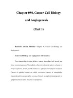
uni- and bi-sporangiate taxa, containing the pollen grains. The microsporangia are surrounded
by an epidermal layer followed on the inside by
the wall layers; the latter are made up of an endothecium, middle layers and a tapetum covering
the sporangial locule (Fig. 17.1).
The anther primordium in transectional view
is almost squarish to rectangular and is made of
homogeneous parenchymatous tissue, covered
by an epidermal layer. The archesporial tissue
differentiates as a single or a group of two to a
few adjacently located cells in the hypodermal
position at the four corners of the anther primordium. This tissue, in fact, extends vertically from
base to the apex of the sporangium. The cells of
this tissue are distinct from the rest of the anther
tissue by their larger size and greater avidity for
nuclear and cytoplasmic stains. The archesporial
cells divide periclinally to form outer primary
parietal cells and inner primary sporogenous
cells. Both these may undergo further periclinal
(and a few anticlinal) divisions to respectively
form the wall layers and the sporogenous cells
(Fig. 17.1); rarely the latter directly function as
sporogenous cells. Based on variations in anther
wall development and the number of wall layers
present, four types are recognized by Davis
(1966): basic, dicot, monocot and reduced types.
One of the earliest genes required for cell division and differentiation in the anther is the
SPOROCYTELESS (SPL)/NOZZLE (NZZ) gene
(Schiefthaler et al. 1999; Yang et al. 1999).
In the spl/nzz mutant, archesporial initiation
occurs normally, but male sporocyte differentiation is halted and anther development fails to
continue. The mutant genes of EXTRA
SPOROGENOUS
CELLS
(EXS)/EXCESS
MICROSPOROCYTES1 (EMS1) alter the number of archesporial cells. Two other genes
SOMATIC EMBRYOGENESIS RECEPTORLIKE KINASE1 (SERK1) and SERK2 also have
redundant functions during the earlier stages of
anther development and, when mutated, result in
more sporogenous cells (Albrecht et al. 2005;
Colcombet et al. 2005).
17.2.1 Endothecium
The endothecium forms a single layer of hypodermal wall tissue; occasionally, more than one
layer may be present in some taxa or may be
totally absent as in cleistogamous flowers, aquatic
plants and extreme saprophytes. The cells of
17
Pre-fertilization: Reproductive Growth and Development
411
Fig. 17.1 (a–t), (a–n) Trachyspermum ammi, (o–t)
Cuminum cyminum. Microsporangium (a, c, e, f, j, k, m).
Outline diagrams for (b, d, f, h, j, l) and (n), respectively,
showing development of anther. (b, d, f, h, j, l, n)
Enlargements of portions marked X, X1, X2, X3, X4, X5
and X6 in (a, c, e, g, i, k) and (m), respectively. (o, p)
Endothecial cells showing thickenings (from whole
mounts) (q, r) lateral and surface views of endothecial
thickenings. (s) Outline diagram of mature anther (t, s). (t)
Same, enlargement of portion marked (Sehgal 1965)
endothecium are often radially elongated and
develop special banded thickening in the inner
tangential walls and rarely on radial walls also
when the sporangium fully matures (Fig. 17.1).
The thickening material is not callose but an
α-cellulose; in some it may be slightly lignified.
Transcriptional activity is required for the differ-
entiation of endothecium as is evident from the
localization of poly(A)-RNA in rice microsporangia by in situ hybridization using [3H] poly(U)
as a probe (Raghavan 2000). Just before meiosis
poly(A)-RNA concentration decreases sharply in
the epidermis and middle layers, a large amount
of this is retained in the endothecium. Even after
412
the completion of meiosis in the microspore
mother cell, some amount of poly(A)-RNA is
retained in the endothecium. In rice and wheat
anthers, the histone H3 gene also activates the
endothelial differentiation, particularly in the
wild-type and transgenic rice; however, the
mechanism of this differentiation is not yet clear.
The importance of endothecium in anther dehiscence and the way in which the latter occurs are
detailed on a subsequent page of this article.
17.2.2 Tapetum
As already stated, the innermost wall layer of the
microsporangium is the tapetum. To start with, it
borders on the sporogenous cells, and because of
its strategic position between the other wall layers and the sporogenous cells, it assumes great
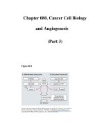
significance and importance. Although it is found
as a single layer all around the sporogenous tissue, it has been shown to have a dual origin
(Fig. 17.2). The tapetal cells towards the outer
sector of the microsporangium are derived from
the primary parietal tissue, while those towards
the centre of the anther are derived from the connective tissue. Although evidences of dual origin
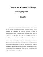
Fig. 17.2 Development of
anther (1–4) to show dual
origin of anther tapetum.
Single-hatched portion of
the anther tapetum is of
parietal origin, while
double-hatched portion is
derived from the connective tissue (Periasamy and
Swamy 1966)
K.V. Krishnamurthy
of tapetum are lost eventually and become a
homogeneous layer in many taxa, there are differences in cell size, shape, number of cell layers,
nuclear size, shape and ploidy or time of differentiation, etc. between proximal and distal tapeta
(Periasamy and Swamy 1966).
Two distinct types of tapeta are known in
angiosperms: (1) glandular, secretory or parietal tapetum in which the cells retain their walls
and persist in situ without much change in shape
and position until they perish by programmed
cell death (PCD) (Fig. 17.1). The tapetal PCD,
as the PCD seen in many other plant cells, is a
highly orchestrated event that occurs synchronously with pollen mitotic division and formation of pollen exine (Sanders et al. 1999). It is
relatively rapid and shows chromatin condensation, DNA fragmentation and mitochondrial and
cytoskeletal disintegration (Papini et al. 1999;
Love et al. 2008); (2) periplasmodial tapetum, in
which the cells lose their inner tangential and
radial walls due to enzymatic action of the tapetal cells themselves followed by the coalescence
of the protoplasts of all tapetal cells to form a
viscous fluid that flows into and fills the sporangial cavity all around the developing microspore
mother cells. The former type is more common
17
Pre-fertilization: Reproductive Growth and Development
in dicots, while the latter in the monocots. The
glandular tapetal cells are richly protoplasmic,
and their nuclei are prominent and metabolically
active; in some taxa, nuclei increase in number
(two to eight), become polyploidal (due to
nuclear fusion or endomitosis) or become polytenic (up to 16 times increase in DNA content).
Crystals, starch, lipids, mitochondria, Golgi
bodies, ER, membrane-bound ribosomes, plastids, etc. are reported in the tapetal cells. The cell
walls are cellulosic. The walls of periplasmodial
tapetal cells, before the formation of periplasmodium, have more pectin than cellulose. The periplasmodium is an organized structure. It gets
dehydrated before its complete degradation. A
third type of tapetum is often recognized and is
named amoeboid tapetum (some botanists mistakenly call the periplasmodial tapetum as amoeboid tapetum; see Swamy and Krishnamurthy
(1980) for discussion on this). In this type, the
cells radially elongate conspicuously and protrude into the sporangial cavity, without, however, losing their cell walls. This type is
associated with some types of male sterility.
The tapetum has been considered as a nurse as
well as a regulatory tissue for the developing
male gametophyte. Many indirect evidences are
there to implicate the tapetal cells as sources of
deoxyribosides which would then be used for
DNA synthesis by the microspores, although
actual transfer of these from tapetal cells could
not be directly demonstrated. There are circumstantial evidences to indicate that carbohydrates
and pollen reserves may result, at least partially,
from the transfer of soluble sugars and peptides
or amino acids from the tapetal cells. In many
plants, there is a close correspondence between
tapetal disintegration and the appearance of pollen reserves.
The most important function of the tapetum is
to supply pollen wall and pollen coat polymers
(Piffanelli et al. 1998). The glandular tapetal cells
contain in their cytoplasm numerous bodies,
often attached to the lipid membrane-bound,
electron-dense organelles known as pro-ubisch,
pro-sphaeroid or proorbicule bodies. The shape
of these bodies varies considerably: granular,
rod-shaped, star-shaped, circular, perforated
413
disc-like or compound multiperforate platelike.
They accumulate as ubisch bodies near the
plasma membrane before disappearing from
inside the cell. They are then immediately seen
on the exine of the microspores, where they get
integrated as sporopollenin (Fig. 17.3). Hence,
ubisch bodies are often considered as transport
forms of sporopollenin. The periplasmodial tapetum, after excessive dehydration, gets deposited
on the surface of microspores/pollen grains to
form tryphine, a complex mixture of lipoidal substances. There is also a deposition of pollenkitt.
Tapetum controls male fertility/sterility through
its timely/untimely production of the enzyme
callase (=β-1,3-glucanase). In fertile anthers, it is
produced by the tapetum when the callose wall
around the microspore tetrad needs to be dissolved to release the individual microspores,
while in sterile anthers, the enzyme is often produced precociously to dissolve the callose wall
around the microspore mother cell before it
undergoes meiosis. Some tapetum sequences
from anther cDNA libraries of Brassica napus
and Arabidopsis specify β-1,3-glucanase. Genes
that encode proteinase inhibitors of β-1,3glucanase action have been isolated from anthers.
In situ hybridization with [3H] poly(U) has
revealed that mRNA accumulation is one of the
metabolic activities that prepares tapetal cells for
their function. Commensurate with this high metabolic activity, the tapetal cells show the activities of a number of genes. At least five
tapetum-specific mRNAs and two mRNAs that
are also seen in other anther tissues (TA series
mRNAs) were demonstrated by in situ hybridization and by the use of chimeric gene constructs in
transgenic plants even as early as 1990 (Koltunow
et al. 1990). These mRNAs get accumulated and
lost in the same temporal sequence during tapetum ontogeny and have been identified from a
cDNA library of tobacco. One of these is TA29
whose product is a glycine-rich cell wall protein
that is likely to be involved in exine formation.
Subsequent studies have revealed the expression
products of several other genes.
An Arabidopsis gene, MALE STERILITY2
(MS2) (Wilson et al. 2001; Ito and Shinozaki
2002), is expressed in the tapetum, and the
K.V. Krishnamurthy
414
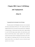
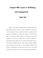
Fig. 17.3 Summary of
pollen wall developmental
stages (1–7) (sporoderm)
ontogeny of Sorghum
bicolor. Corresponding
developmental stages in
the anther locule are also
mentioned opposite to each
figure (Adapted from
Christensen et al. 1972;
Swamy and Krishnamurthy
1980)
DEVELOPMENTAL STAGE
primary wall
1
cytoplasm
primary wall
callose
Sporogenous mass
2
Melosis
3
Dyad − Early tetrad
4
primexine
bacula
5
6
7
−exine
tectum
columella
foot layer
endexine
Late tetrad
Earty vacuolate
microspore
Late vacuolate
microspore
Engorged pollen grain
intine
sequence similarity of this gene’s product to a
protein that converts fatty acids to fatty alcohols
has implicated this gene to pollen exine formation (Aarts et al. 1997). Its rice orthologue is
DEFECTIVE POLLEN WALL (DPW) (Shi et al.
2011). Loss of function of the FACELESS
POLLEN1/WAX2/YRE/CER3 gene causes defects
in exine; this gene is likely to encode a putative
enzyme of unknown function presumably
involved in pollen wall formation (Ariizumi et al.
2003). The other rice genes important in tapetal
function are WAX-DEFICIENT ANTHER1
(WDA1), OsC6 and PERSISTENT TAPETAL
CELL1 (PTC1). Fairly recently, Arabidopsis
genes encoding the cytochrome P450 enzymes of
CYPTO3A2 and CYP704B1 have been shown to
be involved in the biosynthesis of sporopollenin
(mutants have severe to moderate defects in exine
deposition) (Morant et al. 2007; Dobritsa et al.
2009). De Azevedo Souza et al. (2009) have
shown that ACYL CoA SYNTHETASE5 (ACoS5)
encodes a fatty acyl synthetase that plays a vital
role in exine formation and sporopollenin
biosynthesis in Arabidopsis; the acos5 mutant is
totally male sterile with pollen lacking recognizable exine. Genes that co-regulate along with
ACoS5 in pollen exine formation in Arabidopsis
such as DIHYDROFLAVONOL4-REDUCTASE
LIKE1 (DRL1)/TETRAKETIDE α-PYRONE
REDUCTASE1 (TKPR1) (Grienenberger et al.
2010) are also very important, as they affect male
sterility (Tang et al. 2009). DRL1/TKPR1 is
involved in flavonoid metabolism and plays a
pivotal role in sporopollenin precursor biosynthesis. It was also reported recently that the
enzymes closely related to chalcone synthase
(CHS) encoded by At1gO2050 [LESS ADHESIVE
POLLENS (LAP6)/POLYKETIDE SYNTHASEA
(PKSA)] and At4g34850 (LAP5/PKSB) catalyses
the sequential condensation of a starter acyl-CoA
substrate with malonyl-CoA molecules to produce alkylpyrone in vitro (Dobritsa et al. 2010).
PKSA and PKSB are specifically and transiently
expressed in tapetal cells during microspore
development in Arabidopsis anthers, mutants of
PKS genes displayed exine defects and a double
17
Pre-fertilization: Reproductive Growth and Development
pksa pksb mutant was completely male sterile
with no apparent exine; these results show that
hydroxylated α-pyrone polyketide compounds
generated by the sequential action of ACoS5 and
PKSA/B are potential and previously unknown
sporopollenin precursors (Kim et al. 2010).
The other genes which are involved in tapetum development and function are ABORTED
MICROSPORES (AMS) (Sorensen et al. 2003),
the rice orthologue TATETUM DEGENERATION
RETARDATION (TDR) (Li et al. 2006), TAPETAL
DETERMINANT1 (TPD1) (Yang et al. 2003),
DYSFUNCTIONAL TAPETUM (DYT1) (Zhang
et al. 2006), the rice orthologue UNDEVELOPED
TAPETUM (Jung et al. 2005), DEFECTIVE IN
TAPETAL DEVELOPMENT AND FUNCTION1
(TDF1) (Zhu et al. 2008), MYB80 (formerlyMYB103) (Higginson et al. 2003; Li et al. 2007;
Zhang et al. 2007), ECERIFERUM1 (CER1) (Shi
et al. 2011) and MS1 (Wilson et al. 2001). TDF1
encodes MYB; tdf1 mutant also shows enlarged
tapetum with increased vacuolation (Phan et al.
2011) and causes arrest of microspore development. Early tapetal initiation is affected by the
downstream genes EXTRA SPOROGENOUS
CELLS (EXS)/EXCESS MICROSPOROCYTES1
(EMS1) (Cannales et al. 2002; Zhao et al. 2002)
and TPD1. Mutants in these genes have an
absence of tapetal and middle layers. Mutations
in SERK1 and SERK2 genes result in the lack of
a tapetal layer. MYB33 and MYB65 also act
redundantly to facilitate tapetal development
around meiosis stage; it has been shown that the
expression of MYB33 is regulated by miRNAs
(Millar and Gübler 2005). These genes are not
affected in the dyt1 mutant indicating that they
are upstream of DYT1 (Zhang et al. 2006). In the
dyt1 mutant, tapetum occurs (also meiosis), but
tapetum development is abnormal with enlarged
vacuoles in its cells. DYT1 (by encoding basichelix-loop-helix proteins) has been proposed to
be involved in the regulation of many tapetal
genes, either directly or indirectly, including
AMS and MS1 (Zhang et al. 2006). The ams (its
wild gene AMS also encodes basic-helix-loophelix proteins) mutant has premature tapetal
degeneration because of its abnormally enlarged
and vacuolated cells.
415
Detailed studies have been done on the role of
MS1 gene in tapetal development and pollen wall
biosynthesis (Yang et al. 2007). Early events in
anther development in ms1 mutant are normal
and that the MS1 acts, through encoding PHD
transcription factors, late in pollen development
after tapetal initiation and is downstream of DYT1
(Zhang et al. 2006). MS1 coordinates the expression of late genes associated with pollen wall formation and which are involved in the biosynthesis
of components of the phenyl-propanoid pathway,
long-chain fatty acids and phenolics, which are
required for sporopollenin biosynthesis. In the
ms1 mutant, tapetal PCD does not occur, but
tapetal degeneration occurs by necrosis (VizcayBarrena and Wilson 2006); there is also downregulation in the expression of a member of cys
proteases in ms1 mutants. These proteases are
likely to be critical to the progression of PCD,
and in their absence, possibly in association with
a lack of tapetal secretion, PCD does not occur.
MS1 also controls the synthesis of pollen coat
(oleoresin gene family, lipid transfer proteins or
LTPs, ACP lipids and phenyl-propanoid pathway); it does not directly regulate genes associated with pollen wall biosynthesis (due to its
timing of expression) but acts via one or a number of additional transcriptional factors (TFs)
including MYB99 and two NAM genes that contain a conserved NAC domain (Yang et al. 2007).
Based on an analysis of transcript levels within
tdf1 and ams mutants, Zhu et al. (2008) suggested
that TDF1 functions upstream of AMS and that
AMS is upstream of MYB80. Xu et al. (2010)
identified 13 genes as direct targets of AMS, but
MYB80 was not among them. Transcript levels of
MS1, MS2 and A6 are downregulated in the
MYB80 mutant, suggesting that they act downstream of myb80. It is not known if the three
genes are directly or indirectly regulated by
MYB80. MYB80 is recently shown (Phan et al.
2011) to directly target a glyoxal oxidase
(GLOX1),
a
pectin
methyl
esterase
(VANGUARD1) and an A1 aspartic protease
(UNDEAD), all of which are expressed in the
tapetum and microspores. The timing of PCD in
tapetum is likely to be regulated by
MYB80/UNDEAD system. The overall genetic
416
K.V. Krishnamurthy
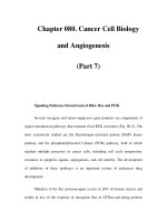
Fig. 17.4 Successive divisions of microspore mother cell of Lilium regale (Gerassimova-Navashina 1951)
regulation of sporopollenin synthesis and pollen
exine development is reviewed by Ariizumi and
Toriyama (2011).
17.2.3 Microsporogenesis
and Microgametogenesis
The sporogenous cells either directly or after a
few divisions give rise to microspore mother cells
(MMCs). The MMCs possess thin cellulosic cell
walls with plasmodesmal connections, not only
between themselves but also with the tapetal
cells. Dictyosomes and plastids (without starch
grains) are characteristically present in the cells.
Most DNA synthesis in MMCs is done during
premeiotic interphase, but a meager amount is
also synthesized during zygotene-pachytene.
Similarly, active RNA and protein synthesis takes
place during premeiotic stage with a fall during
meiotic prophase. There is a decline in ribosomal
population after the initiation of meiosis, but the
population is restored after homotypic division.
There is also a reorganization of mitochondria
and plastids in the microspore, as they are partly
degraded during meiosis. Just at the onset of meiosis in MMCs, a callose wall is deposited inner to
the original cellulosic wall. Any irregularity in
callose deposition/metabolism results in male
sterility. Callose deposition starts on the walls of
MMCs close to tapetum and gradually extends to
the more centrally located cells of the anther.
Initially, the callose wall is incomplete leaving
many gaps in the wall through which massive
cytoplasmic channels between adjacent MMCs
(but not with tapetum cells) are established.
These channels reach their maximum development during zygotene-pachytene and help establishing near synchronicity in meiosis in all
MMCs of a sporangium. Callose deposition is
considered as a necessary prerequisite for meiotic induction and continuance (Krishnamurthy
1977, 2015). Callose is highly impervious to
most molecules and thus is a highly isolating and
insulating material. The plasmodesmal connections are sealed off towards the end of metaphase
I in taxa with successive division and at anaphase
II in plants with simultaneous division.
Two types of meiotic division are known in
MMCs, either of which results in the formation
of a tetrad of four microspores. In successive
division, a centrifugally extending cell plate and
then a wall are promptly laid down between the
daughter nuclei at the end of each of the two divisions (Fig. 17.4). In the simultaneous division,
the separation of all four microspore nuclei is
17
Pre-fertilization: Reproductive Growth and Development
417
Fig. 17.5 Trachyspermum ammi. Microsporogenesis and
male gametophyte; (a–j). Simultaneous meiotic division
in microspore mother cell leading to tetrad formation;
(k–n) Uninucleate microspore. (o–p) Two-celled pollen.
(q) Three-celled pollen; (r) Palynogram (Sehgal 1965)
effected through centripetally extending furrows
at the end of the second division (Fig. 17.5a–j).
The callose wall around the tetrad is heterogeneous and layered. The outermost layer is the
most well developed. Three more concentric layers follow this on the inside distinguished from
each other by their variable density. The fifth
layer is the innermost and the least dense of all. It
surrounds and isolates the four microspores and
cell plates. Each microspore is individually surrounded by the primexine. Soon after meiosis,
callose wall around the microspore tetrad is
degraded by β-1,3-glucanase into D-glucose and
oligomers of D-glucose of different lengths,
which may be used by the microspores for various purposes (such as nutrition and pollen wall
formation). As a result of callose degradation, the
individual microspores are separated out of the
418
tetrads. β-1,3-glucanase is present in low quantities in the tapetum even during meiosis in MMCs,
but increases suddenly during late tetrad stage to
cause the separation of microspores. In some
angiosperms, failure of microspores to separate
out of the tetrads results in the formation of permanent tetrads or compound pollen grains. In
some Mimosaceae and Orchidaceae, polyads of
8–32 grains called massulae are formed. An
extreme case of adherence of all pollen grains of
an entire microsporangium is seen in many
Asclepiadaceae and the resultant structure is
called a pollinium.
The studies made so far show that both the
diploid sporophytic tapetal cells and the haploid
gametophytic microspore contribute to pollen
wall synthesis (Ariizumi and Toriyama 2011).
Exine formation is stated to commence from the
late tetrad stage with the laying down of the primexine between the callose wall and the plasma
membrane of the microspore (Paxson-Sowders
et al. 1997) (except at the germinal pore region
where it is absent). The microspore just released
from the tetrad does not have an exine (the outer
wall of the pollen). The primexine is distinguished from the callose by its electron opacity.
It has a matrix, presumably made up of cellulose,
and radially directed rods, the probaculae and
profoot layer. The deposition of sporopollenin
begins immediately after release of microspores
from the tetrad, and its source is from the tapetum, as already detailed. The characteristic pattern of the sporoderm is determined by features
already imprinted in the primexine during the
period of enclosure in the tetrad (Blackmore et al.
2007). However, a few investigators believe that
the initial exine pattern laid down in the microspore is controlled by the plasma membrane and
that callose causes this imprinting by acting as a
template (and not the primexine). After the first
division of the microspore, exine formation is
almost complete. At later stages of pollen ontogeny, pectocellulosic intine and tryphine are
deposited (Piffanelli et al. 1998). Intine formation first begins in the vicinity of the germinal
aperture(s) and from there spreads all around the
microspore; this growth is said to be associated
with dictyosome activity in coordination with the
K.V. Krishnamurthy
plasma membrane. Thus, intine is programmed
entirely by the haploid, male gametophytic
genome and is made of pectocellulose, while the
exine is organized both through tapetal inputs
and microspore activity.
Under typical conditions, the microspore
nucleus occupies a central position, while the
cytoplasm has many small vacuoles spread
almost evenly (Fig. 17.5k–n). Just before division, the nucleus moves towards a side that is
generally opposite to the furrows. Mitochondria
and plastids are displaced to the cytoplasm opposite to the nucleus. During interphase, active
ribosomal RNA synthesis takes place. A conspicuously large vacuole appears in the cytoplasm
opposite to the nucleus. The nucleus then divides
followed by a curved callose wall to result in a
small lens-shaped daughter cell (appearing spindle shaped in cross-sectional view) called the
generative cell (GC) and a conspicuously larger
cell called vegetative cell (VC) (Fig. 17.5o, p).
Thus, the division is asymmetric. The callose
wall separating the GC from VC is highly transitional and is retained only for about 10–20 h. GC
soon gets pinched off from the microspore wall
and becomes embedded in the cytoplasm of the
VC, by which time its callose wall is also lost.
This may or may not be accompanied by a change
of shape of the GC. This separation is effected by
the growth of callose wall in between the plasma
membrane of the GC and the intine of pollen
grain. The new location of GC obviously provides a new environment for interaction between
GC and VC. At this stage, the pollen is said to be
mature in most taxa. The GC is surrounded by a
double membrane, by a distinct cellulosic wall or
by the retention of the original callose wall
depending on the species. The GC is less dense
due to very poor or even no RNA and proteins.
Minute vacuoles filled with water or lipid materials are also present. The DNA content of its
nucleus is very high (rises to 2C level), but the
nucleolus is not very conspicuous. Axial microtubules have been recorded and these are important in controlling the shape of the GC. However,
there is some disagreement regarding the cytoplasmic organelles of the GC, probably because
of species-dependent variations. Mitochondria,
17
Pre-fertilization: Reproductive Growth and Development
dictyosomes, lipid bodies and ER have been
reported. Plastids have not been detected in many
species, although reported in a few taxa. In general, GC is poor in organelle content and variety.
In contrast, the VC shows dense cytoplasm due to
greater amount of RNA and proteins. The nucleus
is invariably lobed and poorer in DNA content
(mostly at the 1C level) and has a relatively large
nucleolus. Thus, the nucleus of GC switches on
DNA synthesis, but there is no appreciable RNA
or protein synthesis as transcription is slowed
down, while the nucleus of VC switches off DNA
synthesis but without interfering with transcription (Raghavan 2000). VC may have starch or oil
as a major storage product.
The division of the GC into two male gametes
or sperms takes place either in the pollen itself
(in about 25 % of the angiosperms) (Fig. 17.5q)
or in the pollen tube. Hence, the pollen grains are
liberated from the anther at the two- or threecelled condition. Division of GC in the pollen
grain is due to normal mitosis followed by cytokinesis through cell plate formation or through
furrowing. The mechanism of division of GC in
the pollen tube is not very clear because of
difficulties in studying due to spatial restraints; it
appears to be normal mitosis. The organelles
reported in GC are also recorded in the two
sperms. DAPI staining and fluorescence
microscopy have indicated the absence of plastid
DNA in the sperm cells (in 82 % of species
surveyed).
17.2.4 Genetics of Microsporogenesis
and Microgametogenesis
Cytochemical, autoradiographic, biochemical
and molecular studies on RNA and protein
synthesis have indicated that pollen development is controlled by a temporal and spatial
programme of differential gene expression.
The period leading up to the first division of
the microspore is marked by major contribution of rRNA in the total RNA synthesized.
This is consistent with the opinion that among
the multiple copies of 5s RNA genes that control pollen development, some are switched off
419
after the peak synthesis, while a few persist for
an additional period. Quantitative variation in
mRNA populations has also been noted during
pollen development. The mRNA that gets
accumulated in the mature pollen may serve as
templates for the first proteins in germination.
Both qualitative and quantitative differences
are detected in the proteins synthesized during
different stages of pollen development. Such
proteins include lysine- and arginine-rich histones which accumulate in the GC and sperm
nucleus, and they are linked to transcription of
the haploid microspore/pollen genome. Stress
proteins such as extensins and arabinogalactan-rich proteins, which are important in
incompatibility reactions, are also synthesized
by the developing pollen.
All the above imply active gene expression.
The genes involved in pollen development have
been isolated and characterized, especially in
Arabidopsis, Brassica napus, B. oleracea, cotton,
Lilium, Oenothera, Petunia, tomato, Tradescantia
and Zea mays. The isolated genes were found to
be members of small gene families present in one
or two copies in the genome, and none appeared
to belong to large multigene families (Raghavan
2000). As already indicated, both sporophytic
and gametophytic genes are involved. Transcripts
of two distinct sets of gametophytic genes are
shown to be activated in specific temporal and
spatial patterns. Transcripts of the first set, commonly called early genes, become active at the
tetrad stage or at the latest when microspores are
released from the tetrads, but these have only
short periods of activity. Some of the early genes
are importantly needed for coding cytoskeleton
elements. One of the very early products is the
DEFECTIVE IN EXINE PATTERN FORMATION
1 (DEX1) gene protein; it is a putative membraneassociated protein with predictable proteinbinding domains. The dex1 mutant in Arabidopsis
delays primexine formation, and hence, the sporopollenin synthesized by the tapetum is abnormally deposited on the mutant microspore surface
(Paxson-Sowders et al. 1997). Recently, Kim
et al. (2011) have shown that ER- and Golgilocalized phosphatases gene A2 (PLA2) plays
critical roles in Arabidopsis pollen development
420
and germination. These authors have
characterized three to four Arabidopsis PLA2
paralogues and found that they are expressed during pollen development, germination and pollen
tube growth. Suppression of PLA2 using RNA
interference approach resulted in pollen lethality
and inhibition of tube growth.
It was already shown that there are phenotypic
differences between the GC and VC. The genetic
basis of these phenotypic differences has been
analyzed by using transgenic molecular markers
and in situ hybridization techniques with cloned
genes. An associated asymmetry in gene expression is seen along with the asymmetric cell division that results in GC and VC. Mitotic division
is not a prerequisite for expression of VC-specific
gene(s) but a symmetric division silences gene
expression in GC. Twell (1995) has shown that
ablation of VC of transgenic tobacco pollen by
the cytotoxin DTA gene linked to the tomato
pollen-specific gene inactivates the GC and prevents its function (Raghavan 2000). How the VC
controls the activity of GC is not clear. The late
genes become active after the microspores divide
and their activity continues till pollen tube growth
(Stinson et al. 1987). The proteins encoded by
late genes include pectin lyases, pectin esterase,
polygalacturonase, protein kinases, ascorbate
oxidase, thioredoxins, actin-depolymerizing factors, zinc finger class proteins, RNA helicases,
pollen allergins, ATPase, osmotin, stress proteins,
PR-proteins, malate synthase, superoxide dismutase, etc.
Attention should also be focused on the
MIKC*-type type II MADS-box genes that affect
development of male gametophyte. Combinations
of double and triple mutants of agl65, agl66,
agl104 MADS-box genes give rise to several pollen phenotypes with disturbed viability, delayed
germination and aberrant pollen tube growth
(Adamczyk and Fernandez 2009; Smaczniak
et al. 2012). The gene products form a protein
interaction and regulatory network controlling
pollen maturation. These also regulate transcriptome dynamics during pollen development.
A detailed account on tapetal genes (i.e. sporophytic genes) was already provided. Most, if
not all, of them affect the pollen development in
K.V. Krishnamurthy
diverse ways, either directly or indirectly. For
example, mutation in AMS and DYT1 genes
causes degeneration of microspores. In
Arabidopsis, a candidate gene called QUARTET
(QRT) is required for the separation of microspores from the tetrads. It is probably a tapetal
gene. A mutation of this gene causes a patchy
formation of callose between the microspores in
the tetrad (Preuss et al. 1994); there is also a
fusion of the microspores through their developing exine due to a failure of pectin degradation.
Another well-studied gene is the DUO POLLEN1
(DUO1) gene (see Zheng et al. 2011). It encodes
a male germ cell-specific R2R3Myb protein
(Rotman et al. 2005) that is required for the
expression of the Arabidopsis thaliana G2/M
regulator cyclin B1;1 (CYCB1;1) in the male
germline (Brownfield et al. 2009a, b), suggesting
an integrative role for DUO1 in cell specification
and cell cycle progression that is necessary for
twin sperm cell production. DUO1 mRNA is
directly targeted by miRNA159, which leads to
its degradation. Whether APC/C is required for
DUO-1-dependent CYCB1;1 regulation is
unknown (Zheng et al. 2011). Mutants in both
APC8 and APC13 had pleiotropic phenotypes
resembling those of mutants affecting miRNA
biogenesis. Zheng et al. (2011) have shown that
these apc/c mutants have reduced miR159 levels
and increased DUO1 and CYCB1;1 transcript
levels and that APC/C is required to recruit RNA
polymerase II to MIR159 promoters. Thus, in
addition to its role in degrading CYCB1;1,
APC/C stimulates production of miR159, which
downregulates DUO1 expression, leading to
reduced CYCB1;1 transcription. Both MIR159
and APC8 protein accumulated in unicellular
microspores and bicellular pollen, suggesting
that spatial and temporal regulation of miR159
by APC/C ensures mitotic progression.
Consistent with this, the percentage of mature
pollen with no or single sperm-like cells
increased in apc/c mutants and plants overexpressing APC8 partially mimicked the DUO1
phenotype. Thus, APC/C is an integrator that
regulates both miRNA-mediated transcriptional
regulation of CYCB1;1 and degradation of
CYCB1;1 (Zheng et al. 2011) (Fig. 17.6).
17
Pre-fertilization: Reproductive Growth and Development
Fig. 17.6 A model for the dual roles of APC/C in regulating cyclin B1;1 during male gametophyte development
(Based on Zheng et al. 2011)
17.2.5 Anther Dehiscence
Almost simultaneously with the maturation of
microsporangia, the wall layers aligned in the
groove between the adaxial and abaxial pairs of
sporangia fail to undergo histological modifications. This linear strip of tissue is the stomium
which predetermines the place of future anther
dehiscence. Due to the continued bulging out of
the distal anther wall, the stomium appears to be
seated in a furrow. The cells of the stomium form
the weak zone in the anther wall. The few parenchyma cell layers that separate the adaxial and
abaxial sporangia that form a septum are resorbed
towards anther maturity causing the merger of
the sporangia on either side of the anther. At
about this time, the stomial cells slightly elongate
radially obviously due to the pressure exerted by
the bulging anther wall on either side. Meanwhile,
the mechanical action of the endothecium causes
an evagination of the anther wall along the stomial direction. Finally, the anther dehisces to
release the pollen. In some taxa, pollen is released
through apical pores or valves in the sporangia,
while in cleistogamous and aquatic taxa, the pollen is released through the disintegration of the
anther wall.
A critical analysis of anther-specific cDNA
clones in tobacco supports the contention that the
whole programme of anther wall differentiation,
degeneration of middle layers and anther dehiscence consists of a cascade of temporal and spatial gene expression events in the anther wall
(Raghavan 2000; Krishnamurthy 2015). The
cDNA clones implicated in this programme are
TA56, encoding a thiol endopeptidase, and TA20,
421
encoding an unknown protein. By following the
expression of these two clones by in situ hybridization, it was shown that TA56 transcripts accumulated in the anther wall in the prospective
stomial region at a very early stage of anther
development. As the anther matures, there is an
appreciable decrease in the intensity of hybridization signals in the cells in and around the stomium. At the same time, the cells around the
connective tissue acquire the hybridization signal. TA20 transcripts are seen in all layers of
anther wall to start with, but with anther maturation, they are concentrated in the connective cells
around the vascular bundles. Selective expression
of TA56 gene transcripts in the stomial region
suggests a role for endopeptidase in anther dehiscence (Koltunow et al. 1990). When the stomial
region alone is ablated with a cytotoxic gene
fused to the TA56 gene promoter, anther development was normal, but fails to dehisce (Beals and
Goldberg 1997), again supporting the above contention. Transcripts of anther-specific cDNA
clones isolated from tomato anthers are also
expressed in the wall layers, particularly in epidermis and endothecium. The protein encoded by
these genes show homology to Kunitz trypsin
inhibitor (KTI) and pectinase enzyme.
The events in anther dehiscence are also mediated by structural features of the filament since
the former is dependent on the latter for the transport of water and nutrients. Dehiscence is largely
a desiccatory process, and any histological feature promoting rapid water loss from the anther
or disruption of water to the anther might facilitate dehiscence. Open stomata, a weakly developed cuticle, prominent intercellular space
system and xylem lacunae of the filament are
some of these histological features. There are
hygroscopic and cohesive mechanisms involved
in desiccatory anther dehiscence. Hygroscopic
mechanisms depend entirely upon volumetric
changes in the cell walls, whereas cohesion
mechanisms involve volumetric changes in the
cell lumen, the cell walls merely undergoing passive deformation. Cohesive mechanisms largely
involve cohesive forces between water molecules
in the cell lumen. Dehiscence occurs when cohesive forces are exceeded. In hygroscopic
K.V. Krishnamurthy
422
mechanisms, adhesive forces are important.
Although most people accept cohesive mechanism, both appear to be important.
At the time of dispersal, pollen grains are
partly dehydrated with a water content of
10–30 %. Further water loss occurs during pollen
transport resulting in a condition similar to that in
dry seeds. The pollen becomes metabolically
poor in activity due to disorganization of the
membrane systems of ‘vegetative cell’ organelles
and plasmalemma. The effect of dehydration is
also evident on the cell walls. Apparently, this
dehydration helps the dispersal capability of pollen. Proline accumulation in the pollen cytoplasm
and the presence of some stress proteins in the
cell walls characterize such dehydrated pollen.
17.3
Ovule and Female
Gametophyte
The female gametophyte develops in a structural
unit called megasporangium, the female counterpart of the microsporangium. This term is generally employed for the megaspore (sometimes also
called the macrospore) bearing units of vascular
plants, but the same unit of seed-bearing plants
(spermatophyta) is called the ovule, which contains the nucellus, enclosed by one or two integuments. It is in the nucellus that the female
gametophyte gets differentiated. Unlike the nonovule-bearing vascular plants (pteridophytes),
fertilization of the egg (the female gamete developed inside the female gametophyte) and the
consequent development of the embryo is initiated while the ovule (future seed) is still attached
to the parent sporophyte.
17.3.1 Configuration of Ovule
The ovule primordium is initiated as a tiny protuberance on the placental tissue of the ovary.
While the primordium is growing in size, a small
annular tissue thickening appears just above the
point of attachment of the primordium to the placenta. This point corresponds to the location of
the chalaza or base of the ovule. This annular belt
grows at a relatively faster rate than the protuberance and soon encloses the latter leaving a pore at
the apex called the micropyle. The central protuberance becomes the nucellus, while the annular
belt becomes the integument; in some taxa, an
additional integument is formed in the same way
as the first. If the primordium continues to grow
straight throughout its course without showing
any change of direction, the configuration of such
an ovule is said to be orthotropous or atropous. In
such an ovule, at maturity, the funicle, or stalk of
the ovule, the chalaza [that part of the ovular tissue adjacent to the base of the integument(s)] and
the micropyle lie along the same vertical axis.
Changes in the direction of growth of the ovule
primordium result in other ovular configurations
such as anatropous, campylotropous, hemitropous and amphitropous where the imaginary
lines connecting the positions of funicle, chalaza
and micropyle form different types of triangles.
Details on structural variations in the ovules of
angiosperms are summarized in Kapil and Vasil
(1963), Swamy and Krishnamurthy (1980) and
Bowman (1984).
17.3.2 Nucellus
As already stated, the ovular tissue enclosed by
the integument(s) forms the nucellus. Depending
on the extent of this sporophytic tissue, two major
types of ovules are recognized: (1) tenuinucellate, where the nucellus is represented only by a
few cells, and (2) crassinucellate, where the
nucellus is massive. In the former type, the hypodermal female archesporial cell directly functions as the megaspore mother cell, while in the
latter it cuts off a parietal cell which undergoes
repeated divisions to not only form a massive
nucellus but also to push the megaspore mother
cell deep inside the nucellus. Crassinucellate
condition may also be contributed by active cell
division of apically located chalazal cells. In
some cases, the nucellar epidermis at the micropylar pole divides repeatedly periclinally to add
to the mass of the nucellus. This condition is
17
Pre-fertilization: Reproductive Growth and Development
423
Distal
D
C
Proximal
Axis (PD axis)
formation
P
Fig. 17.7 Diagrammatic representation of the proximodistal polarity in a developing ovule showing the three
pattern elements. The proximal (P) domain forms the
funicle, the central or chalazal (C) domain forms the cha-
laza-integument complex and the distal (D) domain forms
the nucellus and embryo sac. C domain may possibly have
two subdomains (Modified from Grossniklaus and
Schneitz 1998)
called pseudocrassinucellate (Davis 1966); here,
also the megaspore mother cell is pushed deep in
the nucellus. Nucellus is totally lost after fertilization in all tenuinucellate ovules and in many
crassinucellate and pseudocrassinucellate taxa,
but in a few as in Piper nigrum, it may persist in
the seed as perisperm.
laza. The vascular trace to the ovule also
terminates at the base of the chalaza; further
branching and ramification of the integumentary
vasculature, if present, is also seen in the chalaza
both before and after fertilization (Krishnamurthy
2015). Most pronounced growth of the embryo
sac invariably takes place only along the chalazal direction. Ovules of some species exhibit the
differentiation of a histologically distinctive pad
of cells in the chalazal region called hypostase
(also called postament, podium or pseudochalaza) whose cells are often thick walled; it is
believed to play a role in the supply of nutrients
to the growing embryo sac, as well as in stabilizing the moisture status of the ovule.
Thus, the chalaza serves as an important topocentre of the ovule all throughout the development of the ovule and the seed. Data on molecular
biology of ovule/seed development have indicated that the ovules develop and mature into
seeds by maintaining a distinct proximo-distal
axis (Grossniklaus and Schneitz 1998) and that
this axis is characterized by a three-tiered
arrangement of pattern elements: distal (or nucellar), chalazal (or central) and proximal (funicular) domains (Fig. 17.7). Chalazal domain can be
further subdivided into two subdomains (Baker
et al. 1997), based on its role in the production of
either the inner or outer integument. About a
dozen genes are already known to affect the
integuments in Arabidopsis by operating at the
chalazal domain (Table 17.1).
17.3.3 Chalaza
That part of the ovule that is subjacent to the
base of the integument may be designated as the
chalaza. It is the region of the ovule from where
the integuments originate and where there is no
distinction into nucellus and integument(s). The
chalaza indicates a pole of the ovule that serves
as a seat of very vital metabolic activities from
the very beginning of ovule organization to even
during post-fertilization stages. It is very difficult to delimit the boundaries of chalaza either
morphologically or physiologically. The importance of chalaza in the establishment of different
ovular configurations was already drawn attention to. Its importance as the point of origin of
the integument(s) has also been indicated. It is
also the location from where additional ‘integumentary’ structures like arils arise in arillate
taxa. Attention may also be drawn to the already
indicated fact that the basal increase in nucellar
volume in some crassinucellate ovules is contributed by the apically located cells of the cha-
424
Table 17.1 Chalaza as a topocentre of operation of
genes involved in ovule/seed development
S. no. Wild-type genes
Effect seen in mutants
whose mutant
forms are known to
be involved
1.
Inner no outer
Only inner integument
integument (INO)
formed; no outer
integument
2.
Both the integuments not
Bell 1 (BEL 1)
organized; inner totally
absent, while the outer
represented by an
amorphous entity or
collar-like structure
3.
Development of outer
Superman (SUP)
integument affected;
(also known as
grows asymmetrically
FLO10 gene)
around the ovule
4.
Short integument1 Integument development
affected; no clear
(SIN1) (maternal
distinction between inner
gene)
and outer integuments;
they are extremely short.
The gene also causes
defects in embryo-like
funnel-shaped cotyledon
or masses of unorganized
tissue
5.
Lacks integuments. Also,
Aintegumenta
there is no embryo sac
(ANT)
formation
6.
Lacks integuments. Also,
Huellenlos (HLL)
there is no embryo sac
formation
7.
Produces a single-fused
Aberrant testa
integument and hence no
shape (ATS)
distinction between the
two integuments
8.
Produces supernumerary
Unicorn (UCN)
integuments
9.
Affect cell division and
Blasig (BAG)
cell shape in integuments
10.
Affect cell division and
Strubbelig
cell shape in integuments
Affect ovule
11.
FBP7, FBP11
(MADS-box genes) determination and
development in carpel;
(in Barley and
no endosperm
Petunia)
development
All the genes in mutant form affect development (data
compiled from different sources)
K.V. Krishnamurthy
17.3.4 Archesporium,
Megasporogenesis
and Female Gametophyte
Although comprehensive reviews on the female
gametophyte have been published previously
(Maheshwari 1950; Swamy and Krishnamurthy
1980; Willemse and van Went 1984; Haig 1990;
Huang and Russell 1992; Russell 2001; Yadegari
and Drews 2004), in this article a consolidated
account is provided laying emphasis on molecular biological aspects. The female archesporium
is always differentiated at the nucellar apex in the
hypodermal position. Invariably only one archesporial cell gets differentiated, although there are
taxa where more than one cell may be formed in
an ovule. It is a very conspicuous cell, larger than
other nucellular cells, with a deeply staining
cytoplasm, a large nucleus and high nucleolar
RNA and with plasmodesmal connections with
adjacent nucellar cells. It cuts off through a periclinal wall an outer parietal cell and an inner sporogenous cell as already detailed; it remains near
the surface or fairly deep inside depending,
respectively, on tenuinucellate or crassinucellate
ovules. The sporogenous cell increases in size
and becomes the megaspore mother cell (MMC)
or megasporocyte (Fig. 17.8).
The MMC elongates parallel to the long axis
of the nucellus. Just prior to the initiation of meiosis, the plasmodesmal connections that the
MMC had with the nucellar cells are cut off, and
a callose wall is deposited around it. A failure of
callose deposition results either in female sterility or in unreduced apomictic development
(Krishnamurthy 1977, 2015). The deposition of a
callose wall is intrinsically related to the position
of the functional megaspore and the type of
embryo sac (female gametophyte) development.
There is an unequal deposition of callose on the
MMC, which, in fact, is related to the polarization of its organelles and the cell as a whole.
Callose is laid down first and thickest on the portion of the wall of MMC where the functional
17
Pre-fertilization: Reproductive Growth and Development
megaspore is destined to be formed, i.e. in the
chalazal pole in monosporic Polygonum, chalazal
half in bisporic Allium, micropylar pole in monosporic Oenothera, chalazal half in bisporic
Endymion and all around MMC in tetrasporic
types of embryo sacs. Very significant ultrastructural changes and regroupings of organelles of
MMC form the hallmark of the changeover from
the diploid to the subsequent haploid state. The
mitochondria and plastids dedifferentiate at the
early meiotic prophase only to redifferentiate at
the megaspore tetrad stage; they also became
highly dispersed and far removed from one
another in early prophase and again get regrouped
at the tetrad stage. The cytoplasmic and nucleolar
RNA concentrations decrease with the onset of
meiosis due to a prophase diminution or total loss
of ribosome content. A new array of ribosomes is
formed with the initiation of meiosis along with a
steady increase in the number of nucleoli of the
nucleus of MMC through budding. Initiation of
the production of polysomes and appearance of
paracrystalline inclusions are also seen towards
the end of meiosis.
Three basic types of embryo sac development
are conventionally recognized in angiosperms. In
the monosporic type, the MMC undergoes the
heterotypic division of meiosis to form two cells
separated by a thick callose wall, each of which
again divides (homotypic division) to form a tetrad of four haploid cells or megaspores. In the
monosporic Polygonum type, the chalazal megaspore alone is functional, while in Oenothera
type, the micropylar megaspore alone is functional. In the bisporic type, the MMC undergoes
the heterotypic meiotic division to form a dyad
where homotypic division proceeds either in the
chalazal (Allium type) or micropylar dyad
(Endymion type) only to form a binucleate (two
megaspores) functional cell. In both the monoand bisporic types, the non-functional megaspores/cells undergo PCD. The only functional
megaspore (nucleus) in monosporic and the two
functional megaspores (nuclei) in bisporic types
425
further divide to form the mature embryo sac
(female gametophyte). The major unanswered
question in the above four subtypes of female
gametophytic ontogeny is the following: what
determines the selection of the functional megaspore (cell)? Although, as stated earlier, there are
differences in the pattern of callose deposition, it
is not known whether it is the cause or the effect
of this determination. It is also to be mentioned
that the non-functional megaspore-containing
cells are always smaller than the functional cells
and that there is a definite asymmetry involved in
their production; asymmetric division is definite
to decide the different fates of the two resultant
cells. In the tetrasporic types, both the heterotypic and homotypic divisions of meiosis are not
accompanied by cell walls, and hence, a cell with
four megaspore (nuclei) called coenomegaspore
is formed. All the four megaspores here contribute to the formation of the mature embryo sac.
Thus, in these three basic types of female gametophytic ontogeny, the formation/identification of
the functional megaspore(s) is varied with reference to the heterotypic or homotypic meiotic
divisions; hence, these two divisions form an
important criterion in embryo sac ontogenies. In
view of this, a modified classification of embryo
sac types was proposed by Swamy and
Krishnamurthy (1975, 1980). Among the tetrasporic types, further classification is based on the
total number of nuclei, their relative position in
the mature embryo sac, nuclear fusion and the
consequent ploidy of the involved components of
the embryo sac, etc.
Once the required number of nuclei is formed
during megagametogenesis, depending on the
type of female gametophyte, their organization
sets in (Fig. 17.8). Invariably, three of the nuclei
at the micropylar pole organize into the cells of
the egg apparatus, with an egg cell in the centre
with two synergids, one on either side of the egg.
Two nuclei move to the middle part of the embryo
sac and get organized as part of the central
cell; these two nuclei are the polar nuclei. At the
426
K.V. Krishnamurthy
Fig. 17.8 Monosporic Polygonum type of embryo sac
development as found in Trachyspermum ammi and
Cuminum cyminum. (a) L.S. of ovule primordium. (b)
L.S. of ovule primordium with three archesporial cells. (c)
L.S. of ovule with enlarged archesporial cell. (d) Division
of archesporial cell. (e) Megaspore tetrads. (f) Functional
megaspore along with three degenerating megaspores. (g)
Two nucleate embryo sac. (h) Four nucleate embryo sac.
(i) Mature embryo sac showing egg apparatus at the
micropylar end three antipodals at the chalazal end and a
central cell with two polar nuclei (Sehgal 1965)
chalazal end of the embryo sac, three nuclei organize themselves into the antipodal cells. The egg
nucleus is towards its chalazal end, while a large
vacuole occupies its opposite end. The egg cell
normally has a wall only at its micropylar facet
with the chalazal region devoid of it. Whether a
thin wall is formed at the chalazal region of the
egg initially and then disappeared or whether
from the beginning there is no wall in this region
is a matter of controversy, since both conditions
17
Pre-fertilization: Reproductive Growth and Development
have been known in literature. The absence of a
wall is necessary for the easy transfer of male
gamete from the synergid. The ER of the egg cell
is oriented parallel to its plasmalemma over a
large part of it, but in more numbers around the
nucleus; ribosomes are also concentrated near the
nucleus. A very prominent nucleus is seen in the
egg cell. Large amount of cytoplasmic DNA is
present in the egg cell along with the nuclear protein, histone.
The two synergids are saccate and pyriform,
and their nucleus is situated at the micropylar
end of the cell with a large vacuole occupying at
its chalazal pole. The micropylar region has a
characteristic structure called filiform apparatus
(FA). The FA is a special type of wall ingrowth
that is a characteristic of transfer cells. The wall
ingrowths are finger- or platelike and are made
of fibrous polysaccharides. They have a central
core with tightly packed cellulose microfibrils
surrounded by a sheath of non-microfibrillar
material. The presence of FA functionally implicates the synergids as transfer cells, but the
direction of translocation, whether towards or
out of these cells, is not clear; it is more probable
that it is out of these cells that substances, especially chemotropic substances, are released
towards the micropyle probably to attract and
direct the pollen tubes. Cell ablation studies in
synergids of Torenia fournieri show their control
on both pollen tube guidance and reception
(Higashiyama et al. 2001). The material synthesized and released by the synergid consists of a
homogeneous osmiophilic substance indicating
its non-cellulosic composition, but it is likely to
be a carbohydrate that shows positive reaction to
periodic acid-Schiff’s reagent. In Nicotiana, one
of the two synergids becomes receptive to pollination and starts to accumulate more loosely
bound calcium (Tian and Russell 1997).
Synergids have a maximum concentration of ER
towards their micropylar end, and its density
gradually gets reduced towards the chalazal end;
dictyosomes are more concentrated in the middle part of the cell, while spherosomes are evenly
spread over the entire synergid cytoplasm
(Swamy and Krishnamurthy 1980). Only one
427
synergid is present in Peperomia type, while
they are absent in the Plumbago and Plumbagella
types of embryo sacs. In the latter two types, the
egg cell itself has a filiform apparatus. Synergid
haustoria are seen in some species.
The central cell is the largest cell of the female
gametophyte. It normally has two polar nuclei
lying juxtaposed to each other or exhibiting various degrees to fusion or total fusion to form a
diploid secondary nucleus. Only one polar
nucleus is present in the Oenothera type, while
more than two nuclei occur in Penaea, Plumbago
and Peperomia types. The central cell is highly
vacuolated, and its cytoplasm has a high amount
of active dictyosomes and ER, especially around
the nuclei. There is abundant cytoplasmic RNA,
mostly ribosomal. Free ribosomes and many
mitochondria are present, especially near its
micropylar end. The vacuole(s) of central cell is a
major reservoir of sugars, amino acids and inorganic salts. Starch is abundantly present in the
cytoplasm of the central cell. The wall of central
cell in maize is multilayered and pectocellulosic,
while in cotton it is rich in pectic substances. In
many taxa, the lateral walls show transfer cell
morphology. Normally, there are three antipodal
cells or nuclei located at the chalazal end of the
embryo sac. Antipodal cells with more than one
nucleus, syncytial antipodals, endoreduplicated
antipodal nuclei (reaching up to 1024C level)
showing polytenic chromosomes and/or nucleolar DNA amplification, increased number of
antipodals (up to about 300 reported in the grass,
Sasa paniculata) or ephemeral antipodals characterize some taxa. The antipodal cells often have
been suggested to play some role in the control of
endosperm development, at least in some grasses,
although often considered as inert structures
without any obvious function. Some fairly recent
studies indicate their involvement in secretion,
absorption and transport of nutrients to the central cell before and after fertilization. This is evident from the presence of active nucleoli,
ribosome-polysome pattern, active ER, transfer
cell morphology, plasmodesmata (between antipodals and central cell), etc. Antipodal haustoria
develop in some taxa.
K.V. Krishnamurthy
428
17.3.5 Gene Expression in Female
Gametophyte Development
and Function
The developing and the fully developed female
gametophytes are physiologically very active and
probably express hundreds of genes. Gene
expression during megagametogenesis has a dual
function: (1) orchestrating the female gametophytic programme during the division of the
functional megaspores and (2) assigning characteristic fates to the female gametophytic cells
formed (see Raghavan 2000). Although until the
mid-1990s the gametophytic factor1 (gf1) mutant
alone was described in Arabidopsis, in the subsequent years, a large number of gametophytic
mutants have been isolated; such mutants are
very difficult to identify since half of the genome
of embryo sac carries a wild-type allele such that
the plants appear fertile (Brukhin et al. 2005).
These mutants are deficient in one or more of the
developmental processes/functions ascribed to
the embryo sac. With the introduction of protocols for marked insertional mutagenesis and
studying chromosomes carrying multiple markers, the process of identification of gametophytic
mutants has become easier (see Brukhin et al.
2005 for more details on the protocols). There is
a differential gene expression in the different
cells of the embryo sac, although derived from a
single source, as indicated by differences in
mRNA profiles and ribosomal populations of
these cells. There is also a differential gene
expression at different stages of gametophytic
development such as megaspore specification,
initiation of megagametogenesis, mitotic progression, establishment of polarity, migration of
polar nuclei to the centre of the embryo sac,
fusion of polar nuclei, cellularization of the components nuclei of the embryo sac, antipodal cell
death and degeneration of synergids (Brukhin
et al. 2005). Steffen et al. (2007) have identified
71 Arabidopsis genes through a differential
expression screen based on reduced expression in
determinant infertile1 (dif1) ovules which lack
female gametophytes. Of these, 11 were exclusively expressed in the antipodals, 11 exclusively
or predominantly in central cells, 17 exclusively
Table 17.2 Embryo sac mutants known
S. no Arabidopsis
thaliana mutant
class
1.
Mitotic
2.
3.
4.
5.
6.
7.
8.
9.
Name of mutants
ada, agp18, ana, ant, apc2,
astlik (alk), bel1, cki1,
eda1-eda23, emd fem2,
fem3, fem5, fem9-fem16,
fem18-26, fem 29-fem31,
fem33-fem38, gfa4, gfa5, gfl,
hma, kupalo (kuo), msd,
nomega, prolifera (prl),
rbr1, sin, swa1, tya
Karyogamy
amon (amn), apis (aps),
eda24-eda41, gfa2, gfa3,
gfa7, nan, pri.
Cellularization
dam, fem4, fem6-fem8,
fem11, fem13, fem15, gfa2,
gfa3, jum, nja, wlg.
Degeneration
gfa2, fem1, fem14, nan,
yarilo (yar)
Fertilization
fer, srn, une1-une18
Maternal effect
ash, aya, bga, cap1, cap2,
ctr1, didilia (did), dme, fie,
fis1 (mea), fis2, fis3 (fie),
kem, lpat2, mea, mea1mea70, prl, zal
Zea mays mutant Name of the mutants
class
Mitotic
ig, hdd
Fertilization
zmeal
Maternal effect
mel1
or predominantly in the synergid cells, one exclusively in egg and three in multiple cells. Most of
the gametophytic mutants have been isolated
from Arabidopsis, but a few have also been
known from maize (Table 17.2).
Most mutants fall under the mitotic class.
These mutants affect the initiation or control and
regulation of any of the three mitotic divisions
involved in embryo sac ontogeny from the functional megaspore in Arabidopsis (Polygonum
type). The phenotypic effects of these mutants
are the unusual number of nuclei or an aberrant
distribution of nuclei in the developing embryo
sac (Christensen et al. 1997, 1998, 2002;
Pagnussat et al. 2005). The most important and
interesting mutant of this class is nomega, where
the embryo sac is arrested at the two-nucleate
stage. This mutant illustrates how variable
expressivity of a mutation influences the degree
17
Pre-fertilization: Reproductive Growth and Development
of segregation ratio distortion and ovule sterility.
Although nomega is a gametophytic mutant and
50 % of the ovules should contain mutant embryo
sacs, only 30 % of ovules were aborted (Kwee
and Sundaresan 2003). The NOMEGA gene
product shows a high degree of homology to the
APC6/CDC16 subunit of the anaphase-promoting
complex and is involved in chromosome separation (and cytokinesis). It is to be mentioned here
that APC/C functions as an E3 ubiquitin ligase in
the ubiquitin-mediated proteolysis pathway,
which controls several key steps in the cell cycle.
Another mutant of this class is hadad (hdd)
(Moore et al. 1997). Cellularization and mitotic
progression are not coupled to each other in this
mutant indicating that the developmental programmes controlling nuclear division and cellularization are independent. The second class of
embryo sac mutants is the karyogamy class
mutants. In these mutants, the fusion of polar
nuclei is arrested, and there is often a delay in the
degeneration of antipodals and synergids (for a
feature of another class of mutants, see below)
(Christensen et al. 2002). The most important
mutant of this class is the gfa2 mutant. The GFA2
gene encodes a J-domain-containing protein
which is associated with mitochondrial function
involved in nuclear fusion. The cellularization
class of mutants forms the third class of mutants
and they show defects in cell formation around
embryo sac nuclei once they are formed; they
also affect cell polarity and cell shape (especially
of the egg and synergids) depending on the
mutant (Moore 2002; Christensen et al. 1998).
The fem4 mutant is a good example of this class;
in this mutant, the egg cell is not pear shaped and
the synergids have altered polarity and shape.
The degeneration class mutants show defects in
the degeneration of three non-functional megaspores, the synergids and/or the antipodals and
are probably involved in the PCD process. The
gfa2 mutation that affects karyogamy can also
belong to this class. It affects synergid degeneration and the failure of polar nuclei fusion (Moore
2002). In the fertilization class of mutants, the
pollen tube does not stop after entering into one
of the synergids but continues to grow and fails to
release its contents; or many tubes enter into the
429
embryo sac, but none is involved in fertilization
(Huck et al. 2003). The mutants feronia (fer) and
sirene (srn) are examples of this class (Rotman
et al. 2003); in these, the embryo sac development is normal. The maternal-effect class of
mutants shows their effects after double fertilization and details on these are provided in Chap. 18
of this volume.
Genetic studies have revealed functions for
several type I MADS-box genes in embryo sac
development (Masiero et al. 2011) (type I
MADS-box genes are a heterogeneous group and
have only the ~180 bp DNA sequence encoding
the MADS domain in common – see Smaczniak
et al. 2012). A large-scale expression analysis
revealed that 38 out of 61 type I MADS-box
genes are active in female gametophyte (and
seed) development (Bemer et al. 2010; Wuest
et al. 2010). Some of them exhibit highly specific
expression patterns in particular cells. However,
for many of them, no direct function has been
given so far, probably due to genetic redundancy.
The AGL80 protein and DIANA (DIA; AGL61)
protein form a functional protein dimer and control the differentiation of the central cell (Steffen
et al. 2008).
17.4
Double Fertilization
Fusion of male and female gametes is fertilization. In the gymnosperms, of the two male gametes contained in the pollen tube, one fuses with
the egg and the other degenerates, and hence,
there is only single fertilization. In contract, in
angiosperms, both the sperms in the pollen tube
are involved in fertilization, the first with the egg
(syngamy) to result in zygote and the second with
the polar nuclei/secondary nucleus (triple fusion)
to result in primary endosperm nucleus (PEN).
This process is double fertilization, and it is an
exclusive and defining feature of the angiosperms
(Raghavan 2003). Double fertilization provides
not only the required stimulus for embryo and
endosperm development but also for the development of the ovary into fruit and of the ovule into
seed. Events that happen prior to double fertilization in the stigma, style, pollen on the stigma,
430
pollen tube in the style and ovule and in the different components of the embryo sac are all very
important for viable double fertilization and postfertilization changes. These changes are due to
long-distance signalling between pollen (on the
stigma) and the female tissues. These events are
often covered under progamic phase (Raghavan
2000). Auxin and ACC, the precursor of ethylene, can partly mimic the progamic event effect,
but other yet unidentified pollination factors are
needed to induce the full postpollination syndrome (Zhang and O’Neill 1993; O’Neill 1997).
A large body of knowledge concerning double
fertilization have already been reviewed (Lord
and Russell 2002; Willemse and van Lammeren
2002; Higashiyama et al. 2003; Raghavan 2003;
Weterings and Russell 2004), and only the most
notable information are provided here.
17.4.1 Pollen on the Stigma
Pollination brings the pollen to the stigma. Pollen
adhesion to the stigmatic surface is determined by
the degree of wetness and/or the surface features
of the stigma and pollen exine. Stigmas are classified into wet stigma, characterized by stigmatic
exudates produced either by the stigma itself or by
the stylar canal cell from where it gets transported
to the stigma surface, and dry stigma, where the
stigmatic papillae are invested on their surface by
a proteinaceous pellicle (a physiological equivalent of stigmatic exudate). Wet stigma is generally
important for two-celled pollen, while the dry
stigma is important for three-celled pollen. The
pellicle contains, in addition to proteins, amino
acids, lipids, phenolics, sugars, minerals, water,
CA2+, etc. and shows high esterase, acid phosphatase and a few other enzyme activities, which are
also reported in stigmatic exudates.
The time interval between pollination and pollen germination varies in different species, relatively immediately in herbaceous taxa but
generally after a long time in arborescent taxa.
Correspondingly, the rate of pollen tube growth is
faster in herbs than in trees, and the time between
pollination and fertilization is shorter in the former and prolonged (over days or even months) in
K.V. Krishnamurthy
the latter. It was already mentioned that the pollen grains are dehydrated during release from the
anther, but once they land on the stigma, they get
hydrated from stigmatic exudates/pellicle and
undergo volumetric increase or harmomegathy.
The rapidity of pollen hydration depends on the
nature of stigma, gradual and slow in dry type
and rapid in wet type. For example, in rye, within
3 min after arriving on the stigma, the pollen may
take up 6 × 10⁻⁸ cm3 of water indicating a flow of
3.5 × 10⁻¹⁰ cm3/s−1 at the pollen-stigma interface.
Soon after hydration, rapid changes take place in
the vegetative cell of the pollen. The pollen wall
proteins are released onto the stigma and the
range of proteins released is very wide. Around
26 different proteins have been known to be
released from rye and around 40 proteins in
Brassica oleracea, when compared to control
pollen not kept in contact with stigma. Since
some of these proteins released by pollen are
highly phosphorylated, it is possible that protein
phosphorylation could account for signal transduction in compatible pollen transfer. To start
with, these proteins do not get bound to the
stigma but soon do so to initiate a close interaction. Since the stigma receives various kinds of
pollen grains, some compatible and most others
incompatible, there must exist some kind of a
physiological mechanism to ensure that only
compatible pollen grains are allowed by the
stigma to germinate and produce an effective pollen tube. This mechanism is often referred to by
the term recognition. Compatibility or otherwise
has been shown to be mainly the result of interaction of the proteins released by the pollen with
the proteins of the stigma (pellicle or exudate),
and this reaction is similar to the antibodyantigen reaction. Ca2+ is also involved in pollen
recognition-rejection reaction by serving in cell
signalling. Hence, compatible pollen grains produce transient CA2+ peaks in the stigmatic papillae (for instance, in Brassica napus) adjacent to
the applied pollen grains. In some cases of
incompatibility, pollen tube may pass the stigma
but are arrested in the style. Studies in Arabidopsis
mutants have shown that pollen-stigma interactions are regulated by specific components of the
pollen wall tryphine. In the male-sterile pop1 (for
17
Pre-fertilization: Reproductive Growth and Development
‘defective in pollen-pistil interaction’) mutant, the
pollen grains fail to get hydrated on the stigma and
germinate, although under in vitro conditions they
are able to germinate and hence non-germinability
is not due to loss of viability/fertility. The mutant
grains lack long-chain lipids as well as tryphine.
CER1 locus mutants of Arabidopsis also have pollen that do not hydrate on the stigma and lack tryphine with the normal component of lipids. The
cytological changes in the pollen immediately
after hydration are also striking. The plasma membranes and other membrane systems become more
resolvable, the mitochondria regain normal
appearance, profiles of ER appear, vacuolation
begins with normal tonoplasts around the vacuoles, protoplasmic streaming revives, etc. The vegetative nucleus moves towards the germinal
pore – just before the formation of the pollen tube.
17.4.2 Pollen Germination
and Pollen Tube
The metabolic changes associated with pollen
germination include efflux of metabolites and
increased respiration and rates of RNA and protein synthesis. In many species, the mature pollen
also contains stored mRNA that codes for the
first proteins needed for germination and pollen
tube growth, although they are not enough to
code for all the essential proteins required. In
some taxa, rRNA and tRNA are produced during
germination. A battery of genes that are needed
to produce cell wall degrading enzymes like
polygalacturonase, pectin lyase and pectin esterase as well as other enzymes like ascorbic acid
oxidase, receptor kinases, etc. is also activated.
Pollen germination also involved the formation
of a pollen tube and, hence, the nature and mode
of action of cytoskeletal elements (actin filaments
or MFs and microtubules or MTs) that provide
the motive force for germination are of great
importance (Tiwari and Polito 1990a, b). Both
cytoskeletal elements are organized as short
fibres inside the pollen grain, and these represent
their precursors either as reservoirs of protein
subunits or as units for the assembly of longer
filaments (Cai et al. 2005a, b). The loose network
431
of MFs occurring throughout the vegetative cell
is soon replaced by an entangled web of fibrils
converging towards the germinal aperture. Both
these cytoskeletal elements organize themselves
as longer bundles and enter into the emerging
tube. In the tube, they are mainly structured in
bundles that approximately have the same direction as the tube axis. MTs are more abundant in
the terminal part of the tube close to the growth
region. Although the synthesis of new actin and
tubulin may take place during tube growth
(Mascarenhas 1990; Sorri et al. 1996), the level
of actin and tubulin during tube elongation is
steady. The polarization of both cytoskeletal filaments is initiated with the emergence of the pollen tube. We, however, do not have any
information on the organization sites of these.
A part of the intine confronting the germinal
area protrudes and grows out as the pollen tube.
Local secretion of hydrolytic enzymes is involved
in wall dissolution confronting this area. Thus,
pollen tube is not a real cell but a transporter of
the sperms to the female gametophyte. Most generally, a single tube is formed from a pollen grain,
but more than one tube (up to 14) as well as
branching of the single tube (but only one branch
carries the sperms) are reported in some taxa. A
fairly recent model recognizes four distinct overlapping cytological zones (but not rigidly fixed
zones) in the pollen tube (although not in all
plants): an apical growth zone, a nuclear zone, a
zone of vacuoles and a callose plug zone. The unipolar growth of the tube is restricted to the apical
growth zone of about 4–7 μm where local secretion of wall materials takes place. The tube wall is
made up of cellulose embedded in a noncrystalline polysaccharide matrix, probably pectin.
Callose is absent in the wall at tube tip, but forms
a layer inside the tube wall a little behind the tip.
Thus, the wall at the proximal part of the tube has
pectin in the outer, cellulose in the middle and callose in its inner layers. Immunofluorescence cytochemical studies using pectin monoclonal
antibodies have shown two patterns of pectin
deposition, one as periodic annular deposits found
coating the pollen tube walls of species with solid
styles and the other as a more uniform sheath as in
tubes of species with hollow styles. Pollen tube
432
tip is richer in esterified pectins than the proximal
parts. The plasma membrane at the growing tube
tip is connected to the tubular and smooth
ER. Mitochondria, amyloplasts and secretory
Golgi bodies and vesicles produced by them are
present in abundance. Golgi-derived vesicles are
of two types: one is 0.1–0.3 μm in diameter,
bound by unit membranes and rich in polysaccharides and the other is 0.01–0.05 μm in diameter
and rich in RNA. The former play an important
role in building the wall at the growing tip (3,000–
5,000 vesicles are produced per minute), while
the function of the latter is unknown. The wall
materials are polar transported to the tube tip
through cytoplasmic streaming, as evidenced by
studies using cytochalasin B which inhibits wall
deposition, tube growth and streaming but not
vesicle production; also the apical zone is without
streaming as otherwise the vesicles would be
retransferred to the basal part of the tube and
would not be available for wall growth at the tube
tip. Proton microprobe analysis, fluorescence
using chlortetracycline that selectively fluoresces
Ca2+ ions, and 45 Ca autoradiography reveal that
the pollen tube growth is also associated with
polar electric currents and polar distribution of
CA 2+ ions. The tube tip cytoplasm also has
enzymes like phosphatases, amylases, dehydrogenases, invertase, oxidases, transferases, pectinase,
synthetases, lyases, ligases and lipases.
Attention was drawn already to the cytoskeletal elements that accumulate towards prospective
tube-emerging germ pore region of the pollen
and the way in which they are present. Thus, the
pollen tube, from the beginning, contains filamentous cytoskeletal components that form the
structural basis of its internal organization (Cai
et al. 2005b). They regulate and promote most of
their biological functions, the most important of
which is transporting the sperms towards their
correct destination. They control tube growth and
help the tube’s cytoplasm to dynamically reorganize itself during tube growth. The presence of
microtubules in the growth region is debated.
Immunocytochemical studies have demonstrated
their presence as short and twisted structures. EM
studies have not demonstrated them there; probably they are not produced there (Del Casino
K.V. Krishnamurthy
et al. 1993; Lancelle et al. 1987; Cai et al. 2005b).
Actin bundles that are present in the tube cytoplasm (Tang et al. 1989) are likely to be generated by the action of villin-like proteins (Vidali
et al. 1999) but are not present in the tube apex
where only a mesh of short actin filaments
(G-actin) are present (Miller et al. 1996). The
main aspect of pollen tube growth regulation process is the transformation of the G-actin of the
tube axis to the actin bundles of pollen tube body.
Ca 2+ is a central factor in this transition as it controls many distinct activities such as helping in
fusion of secretory vesicles in the tube tip, polymerization of actin in cooperation with other protein factors, conversion of actin filaments into
bundles, inhibition of myosin activity, etc. The
model of Cai et al. (2005b) based on the above
details is given in Fig. 3.8, but the role of microtubules is not included in it for want of sufficient
data. Microfilaments, however, are implicated by
Cai et al. (2005a, b) in apical secretion and thus
in tube elongation, cytoplasmic streaming and
organelle transport, directional movement of cell
wall materials and transport of sperms.
17.4.3 Pollen Tube Growth Through
Gynoecial Tissue
The pollen tube grows penetrating the cuticle of
stigma surface cells, perhaps through the productions of cutinase, and enters into the stigma.
Some studies indicate that the tube does not penetrate the cuticle of stigma papillary cells from
which the surface exudates or pellicle is removed
enzymatically indicating that the pollen grain
produces a precursor of the cutinase enzyme,
which then gets activated by a ‘factor’ present in
the stigmatic exudate/pellicle. If the stigma cells
are ablated by the introduction, a stigma-specific
gene fused to the cytotoxic BARNASE gene, a
few pollen grains germinate on the stigmatic surface, but the pollen tubes do not penetrate into the
style. This block to penetrate into style becomes
totally restored by the application of an exudate
of the wild-type stigma. The vital factor(s) in the
exudates necessary for pollen tube penetration
appears to be lipids, probably cis-unsaturated tri-
17
Pre-fertilization: Reproductive Growth and Development
433
Fig. 17.9 (a) T.S. of hollow- or open-type style of showing canal cells or transmitting tissue living the canal, over
the surface of which pollen tubes grow towards the ovule.
(b) T.S. of solid- or closed-type style of showing loosely
packed transmitting tissue cells in between which the pollen tubes grow towards the ovule (Leins and Erbar 2010)
glycerides. In the presence of these lipids, pollen
tubes are even able to penetrate leaves, and hence,
lipids appear to decide the recognition/rejection
reaction. Studies carried out also indicate that the
components of this pollen recognition system are
present even in other floral organ, but are segregated to the stigmatic surface by the action of
genes such as FIDDLEHEAD (FDH) of
Arabidopsis.
Once the pollen tube crosses the stigma to
enter into the apical part of the style, its further
growth depends on the structure of the style. In
styles with a centrally located canal (hollow or
open style), the glandular cells lining the canal,
called canal cells, act as the transmitting tissue to
guide the pollen tube’s ectotrophic growth along
their surface (Fig. 17.9a) (e.g. Liliaceae members). The canal cells have an 8–14 um thick
secretory zone on the side facing the canal. The
canal cells are often multinucleate/polyploidal
commensurate with their secretory activity. Their
thin transverse walls have plasmodesmata, while
the longitudinal walls are thick. The material
secreted by the canal cells are released into the
canal. In solid or closed styles (e.g. Malvaceae,
Solanaceae), the pollen tube passes in the intercellular substance present in between cells
located in the central region of the style, and all
these cells are considered as making up the transmitting tissue (Fig. 17.9b). A half-closed type of
style is reported in some members of Cactaceae
and in Artabotrys (Annonaceae), where there is
only a rudimentary type of transmitting tissue.
Irrespective of the style type, the style supplies
regulatory substances, boron and nutrients in the
form of sugars, proteins, lipids and minerals necessary for pollen tube growth. The intercellular
substance of solid style and the secretion of hollow style are also very important in controlling
incompatibility reactions. Especially important
in this connection are the arabinogalactan-rich
and hydroxyproline-rich proteins (extensins),
which are found through immunocytochemistry
in the extracellular matrix of transmitting cells
and which are very important players in deciding
the compatibility or otherwise of the tubes.
Some experiments have shown that the growth
of the pollen tube through the style is mediated
not only by the stylar matrix and its chemicals but
also by electrical or mechanical signals that interact with the stylar matrix (Raghavan 2000). As
per a model for tube direction proposed, pollen
tube growth through style is reckoned as an
authentic directional cell substrate adhesion molecule present in the stylar matrix. This adhesion
molecule is shown to be homologous to human
vitronectin.
Soon after crossing the style, the pollen tube
generally grows along the placenta inside the
ovary and reaches the funicular region from
where it grows into the micropyle. Such a growth
into the micropyle is often guided by a special
glandular structure called obturator, which may
be funicular, ovary wall or placental in origin