Báo cáo khoa học: Caspase-2 is resistant to inhibition by inhibitor of apoptosis proteins (IAPs) and can activate caspase-7 pot
Bạn đang xem bản rút gọn của tài liệu. Xem và tải ngay bản đầy đủ của tài liệu tại đây (1.11 MB, 14 trang )
Caspase-2 is resistant to inhibition by inhibitor of
apoptosis proteins (IAPs) and can activate caspase-7
Po-ki Ho
1,2,3
, Anissa M. Jabbour
1,2,3
, Paul G. Ekert
1,4,5
and Christine J. Hawkins
1,2,3
1 Murdoch Children’s Research Institute, Parkville, Australia
2 Children’s Cancer Centre, Royal Children’s Hospital, Parkville, Australia
3 Department of Paediatrics, University of Melbourne, Parkville, Australia
4 Department of Neonatology, Royal Children’s Hospital, Parkville, Australia
5 The Walter and Eliza Hall Institute, Royal Melbourne Hospital, Parkville, Australia
The caspases are a family of cysteine proteases that
typically cleave their substrates at aspartate residues
[1]. Subclassification of family members has been based
on various criteria including substrate specificity or
structural features. For example, caspases-1, -4 and -5
are involved in the proteolytic maturation of cytokines
including pro-interleukin-1b [2] and pro-interleukin-18
[3]. Caspases-8 and -9 are components of cell death
signal transduction pathways and are classified as api-
cal caspases. The primary role of these proteases, each
of which has a long prodomain containing a protein
interaction motif, is to proteolytically activate distal
caspases (such as caspase-3 and caspase-7), which then
catalyse the cleavage of numerous cellular substrates
[4]. Despite being the second identified member of the
caspase family, the function of caspase-2 (Nedd-2 ⁄
Ich-1) remains somewhat elusive. Its substrate prefer-
ence more closely aligns with that of the pro-apoptotic
caspases than their cytokine processing relatives [5]. Of
the mammalian caspases, caspase-2 is the most similar
to the nematode apoptotic caspase, CED-3. This
would also tend to imply that caspase-2 plays a pro-
apoptotic role, yet caspase-2 deficient mice have
an extremely subtle phenotype, arguing against a non-
redundant role in programmed cell death [6,7].
Caspase-2 has recently received considerable
attention, as several groups have sought to define its
biological role in apoptosis signalling. Overexpressing
caspase-2 provoked the release of pro-apoptotic mole-
cules (including cytochrome c) from mitochondria [8],
Keywords
caspase-2; protease; caspase-7;
S. cerevisiae; enzyme activity
Correspondence
C. Hawkins or P. Ekert, Murdoch Children’s
Research Institute, Royal Children’s
Hospital, Flemington Road, Parkville, VIC
3052 Australia
Fax: +61 3 9345 4993 (CH); +61 3 9347
0852 (PE)
Tel: +61 3 9345 5823 (CH); +61 3 9345
2548 (PE)
E-mail: ;
(Received 10 November 2004, revised 7
January 2005, accepted 18 January 2005)
doi:10.1111/j.1742-4658.2005.04573.x
Caspases are a family of cysteine proteases with roles in cytokine matur-
ation or apoptosis. Caspase-2 was the first pro-apoptotic caspase identified,
but its functions in apoptotic signal transduction are still being elucidated.
This study examined the regulation of the activity of caspase-2 using
recombinant proteins and a yeast-based system. Our data suggest that for
human caspase-2 to be active its large and small subunits must be separ-
ated. For maximal activity its prodomain must also be removed. Consistent
with its proposed identity as an upstream caspase, caspase-2 could provoke
the activation of caspase-7. Caspase-2 was not subject to inhibition by
members of the IAP family of apoptosis inhibitors.
Abbreviations
AFC, 7-amino-4-trifluoromethyl coumarin; CARD, caspase activation and recruitment domain; GST, glutathione-S-transferease.
FEBS Journal 272 (2005) 1401–1414 ª 2005 FEBS 1401
whilst diminished caspase-2 expression or a peptide
caspase-2 inhibitor blocked etoposide-induced cyto-
chrome c release from mitochondria [9]. This suggests
that caspase-2 may function upstream of the mitoch-
ondrial changes associated with stress-induced apopto-
sis. This could be recapitulated in vitro [10] and has
been proposed to occur via direct caspase-2-mediated
permeabilization of mitochondrial membranes [11].
Lassus et al . found that suppression of caspase-2
expression provided equivalent protection to that con-
ferred by Apaf-1 downregulation, against apoptosis
induced by DNA damage [12]. The involvement of
caspase-2 in TRAIL-induced apoptosis has also been
reported recently, placing this enzyme upstream of Bid
cleavage in the pathway [13].
Like caspase-9, caspase-2 bears a caspase activation
and recruitment domain (CARD) in its amino-terminal
prodomain. The role of the CARD (in caspase-9 at
least) is to permit binding to aggregated adaptor pro-
teins, leading to autoactivation through ‘induced proxi-
mity’ [14]. Consistent with this, forced dimerization of
caspase-2 provoked its activation [15], and fusing the
caspase-2 prodomain to caspase-3 resulted in caspase-3
autoactivation [16]. Recent findings by Baliga et al.
indicated that dimerization is the key determinant for
initial activation of murine pro-caspase-2 [17]. The phy-
siological mechanism through which the prodomain
might trigger activation of caspase-2 is still unclear. A
molecular pathway has been proposed to link caspase-
2 to members of the tumour necrosis factor receptor
family via an adaptor molecule (RAIDD ⁄ CRADD)
and intermediaries (RIP, TRADD, FADD and
TRAFs) [18,19]. However, this has not been directly
demonstrated and death ligand-mediated apoptosis
proceeds normally in caspase-2-deficient cells [7]. Other
putative caspase-2 adaptors have been proposed
[20,21], but verification of their relevance in physiologi-
cal settings has not yet been published. Tinel and
Tschopp recently reported a complex they designated
the ‘PIDD-osome’ comprising caspase-2, RAIDD and
PIDD, the formation of which promoted apoptosis
following p53-dependent DNA damage [22]. Further,
caspase-2 is recruited into a high molecular weight
complex independent of the apoptosome components
Apaf-1 and cytochrome c [23]. It has also been recently
postulated that caspase-2 may influence apoptosis [24]
and ⁄ or nuclear factor-jB activation [25] through mech-
anisms unrelated to its enzymatic activity.
If caspase-2 functions as an apical caspase, it may
process and activate downstream caspases. We sought
to characterize the molecular events downstream of
human caspase-2 activation. In particular we focused
on the susceptibility of caspase-2 to suppression by
known caspase inhibitors and the ability of caspase-2
to activate effector caspases. In addition, we explored
the relationship between proteolytic processing of
caspase-2 and its enzymatic activity. Our data suggest
that processing of human caspase-2 is required for
maximal activity. Unlike other caspases, caspase-2
could not be inhibited by mammalian inhibitor of
apoptosis proteins (IAPs). Caspase-2 was able to acti-
vate caspase-7, suggesting that caspase-2 can function
as an apical caspase.
Results
High level expression of pro-caspase-2 is lethal
in yeast
Properties of caspase-2 were assessed using a yeast-
based system we have previously exploited to character-
ize other caspases and apoptotic pathways [26–28].
This system capitalizes on the observation that some
caspases kill yeast upon enforced high-level expression.
In order for caspases to kill yeast, they must both be
able to autoactivate and their proteolytic specificity
must permit cleavage of essential yeast proteins. To
assess the activity of caspase-2 in yeast, various con-
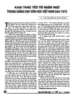
structs encoding different forms of the protein (Fig. 1)
were transformed into yeast (Fig. 2A). Expression of
pro-caspase-2 using the Gal 1 ⁄ 10 promoter affected
yeast growth only marginally (compare growth in lane
2 to that of an empty vector transformant in lane 1).
Increasing the pro-caspase-2 expression level, by intro-
ducing an additional expression construct under dif-
ferent nutritional selection, elicited more substantial
lethality (lane 3). A caspase-2 cleavage site mutant
(D152A), from which the prodomain could not be
removed, was also expressed at a high level using two
plasmids with different nutritional selections. Com-
pared with equivalent expression of wild-type pro-
caspase-2 (lane 3), this mutant exhibited only marginal
toxicity (lane 4) suggesting that removal of the prodo-
main contributes to full enzymatic activity. Consistent
with this observation, a truncation mutant lacking
almost all of the caspase-2 prodomain (caspase-2
D1)149
)
killed yeast more efficiently than full-length caspase-2
(compare lane 7 with lane 2). An artificially autoacti-
vating version of caspase-2 (rev-caspase-2), in which
the small subunit precedes the prodomain and large
subunit [29], killed yeast readily (lane 6). The catalyti-
cally inactive mutant pro-caspase-2
C303A
was unable to
kill yeast (lane 5) implying that the lethality of wild-
type caspase-2 in yeast was due to its enzymatic activity.
The expression of the prodomain (caspase-2
D150)435
)
had no effect on yeast viability (lane 8).
Caspase-2 can activate caspase-7 and is resistant to IAPs P k. Ho et al.
1402 FEBS Journal 272 (2005) 1401–1414 ª 2005 FEBS
To investigate the auto-processing of pro-caspase-2
in yeast, we immunoblotted lysates obtained from
yeast expressing these different forms of caspase-2 with
an antibody recognizing an epitope in the large
subunit. In lysates from yeast expressing wild-type
pro-caspase-2, a partial cleavage product was detected,
in addition to the fully processed large subunit
(Fig. 2B). Like the wild-type enzyme, the cleavage site
mutant pro-caspase-2
D152A
was processed efficiently
between the large and small subunits, however, the
mutation at D152 prevented it from being further
processed to separate the prodomain from the large
subunit. Caspase-2
C303A
remained intact as a result of
the abolished catalytic activity. Rev-caspase-2, despite
its ability to efficiently kill yeast, was only incompletely
processed. A proportion of caspase-2
D1)149
was cleaved
to remove the small subunit, thereby permitting detec-
tion of the dissociated large subunit.
The activities of these different forms of caspase-2
were also analysed biochemically using a fluorogenic
caspase-2 substrate. In this assay, the activity of an
enzyme is reflected by the efficiency with which it
cleaves the substrate to release free 7-amino-4-trifluoro-
methyl coumarin (AFC). The caspase-2-specific fluoro-
genic synthetic peptide Z-VDVAD-AFC was used as a
substrate to assess caspase-2 activity [5]. VDVADase
activity was detected in lysates from yeast expressing all
forms of caspase-2 that were capable of autoprocessing
(Fig. 2C). The most lethal forms of caspase-2 had the
highest VDVADase activity (lanes 3, 6 and 7), while ly-
sates from yeast that survived (lanes 1, 5 and 8) did not
cleave the peptide substrate. Yeast transformed with
one wild-type caspase-2 plasmid or the D152A mutant
were killed only inefficiently, however, their lysates
exhibited significant VDVADase activity. This may indi-
cate that the biochemical assay is a more sensitive meas-
ure of caspase-2 activity than the yeast death assay.
Caspase-2 is not inhibited by mammalian IAP
proteins
Members of the mammalian IAP family contribute to
the regulation of apoptotic pathways in part by their
inhibition of caspases-3, -7 and -9 [30]. Other mamma-
lian caspases (-1, -6, -8 and -10) are known to be resist-
ant to inhibition by IAPs [30], but the susceptibility of
caspase-2 to direct inhibition by IAPs has not been
reported to date. To explore the sensitivity of caspase-2
to IAP inhibition, we tested whether coexpression of
IAPs would suppress caspase-2-dependent yeast death.
Fig. 1. Schematic illustration of the caspase-
2 proteins used in this study. Mutated resi-
dues are listed above wild-type caspase-2
and are depicted with black circles.
P k. Ho et al. Caspase-2 can activate caspase-7 and is resistant to IAPs
FEBS Journal 272 (2005) 1401–1414 ª 2005 FEBS 1403
We had previously established that the inhibitors p35
and p49 could rescue yeast from caspase-2 mediated
death [31], so these baculoviral proteins were used as
positive controls. Caspase-3 effectively killed yeast and
this could be blocked by XIAP (also known as hILP),
MIHB (cIAP-1 ⁄ hIAP-2⁄ BIRC2) and MIHC (cIAP-
2 ⁄ hIAP-1), as well as p35 and p49 (Fig. 3A). In contrast,
the mammalian IAPs could not inhibit yeast death
induced by expression either of full-length pro-caspase-2
(Fig. 3B) or of truncated caspase-2 lacking the prodo-
main (Fig. 3C). As expected, the baculoviral caspase
inhibitors p35 and p49 protected caspase-2-expressing
yeast (Fig. 3B,C).
To confirm these observations using a biochemical
approach, purified caspase-2 was mixed with recombin-
ant XIAP or the inactive mutant XIAP
D148A
[32], then
assayed for its ability to cleave the fluorogenic penta-
peptide substrate Z-VDVAD-AFC. Caspase-2 activity
was not affected by the presence of XIAP (Fig. 3D),
whereas XIAP significantly reduced the activity of
caspase-3, as demonstrated previously [33]. The pres-
ence of p35 led to a decrease in both caspase-2 and
caspase-3 activities. Inactive mutants of p35 (p35
D87A
)
and XIAP (XIAP
D148A
) were unable to inhibit either
caspase.
Caspase-2 can promote caspase-7 catalytic
activity
To explore the potential for caspase-2 to functionally
interact with other caspases, we exploited the dose-
dependent caspase-2-mediated yeast toxicity illustrated
in Fig. 2. Caspase-2 was coexpressed in yeast from a
single plasmid either alone (yielding weak lethality) or
A
B
C
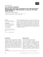
Fig. 2. Caspase-2 kills yeast. (A) A semi-
quantitative assay compares the effect of
transgenes on yeast growth and viability.
Yeast cells were transformed with the indi-
cated plasmids. Suspensions of each trans-
formant were prepared at standardized
concentrations. Serial dilutions were made
and spotted onto solid inducing minimal
media vertically down the plate. Colony size
indicates growth rate and colony number
reflects cell viability. (In every experiment,
each dilution was also spotted onto a
repressing plate to verify that equivalent
numbers of each transformant were spot-
ted; data not shown). (B) Anti-caspase-2
immunoblotting of lysates from the indica-
ted transformants. The presumed identities
of each band are shown to the left (pro, pro-
domain; L, large subunit; S, small subunit).
(C) The ability of caspase-2 to cleave the flu-
orogenic peptide substrate Z-VDVAD-AFC.
Native lysates obtained from yeast were
incubated with Z-VDVAD-AFC. Fluorescence
was monitored over time and the maximal
rate of increase in free AFC was calculated
and graphed. Error bars indicate SD (n ¼ 4).
Caspase-2 can activate caspase-7 and is resistant to IAPs P k. Ho et al.
1404 FEBS Journal 272 (2005) 1401–1414 ª 2005 FEBS
together with the nonlethal caspases-3, -4, -6, -7 and -9
(Fig. 4A). Yeast death was used as an indicator of
caspase activity. Co-expression of caspase-2 with
caspase-7 led to a pronounced increase in yeast death,
compared to that triggered by either caspase alone
(compare lane 12 with lanes 2 and 11). Much weaker
synergy was also reproducibly observed between
caspase-2 and -3 (compare lane 6 with lanes 2 and 5).
We then tested the ability of lysates from these yeast
to cleave a fluorogenic caspase-3 substrate (Ac-DEVD-
AFC) or a caspase-2 substrate (Z-VDVAD-AFC).
Caspase-2 activity was not enhanced by coexpression
of caspases-3 or -7. However, significantly more clea-
vage of Ac-DEVD-AFC was observed when caspase-2
was coexpressed with caspase-7 (or, to a lesser extent
with caspase-3) (Fig. 4B).
To further investigate the apparent synergy between
caspase-2 and caspase-7, plasmids encoding different
forms of these enzymes were transformed into yeast in
various combinations and their effects on enzyme clea-
vage, enzyme activity and yeast growth determined
(Fig. 5). As before, high level expression of caspase-2
resulted in an active enzyme, able to efficiently kill
yeast, whereas lower expression levels of caspase-2 had
in vitro activity but weak killing activity (compare
lanes 2 and 4 in Figs 5A–C). Full length caspase-7 was
unprocessed and did not kill yeast (lane 9), whereas
caspase-7 coexpressed with caspase-2 was activated
and toxic to yeast (lane 5). The activation of caspase-7
by caspase-2 depended on caspase-2 catalytic activity
since coexpression of catalytically inactive caspase-2
with caspase-7 did not yield enzymatic activity (neither
VDVADase nor DEVDase) and did not kill yeast
(Figs 5A–C, lane 6). However, caspase-2 activation
was independent of caspase-7 as caspase-2 proteolytic
activity was the same in the presence of active or enzy-
matically inactive caspase-7 (compare Fig. 5B and C
lanes 5 and 7). Two positive controls were used for
caspase-7 activation. First, caspase-7
D1)53
, which lacks
the prodomain region and is constitutively active in
mammalian cells [34] and in yeast [35] (lane 10).
Second, as previously reported for caspase-3 [27],
caspase-7 was activated by a constitutively active
caspase-9 (rev-caspase-9) (lane 11). This autoacti-
vating caspase-9 protein, which could activate caspase-
3 [27] or caspase-7 (Fig. 5A–C, lane 11), was not able
to co-operate with caspase-2 to kill yeast (Fig. 4A, lane
14). Together, these data suggest that caspase-2 may
lie upstream of caspase-7, and not downstream of
caspase-9, in apoptotic pathways.
A
B
C
D
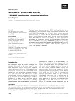
Fig. 3. IAPs do not inhibit caspase-2. The
caspase expression plasmids used to kill
yeast were (A) Caspase-3-lacZ (B) pGALL-
(LEU2)-caspase-2 with pGALL-(URA)-cas-
pase-2 or (C) pGALL-(URA)-caspase-2
D1)149
.
Yeast transformed with the indicated plas-
mids were spotted as described in the
legend to Fig. 1. (D) The indicated combina-
tions of caspase, fluorogenic substrate and
inhibitor were mixed together and the fluor-
escence resulting from the caspase-medi-
ated substrate cleavage was monitored and
calculated as described in the legend to
Fig. 1. Error bars indicate SD (n ¼ 3).
P k. Ho et al. Caspase-2 can activate caspase-7 and is resistant to IAPs
FEBS Journal 272 (2005) 1401–1414 ª 2005 FEBS 1405
The relationship between caspase-2 processing
and enzymatic activity
Previous work had illustrated that human pro-
caspase-2 can be processed at residue D152 to
remove its prodomain, and at residues D316 and
D330 to dissociate the large and small subunits and
release a small linker peptide [36]. To investigate the
impact of these cleavage events on the enzymatic
activity of caspase-2, recombinant caspase-2 proteins
harbouring one or a combination of mutated D152,
D316 or D330 residues were generated and expressed
in bacteria and the protein purified (Figs 1 and 6A).
We observed that full length recombinant caspase-2
has about a 10-fold lower activity than a commonly
used amino-terminal truncation lacking most of the
prodomain (D1–149) [37] (Fig. 6B). We therefore used
this truncated caspase-2 to test the effects on activity
of mutating the D152, D316 and D330 residues. We
immunoblotted the purified caspase-2 enzymes with
an antibody recognizing an epitope within the large
subunit of caspase-2, to determine whether enzyme
autoprocessing had occurred (Fig. 6A) and then tes-
ted cleavage of a caspase-2 specific fluorogenic sub-
strate (Fig. 6B) to determine enzyme activity.
Retention of at least one cleavage site between the
A
B
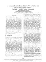
Fig. 4. Co-expression of caspases-2 and -7
enhances yeast lethality. (A) The indicated
combinations of caspases were coexpre-
ssed in yeast and their ability to promote
yeast death was compared to the lethality
arising from expression of single caspases.
(B) The activities of caspases were assayed
in lysates from yeast expressing individual
caspases, or the indicated combinations of
caspases. Substrate cleavage was calcula-
ted from the maximal rate of free AFC
released through cleavage by 30 lg of yeast
lysate. Error bars indicate SD (n ¼ 3).
Caspase-2 can activate caspase-7 and is resistant to IAPs P k. Ho et al.
1406 FEBS Journal 272 (2005) 1401–1414 ª 2005 FEBS
large and small subunits (D316 and ⁄ or D330) permit-
ted autocatalytic separation of the subunits (Fig. 6A)
and yielded active enzymes (Fig. 6B). Fusion of the
linker to the small subunit (D330A) had a slightly
greater deleterious effect on enzyme activity than
fusion to the large subunit (D316A) (Fig. 6B). In
contrast, mutation of both D316 and D330 sites
abolished auto-processing (Fig. 6A) and dramatically
reduced enzymatic activity (Fig. 6B). Using higher
amounts of enzyme (100 nm), it was evident that
mutation of both of these cleavage sites decreased
activity by 840-fold (data not shown). The C303A
A
B
C
Fig. 5. Caspase-2 activates caspase-7 in
yeast. (A) The indicated plasmids were
transformed into yeast and transformants
spotted onto inducing medium to visualize
their impact on yeast growth. (B) Immuno-
blotting was used to detect caspase pro-
cessing. The presumed identities of each
band are shown to the left (pro, prodomain;
L, large subunit; S, small subunit). (C) The
abilities of the yeast lysates to cleave the
caspase-2 (Z-VDVAD-AFC) and caspase-7
(Ac-DEVD-AFC) substrates were assayed.
Substrate cleavage was calculated from the
maximal rate of free AFC released through
cleavage by 30 lg of yeast lysate. Error bars
indicate standard deviations (n ¼ 3).
P k. Ho et al. Caspase-2 can activate caspase-7 and is resistant to IAPs
FEBS Journal 272 (2005) 1401–1414 ª 2005 FEBS 1407
active site mutant was completely inactive (Fig. 6B),
even at 100 nm (data not shown). Full length clea-
vage site mutants were expressed from two plasmids
in yeast and their impact on yeast viability assayed.
The D316A and D330A single mutants were toxic
to yeast, however, yeast expressing the double D316,
330A mutant survived (Fig. 6C). Together, these data
suggest that the proteolytic activity of human
caspase-2 correlates with the degree to which the
large subunit is separated from the small subunit.
We also tested the abilities of the caspase-2 mutant
enzymes to cleave protein substrates. Cellular sub-
strates (Bid, PARP, catalytically inactive pro-caspase-2
and pro-caspase-7) were expressed as glutathione-
S-transferease (GST)-fusion proteins, incubated with
the various caspase-2 enzymes and subjected to
A
B
C
Fig. 6. Caspase-2 processing is necessary
for activation. (A) Caspase-2 enzymes with
the indicated mutations were generated in
bacteria and immunoblotted to determine
the extent of auto-processing. The pre-
sumed identities of each band are shown to
the right (pro, prodomain; L, large subunit;
S, small subunit). (B) The abilities of wild-
type recombinant caspase-2 or the indicated
mutants to cleave the fluorogenic substrate
Z-VDVAD-AFC were monitored as described
in previous legends. Two independent prep-
arations of each enzyme were used. (C) The
indicated plasmids encoding wild-type or
cleavage site mutants of casapse-2 (or
empty vectors) were transformed into yeast
and transformants spotted onto inducing
medium to visualize their impact on yeast
growth.
Caspase-2 can activate caspase-7 and is resistant to IAPs P k. Ho et al.
1408 FEBS Journal 272 (2005) 1401–1414 ª 2005 FEBS
SDS ⁄ PAGE. Cleavage of the substrates was assessed
by staining with Coomassie blue (Fig. 7A) and immu-
noblotting (Fig. 7B). All caspase-2 proteins that were
active in the fluorogenic assay (Fig. 6B) were also able
to cleave Bid at aspartate 60 [10] and a catalytically
inactive GST-tagged pro-caspase-2 (Fig. 7A). The size
of the cleavage product implied that processing
occurred between the large and small subunits. PARP
was cleaved by caspases-3 and -7, but not by caspase-
2. GST-tagged pro-caspase-2
C303A
was not processed
by caspases-3, -7 or -8; cleavage products were not
detected by Coomassie blue staining (Fig. 7A) or by
immunoblotting (Fig. 7B). (Processing of PARP or Bid
by these enzymes confirmed that they were active).
Having observed the activation of caspase-7 by
caspase-2 in yeast, we examined the processing of
GST-tagged pro-caspase-7
C186A
by caspase-2 in this
system. The cleavage of GST-pro-caspase-7
C186A
by
active caspase-2 (as well as by caspases-3 and -8) was
detected by immunoblotting with an antibody that
recognizes cleaved caspase-7 (Fig. 7B).
Discussion
A unique merit of the yeast system used here is that it
is free from the potential interference of other mamma-
lian apoptotic signal transduction pathway compo-
nents, allowing the expression of the gene of interest in
a naive yet eukaryotic cell-based environment. We have
previously used this system to reconstitute caspase-9
activation by Apaf-1 [27] and the core nematode pro-
grammed cell death pathway [28]. Here, we harnessed
this system to analyse the regulation of caspase-2 activ-
ity, exploiting the observation that overexpressed
caspase-2 kills yeast in a concentration dependent man-
ner, requiring a catalytically active enzyme. Purified,
recombinant proteins were also used to verify much of
the data generated from the yeast system.
We have shown that prodomain removal increases
caspase-2 activity, when expressed in yeast or in bac-
teria. For generation of active human caspase-2, pro-
cessing is also required between the small and large
subunits (at D316 and⁄ or D330). Mutation of either
site had little effect on enzyme activity or toxicity to
yeast but mutation of both sites abolished both
enzyme activity and yeast killing. These observations
differ somewhat from previously reported analyses of
murine caspase-2. Firstly, the human and mouse
enzymes vary in their propensity for autoprocessing
between the large subunit and the linker. We have
shown that human caspase-2 almost completely auto-
processes at this point (D316), as indicated by the effi-
cient separation of large and small subunits of the
D330A mutant. In contrast, mutation of the murine
equivalent of the human residue D316 (D333) alone
prevented autoprocessing of caspase-2 [17,38]. This
species difference persisted when the mouse and
human mutants were generated using the identical bac-
terial expression systems (B. Baliga and S. Kumar,
personal communication), ruling out any technical
explanations for the variation. Secondly, human
caspase-2 which was prevented from autoprocessing
between the large and small subunits was almost
totally inactive, however, the D333G mutant of murine
caspase-2 that could not autoprocess retained about
one-fifth of wild-type enzyme activity [17,38]. Further
investigations will hopefully clarify the mechanisms
underlying these curious species differences.
Coexpression of caspase-2 with caspase-7 in yeast
was significantly more toxic than expression of either
protein alone. Although this result could reflect an
additive effect of two mildly lethal stimuli, two pieces of
evidence suggest that caspase-2 activation of caspase-7
accounts for the combined lethality. Firstly, caspase-2
cleaved caspase-7 in vitro. Secondly, lysates from yeast
A
B
Fig. 7. Substrate cleavage by wild-type caspase-2, its cleavage site
mutants and other caspases. (A) GST-tagged, enzymatically inactive
pro-caspase-2 or caspase substrates were incubated with the indi-
cated purified recombinant caspases (as detailed in the experimen-
tal section). The reactions were then subjected to SDS ⁄ PAGE and
the gels stained with Coomassie blue to visualize cleavage. (B) The
more sensitive technique of immunoblotting was used to detect
cleavage of catalytically inactive pro-caspase-2 or pro-caspase-7.
P k. Ho et al. Caspase-2 can activate caspase-7 and is resistant to IAPs
FEBS Journal 272 (2005) 1401–1414 ª 2005 FEBS 1409
expressing caspases-2 and -7 had higher DEVDase
activity (indicative of caspase-7 activity) than those
from yeast only expressing caspase-7. However, the
VDVADase activity (reflecting caspase-2 activity) of the
lysates from double transformants was similar to that
of lysates from yeast only expressing caspase-2.
Previously published data demonstrated that caspa-
ses-1, -2, -3, -4, -6, -7, -11 and CED-3 could all cleave
caspase-2, to varying extents [36,38–40]. In contrast,
our data indicates that concentrations of caspases-3, -7
and -8 capable of efficiently processing known physio-
logical substrates (PARP or Bid) could not cleave an
inactive mutant of pro-caspase-2. This discrepancy
probably relates to differences in the relative concen-
trations and⁄ or purity of the enzymes and substrates
used. The studies cited above used either unspecified
amounts of unpurified enzyme or enzyme concentra-
tions four times [40] or over 11 times [36] that used
here. In the previous studies, reticulocyte lysates con-
taining
35
S-labelled wild-type caspase-2 were used as
substrates. These lysates would contain endogenous
reticulocyte proteins that may potentially influence
the processing of caspase-2. To avoid any such indi-
rect effects, we used purified, catalytically inactive
caspase-2 as a substrate.
The IAP family of apoptosis inhibitors exert their
pro-survival effect, at least in part, through suppres-
sion of caspases-3, -7 and -9 [33,41,42]. The IAPs
XIAP, MIHB and MIHC could not inhibit other casp-
ases including -1, -6, -8 and -10 [30], but their ability
to directly inhibit caspase-2 has not been previously
published. Caspase-2-dependent yeast death was unaf-
fected by coexpression of XIAP, MIHB and MIHC
although, as we previously reported [31], p35 and p49
could inhibit caspase-2 in this system. Furthermore,
XIAP, the most potent caspase inhibitor of the IAP
family, did not impede the ability of recombinant
caspase-2 to cleave a synthetic substrate. It was previ-
ously observed that IAPs partially protected tissue cul-
ture cells from apoptosis induced by caspase-2
overexpression [43]. In the light of our findings, this
inefficient protection was probably due to IAP-medi-
ated inhibition of caspase-7, which was likely activated
by the overexpressed caspase-2.
In summary, this study illustrated that, at least in
the absence of an activating adaptor, generation of
active human caspase-2 requires separation of its large
and small subunits. In the context of autoactivation,
removal of the prodomain also enhances proteolytic
activity. Caspase-2 can act as an apical caspase, pro-
moting the activation of caspase-7. Unlike caspases-3,
-7 and -9, caspase-2 was resistant to inhibition by
members of the IAP family.
Experimental procedures
Plasmid construction
For expression in yeast, coding regions of human genes
were cloned into the pGALL yeast vectors under the regu-
lation of the inducible Gal 1 ⁄ 10 promoter [26]. Yeast
vectors pGALL-(HIS3), pGALL-(LEU2) and pGALL-
(URA) have been described previously [27,35]. Plasmids
pGALL-(LEU2)-caspase-2, pGALL-(URA)-caspase-2, casp-
ase-3-LacZ, pGALL-(LEU2)-caspase-4, pGALL-(URA)-
caspase-7
D1)53
, pGALL-(HIS3)-p35 and pGALL-(HIS3)-
p49 have been reported [27,31,35]. Other plasmids were
constructed as follows: Pro-caspase-2 PCR product, gener-
ated with primers 1 and 2, was cut with BglII ⁄ XbaI and
ligated into BamHI ⁄ XbaI cut vectors to produce pGALL-
(HIS3)-caspase-2 and pGALL-(HIS3)-FLAG-caspase-2. To
make pGALL-(LEU2)-rev-caspase-2, the carboxyl terminal
fragment was amplified with primers 3 and 4, digested with
BglII ⁄ XbaI and ligated into pGALL-(LEU2) to give
pGALL-(LEU2)-rev-caspase-2-C. The amino-terminal frag-
ment was generated with primers 5 and 6, cut with XhoI ⁄
XbaI, and ligated into pGALL-(LEU2)-rev-caspase-2-C
to generate the final construct. pGALL-(LEU2)-caspase-
2
C303A
, pGALL-(URA)-caspase-2
C303A
, pGALL-(HIS3)-
caspase-2
C303A
and pGALL-(HIS3)-FLAG-caspase-2
C303A
were produced by replacing a SpeI ⁄ BamHI cut fragment
with a PCR product generated with primers 1 and 7.
pGALL-(HIS3)-FLAG-caspase-2
D1)149
was cloned by ligat-
ing a NdeI-digested and blunt-ended then BamHI cut frag-
ment from pET23a-caspase-2
D1)149
into SpeI-digested and
blunt-ended then BamHI cut pGALL-(HIS3)-FLAG-ca-
spase-2. A PCR product generated with primers 1 and 8
was cut with BglII ⁄ XbaI and ligated into BamHI ⁄ XbaI cut
vector to produce pGALL-(LEU2)-caspase-2
D153)435
.
pGALL-(HIS3)-FLAG-caspase-2
D152A
and pGALL-(HIS3)-
FLAG-caspase-2
D316A
were made by replacing a SalI ⁄
BamHI cut fragment with PCR products generated with
primer pairs 9, 10 and 11, 12, respectively. pGALL-(HIS3)-
FLAG-caspase-2
D330A
was produced by replacing a Bam-
HI ⁄ XbaI cut fragment with a PCR product generated with
primers 13 and 14. To make pGALL-(HIS3)-FLAG-
caspase-2
C303A; D152,316A
,aSalI ⁄ BamHI fragment in
pGALL-(HIS3)-FLAG-caspase-2
C303A
was replaced with a
PCR product generated using primers 9 and 12. It was sub-
sequently used to produce pGALL-(HIS3)-FLAG-cas-
pase-2
C303A; D152, 316, 330A
byreplacingaSalI ⁄ BamHIfragment
into pGALL-(HIS3)-FLAG-caspase-2
D330A
. SpeI ⁄ BamHI
fragments isolated from pGALL-(HIS3)-FLAG-Caspase-
2
D1)149
or pGALL-(HIS3)-FLAG-Caspase-2
D152A
were used
to replace part of the coding region in pGALL-(HIS3)-
Caspase-2, pGALL-(URA)-Caspase-2 and pGALL-(LEU2)-
Caspase-2 to make pGALL-(HIS3)-Caspase-2
D1)149
and
pGALL-(URA)-Caspase-2
D1)149
or pGALL-(HIS3)-
Caspase-2
D152A
and pGALL-(LEU2)-Caspase-2
D152A
,
Caspase-2 can activate caspase-7 and is resistant to IAPs P k. Ho et al.
1410 FEBS Journal 272 (2005) 1401–1414 ª 2005 FEBS
respectively. pGALL-(HIS3)-Caspase-2
D316A
, pGALL-
(HIS3)-Caspase-2
D330A
, pGALL-(HIS3)-Caspase-2
D316,330A
,
pGALL-(HIS3)-Caspase-2
D152,316,330A
, pGALL-(URA)-Cas-
pase-2
D316A
, pGALL-(URA)-Caspase-2
D330A
, pGALL-
(URA)-Caspase-2
D316,330A
and pGALL-(URA)-Caspase-
2
D152,316,330A
were generated by ligating SpeI ⁄ XbaI
fragments released from pGALL-(HIS3)-FLAG-caspase-
2
D316A
, pGALL-(HIS3)-FLAG-caspase-2
D330A
, pGALL-
(HIS3)-FLAG-caspase-2
D316,330A
and pGALL-(HIS3)-
FLAG-caspase-2
D152,316,330A
into SpeI ⁄ XbaI digested
pGALL-(HIS3)-Caspase-2 and pGALL-(URA)-Caspase-2,
respectively. The coding regions of pro-caspases-3, -6 and -7
were excised with BamHI ⁄ XbaI from pGALL-(URA)-cas-
pase-3 [27], pEF-Mch2 (made from a vector kindly provided
by E. Alnemri [44]) and pCUP1-(LEU2)-caspase-7 [35] and
ligated into pGALL-(LEU2) to generate pGALL-(LEU2)-
caspase-3, pGALL-(LEU2)-caspase-6 and pGALL-(LEU2)-
caspase-7, respectively. pGALL-(URA)-caspase-7 was made
by ligating the coding region of caspase-7 cut with Bam-
HI ⁄ Xba I into pGALL-(URA). To make pGALL-(URA)-cas-
pase-7
C186A
, the carboxyl-terminal fragment was amplified
with primers 15 and 14, digested with BamHI ⁄
XbaI and ligated into pGALL-(URA) to give pGALL-
(URA)-caspase-7
C186A
-C. The amino terminal fragment was
generated with primers 16 and 17, cut with BamHI ⁄ XhoI,
and ligated into pGALL-(URA)-caspase-7
C186A
-C to give the
final construct. pGALL-(HIS3)-XIAP and pGALL-(HIS3)-
MIHB were constructed by ligating EcoRI ⁄ NotI cut PCR
products amplified with primer pairs 18, 19 and 20, 21,
respectively, into pGALL-(HIS3). The coding region of
MIHC was isolated from pADH-(TRP1)-MIHC [27] with
EcoRI ⁄ NotI and cloned into pGALL-(HIS3 ) to give
pGALL-(HIS3)-MIHC.
For expression in bacteria, coding regions of genes were
cloned into pET23a(+) (Novagen, Madison, WI, USA) or
pGEX6P-3 (Amersham Biosciences, Uppsala, Sweden).
pGEX6P3-XIAP [45], pGEX6P3-Bid [46] and pET23a-p35
[28] have been previously described. The coding region of
pro-caspase-2 was amplified with primers 21 and 22, cut
with NdeI ⁄ XhoI and ligated into pET23a to give pET23a-
caspase2. pET23a-caspase2
D1)149
and pET23a-casp-
ase2
D1)149; D152A
were constructed using NdeI ⁄ XhoI cut
PCR products amplified with primer pairs 24, 23 and 25,
23, respectively. HindIII ⁄ EcoRI fragments released from
pGALL-(LEU2)-caspase-2
C303A
, pGALL-(HIS3)-FLAG-
caspase-2
D316A
, pGALL-(HIS3)-FLAG-caspase-2
D330A
,
pGALL-(HIS3)-FLAG-caspase-2
D316,330A
and pGALL-
(HIS3)-FLAG-caspase-2
D152,316,330A
were used to replace
an internal fragment in pET23a-caspase-2
D1)149
or
pET23a-caspase-2
D1)149;D152A
to generate pET23a-caspase-
2
D1)149;C303A
, pET23a-caspase2
D1)149;D316A
, pET23a-
caspase2
D1)149;D330A
, pET23a-caspase2
D1)149;D316,330A
and
pET23a-caspase2D1–149; D152 316 330 A, respectively.
pET23a-p35
D87A
was constructed by ligating a NdeI ⁄
HindIII cut PCR product generated with primers 26 and 14
and pGALL-(HIS3)-p35
D87A
as a template [31]. Using a
template kindly provided by John Silke that contained
XIAP
D148A
, the coding region of XIAP was amplified with
primers 28 and 29, cut with BamHI ⁄ EcoRI and inserted
into pGEX-6P3 to give pGEX6P3-XIAP
D148A
. To construct
pGEX6P3-Bid
D60A
, first a PstI-digested PCR product
amplified with primers 30 and 31 was used to replace an
internal fragment in pBluescriptII(SK+)-Bid [46] to gener-
ate pBluescriptII(SK+)-Bid
D60A
. The coding region was
then amplified with primers 32 and 33, digested with Bam-
HI ⁄ EcoRI and cloned into pGEX-6P3 to give the final
construct. A SpeI ⁄ XbaI fragment excised from pGALL-
(LEU2)-caspase-2
C303A
was ligated into pBluescriptII(SK+)
to produce pBluescriptII(SK+)-caspase-2
C303A
. A blunt-
ended SpeI-cut then NotI-digested fragment isolated from
the above construct was ligated into a blunt-ended EcoRI-
cut then NotI-digested vector to yield pGEX6P3-caspase-
2
C303A
. The coding region of truncated PARP was released
with EcoRI ⁄ NotI from pADH-(TRP1)-mycPARP
D338)1013
[47] and ligated into pGEX-6P3. The construct was then
cut with BamHI, blunt-ended and re-ligated to produce
pGEX6P3-mycPARP
D338)1013
.ABamHI ⁄ XbaI fragment
was excised from pGALL-(URA)-Caspase-7
C186A
and cloned
into pBluescriptII(SK+) to give pBluescriptII(SK+)-
Caspase-7
C186A
, from which a BamHI ⁄ NotI fragment was
isolated and ligated into pGEX-6P3 to yield pGEX6P3-
Caspase-7
C186A
.
The primers used were: 1, 5¢-GGAAGATCTACTAG
TATGGCCGCTGACAGGGGACGC-3¢;2,5¢-GCTCTAG
ACTATGTGGGAGGGTGCCTTGGG-3¢;3,5¢-GCAGAT
CTATGGACCAACAAGATGGAAAG-3¢;4,5¢-CGTCT
AGACTCGAGTCCATCTTGTTGGTCTGTGGGAGGGT
GTCCTGG-3¢;5,5¢-GGCTCGAGATGGCCGCTGACAG
GGGACGC-3¢;6,5¢-GCTCTAGACTAATCTTGTTGGT
CAACCC-3¢;7,5¢-GGGGATCCTGCGTGGTTCTTTCC
ATCTTGTTGGTCAACCCCACGATCAGTCTCATCTCC
ACGGGCGGCCTG-3¢;8,5¢-GCTCTAGATTAATCTTT
ATTGTCTAGGGAGTGTTCC-3¢;9,5¢-GGCGTCGACA
GATACTGTGGAACACTCCCTAGACAATAAAGCTGG
TCCTGTCTGC-3¢; 10, 5¢-GCGGATCCTGCGTGGTTCT
TTCCATC-3¢; 11, 5¢-GGCGTCGACAGATACTGTGGAA
CACTCCC-3¢; 12, 5¢-GCGGATCCTGCGTGGTTCTTTC
CAGCTTGTTGGTCAACCC-3¢; 13, 5¢-GCGGATCCCCC
GGGTGCGAGGAGACTGCTGCCGG-3¢; 14, 5¢-CTTTA
TTATTTTTATTTTATTGAGAGGGTGG-3¢; 15, 5¢-GCG
GATCCCTCGAGAAACCC AAACTCTT CTTC ATTCAG
GCTGCCCGAGGGACCGAGCTTG-3¢; 16, 5¢-CCACTTT
AACTAATACT TTCA ACAT TTTC GG-3 ¢; 17–5¢-GGCCTC
GAGAAGGGTTTTGCATC-3¢; 18, 5¢-GGATTCATGACT
TTTAACAGTTTTGAAGG3¢; 19, 5¢-CCCCCGCGGCCG
CTTAAGACATAAAAATTTTTTGCTTG-3¢; 20, 5¢-GGA
ATTCATGCACAAAACTGCCTCCC-3¢; 21, 5¢-CCCCCG
CGGCCGCTTAAGAGAGAAATGTACGAAC-3¢; 22,
5¢-GGCAGATCTCATATGGCCGCTGACAGGGGACGC-
3¢; 23, 5¢-CCCTCGAGTGTGGGAGGGTGTCCTGGG-3¢;
P k. Ho et al. Caspase-2 can activate caspase-7 and is resistant to IAPs
FEBS Journal 272 (2005) 1401–1414 ª 2005 FEBS 1411
24, 5¢-GAGATCTCATATGAATAAAGATGGTCCTGTC
TGC-3¢; 25, 5¢-GGCAGATCTCATATGAATAAAGCT
GGTCCTGTCTGC-3¢; 26, 5¢-GGAATTCCATATGTGTG
TAATTTTTCCGGTAG-3¢; 27, 5¢-CCCTCGAGTTTAAT
TGTGTTTAATATTAC-3¢; 28, 5¢-GCGGATCCATGACT
TTTAACAGTTTTGAAGG-3¢; 29, 5¢-GAGAATTCTTAA
GACATAAAAATTTTTTGCTTG-3¢; 30, 5¢-GCCTGCAG
ACTGCTGGCAACCGCAGCAGCCACTCGAGG-3¢; 31,
5¢-GCCTGCAGCAGCTGCTCCAGGGCAGTGGCCAGG
TCCCTGTTCCGGTCCTCCTCCGACCGGCTGGTGTTC
CTGAGTTG-3¢; 32, 5¢-GCGGATCCATGGACTGTGAG
GTCAACAACGG-3¢; 33, 5¢-GAGAATTCTCAGTCCAT
CCCATTTCTGGC-3¢.
Yeast transformation and death assays
Saccharomyces cerevisiae strain W303a was used in all yeast
transformation and death assays, as described previously
[26,31].
Preparation of yeast lysates
For immunoblotting, yeast were grown and induced for 7 h
and lysates extracted as described previously [47]. Samples
were resolved by SDS ⁄ PAGE on 12% gels, transferred to
Hybond-P membrane (Amersham Biosciences), and probed
with antibodies against caspase-2 [7] or caspase-7 (Cell
Signaling, Beverly, MA, USA). Blots were washed with phos-
phate-buffered saline ⁄ 0.5% Tween-20, and subsequently
probed with horseradish peroxidase-conjugated goat
anti-rat or donkey anti-mouse secondary Igs (Amersham
Biosciences). Signals were developed using ECL reagents
(Pierce, Rockford, IL, USA).
Native yeast lysates were prepared by resuspension in
ice-cold lysis buffer (50 mm Tris ⁄ HCl, pH 7.0, 150 mm
NaCl, 1 mm EDTA) followed by sonication for 15 s on ice.
Lysates were cleared by centrifugation at 2300 g for 1 min
and protein concentration was measured using the bicincho-
ninic acid kit (Sigma, St. Louis, MO, USA) against a BSA
standard curve.
Expression and purification of recombinant
proteins
Constructs containing the genes of interest were transformed
into Escherichia coli strain BL21(DE3)pLysS. Overnight cul-
tures were grown until D
600
reached 0.6–0.8. For His
6
-
tagged caspase-2 and p35, inductions were carried out at
30 °C for 3 h with 0.2 mm IPTG and for 4 h with 1 mm
IPTG, respectively. For GST-XIAP, GST-XIAP
D148A
,
GST-fusion substrates (Bid and Bid
D60A
) and GST-fusion
substrates (caspase-2
C303A
, myc-PARP
D337)1013
), inductions
were carried out at 25 °C for 3 h with 0.2 mm IPTG, for 4 h
with 1 mm IPTG and for 8 h with 1 mm IPTG, respectively.
Following induction, bacteria were washed once in STE
(10 mm Tris ⁄ HCl, pH 8.0, 150 mm NaCl, 1 mm EDTA),
pelleted and frozen. His
6
tagged caspase-2 and p35 were puri-
fied using Ni
2+
–NTA agarose (Qiagen, Hilden, Germany)
following manufacturer’s instructions. Quantification was
performed by obtaining Western blot ECL signals and com-
parison with standards of known concentration using a
Gel-Doc system and quantity one analysis software
(Biorad, Hercules, CA, USA). GST-fusion proteins were
purified using glutathione sepharose (Amersham Biosciences)
following the manufacturer’s instructions. XIAP and
XIAP
D148A
were released from the GST-fusion partner by
enzymatic cleavage using PreScission protease (Amersham
Biosciences). For GST-fusion substrates, fusion proteins
were left bound on the solid support. Quantification was per-
formed by comparing bands against BSA standards using
SDS ⁄ PAGE and staining with Coomassie blue.
Fluorogenic substrate cleavage assays
Recombinant caspases were preactivated at 37 °C for
30 min prior to the addition of fluorogenic substrates. Fluo-
rogenic substrate cleavage assays were performed in an assay
buffer containing 100 mm Hepes (pH 7.5), 10% sucrose,
0.1% CHAPS, 10 mm dithiothreitol, 25 mm Tris ⁄ HCl
(pH 7.0), 75 mm NaCl, 0.5 mm EDTA and 0.2 mgÆmL
)1
BSA with 50 lm of either Z-VDVAD-AFC or Ac-DEVD-
AFC (Calbiochem, San Diego, CA, USA). For assays using
yeast lysates, 30 lg of yeast lysates were used per 100 lL
assay reaction. Assays using recombinant proteins contained
2nm of preactivated caspase. Inhibition assays contained
2nm of caspase-2 or 0.36 nm of caspase-3 (Biomol,
Plymouth Meeting, PA, USA), with 0.5 lm of either XIAP,
p35 or their inactive mutants per assay reaction.
GST-fusion substrate cleavage assays
Purified GST-fusion substrates bound on sepharose were
used as substrates in the cleavage assays. Each 20-lL reac-
tion contained 0.25 lm to 1.5 lm of GST-fusion protein on
sepharose and 30 nm recombinant caspase (caspases-3, -7
and -8 were purchased from Biomol) in the same assay buf-
fer used in fluorogenic substrate cleavage assays described
above. The reactions were incubated at 37 °C for 90 min
and stopped by the addition of SDS ⁄ PAGE sample buffer,
then boiled and subjected to SDS ⁄ PAGE. Gels were stained
with Coomassie blue or immunoblotted with antibodies
against caspase-2 or cleaved caspase-7 (Cell Signaling).
Acknowledgements
We thank B. Baliga, S. Read and S. Kumar for helpful
comments on the manuscript and for permission to
cite their unpublished data. We also thank E. Alnemri
Caspase-2 can activate caspase-7 and is resistant to IAPs P k. Ho et al.
1412 FEBS Journal 272 (2005) 1401–1414 ª 2005 FEBS
for the Mch-2 plasmid. This work was supported by
the Australian Research Council and a University of
Melbourne Postgraduate Scholarship (to P K. H).
References
1 Degterev A, Boyce M & Yuan J (2003) A decade of cas-
pases. Oncogene 22, 8543–8567.
2 Thornberry NA, Bull HG, Calaycay JR, Chapman
KT, Howard AD, Kostura MJ, Miller DK,
Molineaux SM, Weidner JR, Aunins J et al. (1992)
A novel heterodimeric cysteine protease is required for
interleukin-1 beta processing in monocytes. Nature
356, 768–774.
3 Fantuzzi G & Dinarello CA (1999) Interleukin-18 and
interleukin-1 beta: two cytokine substrates for ICE (cas-
pase-1). J Clin Immunol 19, 1–11.
4 Fischer U, Janicke RU & Schulze-Osthoff K (2003)
Many cuts to ruin: a comprehensive update of caspase
substrates. Cell Death Differ 10, 76–100.
5 Thornberry N, Rano T, Peterson E, Rasper D, Timkey
T, Garcia-Calvo M, Houtzager V, Nordstrom P, Roy S,
Vaillancourt J, Chapman K & Nicholson D (1997) A
combinatorial approach defines specificities of members
of the caspase family and granzyme B. Functional rela-
tionships established for key mediators of apoptosis.
J Biol Chem 272, 17907–17911.
6 Bergeron L, Perez GI, Macdonald G, Shi L, Sun Y,
Jurisicova A, Varmuza S, Latham KE, Flaws JA, Salter
JC et al. (1998) Defects in regulation of apoptosis in
caspase-2-deficient mice. Genes Dev 12, 1304–1314.
7 O’Reilly LA, Ekert P, Harvey N, Marsden V, Cullen L,
Vaux DL, Hacker G, Magnusson C, Pakusch M et al.
(2002) Caspase-2 is not required for thymocyte or neur-
onal apoptosis even though cleavage of caspase-2 is
dependent on both Apaf-1 and caspase-9. Cell Death
Differ 9, 832–841.
8 Robertson JD, Enoksson M, Suomela M, Zhivotovsky
B & Orrenius S (2002) Caspase-2 acts upstream of
mitochondria to promote cytochrome c release during
etoposide-induced apoptosis. J Biol Chem 277, 29803–
29809.
9 Paroni G, Henderson C, Schneider C & Brancolini C
(2002) Caspase-2 can trigger cytochrome c release and
apoptosis from the nucleus. J Biol Chem 277, 15147–
15161.
10 Guo Y, Srinivasula SM, Druilhe A, Fernandes-Alnemri
T & Alnemri ES (2002) Caspase-2 induces apoptosis by
releasing proapoptotic proteins from mitochondria.
J Biol Chem 277, 13430–13437.
11 Enoksson M, Robertson JD, Gogvadze V, Bu P, Krop-
otov A, Zhivotovsky B & Orrenius S (2004) Caspase-2
permeabilizes the outer mitochondrial membrane and
disrupts the binding of cytochrome c to anionic phos-
pholipids. J Biol Chem 279, 49575–49578.
12 Lassus P, Opitz-Araya X & Lazebnik Y (2002) Require-
ment for caspase-2 in stress-induced apoptosis before
mitochondrial permeabilization. Science 297, 1352–1354.
13 Wagner KW, Engels IH & Deveraux QL (2004) Cas-
pase-2 can function upstream of Bid cleavage in the
TRAIL apoptosis pathway. J Biol Chem 279, 35047–
35052.
14 Salvesen GS & Dixit VM (1999) Caspase activation: the
induced-proximity model. Proc Natl Acad Sci USA 96,
10964–10967.
15 Chang DW, Ditsworth D, Liu H, Srinivasula SM,
Alnemri ES & Yang X (2003) Oligomerization is a gen-
eral mechanism for the activation of apoptosis initiator
and inflammatory procaspases. J Biol Chem 278, 16466–
16469.
16 Colussi PA, Harvey NL, Shearwin-Whyatt LM &
Kumar S (1998) Conversion of procaspase-3 to an auto-
activating caspase by fusion to the caspase-2 prodo-
main. J Biol Chem 273, 26566–26570.
17 Baliga BC, Read SH & Kumar S (2004) The biochem-
ical mechanism of caspase-2 activation. Cell Death
Differ 11, 1234–1241.
18 Duan H & Dixit VM (1997) Raidd is a new death adap-
tor molecule. Nature 385, 86–89.
19 Ahmad M, Srinivasula SM, Wang LJ, Talanian RV,
Litwack G, Fernandes-Alnemri T & Alnemri ES (1997)
Cradd, a novel human apoptotic adaptor molecule for
caspase-2, and FasL tumor necrosis factor receptor-
interacting protein RIP. Cancer Res 57, 615–619.
20 Koseki T, Inohara N, Chen S & Nunez G (1998) ARC,
an inhibitor of apoptosis expressed in skeletal muscle
and heart that interacts selectively with caspases. Proc
Natl Acad Sci USA 95, 5156–5160.
21 Bonfoco E, Li E, Kolbinger F & Cooper NR (2001)
Characterization of a novel pro-apoptotic caspase-2 and
-9 binding protein. J Biol Chem 276, 29242–29250.
22 Tinel A & Tschopp J (2004) The PIDDosome, a protein
complex implicated in activation of caspase-2 in
response to genotoxic stress. Science 304, 843–846.
23 Read SH, Baliga BC, Ekert PG, Vaux DL & Kumar S
(2002) A novel Apaf-1-independent putative caspase-2
activation complex. J Cell Biol 159, 739–745.
24 Robertson JD, Gogvadze V, Kropotov A, Vakifahmeto-
glu H, Zhivotovsky B & Orrenius S (2004) Processed
caspase-2 can induce mitochondria-mediated apoptosis
independently of its enzymatic activity. EMBO Report
5, 643–648.
25 Lamkanfi M, D’Hondt K, Vande Walle L, van Gurp
M, Denecker G, Demeulemeester J, Kalai M, Declercq
W, Saelens X & Vandenabeele P (2004) A novel cas-
pase-2 complex containing TRAF2 and RIP1. J Biol
Chem doi: 10.1074/jbc.M411180200.
26 Hawkins CJ, Wang SL & Hay BA (2000) Monitoring
activity of caspases and their regulators in yeast Sac-
charomyces cerevisiae. Methods Enzymol 322, 162–174.
P k. Ho et al. Caspase-2 can activate caspase-7 and is resistant to IAPs
FEBS Journal 272 (2005) 1401–1414 ª 2005 FEBS 1413
27 Hawkins CJ, Silke J, Verhagen AM, Foster R, Ekert
PG & Ashley DM (2001) Analysis of candidate antago-
nists of IAP-mediated caspase inhibition using yeast
reconstituted with the mammalian Apaf-1-activated
apoptosis mechanism. Apoptosis 6, 331–338.
28 Jabbour AM, Ho P-K, Puryer MA, Ashley DM, Ekert
PG & Hawkins CJ (2004) The Caenorhabditis elegans
CED-9 protein does not directly inhibit the caspase
CED-3, in vitro nor in yeast. Cell Death Differ 11,
1309–1316.
29 Srinivasula S, Ahmad M, MacFarlane M, Luo Z,
Huang Z, Fernandes-Alnemri T & Alnemri E (1998)
Generation of constitutively active recombinant cas-
pases-3 and -6 by rearrangement of their subunits.
J Biol Chem 273, 10107–10111.
30 Deveraux Q & Reed J (1999) IAP family proteins-sup-
pressors of apoptosis. Genes Dev 13, 239–252.
31 Jabbour AM, Ekert PG, Coulson EJ, Knight MJ, Ash-
ley DM & Hawkins CJ (2002) The p35 relative, p49,
inhibits mammalian and Drosophila caspases including
DRONC and protects against apoptosis. Cell Death Dif-
fer 9, 1311–1320.
32 Sun C, Cai M, Gunasekera AH, Meadows RP, Wang
H, Chen J, Zhang H, Wu W, Xu N, Ng SC & Fesik
SW (1999) NMR structure and mutagenesis of the
inhibitor-of-apoptosis protein XIAP. Nature 401,
818–822.
33 Deveraux Q, Takahashi R, Salvesen G & Reed J (1997)
X-linked IAP is a direct inhibitor of cell-death pro-
teases. Nature 388, 300–304.
34 Duan HJ, Chinnaiyan AM, Hudson PL, Wing JP, He
WW & Dixit VM (1996) Ice-lap3, a novel mammalian
homologue of the caenorhabditis elegans cell death
protein ced-3 is activated during fas- and tumor
necrosis factor-induced apoptosis. J Biol Chem 271,
1621–1625.
35 Hawkins CJ, Wang SL & Hay BA (1999) A cloning
method to identify caspases and their regulators in
yeast: identification of Drosophila IAP1 as an inhibitor
of the Drosophila caspase DCP-1. Proc Natl Acad Sci
USA 96, 2885–2890.
36 Xue D, Shaham S & Horvitz HR (1996) The Caenor-
habditis elegans cell-death protein CED-3 is a cysteine
protease with substrate specificities similar to those
of the human CPP32 protease. Genes Dev 10, 1073–
1083.
37 Garcia-Calvo M, Peterson EP, Rasper DM, Vaillan-
court JP, Zamboni R, Nicholson DW & Thornberry
NA (1999) Purification and catalytic properties of
human caspase family members. Cell Death Differ 6,
362–369.
38 Butt AJ, Harvey NL, Parasivam G & Kumar S (1998)
Dimerization and autoprocessing of the Nedd2 (Cas-
pase-2) precursor requires both the prodomain and the
carboxyl-terminal regions. J Biol Chem 273, 6763–6768.
39 Harvey NL, Trapani JA, Fernandes-Alnemri T, Litwack
G, Alnemri ES & Kumar S (1996) Processing of the
Nedd2 precursor by ICE-like proteases and granzyme
B. Genes Cells 1, 673–685.
40 Van de Craen M, Declercq W, Van den brande I, Fiers
W & Vandenabeele P (1999) The proteolytic procaspase
activation network: an in vitro analysis. Cell Death Dif-
fer 6, 1117–1124.
41 Roy N, Deveraux QL, Takahashi R, Salvesen GS &
Reed JC (1997) The c-IAP-1 and c-IAP-2 proteins are
direct inhibitors of specific caspases. EMBO J 16, 6914–
6925.
42 Deveraux QL, Leo E, Stennicke HR, Welsh K, Salvesen
GS & Reed JC (1999) Cleavage of human inhibitor of
apoptosis protein XIAP results in fragments with dis-
tinct specificities for caspases. EMBO J 18, 5242–5251.
43 Dorstyn L & Kumar S (1997) Differential inhibitory
effects of CrmA, P35, IAP and three mammalian IAP
homologues on apoptosis in NIH3T3 cells following
various death stimuli. Cell Death Differ 4, 570–579.
44 Fernandes-Alnemri T, Litwack G & Alnemri ES (1995)
Mch2, a new member of the apoptotic ced-3 ⁄ ICE cys-
teine protease gene family. Cancer Res 55, 2737–2742.
45 Silke J, Ekert PG, Day CL, Hawkins CJ, Baca M,
Chew J, Pakusch M, Verhagen AM & Vaux DL (2001)
Direct inhibition of caspase 3 is dispensable for the
anti-apoptotic activity of XIAP. EMBO J 20, 3114–
3123.
46 Knight ME, Riffkin CD, Ekert PG, Ashley DM &
Hawkins CJ (2004) Caspase-8 levels affect necessity for
mitochondrial amplification in death ligand-induced
glioma cell apoptosis. Mol Carcinog 39, 173–182.
47 Hawkins CJ, Yoo SJ, Petersen EP, Wang SL, Vernooy
SY & Hay BA (2000) The Drosophila caspase DRONC
cleaves following glutamate or aspartate and is regu-
lated by DIAP1, HID, and GRIM. J Biol Chem 275,
27084–27093.
Caspase-2 can activate caspase-7 and is resistant to IAPs P k. Ho et al.
1414 FEBS Journal 272 (2005) 1401–1414 ª 2005 FEBS