Báo cáo khoa học: Small exterior hydrophobic cluster contributes to conformational stability and steroid binding in ketosteroid isomerase from Pseudomonas putida biotype B pot
Bạn đang xem bản rút gọn của tài liệu. Xem và tải ngay bản đầy đủ của tài liệu tại đây (307.13 KB, 13 trang )
Small exterior hydrophobic cluster contributes to
conformational stability and steroid binding in ketosteroid
isomerase from Pseudomonas putida biotype B
Young S. Yun
1,2
, Gyu H. Nam
1,2
, Yeon-Gil Kim
1,3
, Byung-Ha Oh
1,3
and Kwan Y. Choi
1,2
1 Division of Molecular and Life Sciences, Pohang University of Science and Technology, South Korea
2 National Research Laboratory of Protein Folding and Engineering, Pohang University of Science and Technology, South Korea
3 National CRI Center for Biomolecular Recognition, Pohang University of Science and Technology, South Korea
Hydrophobic residues are rarely found on the surface
of soluble globular proteins because their exclusion
from water is favored by the hydrophobic effect [1].
However, stabilizing effects resulting from the intro-
duction of hydrophobic residues on the surface of a
protein have been observed [2–4]. A hydrophobic sur-
face formed by hydrophobic side-chains was found to
be associated with the formation and stabilization of
the overall b-sheet structure [5]. A structural motif
known as the small exterior hydrophobic cluster
Keywords
conformational stability; ketosteroid
isomerase; small exterior hydrophobic
cluster; steroid binding; surface hydrophobic
residue
Correspondence
K. Y. Choi, Division of Molecular and Life
Sciences, Pohang University of Science and
Technology, Pohang, 790–784, South Korea
Fax: +82 54 279 2199
Tel: +82 54 279 2295
E-mail:
Database
The atomic coordinate and structural factor
of W92A have been deposited in the Protein
Data Bank under the access code 1W6Y.
(Received 5 November 2004, revised 26
January 2005, accepted 24 February 2005)
doi:10.1111/j.1742-4658.2005.04627.x
A structural motif called the small exterior hydrophobic cluster (SEHC)
has been proposed to explain the stabilizing effect mediated by solvent-
exposed hydrophobic residues; however, little is known about its biological
roles. Unusually, in D
5
-3-ketosteroid isomerase from Pseudomonas putida
biotype B (KSI-PI) Trp92 is exposed to solvent on the protein surface,
forming a SEHC with the side-chains of Leu125 and Val127. In order to
identify the role of the SEHC in KSI-PI, mutants of those amino acids
associated with the SEHC were prepared. The W92A, L125A ⁄ V127A, and
W92A ⁄ L125A⁄ V127A mutations largely decreased the conformational sta-
bility, while the L125F ⁄ V127F mutation slightly increased the stability,
indicating that hydrophobic packing by the SEHC is important in main-
taining stability. The crystal structure of W92A revealed that the
decreased stability caused by the removal of the bulky side-chain of Trp92
could be attributed to the destabilization of the surface hydrophobic layer
consisting of a solvent-exposed b-sheet. Consistent with the structural
data, the binding affinities for three different steroids showed that the
surface hydrophobic layer stabilized by SEHC is required for KSI-PI to
efficiently recognize hydrophobic steroids. Unfolding kinetics based on
analysis of the U
U
value also indicated that the SEHC in the native state
was resistant to the unfolding process, despite its solvent-exposed site.
Taken together, our results demonstrate that the SEHC plays a key role
in the structural integrity that is needed for KSI-PI to stabilize the hydro-
phobic surface conformation and thereby contributes both to the overall
conformational stability and to the binding of hydrophobic steroids in
water solution.
Abbreviations
5-AND, 5-androstene-3,17-dione; K
D
, dissociation constant; d-equilenin, d-1,3–5(10),6,8-estrapentaen-3-ol-17-one; KSI, D
5
-3-Ketosteroid
isomerase; KSI-PI, KSI from Pseudomonas putida biotype B; 19-nortestosterone, 17b-hydroxy-4-estren-3-one; SEHC, small exterior
hydrophobic cluster; WT, wild type.
FEBS Journal 272 (2005) 1999–2011 ª 2005 FEBS 1999
(SEHC) was suggested to explain the stabilizing effect
of the hydrophobic residues at a solvent-exposed site
[6]. In nonsequential b-strands, the SEHC may con-
tribute to conformational stability and folding by
organizing a small cluster to fix the b-strands on the
protein surface. Such a hydrophobic cluster on the
protein surface may be used in the rational design of
proteins to increase conformational stability [7,8].
The protein surface is the significant site for inter-
action of the protein with ligands and substrates [9].
Recent studies have shown that solvent-exposed hydro-
phobic residues or clusters are important in protein–
ligand interactions in which hydrophobic residues can
interact directly with the hydrophobic moieties of lig-
ands at solvent-exposed sites [10,11]. In the case of an
enzyme that converts large hydrophobic substrates,
molecular recognition between the hydrophobic sub-
strate and the hydrophobic surface of the enzymes is
required prior to the enzyme reaction. However,
hydrophobic residues that are exposed to solvent for
hydrophobic substrate binding may inevitably destabil-
ize protein stability. Interfacial activation via an
amphiphilic lid has been proposed to explain the bind-
ing and activation of lipids in some lipases [12–14] and
in cholesterol oxidase [15–17]. This type of recognition
involves the opening of the amphiphilic lid to expose
the hydrophobic surface towards the solvent, leading
to the binding of lipids or cholesterols. Simultaneously,
the lid opening can lead to activation of the enzymes
by reorganization of the active site. Steroids are
important hydrophobic molecules that play significant
roles as hormones or transcription factors together
with their receptor proteins. However, the mechanism
that determines binding affinity or specificity is poorly
understood in steroid-binding or steroid-converting
proteins [18].
D
5
-3-Ketosteroid isomerase (KSI; EC 5.3.3.1) has
been reported to contain hydrophobic residues at sol-
vent-exposed sites [19]. KSI catalyzes a reaction from
D
5
-3-ketosteroids to D
4
-3-ketosteroids at a diffusion-
controlled limit (Scheme 1) [20,21]. In animals, this
reaction is essential for the synthesis of steroid
hormones from cholesterol. Two KSIs from different
bacteria – Pseudomonas putida biotype B and Coma-
monas testosteroni – have been studied extensively as
prototypes in order to understand, in greater detail,
the catalytic mechanism of the allylic rearrangement
[21–28]. X-ray crystal structures [19,29] and the NMR
solution structure [30] of KSI have revealed that this
protein folds into a six-stranded b-sheet and three
a-helices in each monomer (Fig. 1A). One of the most
noticeable features of KSI from P. putida (KSI-PI) is
that the bulky side-chain of Trp92 forms a hydropho-
bic cluster with the aliphatic side-chains of Leu125 and
Val127 on the surface. Leu125 and Val127 are located
close to the C-terminal end, and the hydrophobic clus-
ter is located at the center of three b-strands (B4, B5
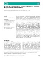
and B7) that are exposed to solvent (Fig. 1B) [19].
Interestingly, this SEHC is located on top of the coni-
cal cleft of the active site in KSI-PI. Even though KSI-
PI exposes the hydrophobic residues to solvent, it
exhibits high thermodynamic stability (a DG
H
2
O
U
value
of 24 kcalÆmol
)1
) and is highly soluble, without aggre-
gation at high concentration [31,32].
In this study, the SEHC in KSI-PI was characterized
to identify its roles in conformational stability and
steroid binding. The SEHC was perturbed by site-
directed mutagenesis in order to investigate the muta-
tional effects on catalysis, stability, unfolding and
binding affinity of KSI-PI. The crystal structure of
W92A in complex with d-1,3–5(10),6,8-estrapentaen-3-
ol-17-one (d-equilenin), determined at 2.1 A
˚
resolution,
provided a structural basis for understanding the roles
of the SEHC. Our studies demonstrate that the SEHC
in KSI-PI is required to stabilize the surface conforma-
tion of solvent-exposed b-strands, thereby contributing
to the overall conformational stability and the binding
affinity of steroids.
O
O
O
O
O
O
H
H
H
H
OOH
O
O
OO
OOH
OO
H
Tyr14 OH
Tyr14 OH
Tyr14 OH
O
OH
Asp99
Asp38
Asp99
Asp38
Asp99
Asp38
4
6
Scheme 1. General catalytic mechanism of D
5
-3-ketosteroid isomerase (KSI). The residues are numbered according to Comamonas testo-
steroni KSI in this scheme.
Role of small exterior hydrophobic cluster in KSI Y. S. Yun et al.
2000 FEBS Journal 272 (2005) 1999–2011 ª 2005 FEBS
Results
Structure of the SEHC
Based on the crystal structure of KSI-PI, the residues
Trp92, Leu125, and Val127 were found to be exposed
to solvent. Using a probe radius of 1.4 A
˚
, the acces-
sible surface areas of the side-chains of Trp92, Leu125,
and Val127 were calculated to be 203.4, 85.7, and
75.8 A
˚
2
, respectively. Given that the maximum access-
ible surface areas for a maximally exposed side-chain
were determined to be 282.5, 197.5, and 179.3 A
˚
2
, the
side-chains of Trp92, Leu125, and Val127 are 72.0,
43.4, and 42.3% exposed, respectively. Moreover, an
SEHC consisting of Trp92, Leu125, and Val127 was
found to be located on the center of three b-strands
(B4, B5 and B7) that occupy the entry site of the ster-
oid-binding pocket (Fig. 1B).
Mutational effect on catalysis
To investigate the mutational effects of W92A,
L125A ⁄ V127A, W92A ⁄ L125A ⁄ V127A, and L125F ⁄
V127F on the catalytic parameters, the k
cat
and K
M
values of the mutant KSI-PIs were determined using
5-androstene-3,17-dione (5-AND) as a substrate
(Table 1). The removal or addition of hydrophobic
moieties from the SEHC affected the K
M
more than
the k
cat
. The k
cat
values decreased by 1.6-, 1.6-, 1.7-
and 1.2-fold for W92A, L125A ⁄ V127A, W92A ⁄
L125A ⁄ V127A and L125F ⁄ V127F while the K
M
values
increased by 1.9-, 2.7-, 2.3- and 1.5-fold, respectively,
indicating that the SEHC could affect the substrate-
binding step as well as the catalytic step in the enzyme
reaction.
Effect of mutations on conformational stability
The unfolding free-energy change, DG
U
, was deter-
mined by monitoring the molar ellipticity of the pro-
teins at 222 nm upon changing the urea concentration
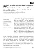
at 25 °C. The transition curves were normalized by
assuming that ellipticities for the native and unfolded
state can be extrapolated linearly into the transition
zone and nicely fitted to a two-state transition model
(Fig. 2). By applying the two-state transition model,
the values of DG
H
2
O
U
, m, and DDG
U
for wild-type
(WT) and mutant enzymes were obtained (Table 2).
Removal of hydrophobic moieties from the SEHC
decreased the DG
H
2
O
U
values by 3.1 and 4.2 kcalÆmol
)1
Table 1. Kinetic parameters of the wild-type (WT) enzyme and its mutants for the isomerization of 5-androstene-3,17-dione to 4-androstene-
3,17-dione. The assays were performed in buffer containing 34 m
M potassium phosphate and 2.5 mM EDTA, pH 7.0.
Enzyme k
cat
(s
)1
) K
M
(lM) k
cat
⁄ k
M
(M
)1
Æs
)1
) Relative k
cat
a
Relative k
M
b
WT (21.2 ± 0.8) · 10
3c
49.9 ± 1.3
c
4.3 · 10
8
1.000 1.000
W92A (12.7 ± 1.9) · 10
3
99.5 ± 23.7 1.3 · 10
8
0.599 1.993
L125A ⁄ V127A (12.9 ± 0.5) · 10
3
137.5 ± 10.3 0.9 · 10
8
0.608 2.755
W92A ⁄ L125A ⁄ V127A (12.0 ± 0.8) · 10
3
118.5 ± 8.7 1.0 · 10
8
0.566 2.374
L125F ⁄ V127F (17.6 ± 0.8) · 10
3
79.5 ± 3.8 2.3 · 10
8
0.827 1.593
a,b
Values relative to those of the WT enzyme.
c
Data from [42].
Val127
Val127
Leu125 Leu125
Trp92
Trp92
d-equilenin
d-equilenin
A
B
Fig. 1. Structure of D
5
-3-ketosteroid isomerase from Pseudomon-
as putida biotype B (KSI-PI). (A) Ribbon diagram of the dimeric
structure of KSI-PI in complex with d-equilenin. (B) Stereoview of
the monomeric structure of the dimeric KSI-PI. Trp92, Leu125,
Val127, and d-equilenin are displayed by a ball-and-stick model. The
figures were drawn using the program
SWISS-PDB VIEWER, Version
3.7 [49].
Y. S. Yun et al. Role of small exterior hydrophobic cluster in KSI
FEBS Journal 272 (2005) 1999–2011 ª 2005 FEBS 2001
in W92A and W92A⁄ L125A ⁄ V127A, respectively.
However, the L125F ⁄ V127F mutation of the SEHC
increased the DG
H
2
O
U
value by 0.9 kcalÆmol
)1
, while
the L125A ⁄ V127A mutation of the SEHC decreased
the DG
H
2
O
U
value by 3.3 kcalÆmol
)1
. These results indi-
cate that the SEHC formed by Trp92, Leu125 and
Val127 contributes to the conformational stability.
Unfolding kinetics
The unfolding of the enzymes was monitored by meas-
uring the fluorescence intensity, as a function of time,
at various urea concentrations. The unfolding curve
was nicely fitted to Eqn (6). When plots of ln k
U
vs.
the urea concentration were made in the range where
the proteins are more than 95% unfolded at equilib-
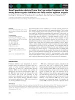
rium, straight lines were obtained (Fig. 3). The unfold-
ing rates of W92A, L125A ⁄ V127A and W92A ⁄ L125A ⁄
V127A were faster than that of the WT enzyme. How-
ever, the unfolding rate of L125F ⁄ V127F was slower
than that of the WT enzyme, suggesting that the hydro-
phobic moieties may play a role during the unfolding
process. The free-energy change of the unfolding trans-
ition state was assessed from the unfolding rate con-
stants (Table 3).
Analyses of the transition-state interaction
The hydrophobic interaction of the SEHC during the
unfolding process was investigated by U
U
value analy-
sis, according to the method described previously [33].
The U
U
value can range from 0 to 1. A high U
U
value
implies that the target region is exposed to solvent in
the transition state to the same extent as in the unfol-
ded state, while a low U
U
value implies that the inter-
action energies in the transition states and folded
states are similar. The W92A, L125A ⁄ V127A and
W92A ⁄ L125A ⁄ V127A mutants gave U
U
values of
0.451, 0.393 and 0.500, respectively, indicating that
50.0–60.7% of the noncovalent interaction energy is
maintained in the transition state for the hydrophobic
cluster (Table 3). However, the L125F ⁄ V127F mutant
had a relatively high U
U
value of 0.777. The ratio of
m
U
à
⁄ m
U
has been reported to indicate the increase in
solvent exposure of the transition state relative to the
native state [34]. The m
U
à
⁄ m
U
values of the WT
enzyme were determined to be 0.147, indicating that
the solvent accessibility of the transition state is very
similar to that of the native state.
Effect of mutations on d-equilenin binding
d-Equilenin has a maximum emission peak at 363 nm
when excited at 335 nm. Addition of the enzyme
Fig. 2. Unfolding equilibrium transition of the wild-type (WT)
enzyme (s), and those of the mutants W92A (n), L125A ⁄ V127A
(·), W92A ⁄ L125A ⁄ V127A (e), and L125F ⁄ V127F (h). The fraction
of unfolded protein at each urea concentration was calculated from
the molar ellipticity at 222 nm after correction for the pre- and post-
transition baselines. The transition curves were obtained by fitting
the data to Eqn (4).
Table 2. Changes in the free energies of unfolding of the wild-type (WT) enzyme and its mutants in the reversible denaturation with urea.
Measurements were performed at 25 °C and pH 7.0. Values were obtained by fitting the data from Fig. 2 according to Eqn (4).
Enzyme
DG
U
H
2
Oa
(kcalÆmol
)1
)
m
b
(kcalÆmol
)1
ÆM)
[Urea]
50%
c
(M)
DG
U
d
(kcalÆmol
)1
)
WT 24.0 ± 0.5 3.41 ± 0.06 5.22 ± 0.12
W92A 20.9 ± 0.4 3.38 ± 0.05 4.20 ± 0.10 ) 3.1
L125A ⁄ V127A 20.7 ± 0.3 3.26 ± 0.04 4.21 ± 0.07 ) 3.3
W92A ⁄ L125A ⁄ V127A 19.8 ± 0.3 3.69 ± 0.02 3.59 ± 0.07 ) 4.2
L125F ⁄ V127F 24.9 ± 0.4 3.44 ± 0.08 5.26 ± 0.09 0.9
a
DG
U
H
2
O
was determined by extrapolation of the data to a concentration of 0 M urea during denaturation.
b
m is the slope of the linear dena-
turation plot, dDG
U
⁄ d[urea].
c
[Urea]
50%
is the concentration of urea at which 50% of the protein is unfolded.
d
Values obtained from Eqn (5).
Role of small exterior hydrophobic cluster in KSI Y. S. Yun et al.
2002 FEBS Journal 272 (2005) 1999–2011 ª 2005 FEBS
caused a decrease in the intensity of this peak owing
to the quenching of the fluorescence in the cavity of
the active site, but no shift of the wavelength at which
the spectral intensity is highest (k
max
) was observed.
Fluorescence intensity at 363 nm was analyzed as a
function of the enzyme concentration (Fig. 4). The K
D
value of d-equilenin for the WT enzyme was found to
be 2.00 lm by fitting the data to Eqn (11) (Table 4).
Removal of the hydrophobic moieties from the SEHC
increased the K
D
value for d-equilenin by 2.20-, 5.40-
and 2.95-fold in W92A, L125A ⁄ V127A and W92A⁄
L125A ⁄ V127A, respectively, suggesting that the hydro-
phobic moieties may be important for the enzyme to
bind d-equilenin. In L125F ⁄ V127F, the small increase
in K
D
indicates that the addition of hydrophobic moi-
eties, such as phenylalanines, does not significantly
affect steroid binding.
Effect of mutations on 17b-hydroxy-4-estren-
3-one (19-nortestosterone) binding
The K
D
value for 19-nortestosterone was determined
by analyzing the changes in UV absorption spectra
upon binding 19-nortestosterone to the enzyme. From
spectral titration at various steroid concentrations, the
K
D
value of 19-nortestosterone was obtained for each
enzyme according to the relationship given in Eqn
(12). The spectral titration for the enzymes is shown
in Fig. 5 and the calculated K
D
values are listed in
Table 4. The K
D
value was determined to be 7.28 lm
for the WT enzyme. The K
D
values for W92A, L125A ⁄
V127A and W92A ⁄ L125A⁄ V127A were increased
by 2.81-, 5.97- and 2.08-fold, respectively, indicating
that the SEHC could affect the affinity towards
19-nortestosterone. The 1.18-fold increased K
D
of
L125F ⁄ V127F suggests that the increased bulkiness of
the phenylalanines did not drastically interfere with
steroid binding.
Structural analysis of W92A
To explain the decreased stability and the increased
K
D
values of W92A towards steroids on a structural
basis, the crystal structure of W92A was determined at
2.1 A
˚
resolution. It belongs to the space group C222
1
with cell dimensions of a ¼ 35.320 A
˚
, b ¼ 95.871 A
˚
and c ¼ 72.970 A
˚
. Crystallographic data and refine-
ment statistics are listed in Table 5. The structure of
W92A was almost the same as that of the WT enzyme,
with an rmsd of 0.46 A
˚
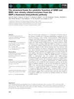
. Two major structural differ-
ences were noticeable (Fig. 6). One is that the b-strand,
including Ala92 in W92A, deviated outwards relative
Fig. 3. Unfolding rate constants (k
U
) at various urea concentrations
for the wild-type (WT) enzyme (s), and those of the mutants
L125F ⁄ V127F (h), W92A (n), W92A ⁄ L125A ⁄ V127A (e ), and
L125A ⁄ V127A (· ). Rate constants were measured in units of s
)1
.
The unfolding process was monitored by measuring the change in
the intrinsic fluorescence intensity of the protein. The excitation
wavelength was 285 nm and the emission wavelength 325 nm.
Table 3. Changes in free energies of the native state (DDG
U
) and the transition state (DDG
U
à
) for unfolding upon mutation of D
5
-3-ketosteroid
isomerase from Pseudomonas putida biotype B (KSI-PI). Measurements were carried out at 25 °C and pH 7.0.
Enzyme
DG
à
U
H
2
Oa
(kcalÆmol
)1
)
m
à
U
b
(kcalÆmol
)1
ÆM)
DDG
U
c
(kcalÆmol
)1
)
DDG
à
U
d
(kcalÆmol
)1
)
DDG
à
U
⁄DDG
U
(in H
2
O) m
à
U
⁄ m
U
WT 27.8 0.504 0.147
W92A 26.4 0.504 ) 3.1 ) 1.4 0.451 0.149
L125A ⁄ V127A 26.5 0.549 ) 3.3 ) 1.3 0.393 0.168
W92A ⁄ L125A ⁄ V127A 25.7 0.621 ) 4.2 ) 2.1 0.500 0.168
L125F ⁄ V127F 28.5 0.595 0.9 0.7 0.777 0.172
a
DG
à
U
H
2
O
was obtained from extrapolation of DG
à
U
to 0 M urea where DG
à
U
was determined from the fit according to the equation: k
U
¼
(k
B
T ⁄ h)Æexp[–DG
à
U
⁄ RT].
b
m
à
U
is the slope of the linear denaturation plot, dDG
à
U
⁄ d[urea].
c
Values obtained from Eqn (5).
d
Values obtained
from Eqn (9).
Y. S. Yun et al. Role of small exterior hydrophobic cluster in KSI
FEBS Journal 272 (2005) 1999–2011 ª 2005 FEBS 2003
to that of the WT enzyme, and the distance between
the a-carbons of Trp92 in the WT enzyme and of
Ala92 in W92A, was 1.47 A
˚
, suggesting that the
b-sheet structure underneath the hydrophobic layer
was largely perturbed by the deletion of the bulky
indole ring of Trp92. Furthermore, the side-chain of
Leu125 in W92A moved towards the hydrophobic cav-
ity as a result of the absence of the bulky indole ring
of Trp92, and the distance between the c-carbons of
Leu125 in the WT enzyme and W92A was measured
to be 2.21 A
˚
. In addition to two structural differences,
the accessible surface areas of the side-chains of
Leu125 and Val127 were increased by 16.8 and
86.5 A
˚
2
compared with those of the WT enzyme,
respectively, indicating that the removal of the bulky
side-chain of Trp92 exposed Leu125 and Val127 to sol-
vent to a greater extent.
Fig. 4. Changes in the fluorescence emission of d-equilenin at
363 nm, with varying enzyme concentration, for the wild-type
enzyme (WT) (s), and for the mutants W92A (n), L125A ⁄ V127A
(·), W92A ⁄ L125A ⁄ V127A (e), and L125F ⁄ V127F (h). The excitation
wavelength was 335 nm. The curves were obtained by fitting the
data to Eqn (11).
Table 4. Dissociation constants (K
D
) on the binding of d-equilenin
and 19-nortestosterone to the the wild-type (WT) enzyme and its
mutants. Measurements were carried out at 25 °C and pH 7.0.
Enzyme
K
D
(lM)
d-Equilenin
a
19-Nortestosterone
b
WT 2.0 ± 0.2 7.2 ± 1.5
W92A 4.4 ± 0.3 20.3 ± 4.1
L125A ⁄ V127A 10.8 ± 1.1 > 43
c
W92A ⁄ L125A ⁄ V127A 5.9 ± 0.2 15.0 ± 3.2
L125F ⁄ V127F 2.8 ± 0.4 8.5 ± 2.1
a
The K
D
for d -equilenin was obtained in a buffer containing 10 mM
potassium phosphate and 5% (v ⁄ v) methanol.
b
The K
D
for 19-nor-
testosterone was obtained in a buffer containing 50 m
M Tris ⁄ HCl
and 100 m
M sodium chloride.
c
The lower limit was indicated owing
to the very low value of the difference spectrum and the inaccuracy
of the K
D
value.
Fig. 5. UV-spectral titrations to measure the dissociation constant
of 19-nortestosterone for the enzymes. For each enzyme, a differ-
ence spectrum was obtained by subtracting the spectra originated
from the steroid and enzyme from that of their mixture. The
absorption maximum (272 nm) of the difference spectrum for the
wild-type (WT) enzyme (s), and for the mutants W92A (n),
L125A ⁄ V127A (·), W92A ⁄ L125A ⁄ V127A (e), and L125F ⁄ V127F (h)
was analyzed at different steroid concentrations. The curves were
obtained by fitting the data to Eqn (12).
Table 5. Crystallographic data and refinement statistics for the
mutant enzyme W92A.
Resolution (A
˚
)2.1
R
sym
(%) 7.2
data completeness, F > 1r (%) 90.0
R
standard
(%) 22.41
R
free
(%) 26.88
No. of refined atoms
Atom ⁄ water 1031 ⁄ 51
Average B factor 22.363
rmsd bond length (A
˚
) 0.006549
rmsd bond angles (deg) 1.23348
Ramachandran plot (%)
Most favored regions 89.4
Additional allowed regions 10.6
Generously allowed regions 0.0
Role of small exterior hydrophobic cluster in KSI Y. S. Yun et al.
2004 FEBS Journal 272 (2005) 1999–2011 ª 2005 FEBS
Discussion
Our study was intended to identify the role of the
SEHC (comprising Trp92, Leu125 and Val127) in
KSI-PI for conformational stability and steroid bind-
ing. The DG
H
2
O
U
values decreased significantly for all
the mutants in which the hydrophobic residue of the
SEHC was replaced with alanine (W92A, L125A ⁄
V127A and W92A ⁄ L125A⁄ V127A). However, when
the hydrophobicity in the SEHC was increased by sub-
stituting leucine and valine with phenylalanines, the
DG
H
2
O
U
value increased. These results indicate that the
SEHC might improve the overall stability by stabil-
izing the solvent-exposed b-sheet constituting the sur-
face hydrophobic layer. The mutational study on
steroid-binding affinity also revealed that the SEHC
plays a role in efficient steroid binding. The crystal
structure of W92A showed that the W92A mutation
disrupts the solvent-exposed b-sheet.
Contribution of the SEHC to conformational
stability
The hydrophobic interaction in the SEHC of KSI-PI
was perturbed by replacing the hydrophobic residues
with amino acids having smaller or larger hydrophobic
side-chains. The 3.1 kcalÆmol
)1
decrease in thermody-
namic stability of W92A is noteworthy given that
amino acid substitution of a surface residue generally
does not affect the stability of a protein [35–38]. The
decreased stability of L125A ⁄ V127A and increased sta-
bility of L125F ⁄ V127F suggest that the hydrophobic
packing of the SEHC is important for the conforma-
tional stability of KSI-PI. The stability of L125A ⁄
V127A was decreased by 0.9 kcalÆmol
)1
upon the
additional mutation of W92A. This additional muta-
tion could be expected to stabilize the protein because
the hydrophobic Trp92 might not be stable in
L125A ⁄ V127A. The marginal decrease of the stability
suggested that Trp92 in L125A ⁄ V127A could interact
with other nearby hydrophobic moieties as a result of
the slight change of local conformation. In previous
studies, the b-sheet structure underneath the hydropho-
bic layer of the thermolysin-like neutral protease of
Bacillus stearothermophilus was found to be stabilized
by utilizing a hydrophobic residue at the solvent-
exposed site [3], and a hydrophobic pocket on the sur-
face of neutral protease of B. subtilis could stabilize
the protease [2]. Hence, the stabilizing effects of KSI-
PI may originate from hydrophobic interaction medi-
ated by the SEHC on the protein surface.
Hydrophobic clusters or residues have sometimes
been found on the surface of b-sheet structure proteins
[5,39]. The SEHC in KSI-PI is located on the center of
three b -strands (B4, B5 and B7) that are exposed to
solvent. Recent studies on the b-sheet structure sugges-
ted that a hydrophobic shield protecting the b-sheet
structure against invading water molecules could be
required to stabilize solvent-exposed b-sheets [2,4,40].
Invading water is critically related to the kinetic stabil-
ity of the protein as protein unfolding can be initiated
from the solvent-exposed region by the invasion of
water. Consistent with this notion, the unfolding rate
constant showed a large increase of 133-fold in
W92A ⁄ L125A ⁄ V127A compared with the WT enzyme
upon increasing the urea concentration up to 7 m.In
the crystal structure of W92A, the deletion of the
bulky indole ring of Trp92 significantly perturbed the
solvent-exposed b-sheet. Given that backbone chain
movement by a single amino acid substitution, especi-
ally in the case of a surface residue, has rarely been
found, the structural perturbation induced by the
W92A mutation is notable. In view of the structural
change in W92A at the solvent-exposed b-sheet, we
may assume that the decreased stability of W92A ori-
ginates from the replacement of the bulky hydrophobic
moiety of tryptophan, resulting in increased access of
the invading water molecules to the b-sheet, ultimately
leading to the acceleration of protein unfolding.
The hydrophobic interaction of the SEHC in KSI-PI
seems to be partially maintained in the transition state
during the unfolding process, as judged by the U
U
val-
ues (Table 3). Solvent-exposed regions, including loops,
usually exhibit high U
U
values, close to 1, because the
exposed region is usually exposed to solvent in the
transition state for the folding process [33,41]. How-
ever, the U
U
values of W92A, L125A ⁄ V127A and
W92A ⁄ L125A ⁄ V127A were found to be below 0.5,
indicating that over 50% of the hydrophobic inter-
Fig. 6. Stereoview of the small exterior hydrophobic cluster (SEHC)
in the wild-type (WT) enzyme and in the mutant W92A after super-
imposition of the backbone atoms of all residues. Trp92, Leu125,
and Val127 are displayed by a ball-and-stick model, and the back-
bone of residues 89–98 and 125–127 are drawn in solid lines. The
structure of the WT enzyme is shown in light grey, and that of the
mutant W92A is shown in dark grey. The superimposition and
drawing were carried out by using the program
SWISS-PDB VIEWER,
Version 3.7 [49].
Y. S. Yun et al. Role of small exterior hydrophobic cluster in KSI
FEBS Journal 272 (2005) 1999–2011 ª 2005 FEBS 2005
action was maintained in the transition state during the
unfolding process. The high U
U
value of L125F ⁄ V127F
seems to be a result of the increased bulkiness caused
by the introduced phenyl rings. In this case, the U
U
value does not seem to properly represent the status of
the transition state in folding, because adding new
functional groups can make other extraneous interac-
tions and cause steric effects in the protein [33,41]. Our
results indicate that the SEHC in KSI-PI contributes to
the resistance to unfolding, despite its solvent-exposed
site. Analysis of the U
U
value also supports that the
SEHC is required to stabilize the surface conformation
in KSI-PI, suggesting that the SEHC can play an
important role in the unfolding process in concert with
the hydrophobic core.
Contribution of the SEHC to recognition
of steroids
Based on the crystal structure, the SEHC comprising
Trp92, Leu125 and Val127 is located on the top of the
hydrophobic layer of the steroid-binding pocket in
KSI-PI (Fig. 1B). The steroid-binding pocket is lined
with hydrophobic residues, which contribute to the
tight binding of hydrophobic steroids [19]. The affinity
of KSI-PI towards steroids was assessed by utilizing
two steroids: d-equilenin and 19-nortestosterone [42].
K
D
values for both steroids increased by over twofold
in all the mutants with decreased hydrophobicity in the
SEHC (i.e. W92A, L125A ⁄ V127A and W92A ⁄ L125A ⁄
V127A). Consistent with the increased K
D
values in
those mutants, the K
M
values of 5-AND increased,
indicating that the SEHC contributes to the steroid
binding in KSI-PI. The decrease of hydrophobicity in
the SEHC, destabilizing the overall hydrophobic layer
along the binding site of the steroid, could lead to a
decrease in affinity towards the steroids 5-AND,
19-nortestosterone and d-equilenin. In the case of
L125A ⁄ V127A, the drastic decrease in the affinity to
steroids could be a result of disruption of the SEHC, as
Trp92 cannot form a hydrophobic cluster without the
aliphatic side-chains of Leu125 and Val127. In
L125F ⁄ V127F, the slight increase in K
D
and K
M
values
suggests that replacing leucine and valine with phenyl-
alanines cannot increase the binding affinity of steroids.
The solvent-exposed hydrophobic residues may con-
tribute to hydrophobic substrate- or ligand-binding to
the protein. It was reported that the hydrophobic sur-
face made by hydrophobic residues could be important
for the binding of phospholipids, vitamin D, lipid and
cholesterol to their respective proteins. These observa-
tions, as well as ours, suggest that solvent-exposed
hydrophobic residues seem to interact with their
ligands or substrates on the protein surface in the
initial binding step. Even if the hydrophobic residues
constituting the SEHC do not directly bind steroids, as
judged by the crystal structure of KSI-PI in complex
with d-equilenin (Fig. 1B), the SEHC seems to indi-
rectly affect the binding process of steroids by stabil-
izing the surface hydrophobic layer or perhaps by
guiding hydrophobic steroids at the top of the hydro-
phobic cleft. The bound mode of the steroid in both
KSI-PI and W92A is almost identical based on the
X-ray crystal data, supporting the fact that the SEHC
might play a role in the initial recognition of hydro-
phobic steroids rather than the binding itself.
In conclusion, the mutational studies on the role of
the SEHC in KSI-PI demonstrate that the SEHC con-
tributes not only to conformational stability, but also
to the binding affinity of steroids, by stabilizing the
hydrophobic surface conformation. Our results suggest
that the SEHC stabilizes the hydrophobic layer by
connecting the solvent-exposed b-strands and helps to
bind hydrophobic steroids. It remains to be investi-
gated whether SEHC, as a structural motif, can con-
tribute to the conformational stability or the binding
of hydrophobic ligands in other proteins.
Experimental procedures
Materials and reagents
5-AND, d-equilenin and 19-nortestosterone were purchased
from Steraloids (Newport, RI, USA). Chemicals for buffer
solutions were from Sigma (St Louis, MO, USA). Oligonu-
cleotides were obtained from Genotech (Daejon, Korea). A
QuickChange Site-Directed Mutagenesis Kit was supplied
by Stratagene (La Jolla, CA, USA). pKK 223–3 plasmid
was from Pharmacia (New York, NY, USA). A Superose
12 gel filtration column was obtained from Amersham
Pharmacia Biotech.
Site-directed mutagenesis
The QuickChange Site-Directed Mutagenesis Kit (Strata-
gene) was used for the mutagenesis. All mutagenesis pro-
cedues were carried out according to the instructions
provided by the supplier. The pKK 223–3 vector, carrying
the KSI-PI gene, was used for the mutagenesis with two
primers for each mutant: 5¢-CGCGTCGAGATGGTC
GCG
AACGGCCAGCCCTGT-3¢ and 5¢-ACAGGGCTGGCCG
TTC
GCGACCATCTCGACGCG-3¢ (W92A); 5¢-TGGAGC
GAGGTCAAC
TTCAGCTTCCGCGAGCCGCAGTAG-3¢
and 5¢-CTACTGCGGCTCGCG
GAAGCTGAAGTTGAC
CTCGCTCCA-3¢ (L125F ⁄ V127F); and 5¢-TGGAGCG
AGGTCAAC
GCCAGCGCGCGCGAGCCGCAGTAG-3¢
Role of small exterior hydrophobic cluster in KSI Y. S. Yun et al.
2006 FEBS Journal 272 (2005) 1999–2011 ª 2005 FEBS
and 5¢-CTACTGCGGCTCGCGCGCGCTGGCGTTGAC
CTCGCTCCA-3¢ (L125A ⁄ V127A); the constructed pKK
223–3 vector carrying the W92A gene was used for the pre-
paration of the triple mutant (W92A ⁄ L125A ⁄ V127A) with
two primers; 5¢-TGGAGCGAGGTCAAC
GCCAGCGCGC
GCGAGCCGCAGTAG-3¢ and 5¢-CTACTGCGGCTCGC
GC
GCGCTGGCGTTGACCTCGCTCCA-3¢; underlined
nucleotides represent those changed by point mutations.
Recombinant plasmids were introduced into Escherichia coli
XL1-Blue supercompetent cell (Stratagene) and purified by
use of a QIAprep Spin Miniprep Kit (Qiagen, ValenciaCA,
USA). The entire KSI-PI gene was then sequenced to con-
firm the desired mutations.
Expression and purification of the KSI-PI proteins
WT and mutant KSI-PIs were overproduced in E. coli
BL21(DE3) utilizing pKK223-3, an expression vector con-
taining the respective KSI-PI gene, and purified by deoxych-
olate affinity chromatography and Superose 12 gel filtration
chromatography, as described previously [26]. The purity of
the protein was confirmed by the presence of a single band
on an SDS ⁄ PAGE gel stained with Coomassie blue. The
protein concentration was determined by utilizing the differ-
ence extinction coefficient between tyrosinate and tyrosine
at 295 nm, as described previously [43]. The accuracy of the
protein concentration was confirmed by the quantitative
analysis of the bands on SDS ⁄ PAGE by use of an imaging
densitometer (Bio-Rad, Hercules, CA, USA; GS-700) and a
software program (molecular analyst ⁄ PC).
Steady-state kinetic analysis
Catalytic activities of the purified enzymes were determined
spectrophotometrically using 5-AND as a substrate, accord-
ing to the procedure previously described [42]. Various
amounts of the substrate dissolved in methanol were added
to a reaction buffer containing 34 mm potassium phosphate
and 2.5 mm EDTA, pH 7.0, at 25 ° C. The concentrations
of 5-AND used were 12, 35, 58, 82 and 116 lm. The final
concentration of methanol was 3.3% (v ⁄ v). The initial reac-
tion rate was obtained within 1 or 2 min after the initiation
of the enzymatic reaction. The fraction of the substrate
converted to the product was below 10% of the substrate
applied to the reaction mixture. The reaction was moni-
tored by measuring the absorbance at 248 nm by using a
spectrophotometer (Shimadzu, Kyoto, Japan; UV-2501
PC). k
cat
and K
M
values were determined by utilizing Line-
weaver–Burk reciprocal plots.
Calculation of accessible surface area
Accessible surface areas were calculated based on the
atomic coordinates (PDB code, 4TSU) obtained by X-ray
crystallography using the program molmol, 2 k2 [44]
according to the method described previously [45]. The
probe radius for the calculation was 1.4 A
˚
.
Equilibrium unfolding
Unfolding of the protein was assessed by measuring the
molar ellipticity at different urea concentrations. Protein
(15 lm) was incubated for at least 48 h in a buffer
containing 20 mm potassium phosphate, pH 7.0, 1 mm
EDTA, 1 mm dithiothreitol and different concentrations
(0–8 m) of urea. A cuvette with a 0.2 cm path length was
used for all CD spectral measurements. The ellipticity at
222 nm was recorded and analyzed. The changes in the
optical properties of the protein were compared by
normalizing each transition curve with the apparent frac-
tion of the unfolded form, F
U
, which was obtained by
Eqn (1):
F
U
¼ðY
N
À Y Þ=ðY
N
À Y
U
Þ; ð1Þ
where Y is the observed molar ellipticity at a given urea
concentration, and Y
N
and Y
U
are the observed values for
the native and unfolded forms, respectively, at the same
denaturant concentration. Linear extrapolations from these
baselines were made to estimate Y
N
and Y
U
in the trans-
ition region. The equilibrium constant (K
U
) and free-
energy change (DG
U
) for denaturation were determined,
according to a two-state model of denaturation, by Eqns
(2) and (3):
K
U
¼ 2P
T
Á½F
2
U
=ð1 À F
U
Þ ð2Þ
and
DG
U
¼ÀRT Á ln ðK
U
Þ¼DG
H
2
O
U
À m Á½urea; ð3Þ
where P
T
is the total protein concentration, DG
H
2
O
U
the
free-energy change in the absence of urea, and m a measure
of the DG
U
dependence on urea concentration. DG
H
2
O
U
and
m values were obtained by fitting urea denaturation curve
data to Eqn (4) [46] using a software program (Abelbeck
Softwae, kaleidagraph version 3.06):
Y ¼ Y
N
ÀðY
N
ÀY
U
ÞÁexp½ðm Á½ureaÀDG
H
2
O
U
Þ=RT
Á½f1 þ8P
T
=exp½ðm Á½ureaÀDG
H
2
O
U
Þ=RTg
1=2
À1=4P
T
:
ð4Þ
The difference in the free-energy change for unfolding,
DDG
U
, between WT and each mutant protein was obtained
by Eqn (5):
DDG
U
¼ DG
m
U
À DG
U
; ð5Þ
where DG
U
and DG
m
U
are the free-energy changes for the
unfolding of WT and mutant proteins, respectively.
Y. S. Yun et al. Role of small exterior hydrophobic cluster in KSI
FEBS Journal 272 (2005) 1999–2011 ª 2005 FEBS 2007
Kinetic analysis of unfolding
The unfolding kinetic experiments for WT and mutant
KSI-PIs were performed by use of a spectrofluorometer
(Shimadzu RF5000) equipped with a thermostatically con-
trolled cell holder. The protein was incubated in a buffer
containing 20 mm potassium phosphate, pH 7.0, 1 mm
EDTA and 1 mm dithiothreitol. Unfolding reactions were
initiated by diluting the protein sample 20-fold into the
same buffer with various concentrations of urea at 25 °C.
The dead time of manual mixing was % 10 s. The kinetics
for unfolding was monitored by measuring the fluorescence
intensity at 325 nm after excitation at 285 nm. The final
protein concentration was 15 lm. The rate constants for
unfolding at each urea concentration were obtained by fit-
ting the data to Eqn (6):
F
t
¼ F
1
þ R½F
i
Á expðÀk
i
Á tÞ; ð6Þ
where F
t
and F
1
are the amplitudes at time t and at the
final state, F
i
is the amplitude of the kinetic phase and k
i
is the rate constant for unfolding. Data fitting was carried
out by using the kaleidagraph program. The unfolding
rate constants, k
U
, obtained at different urea concentra-
tions, were then analyzed according to Eqn (7), as des-
cribed [41]:
ln k
U
¼ ln k
H
2
O
U
þ m
U
z
Á½urea; ð7Þ
where k
H
2
O
U
is the unfolding rate constant in the absence of
urea and m
U
à
the dependence of the unfolding rate con-
stant on urea concentration. The free energy of activation
for the unfolding of KSI-PI was obtained by Eqn (8):
DG
z
U
¼ DG
H
2
Oz
U
À m
U
z
Á½urea; ð8Þ
where DG
H
2
O
U
à
is the free-energy change for the unfolding
transition state in the absence of urea, and m
U
à
represents
a measure of the DG
U
à
dependence on urea concentration.
DG
U
à
was obtained from the relationship, DG
U
à
¼
RTln(k
B
T ⁄ h)–lnk
U
, where k
B
, T and h are the Boltzman
constant, the experimental temperature and the Plank con-
stant, respectively.
Analysis of the F
U
value
The changes in free energy of activation for unfolding,
DDG
U
à
, between WT and mutant proteins were obtained by
Eqn (9):
DDG
z
U
¼ DG
zm
U
À DG
z
U
ð9Þ
where DG
U
à
and DG
U
àm
are the free-energy changes of acti-
vation for the unfolding of WT and mutant proteins,
respectively. The F value of unfolding, F
U
, is the ratio of
the free-energy change determined from the kinetic data to
that determined from the urea equilibrium unfolding experi-
ment, as described in Eqn (10):
U
U
¼ DDG
z
U
=DDG
U
¼ðDG
F
À DG
z
Þ=ðDG
F
À DG
solv
Þ;
ð10Þ
where DG
F
is the difference of the noncovalent interaction
energy between WT and mutant enzymes in the folded
states, DGà the difference in the transition states and DG
solv
the difference in the unfolded states.
Determination of K
D
for d-equilenin
Fluorescence quenching upon the binding of equilenin to
the enzyme was used to determine the dissociation constant,
K
D
, as described previously [42]. Fluorescence measure-
ments were carried out at 25 °C by using a spectroflurome-
ter (Shimadzu, RF5000) in a buffer containing 10 mm
potassium phosphate and 5% (v ⁄ v) methanol at pH 7.0. A
total of 5 lL of the stock solution of d-equilenin was added
to 3.0 mL of the buffer, giving a final concentration of
3 lm. Titrations were carried out by adding 6 lL of the
enzyme solution to give a total volume of 72 lL. After add-
ing the enzyme, the emission spectrum was scanned from
345 nm to 450 nm with an excitation wavelength at
335 nm. After the spectral change caused by the dilution
had been corrected, the fluorescence of d-equilenin at the
emission maximum (363 nm) for each enzyme concentra-
tion was used to calculate the K
D
for d-equilenin by nonlin-
ear least-squares fitting, according to Eqn (11), by using the
kaleidagraph program:
E
t
¼ðF
0
À FÞfK
D
=ðF À F
1
Þþ½equilenin=ðF
0
À F
1
Þg; ð11Þ
where E
t
is the concentration of total enzyme in the solu-
tion, F is the fluorescence intensity, F
0
is the intensity in the
absence of enzyme and F
1
is the intensity extrapolated to
infinite enzyme concentration. A binding stoichiometry of
1 per subunit was assumed.
Determination of K
D
for 19-nortestosterone
The K
D
for 19-nortestosterone was determined by UV
absorption spectrometry, as described previously [42]. The
measurements were carried out at 25 °C, using a spectro-
photometer (Shimadzu, UV-2501 PC), in an 1.0 cm quartz
cuvette with a total volume of 1 mL. The spectra from
320 to 220 nm were obtained in a buffer containing
50 mm Tris ⁄ HCl and 100 mm sodium chloride at pH 7.0.
19-Nortestosterone was added to the enzyme from a
10 mm stock solution containing 20% (v ⁄ v) methanol. The
absorption change caused by the increased volume was
corrected. Difference spectra were obtained by subtracting
the spectra of total steroid and total enzyme from those
of their mixture. The changes in absorption (DA) at the
respective absorption maxima in the difference spectra
were measured as a function of steroid concentration. K
D
values were determined by fitting the DA plots, with
Role of small exterior hydrophobic cluster in KSI Y. S. Yun et al.
2008 FEBS Journal 272 (2005) 1999–2011 ª 2005 FEBS
respect to total steroid concentration, to Eqn (12), by
using the kaleidagraph program:
DA=DA
max
¼½K
D
þ E
t
þ S
t
ÀfðK
D
þ E
t
þ S
t
Þ
2
À 4E
t
S
t
g
1=2
=2E
t
; ð12Þ
where K
D
represents the dissociation constant, E
t
the con-
centration of total enzyme, S
t
the concentration of total
steroid and DA
max
the maximal change in absorption
observed as S
t
approaches infinity. A binding stoichiometry
of 1 per subunit was assumed.
Crystallization and structure determination
of W92A
The crystals of W92A were obtained by co-crystallizing
with a small amount of d-equilenin dissolved in dimethyl
sulfoxide. A total of 30 mm d-equilenin solution was mixed
with 1 mm enzyme solution. Saturated d-equilenin was
ensured by the presence of white precipitates formed imme-
diately after the mixing. Crystals of W92A were grown in a
solution containing 0.2 m magnesium acetate, 20% (w ⁄ v)
PEG 8000 and 0.1 m sodium carcodylate, pH 6.5, by the
hanging drop method of vapor diffusion at 25 °C. Before
cryocooling, crystals were briefly immersed in the same pre-
cipitant solution containing 10–15% (v ⁄ v) glycerol. All dif-
fraction data were collected at 100 K on the beamline 6B
of the Pohang Accelerator Laboratory (Pohang, Korea).
Data reduction, merging and scaling were carried out using
the programs denzo and scalepack, as described previ-
ously [47]. The structure was determined by the molecular
replacement program cns, using the atomic coordinates of
the WT enzyme (PDB code, 4TSU). Further refinement
was carried out by using the program cns [48].
Acknowledgements
This research was supported by grants from the
National Research Laboratory sponsored by the Korea
Ministry of Science and Technology, from the Korea
Science and Engineering Foundation, and by the Brain
Korea 21 project to Y. S. Y.
References
1 Kauzmann W (1959) Some factors in the interpretation
of protein denaturation. Adv Protein Chem 14 , 1–63.
2 Frigerio F, Margarit I, Nogarotto R, de Filippis V &
Grandi G (1996) Cumulative stabilizing effects of
hydrophobic interactions on the surface of the neutral
protease from Bacillus subtilis. Protein Eng 9, 439–
445.
3 Van den Burg B, Dijkstra BW, Vriend G, Van der
Vinne B, Venema G & Eijsink VG (1994) Protein stabi-
lization by hydrophobic interactions at the surface. Eur
J Biochem 220, 981–985.
4 Machius M, Declerck N, Huber R & Wiegand G (2003)
Kinetic stabilization of Bacillus licheniformis alpha-amy-
lase through introduction of hydrophobic residues at
the surface. J Biol Chem 278, 11546–11553.
5 Nesloney CL & Kelly JW (1996) Progress towards
understanding beta-sheet structure. Bioorg Med Chem 4,
739–766.
6 Tisi LC & Evans PA (1995) Conserved structural fea-
tures on protein surfaces: small exterior hydrophobic
clusters. J Mol Biol 249, 251–258.
7 Street AG & Mayo SL (1999) Computational protein
design. Structure Fold Des 7 , R105–R109.
8 Eijsink VG, Bjork A, Gaseidnes S, Sirevag R, Synstad
B, Burg Bv B & Vriend G (2004) Rational engineering
of enzyme stability. J Biotechnol 113, 105–120.
9 Robertson AD (2002) Intramolecular interactions at
protein surfaces and their impact on protein function.
Trends Biochem Sci 27, 521–526.
10 Desrumaux C, Labeur C, Verhee A, Tavernier J, Van-
dekerckhove J, Rosseneu M & Peelman F (2001) A
hydrophobic cluster at the surface of the human plasma
phospholipid transfer protein is critical for activity on
high density lipoproteins. J Biol Chem 276, 5908–5915.
11 Solomon C, Macoritto M, Gao XL, White JH &
Kremer R (2001) The unique tryptophan residue of the
vitamin D receptor is critical for ligand binding and
transcriptional activation. J Bone Miner Res 16, 39–45.
12 Dodson GG, Lawson DM & Winkler FK (1992) Struc-
tural and evolutionary relationships in lipase mechanism
and activation. Faraday Discuss 93, 95–105.
13 Cambillau C, Longhi S, Nicolas A & Martinez C (1996)
Acyl glycerol hydrolases: inhibitors, interface and cata-
lysis. Curr Opin Struct Biol 6, 449–455.
14 Schmid RD & Verger R (1998) Lipases: interfacial
enzymes with attractive applications. Angew Chem Int
Ed 37, 1608–1633.
15 Li J, Vrielink A, Brick P & Blow DM (1993) Crystal
structure of cholesterol oxidase complexed with a ster-
oid substrate: implications for flavin adenine dinucleo-
tide dependent alcohol oxidases. Biochemistry 32,
11507–11515.
16 Vrielink A, Lloyd LF & Blow DM (1991) Crystal struc-
ture of cholesterol oxidase from Brevibacterium steroli-
cum refined at 1.8 A
˚
resolution. J Mol Biol 219, 533–
554.
17 Yue QK, Kass IJ, Sampson NS & Vrielink A (1999)
Crystal structure determination of cholesterol oxidase
from Streptomyces and structural characterization of
key active site mutants. Biochemistry 38, 4277–4286.
18 Duax WL, Griffin JF & Ghosh D (1996) The fascinat-
ing complexities of steroid-binding enzymes. Curr Opin
Struct Biol 6, 813–823.
Y. S. Yun et al. Role of small exterior hydrophobic cluster in KSI
FEBS Journal 272 (2005) 1999–2011 ª 2005 FEBS 2009
19 Kim SW, Cha S-S, Cho H-S, Kim J-S, Ha N-C, Cho
M-J, Joo S, Kim KK, Choi KY & Oh B-H (1997)
High-resolution crystal structures of D
5
-3-ketosteroid
isomerase with and without a reaction intermediate
analogue. Biochemistry 36, 14030–14036.
20 Batzold FH, Benson AM, Covey DF, Robinson CH &
Talalay P (1976) The D
5
-3-ketosteroid isomerase reac-
tion: catalytic mechanism, specificity and inhibition. Adv
Enzyme Regul 14, 243–267.
21 Pollack RM, Thornburg LD, Wu ZR & Summers MF
(1999) Mechanistic insights from the three-dimensional
structure of 3-oxo-D
5
-steroid isomerase. Arch Biochem
Biophys 370, 9–15.
22 Ha N-C, Choi G, Choi KY & Oh B-H (2001) Structure
and enzymology of D
5
-3-ketosteroid isomerase. Curr
Opin Struct Biol 11, 674–678.
23 Hawkinson DC, Eames TC & Pollack RM (1991)
Energetics of 3-oxo-D
5
-steroid isomerase: source of the
catalytic power of the enzyme. Biochemistry 30,
10849–10858.
24 Hawkinson DC, Pollack RM & Ambulos NP Jr (1994)
Evaluation of the internal equilibrium constant for
3-oxo-D
5
-steroid isomerase using the D38E and D38N
mutants: the energetic basis for catalysis. Biochemistry
33, 12172–12183.
25 Xue LA, Kuliopulos A, Mildvan AS & Talalay P (1991)
Catalytic mechanism of an active-site mutant (D38N) of
D
5
-3-ketosteroid isomerase. Direct spectroscopic evi-
dence for dienol intermediates. Biochemistry 30, 4991–
4997.
26 Choi G, Ha N-C, Kim SW, Kim D-H, Park S, Oh B-H
& Choi KY (2000) Asp-99 donates a hydrogen bond
not to Tyr-14 but to the steroid directly in the catalytic
mechanism of D
5
-3-ketosteroid isomerase from Pseudo-
monas putida biotype B. Biochemistry 39, 903–909.
27 Choi G, Ha N-C, Kim MS, Hong BH, Oh B-H & Choi
KY (2001) Pseudoreversion of the catalytic activity of
Y14F by the additional substitution(s) of tyrosine with
phenylalanine in the hydrogen bond network of D
5
-3-
ketosteroid isomerase from Pseudomonas putida biotype
B. Biochemistry 40, 6828–6835.
28 Yun YS, Lee TH, Nam GH, Jang DS, Shin S, Oh B-H
& Choi KY (2003) Origin of the different pH activity
profile in two homologous ketosteroid isomerases.
J Biol Chem 278, 28229–28236.
29 Cho H-S, Ha N-C, Choi G, Kim H-J, Lee D, Oh KS,
Kim KS, Lee W, Choi KY & Oh B-H (1999) Crystal
structure of D
5
-3-ketosteroid isomerase from Pseudomo-
nas testosteroni in complex with equilenin settles the cor-
rect hydrogen bonding scheme for transition state
stabilization. J Biol Chem 274, 32863–32868.
30 Wu ZR, Ebrahimian S, Zawrotny ME, Thornburg LD,
Perez-Alvarado GC, Brothers P, Pollack RM & Sum-
mers MF (1997) Solution structure of 3-oxo-D
5
-steroid
isomerase. Science 276, 415–418.
31 Kim DH, Jang DS, Nam GH, Choi G, Kim JS, Ha
N-C, Kim MS, Oh B-H & Choi KY (2000) Contribu-
tion of the hydrogen-bond network involving a tyrosine
triad in the active site to the structure and function of a
highly proficient ketosteroid isomerase from Pseudomo-
nas putida biotype B. Biochemistry 39, 4581–4589.
32 Nam GH, Jang DS, Cha SS, Lee TH, Kim DH, Hong
BH, Yun YS, Oh B-H & Choi KY (2001) Maintenance
of alpha-helical structures by phenyl rings in the active-
site tyrosine triad contributes to catalysis and stability
of ketosteroid isomerase from Pseudomonas putida bio-
type B. Biochemistry 40, 13529–13537.
33 Matouschek A, Kellis JT Jr, Serrano L & Fersht AR
(1989) Mapping the transition state and pathway of
protein folding by protein engineering. Nature 340, 122–
126.
34 Tanford C (1970) Protein denaturation: theoretical
models for the mechanism of denaturation. Adv Protein
Chem 24, 1–95.
35 Rennell D, Bouvier SE, Hardy LW & Poteete AR
(1991) Systematic mutation of bacteriophage T4 lyso-
zyme. J Mol Biol 222, 67–88.
36 Reidhaar-Olson JF & Sauer RT (1990) Functionally
acceptable substitutions in two alpha-helical regions of
lambda repressor. Proteins 7, 306–316.
37 Bowie JU, Reidhaar-Olson JF, Lim WA & Sauer RT
(1990) Deciphering the message in protein sequences:
tolerance to amino acid substitutions. Science 247,
1306–1310.
38 Funahashi J, Takano K, Yamagata Y & Yutani K
(2000) Role of surface hydrophobic residues in the con-
formational stability of human lysozyme at three differ-
ent positions. Biochemistry 39, 14448–14456.
39 Hazes B & Hol WG (1992) Comparison of the hemo-
cyanin beta-barrel with other Greek key beta-barrels:
possible importance of the ‘beta-zipper’ in protein struc-
ture and folding. Proteins 12, 278–298.
40 Perl D, Mueller U, Heinemann U & Schmid FX (2000)
Two exposed amino acid residues confer thermostability
on a cold shock protein. Nat Struct Biol 7, 380–383.
41 Fersht A (1999) Kinetics of protein folding. In Structure
and Mechanism in Protein Science (Julet MR & Hadler
GL, eds), pp. 540–572. W.H. Freeman, New York.
42 Kim D-H, Nam GH, Jang DS, Choi G, Joo S, Kim J-S,
Oh B-H & Choi KY (1999) Roles of active site aromatic
residues in catalysis by ketosteroid isomerase from Pseu-
domonas putida biotype B. Biochemistry 38, 13810–13819.
43 Copeland RA (1993) Methods for protein quantitation.
In Methods of Protein Analysis. pp. 51–54. Chapman &
Hall, New York, NY.
44 Koradi R, Billeter M & Wuthrich K (1996) molmol:a
program for display and analysis of macromolecular
structures. J Mol Graph 14, 29–32.
45 Connolly ML (1993) The molecular surface package.
J Mol Graph 11, 139–141.
Role of small exterior hydrophobic cluster in KSI Y. S. Yun et al.
2010 FEBS Journal 272 (2005) 1999–2011 ª 2005 FEBS
46 Mok Y-K, Gay GD, Butler PJ & Bycroft M (1996)
Equilibrium dissociation and unfolding of the dimeric
human papillomavirus strain-16, E2 DNA-binding
domain. Protein Sci 5, 310–319.
47 Otwinowski Z (1993) Data collection and processing.
In Proceedings of the CCP4 Study Weekend (Sawyer L,
Isaccs N & Bailey S, eds), pp. 56–62. SERC Daresbury
Laboratory, Warrington, UK.
48 Brunger AT, Adams PD, Clore GM, DeLano WL, Gros
P, Grosse-Kunstleve RW, Jiang JS, Kuszewski J, Nilges
M, Pannu NS, et al. (1998) Crystallography and NMR
system: a new software suite for macromolecular struc-
ture determination. Acta Crystallogr D 54, 905–921.
49 Guex N & Peitsch MC (1997) SWISS-MODEL and
the Swiss-PdbViewer: an environment for comparative
protein modeling. Electrophoresis 18, 2714–2723.
Y. S. Yun et al. Role of small exterior hydrophobic cluster in KSI
FEBS Journal 272 (2005) 1999–2011 ª 2005 FEBS 2011