Báo cáo khoa học: Substrate positioning by His92 is important in catalysis by purple acid phosphatase docx
Bạn đang xem bản rút gọn của tài liệu. Xem và tải ngay bản đầy đủ của tài liệu tại đây (271.74 KB, 10 trang )
Substrate positioning by His92 is important in catalysis
by purple acid phosphatase
Enrico G. Funhoff
1,4
, Yunling Wang
2
, Goran Andersson
2
and Bruce A. Averill
1,3
1 Swammerdam Institute for Life Sciences, University of Amsterdam, the Netherlands
2 Karolinska Institutet, Division of Pathology, Huddinge University Hospital, Sweden
3 Department of Chemistry, University of Toledo, OH, USA
4 Institute of Biotechnology, HPT, ETH Ho
¨
nggerberg, Zu
¨
rich, Switzerland
The binuclear metalloenzyme purple acid phosphatase
(PAP) [1], which may be involved in disorders such
as osteoporosis [2–4], Gaucher disease [5], hairy cell
leukemia [6], and AIDS [7] is widely distributed in
mammalian tissues [8,9]. The expression level of PAP
[also referred to as tartrate resistant acid phosphatase
(TRAP) or type 5 acid phosphatase (AcP5; EC
3.1.3.2)] is elevated in these disorders, suggesting a
relationship between the increased levels of the enzyme
and the clinical picture. The presumed role of PAP
makes it important to develop drugs that can inhibit
PAP activity. In order to facilitate this process, the
precise catalytic mechanism must be elucidated, inclu-
ding the function of each residue involved in catalysis.
Although the available crystal structures of PAPs
[10–12] provide structural information about the resi-
dues potentially involved in catalysis, to date only
structures of inactive redox and protonation states of
the enzymes have been reported. All structures show
an active site composed of two metal ions bridged by
a solvent-derived species and a bidentate aspartate resi-
due. In addition, the iron(III) ion is coordinated by a
tyrosinate [resulting in a ligand-to-metal charge trans-
fer (LMCT) transition that is responsible for the
Keywords
kinetics; mechanism; mutagenesis; purple
acid phosphatase; spectroscopy
Correspondence
B. A. Averill, Department of Chemistry,
University of Toledo, 2801 West Bancroft
Road, Toledo, Ohio 43606–3390, USA
Fax: +1 419 5301586
Tel: +1 419 5301585
E-mail:
Website: />FAC_INFO/Bruce/SOURCE.htm
(Received 12 January 2005, revised 13
March 2005, accepted 28 March 2005)
doi:10.1111/j.1742-4658.2005.04686.x
Proteolysis of single polypeptide mammalian purple acid phosphatases
(PAPs) results in the loss of an interaction between the loop residue
Asp146 and the active site residues Asn91 and ⁄ or His92. While Asn91 is a
ligand to the divalent metal of the mixed-valent di-iron center, the role of
His92 in the catalytic mechanism is unknown. Site-directed mutagenesis of
His92 was performed to examine the role of this residue in single polypep-
tide PAP. Conversion of His92 into Ala, which eliminates polar interac-
tions of this residue with the active site, resulted in a 10-fold decrease in
catalytic activity at the optimal pH. Conversely, conversion of this residue
into Asn, which cannot function as either a proton donor or acceptor,
but can provide hydrogen–bonding interactions, resulted in a three-fold
increase in activity at the optimal pH. Both mutant enzymes had more aci-
dic pH optima, with pK
es,1
values consistent with the involvement of an
iron(III) hydroxide unit or a hydroxide in the second coordination sphere
in catalysis. These results, together with EPR data, support a role of His92
in positioning either the nucleophile or the substrate, rather than directly
in acid or base catalysis. The existence of an extensive hydrogen-bonding
network that could fine-tune the position of His92 is consistent with this
proposal.
Abbreviations
kPP, protein phosphatase from phage k; KBPAP, kidney bean PAP; LMCT, ligand-to-metal charge transfer; MOI, multiplicity of infection;
PAP, purple acid phosphatase; p-NPP, para-nitrophenylphosphate; PP, protein phosphatase; recHPAP, recombinant human purple acid
phosphatase; recRPAP, recombinant rat PAP; TRAP, tartrate resistant acid phosphatase.
2968 FEBS Journal 272 (2005) 2968–2977 ª 2005 FEBS
purple color], a histidine, and an aspartate residue.
The site that contains the iron(II) ion in the active
enzyme is coordinated by two histidine residues and an
asparagine. Very recently, a structure of recombinant
human PAP expressed in Escherichia coli was reported
that is very different from the previous structures, in
that Asp147 of the repression loop, rather than a phos-
phate ion, coordinates to the dinuclear metal center in
a bidentate bridging mode [13].
The above-mentioned ligating residues are conserved
in several other phosphatases [14], including the closely
related protein phosphatases (PPs). As shown by sev-
eral crystal structures [15–19], however, PPs lack the
tyrosinate coordinated to the iron(III) ion; in PPs, the
tyrosinate is effectively replaced by a solvent-derived
ligand to the iron(III) ion. Because of the similarity of
their active sites, the catalytic mechanisms of PAPs
and PPs are believed to be similar [20]. The lack of
systematic studies involving the metal-coordinating res-
idues in PAPs makes it difficult to interpret the func-
tions of these residues. In contrast, several site-directed
mutagenesis studies have been performed with PPs,
which makes them potentially valuable for understand-
ing the PAP mechanism.
Three residues in both the PAPs and the PPs have
been proposed to be involved in substrate binding: the
metal coordinating Asn91, and the nonmetal coordina-
ting His195 and His92 (numbering is according to the
human PAP sequence [21]). Mutagenesis of His195 to
alanine in kidney bean PAP (KBPAP) or to alanine and
glutamine in recombinant rat PAP (recRPAP) resulted
in a sharp decrease in activity [22,23]. Consequently,
Asn91 was suggested to be involved in the activation
of PAP, as well as in coordinating phosphate [24].
In PPs and KBPAP, His92 is part of a histidine ⁄
aspartate pair, which together with a solvent molecule
could be analogous to the catalytic aspartate ⁄ histi-
dine ⁄ serine triad of serine proteases [25]. In principle,
His92 could function as: (a) an active site nucleophile;
(b) a general acid catalyst that protonates the leaving
group; or (c) a general base that deprotonates a sol-
vent molecule coordinated to iron [25]. Because direct
transfer of phosphate to water is observed and k
cat
is
independent of the pK
a
of the leaving group, options
(a) and (b) are highly unlikely. Isotope effect studies
on k phage PP (kPP) [26], however, showed no clear
evidence for option (c). Moreover, the Asn mutant of
His76 in kPP showed a basic limb in the pH optimum
that should not have been present [26]. A fourth poss-
ible role for His92, which is as yet unexplored, is posi-
tioning of the nucleophilic hydroxide or substrate for
optimal in-line attack [20]. Thus, the precise role of
His92 is still unclear.
To further study the role of His92 and its interaction
with Asp146 in PAP, we have prepared mutants of this
residue and characterized their kinetics and spectro-
scopic properties. The characteristics of the His92Asn
and His92Ala mutants do not support the proposal
that His92 is involved in the catalytic process as either
a proton donor for the leaving group or as a base that
regenerates the nucleophilic hydroxide. They do, how-
ever, suggest an important role for this residue in posi-
tioning of either the substrate or the nucleophile.
Results
Production of single polypeptide mutant recRPAP
(His92Asn, and His92Ala) in 1 L shaking flask cultures
resulted in good yields of the His92Asn and His92Ala
mutant enzymes. Purification of the mutant enzymes
gave protein samples with A
280
⁄ A
kmax
values of 17–20
(for pure PAP A
280
⁄ A
kmax
% 16), and a single band
with small impurities (% 5%) was observed for each
mutant in SDS ⁄ PAGE gels stained with Coomassie
brilliant blue.
Kinetics characteristics of mutant enzymes
In order to examine the pH dependence of the single
polypeptide mutants, the pH optimum was measured
at a single substrate concentration [50 mm para-nitro-
phenylphosphate (p-NPP)] for both mutants. The pH
optima obtained with this procedure differ slightly
from the actual pH optima (as determined from a plot
of k
cat
vs. pH), due to nonsaturating substrate condi-
tions at higher pH values [27]. Because unexpected val-
ues for k
cat
were observed and because K
M
was high at
the optimal pH, subsequent measurements of k
cat
vs.
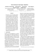
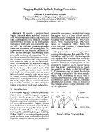
pH were performed (Fig. 1).
The k
cat
vs. pH plot of the His92Ala mutant was
analyzed according to the rapid equilibrium diprotic
model [28] to give the values of pK
es
presented in
Table 1. The k
cat
vs. pH plot showed a broad optimum
at pH 3.8, which is significantly shifted compared to
native recombinant RPAP [24], with apparent pK
es
val-
ues of 2.6 and 5.2 (Fig. 1). The pH optimum and pK
a
values obtained by fitting the data have an error of
approximately ± 0.2 pH units. Because of the low
activity, incubation times during the assay were
increased (from 1 to 5 min), but even at the lowest pH
the enzyme was stable over the time range measured.
Although the k
cat
values have rather small errors, the
plot of K
M
vs. pH suffers from rather large error bars
at each pH. Apparently K
M
parallels the behavior of
k
cat
as a function of pH, rather than simply increasing
with increasing pH. Because of this behavior, k
cat
⁄ K
M
E. G. Funhoff et al. Mutational analysis of His92 in recombinant rat PAP
FEBS Journal 272 (2005) 2968–2977 ª 2005 FEBS 2969
does not show the typical sigmoidal curve from which
apK
e2
value can be determined, as is found for the
native enzyme. The maximal fitted value for k
cat
at
pH 3.8 is 21 s
)1
, less than 10% that of wild-type
enzyme at its optimal pH, while K
M
is 15- to 30-fold
larger compared to the wild-type at pH 4. The value
of k
cat
reported in Table 1 is that obtained by fitting
the data; the apparent maximal k
cat
in Fig. 1 is lower
due to the small difference between the two pK
a
values.
Fig. 1. Plots of k
cat
, k
cat
⁄ K
M
and K
M
vs. pH for single polypeptide recRPAP (upper panel, adapted from [24]), His92Ala-recRPAP (middle
panel) and His92Asn-recRPAP (lower panel) with p-NPP as substrate at 22 °C. The lines represent fits of the data to the rapid equilibrium
diprotic model. The following expression was derived for the observed values of k
cat
: k
cat(obs)
¼ k
cat
⁄ (1 + [H
+
] ⁄ K
es,1
+ K
es,2
⁄ [H
+
]); assuming
that all equilibria are fast compared to k
cat
, k
cat(obs)
⁄ K
M(obs)
¼ k
cat
⁄ K
S
(1 + [H
+
] ⁄ K
e1
+ K
e2
⁄ [H
+
]). K
S
is the dissociation constant of the
enzyme–substrate complex.
Mutational analysis of His92 in recombinant rat PAP E. G. Funhoff et al.
2970 FEBS Journal 272 (2005) 2968–2977 ª 2005 FEBS
The His92Asn mutant shows an optimum at pH 4.4,
with pK
es
values of 3.2 and 5.6 (Fig. 1) and a turnover
number of approximately 760 s
)1
, three times higher
than that of the wild-type recRPAP. K
M
increases with
increasing pH, and is about 10 times larger than that
of the wild type at pH 4.4. Analysis of the k
cat
⁄ K
M
plot shows that values for pK
e,1
and pK
e,2
can be fitted
but the large error bars suggest that these data should
be interpreted with some caution. A value of approxi-
mately 2.2 can be derived from the fits for pK
e,1
, while
for pK
e,2
a value of 4.9 is found.
Spectroscopic characteristics of mutant enzymes
The EPR spectrum of the fully reduced native recR-
PAP shows different species at different pH values
(Fig. 2A). At pH 7, which is above pK
es,2
, a signal
with features at g
xyz
¼ 1.58, 1.74, 1.97 is observed.
Upon decreasing the pH to the optimal pH 5.5, a spe-
cies with g
xyz
¼ 1.59, 1.74, 1.93 is formed, while a
third species is formed at pH values below pK
es,1
, with
g
xyz
¼ 1.60, 1.74, 1.86. This behavior is very similar to
the pH dependency of the EPR spectrum of recombin-
ant human purple acid phosphatase (recHPAP), which
is reported in detail in [29].
The His92Ala mutant shows the presence of a single
species at its pH optimum (Fig. 2B), with apparent
g-values of g
xyz
¼ 1.58, 1.73, 1.97, corresponding to
the species that is observed above pK
es,2
of the native
enzyme. Measuring the spectrum at pH ¼ pK
es,1
was
not possible, due to the instability of the mutant
enzyme during buffer exchange. Increasing the pH to
pH ¼ pK
es,2
gave no change in the EPR spectrum.
Thus, for the His92Ala mutant, a single EPR detect-
able species is present over the pH range 3.7–6.5. The
intensity of the signal due to this species is reduced at
pH 4.1 and 3.7 to % 0.5–0.7 and % 0.1–0.15 spins per
Table 1. Kinetics parameters of single polypeptide His92Asn, and
His92Ala mutants of recRPAP. k
cat
is defined as the number of
substrate molecules hydrolyzed per enzyme molecule per second;
pK
es,1
and pK
es,2
are for deprotonation ⁄ protonation events of a
group of the enzyme–substrate complex; and pK
e
is for a deproto-
nation ⁄ protonation event of a group of the free enzyme. All pK val-
ues reported have an error of ± 0.2 pH units.
pH
opt
pK
es,1
pK
es,2
pK
e,1
pK
e,2
k
cat
(s
)1
) K
M
a
(mM)
RecRPAP
b
5.5 4.5 6.6 – 5.6 240 4.5
His92Asn 4.4 3.2 5.6 2.2 4.9 760 23
His92Ala 3.8 2.6 5.2 – – 21 35
a
At the optimal pH.
b
From [24].
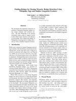
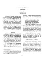
Fig. 2. EPR spectra of native recRPAP (A), His92Ala-recRPAP (B) and His92Asn-recRPAP (C) at different pH values. The spectrum in the lower
panel of Fig. 2C is a five-fold enlargement of the pH 2.7 spectrum. The inset in Fig. 2C shows the superposition of two simulated spectra
(solid black line) with g values of g
xyz
¼ 1.57, 1.70, 1.85 (ÆÆÆÆ), and g
xyz
¼ 1.41, 1.60, 1.74 (dashed line) together with the measured spectrum
(solid gray line). EPR conditions: (A) microwave power 2 mW; microwave frequency, 9.423 GHz; modulation, 12.7 G at 100 kHz; temperature,
4.5–5.5 K. All spectra in (A) were normalized for gain, temperature, and protein concentration; (B) same conditions as for (A) with a tempera-
ture range 4.5–5.7 K and different power. All spectra were normalized for gain, temperature, power, and protein concentration.
E. G. Funhoff et al. Mutational analysis of His92 in recombinant rat PAP
FEBS Journal 272 (2005) 2968–2977 ª 2005 FEBS 2971
molecule, respectively, suggesting a correlation with
the protonation of a catalytically important residue. In
the visible spectra only small shifts in k
max
with pH
are observed: for example, at pH 3.7 k
max
is 504 nm
vs. 508 nm at pH 6.5 (data not shown).
The EPR spectrum of the His92Asn mutant at
pH 6.5 shows apparent g values at 1.96, 1.85, 1.71,
1.60, and 1.41 (Fig. 2C), indicating the presence of
several species. Simulation of the spectra suggests that
two main species are present, with features at g
xyz
¼
1.57, 1.70, 1.85, and g
xyz
¼ 1.41, 1.60, 1.74, respect-
ively. A weak signal with g
xyz
¼ 1.58, 1.74, 1.93 could
also be present. Decreasing the pH to 2.7 caused the
intensity of the g
xyz
¼ 1.57, 1.70, 1.85 species to
decrease relative to that of the g
xyz
¼ 1.41, 1.60, 1.74
species. At pH ¼ pK
es,1
(2.7), the total intensity of the
spectrum decreased from % 0.5 spins per molecule to
< 0.05 spins. Together with the loss of signal intensity
at g
av
¼ 1.74, an increase in the intensity of the signal
due to high-spin Fe
3+
species was observed, from
% 1–2% to % 45% of the total signal intensity, respect-
ively. This was not due to air oxidation, because the
original spectrum was restored simply by raising the
pH of the sample to 6.5. A shift of k
max
to higher
wavelength was observed, which parallels the loss of
signal intensity in the EPR spectrum: k
max
of the
His92Asn mutant shifted from 530 nm at pH 6.5 and
pH 4.4 to 560 nm at pH 2.7. Increasing the pH of the
sample to pH 6.5 restored the original k
max
of 530 nm
(data not shown).
Discussion
To date, two site-directed mutagenesis studies of first
or second coordination sphere active site residues of
PAP have been published. The function of the loop
residue Asp146 in recHPAP has been extensively stud-
ied [24], while preliminary kinetics results on His92 and
His195 mutants have been published [23]. In the latter
study, turnover numbers of 2.4 and 7.8 s
)1
were meas-
ured at pH 4.5 for His92Ala and His92Gln, which is
10–100-fold lower than we observe for the His92Ala
and His92Asn mutants at this pH. Moreover,
Michaelis-Menten constants of approximately 7–15 mm
were observed at pH 4.5, although a K
M
of 40 mm for
the wild-type enzyme was found [23]. The K
M
value of
other single polypeptide PAPs at this pH is significantly
lower, around 1 mm [24,30,31].
Several studies have appeared on the closely related
PPs [14], in which all active site residues have been
mutated [25,26,32–37]. In most of these studies, the
kinetics parameters were determined under only one
set of conditions, which did not result in a clear under-
standing of the function of these residues. Studies by
the Rusnak group have provided more detailed insight
into the role of several active site residues [25,26,37].
Two of the corresponding residues in PAPs, Asn91
and His92, have been proposed to be involved in pro-
teolytic activation of mammalian FeFe-PAPs due to
a possible interaction with the loop residue Asp146
[24,30]. The interaction of these two residues with
Asp146 in an exposed loop reduces the catalytic activ-
ity and decreases pK
es,1
[24] which is the pK
a
of the
metal-coordinated solvent molecule [29]. In the present
study, the His residue that could interact with Asp146
has been mutated to elucidate its role in the catalytic
process.
Mertz et al. [25] suggested that His92 in PPs is not
required for protonation of the leaving group, because
the same relative k
cat
value was found for two sub-
strates with different leaving group pK
a
values.
Instead, they suggested that His92 could function in
concert with the nucleophilic water molecule to either
position a lone pair on the oxygen atom for optimum
in-line attack on the phosphorus atom of the substrate
or serve as a general base to take up a proton concom-
itant with solvent nucleophilic attack. A subsequent
isotope effect study of the wild-type and His76Asn-
kPP showed that the increase in [
15
N](V ⁄ K) and
[
18
O](V ⁄ K)
bridge
isotope effects for the substrate p-NPP
were analogous to those observed for protein tyrosine
phosphatases observed upon mutation of their general
acid, but smaller in magnitude. Thus, these studies did
not clearly answer the question of whether His76 func-
tions as a general base [26].
For recRPAP, the pH optima of both the His92Asn
and His92Ala mutants are shifted 1–1.5 pH units to
lower values due to a 1.5–2 pH unit shift in pK
es,1
;in
addition pK
es,2
decreases by one pH unit. By compar-
ison, the pH optimum of His76Asn-kPP shows more
than a full pH unit decrease [26]. Although it has been
suggested more than once that pK
es,2
is due to depro-
tonation of the His92 imidazole group, the presence of
a basic limb in the pH profiles of mutants of three
enzymes with related active site structures (His92Asn-
recRPAP, His76Asn-kPP [26], and His92Ala-recRPAP)
provides convincing evidence that pK
es,2
is not due to
the (de)protonation of the imidazole group of His92.
The large (15- to 30-fold) increase in K
M
observed
for His92Ala-recRPAP suggests that this residue is
involved in substrate binding in PAPs. In principle,
however, an Asn residue at position 92 should be able
to hydrogen bond to the substrate, but this mutant
also shows significantly increased values for K
M
, argu-
ing against such a role in substrate binding. Previous
site-directed mutagenesis studies of kPP also argue
Mutational analysis of His92 in recombinant rat PAP E. G. Funhoff et al.
2972 FEBS Journal 272 (2005) 2968–2977 ª 2005 FEBS
against such a role, although these results can also be
explained in favor of monoanion binding [25,26]. For
all the PP mutants, k
cat
was ¼ 1% of that of the wild-
type enzyme, in dramatic contrast with the observed
k
cat
values for His92Ala and His92Asn-recRPAP
[25,26,32,34]. The almost three-fold increase in k
cat
of
the His92Asn mutant compared to the wild-type
enzyme is particularly surprising. The shift of pK
a,1
to
lower pH upon mutation of His92 into Ala and the
increase in k
cat
for the His92Asn mutant strongly sug-
gest that the presence of the His92 imidazole group
increases pK
a,1
(the pK
a
of the nucleophile), but does
not significantly affect its reactivity. Thus, His92 does
not function as an acid or a base in the catalytic cycle,
either in regeneration of the nucleophile as has been
proposed for metal-containing enzymes such as argi-
nase or carbonic anhydrase [38], or by abstracting or
donating a proton to the substrate or product. It is
clear, however, that His92 does play a major role in
enzyme catalysis.
Although detailed mechanistic studies are lacking,
the congruence of active site structures strongly sug-
gests that PAPs and PPs catalyze hydrolysis of phos-
phate ester substrates via very similar mechanisms [20];
in particular, it seems very likely that the groups
responsible for pK
a,1
and pK
a,2
are identical in the two
sets of enzymes. The group responsible for pK
a,1
is
a metal-bound solvent molecule whose identity is
unknown [29,39]. Possibilities include a terminally
coordinated Fe
3+
or Fe
2+
hydroxide, a bridging
hydroxide ion, or a second coordination sphere
hydroxide [40,41]. Observed values of pK
a,1
for PAP
range from 5.5 for the proteolytically cleaved wild-type
enzyme to 4.5 for wild-type single polypeptide [24,30]
to 2.6 for the His92Ala mutant. Based on the pK
a
values of hexa-aquo complexes of metal ions [42,43],
it is not feasible to attribute a pK
a,1
of 2.6 (His92Ala)
or 3.2 (His92Asn) to a water group coordinated to
a divalent transition metal ion. Although the most
plausible assumption is that pK
a,1
is due to a solvent
molecule coordinated terminally to the trivalent ion,
there are very strong arguments against a terminal
trivalent coordinated nucleophile; they include the
following:
(a) Replacement of the trivalent metal ion by other
metals has essentially no effect on the kinetics proper-
ties, while substitution of the divalent site results in
significant kinetics changes [44,45]. The involvement of
the divalent site is most pronounced for single poly-
peptide recHPAP, where an increase in k
cat
from 210
to 5000 s
)1
is observed for FeZn-recHPAP. Further-
more, proteolysis does not result in activation or a sig-
nificant change in pK
es,1
and pK
es,2
[46].
(b) Fluoride can replace the hydroxide only if it is
protonated [29]. Moreover, the disappearance of the
NMR spectrum of the enzyme–fluoride complex below
pK
es,1
is consistent with a significant change in the
super-exchange interaction between the two iron ions,
suggesting that fluoride replaces a solvent molecule
bridging both metal ions [27].
(c) ENDOR results on single polypeptide uteroferrin
could not detect a water-derived ligand coordinated to
the trivalent site [41].
Thus, many results point in the direction of a nucle-
ophilic hydroxide that is coordinated to the divalent
metal ion, which is difficult to correlate with the pK
es,1
values observed for the His92Asn and His92Ala
mutants. One possibility that could resolve this contro-
versy is the possibility that a solvent molecule in the
second coordination sphere acts as the nucleophile
[45].
The EPR spectrum of native recRPAP shows the
presence of three different species at different pH
values, and these are also observed for recHPAP.
For recHPAP the change in the EPR spectrum from
a feature with g
xyz
¼ 1.58, 1.74, 1.94 to a feature
with g
xyz
¼ 1.58, 1.74, 1.97 correlates well with pK
es,2
[29]. The only species observed by EPR for
His92Ala-recRPAP over the pH range 4–8 resembles
native recRPAP and recHPAP at pH > pK
es,2
. The
intensity of this species is reduced at pH 3.7 and 4.1
compared to the spectrum at pH 6.5. This suggests
that His92 is apparently capable of interacting with
the metal site in PAP (and, presumably, in kPP and
PP1) either directly or via solvent molecules, as sug-
gested by the structures of uncomplexed forms of
PP1 [16] and calcineurin [15], even though it is 4.5–
5A
˚
away from the metals in PAPs [11,12,47] and
PPs [17–19]. Thus, it could well be that in the wild-
type enzyme at pH ¼ pK
a,2
a change in the position
of His92 is responsible for the observed shift in the
g
z
feature of the EPR spectrum (from 1.93 to 1.97).
Because mutagenesis has shown that His92 itself is
not responsible for pK
es,2
, deprotonation of an as
yet unidentified residue that interacts with His92
might force the latter into a different position, one
that is less favorable for catalysis.
To gain better insight into the structural effects of
the mutations, first approach models with energy
minimization were obtained using SwissModel and
DeepView ⁄ Swiss PdB-viewer. First approach mode-
ling of the His92Asn mutant (Fig. 3) shows that
the amido nitrogen of Asn92 does not coordinate to
the phosphate ion, but points in the direction of
Asp146. A weak interaction could be present, jud-
ging from the distance of 3.34 A
˚
between the Asn92
E. G. Funhoff et al. Mutational analysis of His92 in recombinant rat PAP
FEBS Journal 272 (2005) 2968–2977 ª 2005 FEBS 2973
amido nitrogen and the Asp146 carboxylate, which
could weaken the Asp146–Asn91 interaction that
decreases the Lewis acidity of the iron(II) ion and
thereby the electrophilicity of the activated substrate
molecule [24,30]. This hypothesis would explain the
higher activity of the His92Asn mutant, but it does
not account for the lower pK
es
values.
In conclusion, the observed low pK
es
values sup-
port a model in which the nucleophilic hydroxide is
that coordinated to the iron(III) ion and whose reac-
tivity is tuned by the position of His92. However, the
dramatic effects of metal substitution on the iron(II)
site [46], together with the fluoride inhibition ⁄
spectroscopic studies [27] suggest a bridging hydr-
oxide as nucleophile. Hypothetically, the flexible
character of the bridging hydroxide [48] and its inter-
action with His92 could result in [partial] opening of
the hydroxide bridge, resulting in an equilibrium
between two species: one in which the hydroxide
interacts more strongly with the divalent metal ion,
resulting in higher pK
es
values and a higher k
cat
due
to faster ligand exchange rates: and one in which it
interacts more strongly with the trivalent metal,
resulting in more acidic pK
es
values and lower k
cat
values. High-resolution crystal structures of a mam-
malian PAP in active redox and pH states in the
presence and absence of nonhydrolysable substrate
analogues, such as AMP, perhaps in combination
with ENDOR spectroscopy, will be necessary to elu-
cidate the mechanism of PAPs.
Experimental procedures
General procedures
Enzyme concentrations were determined by measuring the
absorbance of the Tyr
–
to-Fe
3+
charge transfer at k
max
(510–550 nm; e ¼ 4080 m
)1
cm
)1
) [49] on a Cary 50 or
HP8452A photodiode array spectrophotometer (Varian
Inc., Palo Alto, CA, USA).
Generation of mutant proteins
RecRPAP mutant enzymes were prepared as previously
described with the QuickChange
TM
Site-Directed Mutagen-
esis Kit (Stratagene, La Jolla, CA, USA) [24]. Primers used
for specific mutations were as follows: His92Ala, 5¢-CTGG
CTGGAAAC
GCTGATCACCT TGGC-3¢; His92Asn, 5¢-GGC
TGGAAAC
AATGATCACCTTG-3¢. The underlined bases
indicate changes compared to the wild-type sequence.
Recombinant baculovirus stocks containing regions
coding for the His92Asn, and His92Ala mutants were
used to infect High 5 cells cultured in 500–1000 mL Excel
405
TM
SFM at 27 °C; the cell density was 0.7–0.9 · 10
6
cellsÆmL
)1
, and a low multiplicity of infection (MOI;
0.001–0.01) was used. After 5 days the cells were removed
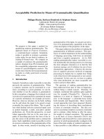
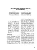
Fig. 3. Active site structure of phosphate-complexed uteroferrin at 1.55 A
˚
resolution (left panel) showing the hydrogen bonding network
between the divalent metal (II), trivalent metal (III), Asn91, and His92. The distances between the residues are given in A
˚
. The backbone of
His92 is stabilized via hydrogen bonds between the amido nitrogen of Asn144 and the backbone carbonyl of Asn91, and between the carb-
oxylate of Asp52 and the backbone nitrogen of His92. The interaction between the amido nitrogen of Asn91 and the carboxylate of Asp146
and the intraloop hydrogen bond between the Ser145 backbone amino group and the side chain oxygen of Asn144 further fine-tune this
hydrogen bonding network. The right panel shows the first approach modeling structure of the His92Asn mutant after energy minimization.
The amido nitrogen of Asn92 is oriented towards Asp146 and shows weak interaction with the Asp146 carboxylate group. The figure was
generated using the D
EEPVIEW ⁄ SWISS-PDBVIEWER program with the coordinates of 1UTE.
Mutational analysis of His92 in recombinant rat PAP E. G. Funhoff et al.
2974 FEBS Journal 272 (2005) 2968–2977 ª 2005 FEBS
by centrifugation (10 000 g), and the fully reduced
enzyme was purified from the medium as previously des-
cribed [24].
Kinetics measurements
The pH dependence of the catalytic activity of His92Asn
and His92Ala was measured in 100 mm buffer (sodium
acetate, Mes and Hepes), 300 mm KCl, 10 mm Na ⁄ K tar-
trate, 6.7 mm sodium ascorbate, 0.37 mm Fe(NH
4
)
2
(SO
4
)
2
,
and substrate concentrations between 1 and 100 mm p-NPP
as previously described [24]. At intervals after enzyme addi-
tion, 250 lL aliquots were removed and quenched with
1.0 mL of 0.5 m NaOH to convert all product to the phe-
nolate form. For each determination of V
max
and K
M
, the
hydrolysis rate was measured using at least six different
p-NPP concentrations with assay times between 1 and
5 min. Enzyme concentrations were varied to ensure a sig-
nificant change in absorbance. After each assay, the pH of
the reaction mixture was measured to ensure that it had
not changed. Values of K
M
and V
max
were obtained by fit-
ting the data to the Michaelis–Menten equation:
v ¼ðV
Ã
max
½SÞ=ðK
M
þ½S Þ
using the program Leonora (Athel-Cornish Bowden, ver-
sion 1).
The pH dependencies of values of k
cat
vs. pH were ana-
lyzed according to the rapid equilibrium diprotic model
[28], which is used if the difference in pK
a
values is less
than 3.5 pH units. The following expressions were derived
for k
cat
and k
cat
⁄ K
M
:
k
catðobservedÞ
¼ k
cat
=ð1 þ½H
þ
=K
es;1
þ K
es;2
=½H
þ
Þ
k
catðobsÞ
=K
MðobsÞ
¼ k
cat
=K
S
ð1 þ½H
þ
=K
e1
þ K
e2
=½H
þ
Þ:
Electron paramagnetic resonance (EPR)
spectroscopy
Samples of fully reduced native and mutant recRPAP
were prepared in a buffer of the appropriate pH [100 mm
sodium acetate, Mes, Hepes; 0.45 m KCl, 20% (v ⁄ v)
glycerol], and frozen in liquid N
2
. X-band EPR spectra
(9.43 GHz) were obtained on a Bruker ECS106 EPR
spectrometer (Bruker-Biospin Corp., Billerica, MA, USA)
equipped with an Oxford Instruments ESR900 helium-flow
cryostat with an ITC4 temperature controller. The magnetic
field was calibrated with an AEG Magnetic Field Meter,
and the frequency was measured with a HP 5350B Micro-
wave Frequency Counter. After recording the spectrum, the
sample was thawed and buffer exchanged into a buffer with
the appropriate pH by repetitive concentration ⁄ dilution
using a Microcon (30 kDa cut-off). Complete buffer
exchange during the complete EPR experiment took several
hours.
Acknowledgements
We thank Robert-Jan Sanders for his initial work on
the mutant enzymes. This research was supported by
grants from the Swedish Research Council (to GA)
and the Research Funds of Karolinska Institutet, and
by a contract from the EU 4th Framework Biotechno-
logy Program (BI04-CT98-0385).
References
1 Averill BA (2003) Dimetal hydrolases. Comprehensive
Coordination Chemistry II (Que JL & Tolman WB, eds),
Elsevier Pergamon, Amsterdam, Netherlands.
2 Andersson G, Ek-Rylander B, Hollberg K, Ljusberg-
Sjoelander J, Lang P, Norgard M, Wang Y & Zhang
S-J (2003) TRACP as an osteopontin phosphatase.
J Bone Min Res 18, 1912–1915.
3 Angel NZ, Walsh N, Forwood MR, Ostrowski MC,
Cassady AI & Hume DA (2000) Transgenic mice over-
expressing tartrate-resistant acid phosphatase exhibit an
increased rate of bone turnover. J Bone Min Res 15,
103–110.
4 Hayman AR, Jones SJ, Boyde A, Foster D, Colledge
WH, Carlton MB, Evans MJ & Cox TM (1996) Mice
lacking tartrate-resistant acid phosphatase (Acp 5) have
disrupted endochondral ossification and mild osteope-
trosis. Development 122, 3151–3162.
5 Schindelmeiser J, Radzun HJ & Munstermann D (1991)
Tartrate-resistant, purple acid-phosphatase in Gaucher
cells of the spleen – immunochemical and cytochemical
analysis. Pathol Res Practice 187, 209–213.
6 Hoyer JD, Li CY, Yam LT, Hanson CA & Kurtin PJ
(1997) Immunohistochemical demonstration of acid
phosphatase isoenzyme 5 (tartrate-resistant) in paraffin
sections of hairy cell leukemia and other hematologic
disorders. Am J Clin Pathol 108, 308–315.
7 Schindelmeiser J, Gullotta F & Munstermann D (1989)
Purple acid-phosphatase of human-brain macrophages
in aids encephalopathy. Pathol Res Practice 185,
184–186.
8 Lang P, Schultzberg M & Andersson G (2001) Expres-
sion and distribution of tartrate-resistant purple acid
phosphatase in the rat nervous system. J Histochem
Cytochem 49, 379–396.
9 Hayman AR, Bune AJ & Cox TM (2000) Widespread
expression of tartrate-resistant acid phosphatase (Acp 5)
in the mouse embryo. J Anat 196 , 433–441.
10 Stra
¨
ter N, Klabunde T, Tucker P, Witzel H & Krebs B
(1995) Crystal structure of a purple acid phosphatase
containing a dinuclear Fe(III)-Zn(II) active site. Science
268, 1489–1492.
11 Uppenberg J, Lindqvist F, Svensson C, EkRylander B
& Andersson G (1999) Crystal structure of a mamma-
lian purple acid phosphatase. J Mol Biol 290, 201–211.
E. G. Funhoff et al. Mutational analysis of His92 in recombinant rat PAP
FEBS Journal 272 (2005) 2968–2977 ª 2005 FEBS 2975
12 Guddat LW, McAlpine AS, Hume D, Hamilton S, de
Jersey J & Martin JL (1999) Crystal structure of mam-
malian purple acid phosphatase. Structure 7, 757–767.
13 Krebs B, Jasper B, Stra
¨
ter N & Averill BA (2004) Struc-
ture analysis of a recombinant human purple acid phos-
phatase. Paper presented at the European Biological
Inorganic Chemistry Conference (EUROBIC 7),
29.08.04–02.09.04, Garmisch Partenkirchen, Germany.
14 Koonin EV (1994) Conserved sequence pattern in a
wide variety of phosphoesterases. Protein Sci 3 , 356–
368.
15 Kissinger CR, Parge HE, Knighton DR, Lewis CT,
Pelletier LA, Tempczyk A, Kalish VJ, Tucker KD,
Showalter RE, Moomaw EW, Gastinel LN, Habuka N,
Chen X, Maldonado F, Barker JE, Bacquet R & Villa-
franca JE (1995) Crystal structure of human calcineurin
and the human FKBP12-FK506-calcineurin complex.
Nature 378, 641–644.
16 Egloff MP, Cohen PTW, Reinemer P & Barford D
(1995) Crystal structure of the catalytic subunit of
human protein phosphatase 1 and its complex with
tungstate. J Mol Biol 254, 942–959.
17 Goldberg J, Huang H-B, Kwon Y-G, Greengard P,
Nairn AC & Kuriyan J (1995) Three-dimensional struc-
ture of the catalytic subunit of protein serine ⁄ threonine
phosphatase-1. Nature 376, 745–753.
18 Griffith JP, Kim JL, Kim EE, Sintchak MD, Thomson
JA, Fitzgibbon MJ, Fleming MA, Caron PR, Hsiao K
& Navia MA (1995) X-Ray structure of Calcineurin
inhibited by the immunophilin-immunosuppressant
FKBP12-FK506 complex. Cell 82, 507–522.
19 Voegtli WC, White DJ, Reiter NJ, Rusnak F &
Rosenzweig AC (2000) Structure of the bacteriophage
lambda Ser ⁄ Thr protein phosphatase with sulfate ion
bound in two coordination modes. Biochemistry 39,
15365–15374.
20 Rusnak F & Mertz P (2000) Calcineurin: form and
function. Physiol Rev 80, 1483–1521.
21 Lord DK, Cross NCP, Bevilacqua MA, Rider SH,
Gorman PA, Groves AV, Moss DW, Sheer D & Cox
TM (1990) Type 5 acid phosphatase: sequence, expres-
sion and chromosomal localization of a differentiation-
associated protein of the human macrophage. Eur J
Biochem 189, 287–293.
22 Vogel A (2001) Heterologe expression, mutationsanalyse
und in vivo expression der violetten sauren phosphatase
der roten kidneybohne. PhD thesis, University of Mun-
ster, Germany.
23 Kaija H, Alatalo SL, Halleen JM, Lindqvist Y, Schnei-
der G, Vaananen HK & Vihko P (2002) Phosphatase
and oxygen radical-generating activities of mammalian
purple acid phosphatase are functionally independent.
Biochem Biophys Res Comm 292, 128–132.
24 Funhoff EG, Ljusberg J, Wang Y, Andersson G &
Averill BA (2001) Mutational analysis of the interaction
between active site residues and the loop region in mam-
malian purple acid phosphatases. Biochemistry 40,
11614–11622.
25 Mertz P, Yu L, Sikkink R & Rusnak F (1997) Kinetic
and spectroscopic analyses of mutants of a conserved
histidine in the metallophosphatases calcineurin and
lambda protein phosphatase. J Biol Chem 272, 21296–
21302.
26 Hoff RH, Mertz P, Rusnak F & Hengge AC (1999) The
transition state of the phosphoryl-transfer reaction cata-
lyzed by the lambda Ser ⁄ Thr protein phosphatase. JAm
Chem Soc 121, 6382–6390.
27 Dikiy A, Funhoff EG, Averill BA & Ciurli S (2002)
New insights into the mechanism of purple acid phos-
phatase through
1
H NMR spectroscopy of the recombi-
nant human enzyme. J Am Chem Soc 124, 13974–13975.
28 Segel IH (1993) Enzyme Kinetics: Behaviour and Analy-
sis of Rapid Equilibrium and Steady-State Enzyme Sys-
tems. John Wiley & Sons, New York.
29 Funhoff EG, de Jongh TE & Averill BA (2005) Direct
observation of multiple protonation states in recombi-
nant human purple acid phosphatase. J Biol Inorg
Chem, in press.
30 Funhoff EG, Klaassen CHW, Samyn B, Van Beeumen
J & Averill BA (2001) The highly exposed loop region
in mammalian purple acid phosphatase controls the cat-
alytic activity. Chembiochem 2, 355–363.
31 Valizadeh M, Schenk G, Nash K, Oddie GW,
Guddat LW, Hume DA, de Jersey J, Burke TRJ &
Hamilton S (2004) Phosphotyrosyl peptides and analo-
gues as substrates and inhibitors of purple acid phos-
phatases. Arch Biochem Biophys 424, 154–162.
32 Zhang J, Zhang ZJ, Brew K & Lee EYC (1996) Muta-
tional analysis of the catalytic subunit of muscle protein
phosphatase-1. Biochemistry 35, 6276–6282.
33 Huang HB, Horiuchi A, Goldberg J, Greengard P &
Nairn AC (1997) Site-directed mutagenesis of amino
acid residues of protein phosphatase 1 involved in cata-
lysis and inhibitor binding. Proc Natl Acad Sci USA 94,
3530–3535.
34 Zhuo S, Clemens JC, Stones RL & Dixon JE (1994)
Mutational analysis of a Ser ⁄ Thr phosphatase: identifi-
cation of residues important in phosphoesterase
substrate binding and catalysis. J Biol Chem 269,
26234–26238.
35 Mondragon A, Griffith EC, Sun L, Xiong F, Armstrong
C & Liu JO (1997) Overexpression and purification of
human calcineurin alpha from Escherichia coli and
assessment of catalytic functions of residues surrounding
the binuclear metal center. Biochemistry 36, 4934–4942.
36 Zhang LF & Lee EYC (1997) Mutational analysis of
substrate recognition by protein phosphatase 1. Bio-
chemistry 36 , 8209–8214.
37 White DJ, Reiter NJ & Sikkink RA, Yu L & Rusnak F
(2001) Identification of the high affinity Mn
2+
binding
Mutational analysis of His92 in recombinant rat PAP E. G. Funhoff et al.
2976 FEBS Journal 272 (2005) 2968–2977 ª 2005 FEBS
site of bacteriophage lambda phosphoprotein phospha-
tase: effects of metal ligand mutations on electron para-
magnetic resonance spectra and phosphatase activity.
Biochemistry 40, 8918–8929.
38 Christianson DW & Cox JD (1999) Catalysis by metal-
activated hydroxide in zinc and manganese metalloen-
zymes. Annu Rev Biochem 68, 33–57.
39 Dietrich M, Mu
¨
nstermann D, Suerbaum H & Witzel H
(1991) Purple acid phosphatase from bovine spleen,
Interactions at the active site in relation to the reaction
mechanism. Eur J Biochem 199, 105–113.
40 Merkx M, Pinkse MWH & Averill BA (1999) Evidence
for nonbridged coordination of p-nitrophenyl phosphate
to the dinuclear Fe(III)-M(II) center in bovine spleen
purple acid phosphatase during enzymatic turnover.
Biochemistry 38, 9914–9925.
41 Smoukov SK, Quaroni L, Wang X, Doan PE, Hoffman
BM & Que LJ (2002) Electro-nuclear double resonance
spectroscopic evidence for a hydroxo-bridge nucleophile
involved in catalysis by a dinuclear hydrolase. JAm
Chem Soc 124, 2595–2603.
42 Yatsimirskii KB & Vasilev VP (1960) Instability Con-
stants of Complex Compounds. Pergamon, Elmsford.
43 Lippard SJ & Berg JM (1994) Principles of Bioinorganic
Chemistry, 1st edn. University Science Books, Mill
Valley.
44 Merkx M & Averill BA (1998) Ga
3+
as a functional
substitute for Fe
3+
: preparation and characterization of
the Ga
3+
Fe
2+
and Ga
3+
Zn
2+
forms of bovine spleen
purple acid phosphatase. Biochemistry 37, 8490–8497.
45 Merkx M & Averill BA (1999) Probing the role of the
trivalent metal in phosphate ester hydrolysis: Prepara-
tion and characterization of purple acid phosphatases
containing (AlZnII)-Zn-III and (InZnII)-Zn-III active
sites, including the first example of an active aluminum
enzyme. J Am Chem Soc 121, 6683–6689.
46 Funhoff EG, Bollen M & Averill BA (2005) The FeZn
from of recombinant human purple acid phosphatase is
not activated by proteolysis. J Inorg Biochem 99, 521–529.
47 Lindqvist Y, Johansson E, Kaija H, Vihko P & Schnei-
der G (1999) Three-dimensional structure of a mamma-
lian purple acid phosphatase at 2.2 angstrom resolution
with a mu-(Hydr) oxo bridged di-iron center. J Mol Biol
291, 135–147.
48 Yang YS, McCormick JM & Solomon EI (1997) Circu-
lar dichroism and magnetic circular dichroism studies of
the mixed-valence binuclear non-heme iron active site in
uteroferrin and its anion complexes. J Am Chem Soc
119, 11832–11842.
49 Davis JC, Lin SS & Averill BA (1981) Kinetics and
optical spectroscopic studies on the purple acid phos-
phatase from beef spleen. Biochemistry 20, 4062–4067.
E. G. Funhoff et al. Mutational analysis of His92 in recombinant rat PAP
FEBS Journal 272 (2005) 2968–2977 ª 2005 FEBS 2977