A non-doped microporous titanosilicate for bimodal adsorption-photocatalysis based removal of organic water pollutants
Bạn đang xem bản rút gọn của tài liệu. Xem và tải ngay bản đầy đủ của tài liệu tại đây (3.93 MB, 9 trang )
Microporous and Mesoporous Materials 345 (2022) 112276
Contents lists available at ScienceDirect
Microporous and Mesoporous Materials
journal homepage: www.elsevier.com/locate/micromeso
A non-doped microporous titanosilicate for bimodal
adsorption-photocatalysis based removal of organic water pollutants
Ayomi S. Perera a, *, Patrick M. Melia a, Reece M.D. Bristow a, James D. McGettrick b,
Richard J. Singer a, Joseph C. Bear a, Rosa Busquets a
a
b
Kingston University London, Faculty of Science, Engineering and Computing, Kingston Upon Thames, KT1 2EE, UK
SPECIFIC IKC, Materials Research Centre, College of Engineering, Swansea University, Bay Campus, Fabian Way, Swansea, SA1 8EN, UK
A R T I C L E I N F O
A B S T R A C T
Keywords:
Photocatalysis
TiO2 alternative
Advanced oxidation
Water treatment
Porous adsorbent
Access to clean drinking water is limited for millions around the world and lead to dire health and economic
ramifications, particularly in developing nations. This study explores a recyclable, low-cost, non-doped, micro
porous titanosilicate for effective removal of organic water pollutants. Rhodamine B was utilized as a modal
pollutant to explore and optimize the activity of the titanosilicate, which evidently occurred via an adsorption
and subsequent photocatalytic degradation based bimodal mechanism. The novel titanosilicate has high surface
area (SBET of 468 m2/g), is microporous (~1.3 nm pore diameter), achieved via a surfactant templating tech
nique. Its’ physicochemical properties were characterised using FTIR, Raman, BET, SEM, PXRD and XPS. The
photocatalytic activity of the material was studied under a solar simulator via time dependent UV–vis absorption
measurements. The material showed 97% removal of Rhodamine B (5 mg/L) within 3 h, and outperformed
nanosized titanium dioxide (anatase:rutile 4:1), the most conventionally used photocatalyst in tertiary water
treatment. Interestingly, the titanosilicate displayed a dual mechanism of pollutant removal: an initial rapid
removal of 59% due to adsorption during a 30 min equilibrating step in the dark, followed by near complete
removal within 3 h. Additionally, a >90% efficiency of Rhodamine B removal by the titanosilicate catalyst was
achieved consistently throughout 4 cycles, demonstrating its ability for regeneration and reusability. Such ac
tivity has not been previously reported in non-doped or non-composite titanosilicates, and opens up pathways to
efficient, low-cost water treatment materials, consisting only of environmentally benign raw materials and
synthetic procedures.
1. Introduction
Water pollution due to organic contaminants such as pesticides,
antibiotics, dyes, plasticisers and pharmaceuticals is a growing concern
around the world [1–5]. Developing countries in particular suffer grim
environmental, health and economic consequences from pollution of
surface and ground water, as a result of increased industrial production
and usage of organic pollutants. These issues are unfortunately inten
sified by poor wastewater treatment capacities within such countries [6,
7]. It therefore becomes imperative to develop affordable yet effective
technologies that can remove these contaminants in a sustainable
manner. Recent advances in decontamination of surface water include
adsorption with activated carbons [3,8]; membrane techniques [9];
biological treatment [10]; and advanced oxidation methods, among
others [11,12]. Photocatalysis is one such technique that is gaining
prominence as a potentially effective water purification tool [13].
Sunlight driven photocatalytic degradation of organic contaminants has
several advantages over other competing technologies, including
providing complete mineralisation of contaminants, minimising waste
and not requiring additional energy or chemical input [14].
One candidate class of materials that encompass the above charac
teristics, along with other useful advantages are titanosilicates. They are
a structurally diverse class of zeolite-derivative materials that are
currently used industrially as heterogeneous catalysts for alkene
oxidation [15,16], and adsorbents/ion exchange agents for water
filtration [17–19]. Their excellent catalytic properties are derived from
the isolated, tetrahedrally coordinated Ti4+ active site centres,
embedded within a silica matrix [20]. Titanosilicates also show photo
activity, reported to occur via highly dispersed titanium oxide species
found within its structure [21], which were subsequently identified to be
* Corresponding author.
E-mail address: (A.S. Perera).
/>Received 18 July 2022; Received in revised form 1 October 2022; Accepted 7 October 2022
Available online 12 October 2022
1387-1811/© 2022 The Authors. Published by Elsevier Inc. This is an open access article under the CC BY-NC-ND license ( />
A.S. Perera et al.
Microporous and Mesoporous Materials 345 (2022) 112276
Ti4+ centres with tetrahedral coordination [22,23], now known to
consist of -Ti-O-Si- linkages (unlike the -Ti-O-Ti- linkages found in TiO2
which have octahedral geometry). Titanosilicates can be synthesised to
have zeolite framework structures with highly customizable pore net
works to target high product selectivity or reagent conversion depending
on the desired reaction, which is a key advantage over TiO2 [22,24].
Moreover, titanosilicates have displayed superior photocatalytic activity
and selectivity compared to bulk TiO2 [25], due to increased charge
transfer and stabilization of photoactive species within their semi
conductor framework [21,26]. These properties can be utilized for
organic contaminant degradation, providing advanced avenues for the
degradation of a myriad of organic contaminants. Notably, titanosili
cates could become an alternative to TiO2, which has been banned from
use in food in Europe due to their suspected carcinogenicity in humans
[27] – a trend that would likely expand on to water treatment.
Studies investigating titanosilicates as photocatalysts for the degra
dation of organic contaminants are scarce, compared to those focused on
conventional heterogeneous catalysis. The ones that do, with activities
comparable or surpassing TiO2, often include either titanosilicates
doped with toxic metals [26], or made into composite materials, such as
with graphitic carbon nitride [28,29]. These extra components can be
expensive and they are typically produced via hydrothermal synthesis,
which requires heating at high temperatures over long periods of time,
further adding to the cost of production. As a result, after three decades
of promising research, titanosilicates are yet to surpass TiO2 as
commercially and industrially viable photocatalysts, particularly in
water purification, despite having superior activity. It is therefore,
imperative to focus research on developing cost effective, environmen
tally benign, yet efficient categories of titanosilicates for applications in
advanced photocatalysis.
With the above goal in mind, this study was aimed to explore another
well-known property of titanosilicates – adsorption, to be used in
conjunction with photocatalytic ability, in order to enhance their
capability in removal of organic impurities for applications in water
treatment. Recent research has indicated a trend towards development
of novel titanosilicates as adsorbents for removal of organic impurities,
heavy metals and radioactive pollutants from water [28–32]. However,
the preparation cost and use of non-environmentally benign reagents/
conditions on doped and nanocomposite titanosilicates in such works
will likely prevent their use in real-world applications. The current study
intended to address such drawbacks by development of non-doped,
non-composite titanosilicates with high porosity and optimal active
site concentration as key strategies in advancing sustainable Ti-based
photocatalyst design. Templating with a surfactant/oil mixture was
investigated as a cost-effective, facile technique to improve material
porosity. This method has successfully been used previously to develop
various types of titanosilicates with a wide range of structural properties
and advanced pore structures to catalyse industrially relevant reactions
[24,33,34].
Herein we outline the development of a new microporous titanosi
licate (MiTS) in microbead morphology, without utilisation of any
organic/inorganic dopants. Through the obtained results and analysis,
we demonstrate that this material has potential relevance within the
existing water treatment infrastructure, due to its ability to effectively
remove organic contaminants, along with high recyclability, costeffectiveness and environmental compatibility.
(15 MΩ‧cm) was obtained with an ELGA Purelab system and used in all
experiments.
2.2. Preparation of microporous titanosilicate microbeads
The microporous titanosilicate microbeads (MiTS) were prepared
using an oil-water emulsion –mediated surfactant templating, adapted
from Perera et al. [24], and modified to achieve high porosity. Initially,
1 mL of Ti(IV) n-butoxide (97%) was added dropwise to 30 mL of ul
trapure water at 4 ◦ C whilst stirring. The prepared Ti(OH)4 precipitate
was washed with ultrapure water and separated via filtration, before
dissolving in 4 mL of 4 M HNO3, producing the TiO(NO3)2 species. The
active TiO(NO3)2 specie was stirred vigorously together with 6.6 mL of
TEOS (98%) and 2 mL ethanol for 30 min before the microbead for
mation. The titanosilicate mixture was added to a mixture of 26.1 g
kerosene and 7.9 g Span 80, and homogenized with a Heidolph RZR 1
homogeniser, at 2000 rpm for 2 h, at 80 ◦ C. The beads were washed and
vacuum-filtered with ultrapure water and acetone before drying at 50 ◦ C
under vacuum for 2 h. The beads were then calcined in a tube furnace
(Carbolite Gero CWF 1200) at 750 ◦ C for 6 h using a heating rate of 1 ◦ C
min− 1. The as prepared MiTS beads were stored in a desiccator and dried
overnight at 50 ◦ C under vacuum before use.
2.3. Characterisation
The titanosilicate microbeads were characterised using SEM, FTIR,
Raman, PXRD, XPS and N2 adsorption/desorption isotherms. For SEM
analysis, the titanosilicate microbeads was mounted on specimen stubs
fitted with adhesive carbon pads, sputter-coated with gold-palladium
and examined using a Zeiss Evo50 (Oxford Instruments, Cambridge,
UK) scanning electron microscope, where micrographs were obtained at
an acceleration voltage of 20 kV. FTIR analysis was conducted with a
Nicolet iS5 spectrometer with an iD1 transmission attachment (Thermo
Scientific, UK). The analysis consisted of 20 scans with a resolution of 1
cm− 1. Raman spectra were obtained via a Renishaw InVia system (UK)
along 100-1500 cm− 1 with a green laser. PXRD analysis was conducted
on a Bruker-AXS diffractometer, model D-8, using Cu K α radiation (λ =
1.54184 Å). N2 adsorption-desorption isotherms were carried out at 77 K
using a BELSORP-miniII porosimeter (MicrotracBEL, Japan). The tita
nosilicate microbeads were degassed for 24 h at 150 ◦ C before isotherm
measurements. The specific surface area (SBET) was calculated using the
standard BET (Brunauer-Emmett-Teller) model [35] and the BJH (Bar
rett-Joyner-Halenda) and MP/NLDFT models were used for pore char
acterisation. Total pore volume, Vp, was estimated at P/P0 ~ 0.99,
where P and P0 denote equilibrium pressure and saturation pressure of
N2 at 77 K respectively.
XPS analysis was carried out on the microbeads after synthesis and
after use to determine the composition, active Ti4+ site stability and
degree of Rhodamine B degradation. X-ray Photoelectron Spectroscopy
(XPS) was performed on a Kratos Axis Supra. Wide scans were collected
in triplicate for each sample with a pass energy of 160 eV, with a
monochromated Al Kα X-ray source (AlKα at 15 mA and 225 W). High
resolution scans, at 40 eV pass energy, were undertaken for the Ti2p
(450–470 eV), O1s (523–543 eV), C1s (278–298 eV) and Si2p (97–112
eV) regions, and fitted using the CasaXPS software package (Version
2.3.23rev1.1 K) using the default GL (mixed Gaussian-Lorentzian)
lineshape and Shirley backgrounds unless otherwise stated. For the
Ti2p and Si2p regions, doublet separation values of 5.7 eV35 and 0.63
eV36 respectively. The integral charge neutraliser was used throughout.
For band-gap measurements, Ultra-violet visible (UV–Vis) spectroscopy
was performed in diffuse reflectance mode on a Shimadzu UV–vis 2600
spectrophotometer equipped with an integrating sphere. A few mg of
sample was pressed between two microscope slides for each measure
ment, and spectra were acquired over the 190–1200 nm range. The
Kubelka-Munk function was applied to the data, with the band gap
values determined using Tauc plots (Fig. 4 and S5).
2. Experimental
2.1. Materials
The chemicals used for the preparation of the MiTS microbeads were
tetraethyl orthosilicate (TEOS, 98%), Ti(IV) n-butoxide (97%), kerosene
(b.p. 180–230 ◦ C), Span® 80 (for synthesis), HNO3 and ethanol (99.8%)
purchased from Sigma-Aldrich Ltd. Rhodamine B (>95%), purchased
from Alfa Aesar, was used in the degradation studies. Ultrapure water
2
A.S. Perera et al.
Microporous and Mesoporous Materials 345 (2022) 112276
2.4. Photocatalytic degradation of Rhodamine B
Rhodamine B degradation procedure as described above, except with
the addition of 1.0 mM para-benzoquinone (PBQ), and 1.0% (v/v) of
isopropanol (IPA) to detect O2•− , and •OH radicals, respectively, ac
cording to procedures from Fang et al. [36].
The Rhodamine B degradation studies were carried out within a 250
mL beaker using a 100 mL solution of 5 mg/L Rhodamine B in deionized
water (15 MΩ) and 100 mg of the titanosilicate. A solar simulator
(Newport, Oriel LCS-100, USA) was used to provide the simulated sun
light irradiation and was maintained at 7 inches (i.e., 178 mm) above
the surface of the solution to replicate the intensity and spectrum of 1
sun, corresponding to AM1.5, or 1 kW/m2. The AM1.5G spectral
correction filter was used in order to achieve a light output to closely
match the total (i.e., direct and diffuse) solar spectrum on the Earth’s
surface, at a zenith angle of 48.2◦ (ASTM 892). This generates a Class A
irradiance spectrum suitable for photovoltaic cell testing. The solution
containing the titanosilicate beads was stirrer under moderate condi
tions to ensure the equal contact of titanosilicate surface with the
Rhodamine B solution. Aliquots (4 ml) were taken at various time in
tervals, centrifuged for 3 min at 3500 rpm, before analysis using UV–vis
(Jenway 7315 Spectrophotometer, UK). After the measurement, the 4
mL aliquot was recombined using a vortex stirrer and returned to the
beaker. A dark control (containing the catalyst) and light control (not
containing the catalyst) were also conducted under the same conditions.
Rhodamine B degradation was carried out over 4 cycles to investigate
catalyst recyclability. The titanosilicate beads were recovered after each
cycle through centrifugation and were kept in the dark and dried
overnight before the subsequent cycle. The volume of solution was kept
proportional to the mass of titanosilicate material remaining after each
cycle during recycling experiments. The lamp height was also adjusted
accordingly. For comparison purposes, the exact same experiments
under simulated sunlight and in the dark were conducted with TiO2
nanopowder (from US Nano, TX, USA) which was a 4:1 mixture of
anatase and rutile with a 4 nm particle diameter.
Radical scavenging experiments were accomplished using the same
3. Results and discussion
3.1. Characterisation of microporous titanosilicate microbeads
Physicochemical characterisation of the MiTS material was per
formed with SEM, Raman, FTIR, PXRD, BET and XPS.
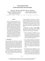
The SEM results (Fig. 1 A) show clustered microbeads, which are
approximately 20–30 μm in diameter. Whilst most of the microbeads are
isolated and spherical, some amalgamation of material in the form of a
fusion of beads are evident. SEM micrographs also show micron sized
pores, suggesting the presence of the oil/surfactant templating mixture
prior to calcination, leaving behind a complex mesoporous/microporous
structure, which will shift (in this case, to a predominantly microporous
structure) based on homogenizing conditions, similar to previous re
ports [24,33].
The titanosilicate Raman spectrum (Fig. 1 B) showed prominent
peaks at 960 nm and at 1107 nm, which are typical of Ti–O–Ti sym
metric and asymmetric stretching, respectively [37]. This was compared
against a commercial TiO2 (anatase: rutile 4:1) which did not show the
above two peaks but showed characteristic anatase bands at 144 nm,
395 nm (shoulder), 530 nm, 635 nm (broad) and rutile bands at 245 nm,
450 nm and 615 nm [38,39]. These peaks were not discernible on the
titanosilicate spectrum, however, two broad shoulders at 140 nm and
450 nm were visible and likely correspond to silica phases [40] within
the titanosilicate, in accordance with previous reports [24]. FTIR spec
trum for the titanosilicate (Fig. 1 C) showed peaks at 945 cm− 1 indica
tive of the Ti–O–Si asymmetric stretching, characteristic of
Fig. 1. Characterisation of titanosilicate microbeads. A - Scanning electron micrograph of titanosilicate beads, B - Raman spectra for anatase and titanosilicate, C FTIR spectra for anatase, silica and titanosilicate, D – X-Ray Diffraction data for titanosilicate before and after photocatalytic reaction with Rhodamine B.
3
A.S. Perera et al.
Microporous and Mesoporous Materials 345 (2022) 112276
titanosilicates. Additionally, peaks at 1070 cm− 1 for typical Si–O–Si
asymmetric stretching and at 804 cm− 1 for the O–Si–OH bending mode
indicated the presence of the silica matrix, that were comparable to the
reference SiO2 sample spectrum and evidence the silica matrix present
within the titanosilicate framework [24,33]. Anatase peaks seen in the
commercial sample were not visible in the titanosilicate FTIR spectrum.
The fingerprint region of the titanosilicate had a broad peak at ~460 nm
corresponding to the analogous silica reference peak, indicating silica
bending mode [41]. The PXRD results (Fig. 1 D) suggest an amorphous
crystal structure [24], both before and after use, indicating the robust
ness of the material.
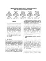
The pore structure of the titanosilicate microbeads generated via
surfactant templating and subsequent calcination at 750 ◦ C for removal
of the oil/surfactant phase [33], was analysed via BET porosimetry. The
size and volume of the pores were characterized via the BET (Bru
nauer-Emmett-Teller) and BJH (Barrett-Joyner-Halenda) methods
respectively (Fig. 2 A and B). N2 adsorption/desorption isotherms
indicated that the titanosilicate microbeads had an SBET surface area of
468 m2/g, total pore volume of 0.459 cm3/g and an average pore
diameter of ~1.3 nm, with a predominantly microporous structure. The
adsorption-desorption isotherms and BJH plots of the material both
before and after its use as a photocatalyst, indicated that the majority of
the material’s pore and surface characteristics are retained after one
reaction cycle (SBET 418 m2/g) with a slight increase to the degree of
mesoporosity (average pore diameter ~2 nm) after use. This may be due
to these pores becoming unblocked through use or relative disintegra
tion of micropores into larger pores due to the mechanical impact of
magnetic stirring of the titanosilicate beads over several catalytic cycles.
Therefore, use of this material could be further optimised through
immobilisation on a solid support, thus eliminating their mechanical
breakdown and improving catalyst lifespan, which is currently under
investigation in our laboratory.
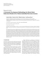
time, indicating that degradation products formed did not contain any
chromophores or large conjugated molecular fragments (ESI Figure S1).
The minor peaks present in the beginning of the reaction (i.e., at 521 nm,
355 nm, 314 nm and 216 nm) were all degraded over time. This
observation is in accordance with previous reports on photocatalytic
degradation of Rhodamine B. [43,44]. Enhancement of light absorbance
along 200–300 nm range with time does indicate that the fused micro
spheres of the titanosilicate catalyst maybe broken down into smaller
fragments, which can easily be suspended in solution and typically
absorb light in this range [24]. This was verified via SEM analysis of used
catalyst, which indicated that the microspheres had partially frag
mented during the reaction (ESI Figure S2).
Simulated sunlight generated via a solar simulator with irradiation
intensity equivalent of 1 sun (or AM1.5, or 1 kW/m2) was used to
evaluate the photocatalytic efficiency of the titanosilicate against
Rhodamine B removal. The changes in Rhodamine B concentration were
monitored via UV–vis spectroscopy (Fig. 3 C). Three types of experi
ments were conducted with the MiTS material in order to assess the
extent of both adsorption and photocatalysis on Rhodamine B removal:
1) control without the catalyst in light, 2) with catalyst in the dark, 3)
with catalyst under irradiation. The concentration of Rhodamine B did
not show any significant change without the titanosilicate catalyst, in
the control experiment with irradiation, indicating that Rhodamine B
does not degrade by sunlight alone. However, during the light experi
ment, the initial homogenizing step (t = − 30 min–0 min, in the dark)
indicated a drastic 59% reduction of Rhodamine B concentration in
solution indicating significant adsorption onto the microporous titano
silicate, followed by 88% within 60 min, 94% within 120, and finally
97% in 180 min wit irradiation. Interestingly, during the dark experi
ment with catalyst, an identical level of adsorption was evident during
the − 30 min initial homogenizing step, followed by significant levels of
Rhodamine B reduction within the 3 h, with a final value of 86%. Thus,
the impact of adsorption during the process was evident. Commercial
TiO2 consisting of 4 nm diameter particles of anatase: rutile 4:1 mixture
was also studied and was compared against the titanosilicate. This ma
terial did not show any adsorption during the − 30 min homogenizing
step and its overall Rhodamine B removal after the 3-h reaction time was
comparable to that of the titanosilicate (Fig. 3 C).
The C/C0 normalised data graph gives further insight into the impact
of adsorption on the process of Rhodamine B removal (Fig. 3 D). Once
represented in this manner, impact of the initial adsorption seen during
the -30-0 min homogenizing step is essentially removed from the data,
and the process of photocatalysis becomes clearer. Hence, the differ
ences between light and dark experiments become prominent for MiTSbased removal of Rhodamine B. Moreover, the commercial TiO2 appear
to be the superior material based on photocatalysis alone. However, the
MiTS would be a better option in application in real life water purifi
cation as the 4 nm particle size of commercial TiO2 could escape from
treatment units and would risk contamination of water. In contrast, the
20–30 μm size the MiTS particles are a safer option to for water filters.
Moreover, it is a non-doped material, which can match the best
3.2. Adsorption-photocatalysis based removal of Rhodamine B from
water: A bimodal mechanism
Rhodamine B (Fig. 3 A) was utilized as a model pollutant to study the
dual effects of adsorption-photocatalysis based removal from water with
the novel titanosilicate (MiTS). Due to its high UV–vis absorbance, water
solubility and polarity, Rhodamine B is an ideal candidate to study the
targeted dual effect of MiTS on pollutant removal. However, it has a size
of ~1.5–1.79 nm (depending on dimension side) [42], which hinders its
ability to reach certain micropores within the titanosilicate structure.
Thus, strong adsorption onto titanosilicate surface becomes an impor
tant factor in its degradation with the MiTS material discussed herein.
The experiments were conducted in deionized water with a 5 mg/L
initial concentration of Rhodamine B, for accurate quantification of the
bimodal-mechanism. The main absorbance of Rhodamine B occurred at
a maximum of 554 nm, the intensity of which was monitored to detect
and quantify its removal from the water medium (Fig. 3 B). There were
no new by-product peaks discernible via UV–vis during the 3-h reaction
Fig. 2. A – N2 adsorption-desorption isotherms of titanosilicate microbeads before and after use indicating a microporous structure before reaction and a shift
towards mesoporosity after reaction, B – their respective BJH plots, confirming an increase in average pore diameter after reaction.
4
A.S. Perera et al.
Microporous and Mesoporous Materials 345 (2022) 112276
Fig. 3. Demonstration of photocatalytic activity of titanosilicates against Rhodamine B. A - Chemical structure of Rhodamine B; B – UV–vis absorbance of Rhodamine
B with time under photocatalysis with titanosilicate; C - degradation of Rhodamine B concentration against time with titanosilicate and commercial TiO2 (anatase:
rutile 4:1). The − 30 to 0 min step indicates homogenizing in the dark leading to significant and fast adsorption of dye on the porous titanosilicate catalyst; D normalised graph of Rhodamine B degradation against time, where C0 is the starting concentration of Rhodamine B and C is the Rhodamine B concentration after its
removal against time.
conventionally known photocatalyst TiO2. Additionally, the MiTS
catalyst are capable of removing pollutants in the dark and during poor
light conditions due to adsorption, whereas the TiO2 tested above
showed no such activity (ESI Figure S3). The favourable particle size and
non-use of expensive dopants together with superior adsorbentphotocatalytic activity, paves a new path for advanced water purifica
tion using titanosilicates.
The considerable adsorption of Rhodamine B onto the titanosilicate
catalyst that takes place during this bimodal-mechanism-based photo
catalytic reaction (5 mg of Rhodamine B per 100 mg of titanosilicate)
plays a key role in its kinetic progression. Considering typical LangmuirHinshelwood kinetics for a porous catalyst and one reactant reaction
where adsorption is significant [45,46], the reaction appears to fit
within pseudo first order kinetics (ESI Figure S4). When the − 30 min
homogenizing step where high adsorption takes place was included, the
rate constants were found to be 0.023 mgl− 1min− 1 within the first hour,
0.019 mgl− 1min− 1 within the second hour and 0.016 mgl− 1min− 1
within the total 3-h reaction time. However, if the latter were to be
eliminated, the rates change to 0.020 mgl− 1min− 1, 0.016 and 0.014
mgl− 1min− 1 within the first, second and third hours respectively, rep
resenting 13.0%, 15.8% and 12.5% drops in respective rates. Consid
ering this discrepancy and the dual mechanism, a further inspection of
factors that influence kinetic parameters of photocatalytic degradation
needed to be considered. The rates of photocatalytic reactions are found
to be dependent on light intensity, reactant concentration, pH of the
medium, temperature and catalyst concentration [47,48], all of which
are kept constant in our study. Since our light source is kept at optimum
height to the reaction mixture to receive maximum intensity, and that
the reaction is small scale, it can be assumed that the local light intensity
experienced by the mixture is high and uniform. In such a case the
photocatalyst is found to behave as a typical heterogeneous catalyst and
not be influenced by kinetics related to the flux of absorbed photons on
its surface [47]. This theory however, considers that all available active
sites are exposed to light, which may not be the case for our 20–30 μm
sized catalyst, which has pores consisting predominantly of ~1.3 nm.
Hence, the calculated reaction rates should be taken as an approxima
tion on reaction kinetics. Nevertheless, they shed light into the potential
application of the material in water purification with regards to its
effectivity and stability. It must be noted that experiments conducted in
the dark are now particularly useful in determining the effect of
adsorption-based removal of Rhodamine B.
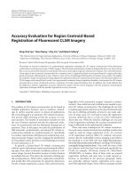
Diffuse reflectance studies of the titanosilicate followed by the
Kubelka-Munk analysis, indicated a direct band gap of 3.63 eV for the
material (Fig. 4). This is higher than the typical band gap value reported
for commercial P25 semiconductor, which is 3.2 eV Eg. However it is
closer to higher end of TiO2 prepared by sol–gel method (3.4–3.1 eV),
which are considered the best semiconductor options for environmental
Fig. 4. Band gap energy data generated via Kubelka-Munk analysis of diffusereflectance UV–vis data for the titanosilicate.
5
A.S. Perera et al.
Microporous and Mesoporous Materials 345 (2022) 112276
applications [49]. It is important to note that the titanosilicate reported
herein was synthesised via a sol-gel method [24]. The 4 nm TiO2
reference used herein indicated a band gap of 3.18 eV (ESI Figure S5),
which is considerably lower than the titanosilicate. Despite the higher
band gap, the tianosilicate gives very close Rhodamine B degradation
results to that of the TiO2 nanopowder, further evidencing the impor
tance and advantage of adsorption that occur during this process.
time (Fig. 7) – after turning pink upon adsorption of the dye (after 3 h
equilibration) the MiTS return to its original white colour after further
irradiation (19 h). This further indicates the potential for regeneration
and reuse of the MiTS material within multiple cycles.
3.5. Elemental analysis: further mechanistic insights
Further to Raman and IR analyses, the composition of the titanosi
licate samples were analysed by X-ray photoelectron spectroscopy
(XPS). Survey scans (taken over 0–1350 keV) revealed the samples were
composed of titanium, silicon and oxygen as expected for as-synthesised
titanosilicate samples. It was also evident that the calcining step
removed nearly all carbon contamination. During the photocatalytic
experiments, titanosilicate samples were isolated after 3 and 19 h by
centrifugation, before drying in vacuo and analysis by XPS. The amount
of carbon in the sample increased, and as XPS is a surface-sensitive
technique, this is good evidence for dye adsorption to the surface of
the titanosilicates. The average elemental composition in the titanosi
licates show some variation before and after reactions (Table 1, ESI
Figure S6). Samples X, Y and Z correspond to the as-synthesised tita
nosilicate powder (X), after 3 h of Rhodamine B photocatalytic degra
dation (Y) and after 19 h of Rhodamine B photocatalytic degradation
(Z). (These labels correspond to the high-resolution C1s scans given in
Fig. 7 C.)
The compositions listed in Table 1 (and indeed supported by the C1s
scans in Fig. 7) show a marked increase in the amount of carbon in the
sample as soon as the photocatalytic degradation of Rhodamine B was
underway. The carbon content increase from 6.1 atom % in the assynthesised titanosilicate sample (X) to 20.9 atom % after 3 h of pho
todegradation of Rhodamine B. Due to the way in which the samples
were prepared for XPS (i.e. centrifugation before drying), any adsorbed
material (i.e. dye) would be included in the analysis. This accounts for
the significantly higher proportion of carbon in Sample Y when
compared to Samples X and Z (after degradation was complete). This
observation supports the notion that surface adsorption of Rhodamine B
is a key part of the bimodal mechanism of pollutant degradation in the
MiTS titanosilicate material. After 19 h of reaction however, the carbon
content has significantly reduced, indicating a return to the surface
chemistry of the as-synthesised material. This is a favourable factor for
considerations in recycling and reusing of the material.
The model used to fit high resolution scans of the Ti2p region con
sisted of 2 components, with a more oxidised component (at higher
binding energy) attributed to a titanosilicate TiO2/SiO2 or tetrahedrally
coordinated environment, similar to that found in literature [51,52] and
confirmed by us in a previous study [24]. This likely corresponds to the
Ti–O–Si bonds in the active sites of the titanosilicate. The more reduced
component has a similar binding energy to titanium in a TiO2 or octa
hedrally coordinated environment [53], and this coupled with the more
oxidised component, forms the basic structure of the titanosilicate
3.3. Radical quenching experiments
In order to gain insight into the mechanism of photocatalytic
degradation of Rhodamine B, experiments were conducted in the pres
ence of para-benzoquinone (PBQ) and isopropanol (IPA), which are
known to quench radicals O2•− and •OH respectively. It was apparent
that both PBQ and IPA inhibited the degradation of Rhodamine B
significantly, compared to the experiments conducted without them,
and under the same irradiation conditions (Fig. 5). Thus, it was apparent
that both O2•− and •OH radicals are produced during the photocatalytic
degradation of Rhodamine B at the conditions given under visible light
irradiation, in accordance with previous studies. [36,50]. Intriguingly, it
was evident that the process of adsorption of Rhodamine B onto the
catalyst, which occurs predominantly during the initial − 30 to 0 min
homogenizing step, is inhibited more by IPA than PBQ (Fig. 5 A).
However, at the end of the 3-h reaction time, the experiments with both
scavenger compounds yielded the same results as the experiments con
ducted in the dark, where only adsorption takes place. The overall
Rhodamine B degradation by the titanosilicate after 3 h for IPA and PBQ
were found to be 86% and 88% respectively, which were close to the
86% removal shown by the experiments in the dark. Once the data was
normalised by plotting C/C0 to remove the effect of the initial adsorption
step (Fig. 5 B), it appeared that •OH scavenger IPA had more of an
impact on reaction inhibition than the O2•− scavenger PBQ. In reality
however, since adsorption does play an integral part of the catalyst’s
mechanism, the concentration vs. time graph should be considered to for
a more accurate depiction of progression this reaction catalysed via our
titanosilicate.
3.4. Recyclability
The MiTS photocatalyst was recycled over 4 cycles in order to
monitor recovery and recyclability. For light based photocatalytic re
action cycles (Fig. 6 A), only 5% loss of activity was evident after the 3-h
reaction time between cycles 1 and 2, indicating the robustness of the
material. There were no significant differences in Rhodamine B removal
evident among cycles 2–4 (>0.5%). The dark control data and indicates
the adsorptive capacity of the titanosilicate for the dye (Fig. 6 B). The
adsorption capacity decreases over time, when four consecutive recy
cling experiments are conducted in the dark. However, this would not be
an issue in real life day-night usage, as the adsorbed dye is degraded over
Fig. 5. Proof of photocatalytic mechanism of Rhodamine B degradation with the titanosilicate, under the presence of radical quencher compounds isopropanol (IPA)
and para-benzoquinone (PBA). A – Rhodamine B concentration against time, B – normalised graph of Rhodamine B degradation against time, where C0 is the starting
concentration of Rhodamine B and C is the Rhodamine B concentration after its removal against time.
6
A.S. Perera et al.
Microporous and Mesoporous Materials 345 (2022) 112276
Fig. 6. Recyclability of the MiTS titanosilicate photocatalyst with respect to Rhodamine B degradation over four reaction cycles; A - under simulated sunlight and B in the dark.
Fig. 7. A: UV–vis spectra of typical photocatalytic degradation of Rhodamine B, which indicates the gradual loss of the peak at ca. 560 nm over time. The dispersions
were centrifuged prior to UV–vis analysis to avoid any scattering effects from the titanosilicate powder. B: Photographs corresponding to the photocatalytic
degradation prior to reaction, after 3 h and after 19 h. C: High-resolution C1s XPS spectra showing the changing carbon environments. Note the increase in intensity
of the C1s environment after 3 h, indicating surface adsorption of Rhodamine B, with a sharp loss of intensity after 19 h of reaction time.
Table 1
Average atom % compositions calculated from XPS of the as-synthesised tita
nosilicate samples (X), after 3 h of photocatalytic degradation of Rhodamine B
(Y) and after 19 h of reaction time (Z). The Si:Ti ratio is given in the final column.
Table 2
The changing ratio of the fitted Ti2p environments in our fitted model for
Samples X, Y and Z. The “TiO2” peak has a lower binding energy (ca. 458.7 eV
for Ti2p3/2) and the higher binding energy “TiO2/SiO2” peak (ca. 459.6 eV for
Ti2p3/2). Ratios are calculated from the area of the Ti2p3/2 peaks.
Average composition (Atom %)
Sample identifier
C1s
O1s
Si2p
Ti2p
Si:Ti ratio
X
Y
Z
6.10
20.9
10.7
75.2
64.8
71.9
16.5
13.2
14.9
2.30
1.10
2.60
7.5
11.7
5.8
Concentration (Atom %)
material with active catalytic Ti–O–Si sites in the vicinity of Ti–O–Ti
phases. During the reaction, the ratio of the two environments changed,
presumably due to occupation of the catalytically active sites by the
Rhodamine B dye (Table 2).
The increase in the ratio of the TiO2:TiO2/SiO2 peaks after 19 h is in
contrast to the decrease in the amount of carbonaceous material seen in
Sample Identifier
TiO2 peak
TiO2/SiO2 peak
Ratio
X
Y
Z
1.13
0.735
2.26
1.37
0.675
0.553
0.830
1.09
4.08
Table 1 and Fig. 5. These data indicated that the surface availability of
the oxidised TiO2/SiO2 component changes post reaction rather than
returning to the ratio seen in Sample X. This could possibly be due to the
partial collapse of the pore structure during the reaction (see Fig. 2),
which made certain sites inaccessible during XPS analysis. In addition,
7
A.S. Perera et al.
Microporous and Mesoporous Materials 345 (2022) 112276
the apparent change in TiO2/SiO2 quantity does not seem to affect the
photocatalytic activity or the rate of degradation in repeat cycles (see
Fig. 6). The material retains its ability to remove Rhodamine B effec
tively for four consecutive catalytic cycles. The role in adsorption pro
cesses may play a key role in maintaining this activity as a way to
mitigate possible changes in the chemical structure of the material, thus
enhancing its’ robustness. These interesting observations warrant
further investigation, along with degradation studies on prevalent
organic water pollutants such as pesticides, antibiotics etc., but is
beyond the scope of the present ‘proof-of-concept’ study.
Data availability
Data will be made available on request.
Acknowledgements
The authors gratefully acknowledge the contributions from Mr
Richard Giddens for his assistance in conducting the SEM experiments,
Mr Simon Crust for assistance with Raman spectroscopy, Dr Andreas
Kafizas of Imperial College London for help with solid-state UV/Vis
measurements and Mr Owen Lawler for technical support with FTIR
analysis.
4. Conclusions
Appendix A. Supplementary data
A novel titanosilicate was prepared in microbead morphology of
20–30 μm diameter, via an oil-water emulsion based surfactanttemplating technique, without the addition of any dopants. The syn
thesised material was found to have a microporous structure with ~1.3
nm pores and a high BET surface area of 468 m2/g. The isolated,
tetrahedrally coordinated Ti4+ sites within the prepared materials,
responsible for its catalytic activity, were confirmed with FTIR, Raman
and XPS, and found to be stable throughout its use. The titanosilicate
was found to be effective in the sunlight driven degradation of the bulky,
model organic pollutant Rhodamine B, degrading over 97% of it within a
first 3-h cycle. This activity was comparable to commercial TiO2 nano
powder (4 nm, anatase:rutile 4:1). However, the titanosilicates being
micron-sized would be a far safer option for water treatment as larger
particles would be less likely to leach out of filtration units. The tita
nosilicate also appeared to be recyclable and reusable indicating >90%
removal of Rhodamine B over 4 reaction cycles, obtained consistently.
Adsorption was found to play a major role in the materials’ ability to
remove Rhodamine B, accounting for 59% of its removal during an
initial − 30 min homogenizing step, in the dark, and a subsequent 86%
removal within 3 h in the dark. These findings suggest that a bimodal
adsorption-photocatalysis based mechanism takes place, allowing for
fast and sustained removal of organic pollutants under simulated sun
light. Due to this activity, process cost-effectiveness and environmental
benignity of raw materials used, this material has potential to be applied
in tertiary water treatment. Further studies must test the ability to
degrade a wider range of emerging contaminants to determine its scope
of application.
Supplementary data to this article can be found online at https://doi.
org/10.1016/j.micromeso.2022.112276.
References
[1] M. Syafrudin, R.A. Kristanti, A. Yuniarto, T. Hadibarata, J. Rhee, W.A. Al-Onazi, T.
S. Algarni, A.H. Almarri, A.M. Al-Mohaimeed, Pesticides in drinking water-A
review, Int. J. Environ. Res. Publ. Health 18 (2) (2021) 468, />10.3390/ijerph18020468.
[2] M.-C. Danner, A. Robertson, V. Behrends, J. Reiss, Antibiotic pollution in surface
fresh waters: occurrence and effects, Sci. Total Environ. 664 (2019) 793–804,
/>[3] U. Shanker, M. Rani, V. Jassal, Degradation of hazardous organic dyes in water by
nanomaterials, Environ. Chem. Lett. 15 (4) (2017) 623–642, />10.1007/s10311-017-0650-2.
[4] O.M. Ogunbanwo, P. Kay, A.B. Boxall, J. Wilkinson, C.J. Sinclair, R.A. Shabi, A.
E. Fasasi, G.A. Lewis, O.A. Amoda, L.E. Brown, High concentrations of
pharmaceuticals in a Nigerian river catchment, Environ. Toxicol. Chem. 41 (3)
(2022) 551–558, />[5] M.J. Whelan, C. Linstead, F. Worrall, S.J. Ormerod, I. Durance, A.C. Johnson,
D. Johnson, M. Owen, E. Wiik, N.J.K. Howden, T.P. Burt, A. Boxall, C.D. Brown, D.
M. Oliver, D. Tickner, Is water quality in British rivers “better than at any time
since the end of the industrial revolution”, Sci. Total Environ. 843 (2022), 157014
/>[6] K. Balakrishna, A. Rath, Y. Praveenkumarreddy, K.S. Guruge, B. Subedi, A review
of the occurrence of pharmaceuticals and personal care products in Indian water
bodies, Ecotoxicol. Environ. Saf. 137 (2017) 113–120, />ecoenv.2016.11.014.
[7] L.C. Pereira, A.O. de Souza, M.F. Franco Bernardes, M. Pazin, M.J. Tasso, P.
H. Pereira, D.J. Dorta, A perspective on the potential risks of emerging
contaminants to human and environmental health, Environ. Sci. Pollut. Res. Int. 22
(18) (2015) 13800–13823, />[8] Z. Li, J. Li, Z. Guo, L.C. Campos, Investigation of metaldehyde removal by
powdered activated carbon from different water samples, Environ. Sci. Water Res.
Technol. 6 (5) (2020) 1432–1444, />[9] W.T. Vieira, M.B. de Farias, M.P. Spaolonzi, M.G.C. da Silva, M.G.A. Vieira,
Removal of endocrine disruptors in waters by adsorption, membrane filtration and
biodegradation. A review, Environ. Chem. Lett. 18 (4) (2020) 1113–1143, https://
doi.org/10.1007/s10311-020-01000-1.
[10] N.A. Zhou, H.L. Gough, Enhanced biological trace organic contaminant removal: a
lab-scale demonstration with bisphenol A-degrading bacteria sphingobium sp.
BiD32, Environ. Sci. Technol. 50 (15) (2016) 8057–8066, />10.1021/acs.est.6b00727.
[11] L.V. Nguyen, R. Busquets, S. Ray, A.B. Cundy, Graphene oxide-based degradation
of metaldehyde: effective oxidation through a modified fenton’s process, Chem.
Eng. J. 307 (2017) 159–167, />[12] E. Kudlek, Decomposition of Contaminants of Emerging Concern in Advanced
Oxidation Processes, Water, 2018, />[13] R. Quesada-Cabrera, C. Sotelo-Vazquez, J.C. Bear, J.A. Darr, I.P. Parkin,
Photocatalytic evidence of the rutile-to-anatase electron transfer in titania, Adv.
Mater. Interfac. 1 (6) (2014), 1400069, />admi.201400069.
[14] S.K. Loeb, P.J.J. Alvarez, J.A. Brame, E.L. Cates, W. Choi, J. Crittenden, D.
D. Dionysiou, Q. Li, G. Li-Puma, X. Quan, D.L. Sedlak, T. David Waite,
P. Westerhoff, J.-H. Kim, The technology horizon for photocatalytic water
treatment: sunrise or sunset? Environ. Sci. Technol. 53 (6) (2019) 2937–2947,
/>[15] US-4410501-A @ Pubchem.Ncbi.Nlm.Nih.Gov.
[16] D.R.C. Huybrechts, L. De Bruycker, P.A. Jacobs, Oxyfunctionalization of alkanes
with hydrogen peroxide on titanium silicalite, Nature 345 (6272) (1990) 240–242,
/>[17] L. Lv, G. Tsoi, X.S. Zhao, Uptake equilibria and mechanisms of heavy metal ions on
microporous titanosilicate ETS-10, Ind. Eng. Chem. Res. 43 (24) (2004)
7900–7906, />
Funding sources
The authors gratefully acknowledge the funding provided by UKRI
HEFCE-GCRF grant and the Department of Chemical and Pharmaceu
tical Sciences at Kingston University.
CRediT authorship contribution statement
Ayomi S. Perera: Writing – review & editing, Writing – original
draft, Supervision, Project administration, Methodology, Investigation,
Funding acquisition, Conceptualization. Patrick M. Melia: Writing –
review & editing, Methodology, Investigation, Formal analysis. Reece
M.D. Bristow: Writing – review & editing, Methodology, Formal anal
ysis. James D. McGettrick: Writing – review & editing, Formal analysis.
Richard J. Singer: Supervision, Methodology, Formal analysis. Joseph
C. Bear: Writing – review & editing, Methodology, Data curation. Rosa
Busquets: Writing – review & editing, Supervision, Project
administration.
Declaration of competing interest
The authors declare that they have no known competing financial
interests or personal relationships that could have appeared to influence
the work reported in this paper.
8
A.S. Perera et al.
Microporous and Mesoporous Materials 345 (2022) 112276
[36] S. Fang, K. Lv, Q. Li, H. Ye, D. Du, M. Li, Effect of acid on the photocatalytic
degradation of rhodamine B over G-C3N4, Appl. Surf. Sci. 358 (2015) 336–342,
/>[37] G. Ricchiardi, A. Damin, S. Bordiga, C. Lamberti, G. Span`
o, F. Rivetti, A. Zecchina,
Vibrational structure of titanium silicate catalysts. A spectroscopic and theoretical
study, J. Am. Chem. Soc. 123 (46) (2001) 11409–11419, />ja010607v.
[38] F.D. Hardcastle, Raman spectroscopy of titania (TiO2) nanotubular water-splitting
catalysts, J. Ark. Acad. Sci. (2011).
[39] S. Challagulla, K. Tarafder, R. Ganesan, S. Roy, Structure sensitive photocatalytic
reduction of nitroarenes over TiO2, Sci. Rep. 7 (1) (2017) 8783, />10.1038/s41598-017-08599-2.
[40] E. Berrier, C. Zoller, F. Beclin, S. Turrell, M. Bouazaoui, B. Capoen, Microstructures
and structural properties of Sol− Gel silica foams, J. Phys. Chem. B 109 (48) (2005)
22799–22807, />[41] T.N. Tran, T. Van Anh Pham, M.L. Phung Le, T.P. Thoa Nguyen, V.M. Tran,
Synthesis of amorphous silica and sulfonic acid functionalized silica used as
reinforced phase for polymer electrolyte membrane, Adv. Nat. Sci. Nanosci.
Nanotechnol. 4 (4) (2013), 45007, />045007.
[42] J. Canning, G. Huyang, M. Ma, A. Beavis, D. Bishop, K. Cook, A. McDonagh, D. Shi,
G.-D. Peng, M.J. Crossley, Percolation diffusion into self-assembled mesoporous
silica microfibres, Nanomaterials (2014), />[43] T. Liu, L. Wang, X. Lu, J. Fan, X. Cai, B. Gao, R. Miao, J. Wang, Y. Lv, Comparative
study of the photocatalytic performance for the degradation of different dyes by
ZnIn2S4: adsorption, active species, and pathways, RSC Adv. 7 (20) (2017)
12292–12300, />[44] H. Yang, J. Yang, Photocatalytic degradation of rhodamine B catalyzed by TiO2
films on a capillary column, RSC Adv. 8 (22) (2018) 11921–11929, https://doi.
org/10.1039/C8RA00471D.
[45] G.W. Roberts, C.N. Satterfield, Effectiveness factor for porous catalysts. Langmuirhinshelwood kinetic expressions, Ind. Eng. Chem. Fundam. 4 (3) (1965) 288–293,
/>[46] T. Sauer, G. Cesconeto Neto, H.J. Jos´e, R.F.P. Moreira, Kinetics of photocatalytic
degradation of reactive dyes in a TiO2 slurry reactor, J. Photochem. Photobiol.
Chem. 149 (1) (2002) 147–154, />[47] J.Z.A. Bloh, Holistic approach to model the kinetics of photocatalytic reactions,
Front. Chem. 7 (2019) 128, />[48] B.O. Burek, D.W. Bahnemann, J.Z. Bloh, Modeling and optimization of the
photocatalytic reduction of molecular oxygen to hydrogen peroxide over titanium
dioxide, ACS Catal. 9 (1) (2019) 25–37, />acscatal.8b03638.
[49] R. L´
opez, R. G´
omez, Band-gap energy estimation from diffuse reflectance
measurements on sol–gel and commercial TiO2: a comparative study, J. Sol. Gel
Sci. Technol. 61 (1) (2012) 1–7, />[50] K. Nagaveni, M.S. Hegde, N. Ravishankar, G.N. Subbanna, G. Madras, Synthesis
and structure of nanocrystalline TiO2 with lower band gap showing high
photocatalytic activity, Langmuir 20 (7) (2004) 2900–2907, />10.1021/la035777v.
[51] S. Contarini, P.A.W. van der Heide, A.M. Prakash, L. Kevan, Titanium coordination
in microporous and mesoporous oxide materials by monochromated X-ray
Photoelectron spectroscopy and X-ray auger electron spectroscopy, J. Electron.
Spectrosc. Relat. Phenom. 125 (1) (2002) 25–33, />[52] G. Moretti, A.M. Salvi, M.R. Guascito, F. Langerame, An XPS study of microporous
and mesoporous titanosilicates, Surf. Interface Anal. 36 (10) (2004) 1402–1412,
/>[53] J.C. Bear, V. Gomez, N.S. Kefallinos, J.D. McGettrick, A.R. Barron, C.W. Dunnill,
Anatase/rutile Bi-phasic titanium dioxide nanoparticles for photocatalytic
applications enhanced by nitrogen doping and platinum nano-islands, J. Colloid
Interface Sci. 460 (2015) 29–35, />
[18] D.M. Poojary, R.A. Cahill, A. Synthesis Clearfield, Crystal structures, and ionexchange properties of a novel porous titanosilicate, Chem. Mater. 6 (12) (1994)
2364–2368, />[19] M.A. Roberts, G. Sankar, J.M. Thomas, R.H. Jones, H. Du, J. Chen, W. Pang, R. Xu,
Synthesis and structure of a layered titanosilicate catalyst with five-coordinate
titanium, Nature 381 (6581) (1996) 401–404, />[20] A.S. Perera, M.-O. Coppens, Titano-silicates: highlights on development, evolution
and application in oxidative catalysis, in: Catalysis, vol. 28, The Royal Society of
Chemistry, 2016, pp. 119–143, />vol. 28.
[21] Z. Juan, Z. Dishun, Y. Liyan, L. Yongbo, Photocatalytic oxidation dibenzothiophene
using TS-1, Chem. Eng. J. 156 (3) (2010) 528–531, />cej.2009.04.032.
[22] M. Anpo, H. Yamashita, K. Ikeue, Y. Fujii, S.G. Zhang, Y. Ichihashi, D.R. Park,
Y. Suzuki, K. Koyano, T. Tatsumi, Photocatalytic reduction of CO2 with H2O on TiMCM-41 and Ti-MCM-48 mesoporous zeolite catalysts, Catal. Today 44 (1) (1998)
327–332, />[23] G.D. Lee, S.K. Jung, Y.J. Jeong, J.H. Park, K.T. Lim, B.H. Ahn, S.S. Hong,
Photocatalytic decomposition of 4-nitrophenol over titanium silicalite (TS-1)
catalysts, Appl. Catal. Gen. 239 (1) (2003) 197–208, />S0926-860X(02)00389-7.
[24] A.S. Perera, P. Trogadas, M.M. Nigra, H. Yu, M.-O. Coppens, Optimization of
mesoporous titanosilicate catalysts for cyclohexene epoxidation via statistically
guided synthesis, J. Mater. Sci. 53 (10) (2018) 7279–7293, />10.1007/s10853-018-2057-2.
[25] H. Yamashita, Y. Ichihashi, M. Anpo, M. Hashimoto, C. Louis, M. Che,
Photocatalytic decomposition of NO at 275 K on titanium oxides included within Yzeolite cavities: the structure and role of the active sites, J. Phys. Chem. 100 (40)
(1996) 16041–16044, />[26] S. Uma, S. Rodrigues, I.N. Martyanov, K.J. Klabunde, Exploration of photocatalytic
activities of titanosilicate ETS-10 and transition metal incorporated ETS-10,
Microporous Mesoporous Mater. 67 (2) (2004) 181–187, />j.micromeso.2003.11.003.
[27] European Commision, Goodbye E171: the EU bans titanium dioxide as a food
additive. (Accessed 18
July 2022).
[28] A.K. Adepu, R. Anumula, V. Narayanan, Photocatalytic degradation of rhodamine
B over a novel mesoporous titanosilicate/g-C3N4 nanocomposite under direct
sunlight irradiation, Microporous Mesoporous Mater. 247 (2017) 86–94, https://
doi.org/10.1016/j.micromeso.2017.03.046.
[29] A.K. Adepu, V. Katta, V. Narayanan, Synthesis, characterization, and
photocatalytic degradation of rhodamine B dye under sunlight irradiation of
porous titanosilicate (TS)/Bismuth vanadate (BiVO4) nanocomposite hybrid
catalyst, New J. Chem. 41 (6) (2017) 2498–2504, />C7NJ00071E.
[30] Y.K. Kim, S. Kim, Y. Kim, K. Bae, D. Harbottle, J.W. Lee, Facile one-pot synthesis of
dual-cation incorporated titanosilicate and its deposition to membrane surfaces for
simultaneous removal of Cs+ and Sr2+, Appl. Surf. Sci. 493 (2019) 165–176,
/>[31] H. Liu, A. Yonezawa, K. Kumagai, M. Sano, T. Miyake, Cs and Sr removal over
highly effective adsorbents ETS-1 and ETS-2, J. Mater. Chem. 3 (4) (2015)
1562–1568, />[32] K. Popa, C.C. Pavel, Radioactive wastewaters purification using titanosilicates
materials: state of the art and perspectives, Desalination 293 (2012) 78–86,
/>[33] A.S. Perera, J.K. Cockcroft, P. Trogadas, H. Yu, N. Kapil, M.-O. Coppens, Titanium
(IV)-Induced cristobalite formation in titanosilicates and its potential impact on
catalysis, J. Mater. Sci. 54 (1) (2019) 335–345, />[34] K. Egeblad, C.H. Christensen, M. Kustova, C.H. Christensen, Templating
mesoporous zeolites, Chem. Mater. 20 (3) (2008) 946–960, />10.1021/cm702224p.
[35] S.J. Gregg, K.S.W. Sing, Adsorption, Surface Area and Porosity, second ed.,
Academic Press, London, 1995.
9