báo cáo hóa học:" In vivo evaluation of a vibration analysis technique for the per-operative monitoring of the fixation of hip prostheses" pdf
Bạn đang xem bản rút gọn của tài liệu. Xem và tải ngay bản đầy đủ của tài liệu tại đây (2.45 MB, 10 trang )
BioMed Central
Page 1 of 10
(page number not for citation purposes)
Journal of Orthopaedic Surgery and
Research
Open Access
Research article
In vivo evaluation of a vibration analysis technique for the
per-operative monitoring of the fixation of hip prostheses
Leonard C Pastrav*
1,2
, Siegfried VN Jaecques
1,3
, Ilse Jonkers
4
, Georges Van
der Perre
1
and Michiel Mulier
5
Address:
1
Division of Biomechanics and Engineering Design (BMGO), Katholieke Universiteit Leuven, Celestijnenlaan 300C, bus 2419, 3001
Heverlee, Belgium,
2
Group T Leuven Engineering College (Association K.U. Leuven), Vesaliusstraat 13, 3000 Leuven, Belgium,
3
BIOMAT Research
Cluster, Katholieke Universiteit Leuven, Kapucijnenvoer 7, 3000 Leuven, Belgium,
4
Dept Biomedical Kinesiology, Katholieke Universiteit Leuven,
Tervuursevest 101, 3000 Leuven, Belgium and
5
Department of Orthopaedics, Katholieke Universiteit Leuven, Weligerveld 1, 3212 Lubbeek,
Belgium
Email: Leonard C Pastrav* - ; Siegfried VN Jaecques - ;
Ilse Jonkers - ; Georges Van der Perre - ;
Michiel Mulier -
* Corresponding author
Abstract
Background: The per-operative assessment of primary stem stability may help to improve the
performance of total hip replacement. Vibration analysis methods have been successfully used to
assess dental implant stability, to monitor fracture healing and to measure bone mechanical
properties. The objective of the present study was to evaluate in vivo a vibration analysis-based
endpoint criterion for the insertion of the stem by successive surgeon-controlled hammer blows.
Methods: A protocol using a vibration analysis technique for the characterisation of the primary
bone-prosthesis stability was tested in 83 patients receiving a custom-made, intra-operatively
manufactured stem prosthesis. Two groups were studied: one (n = 30) with non cemented and one
(n = 53) with partially cemented stem fixation. Frequency response functions of the stem-femur
system corresponding to successive insertion stages were compared.
Results: The correlation coefficient between the last two frequency response function curves was
above 0.99 in 86.7% of the non cemented cases. Lower values of the final correlation coefficient
and deviations in the frequency response pattern were associated with instability or impending
bone fracture. In the cases with a partially cemented stem an important difference in frequency
response function between the final stage of non cemented trial insertion and the final cemented
stage was found in 84.9% of the cases. Furthermore, the frequency response function varied with
the degree of cement curing.
Conclusion: The frequency response function change provides reliable information regarding the
stability evolution of the stem-femur system during the insertion. The protocol described in this
paper can be used to accurately detect the insertion end point and to reduce the risk for intra-
operative fracture.
Published: 9 April 2009
Journal of Orthopaedic Surgery and Research 2009, 4:10 doi:10.1186/1749-799X-4-10
Received: 20 November 2008
Accepted: 9 April 2009
This article is available from: />© 2009 Pastrav et al; licensee BioMed Central Ltd.
This is an Open Access article distributed under the terms of the Creative Commons Attribution License ( />),
which permits unrestricted use, distribution, and reproduction in any medium, provided the original work is properly cited.
Journal of Orthopaedic Surgery and Research 2009, 4:10 />Page 2 of 10
(page number not for citation purposes)
Background
Total hip replacement (THR) is the second most per-
formed surgical procedure with an estimated number of
more than one million operations each year worldwide.
This implies that, despite survival rates of 97% at 3 years
[1] and even up to 10 years follow-up [2] for some pros-
thesis types, a large number of revision operations are
needed every year, most of them because of aseptic loos-
ening. Revision operations are more difficult to perform,
carry more risk for complications and have a poorer prog-
nosis than primary THR [3].
Survival rate is directly related to the long term fixation
stability of the prosthesis stem [4]. Beside the design,
material composition and surface characteristics of the
implant, the initial per-operative fixation of the stem in
the femoral bone has a critical influence on its long term
fixation stability. This is especially the case for non
cemented, press-fit fixated stems. The insertion procedure
results in well-defined contact areas and interface pre-
stresses between the stem and the femoral bone. Under
actual loading, the hip stem displacement and the femoral
stress distribution will strongly depend upon these initial
contact conditions. Primary hip stem stability is not only
important regarding prosthesis migration, but also regard-
ing micro movements that must be limited in order to
allow interfacial bone formation and in-growth [5]. Fem-
oral stress distribution has a crucial influence on bone
remodelling and therefore on the final strength of the
bone-implant structure. Therefore the per-operative char-
acterization of the primary stem-femur contact and the
assessment of primary stem stability in the first place may
help to improve the survival rate of THR.
Nowadays objective intra-operative assessment of pri-
mary stem stability is a challenge, as surgeons have to rely
mainly on their clinical experience, which consists mainly
of a sense of mechanical stability when exerting axial force
and/or torque on the prosthesis. Moreover, excessive
press-fitting of a THR femoral component can cause intra-
operative fractures with an incidence of up to 30% in revi-
sion cases [6].
Vibration analysis has been successfully used to determine
bone mechanical properties [7-9]. Clinical applications of
this method were monitoring of fracture healing and in
vivo assessment of bone mechanical properties [10-14].
Vibration analysis was also successfully used to quantify
the fixation of oral implants [15]. A limited number of
studies prove the feasibility of detecting several forms of
femoral implant loosening, in vitro and in vivo using
techniques based on harmonic distortion [16-19].
In vitro, the analysis of frequency response function (FRF)
was used to discriminate between well fixed and quasi-
well fixed femoral stems [20].
This paper presents a series of cases where a per-operative
vibration analysis technique was used for the mechanical
characterization of the primary bone-prosthesis stability.
In a previous study we demonstrated the feasibility and
validity of a vibration analysis technique for the assess-
ment of the femur-stem fixation in vitro [21-24]. The stem
insertion process was performed on a dry cadaver femur
and synthetic composite femurs and the FRF change was
analysed. In a recent study a finite element model was cre-
ated to gain insight into the dependence of the FRF on sys-
tem parameter variations [25].
The imperfections in the connection between a THR pros-
thetic stem and a femur can most sensitively be detected
by observing shifts in the resonance frequency of the
higher vibration modes of the femur-prosthesis system.
This observation is in accordance with the work of Qi et
al. who stated that the most sensitive frequency band for
observing defects in the femur-prosthesis connection is
above 2500 Hz [26].
In the present study the vibration analysis technique was
applied for the per-operative assessment of fixation stabil-
ity in 83 THR patients who obtained an intra-operatively
manufactured prosthesis (IMP) provided by Advanced
Custom Made Implants, Leuven, Belgium (see appendix
1). The IMP approach aims at optimal stem stability
through a maximum fit and fill of the femoral cavity [27].
The objective of the present study was to apply and evalu-
ate an endpoint criterion for the insertion of the stem by
successive surgeon-controlled hammer blows. The end-
point-of-insertion criterion was based upon the Pearson's
correlation coefficient R between the FRFs of two succes-
sive insertion stages.
Methods
From the previous in vitro studies a protocol was derived
to be applied in per-operative conditions.
The prosthesis neck was attached to a shaker (Bruel &
Kjaer, Naerum, Denmark, model 4810) using a stinger
provided with a clamping system. The excitation was real-
ized through white noise in the range 0–12.5 kHz. The
input force and the response acceleration were measured
in the same point with an impedance head (PCB Piezo-
tronics, Depew, New York, USA, model nr 288D01)
mounted between the shaker and the stinger. The stinger
is a slender rod threaded at both ends, one of them being
connected to the impedance head and the other one to a
Journal of Orthopaedic Surgery and Research 2009, 4:10 />Page 3 of 10
(page number not for citation purposes)
kind of claw forming a clamping system that is firmly
attached to the prosthesis neck.
The excitation system used low amplitude vibrations and
introduced approximately 0.5 W of power into the femur-
prosthesis system. This is considered as safe and no
adverse effects have been reported by other authors using
a similar excitation system [11-13]. The experimental
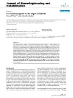
setup is shown in Figure 1.
The FRF was measured and recorded by a Pimento vibra-
tion analyser (LMS International, Haasrode, Belgium)
connected to a portable computer provided with the
appropriate software (Pimento 5.2, LMS International,
Haasrode, Belgium). The vibration analyser generates the
excitation signal which is amplified and sent to the shaker.
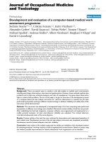
The vibration analyser, the portable computer and the
amplifier were installed in the surgical theatre but outside
the so-called laminar flow area (Figure 2).
Patients, eligible for THR, received full information rela-
tive to the surgical intervention and the study objectives,
including the scheme for follow-up visits. The study pro-
tocol was approved by the institutional review board.
Patients were included after giving written informed con-
sent. Thirty patients received non cemented IMP stems
and fifty three patients received distally cemented IMP
stems. The decision between the two procedures was
made by the surgeon on clinical criteria. All stems were
proximally coated with hydroxyapatite.
Before starting the measurements on patients the full pro-
tocol was tested in a cadaver study.
Non cemented prostheses
The surgeon inserted the implant in the femoral canal
through successive controlled hammer blows. After each
blow, the FRF of the implant-bone structure was meas-
ured directly on the prosthesis neck in the range 0–10
kHz.
During the insertion the assembly composed by shaker,
impedance head and stinger with clamping system was all
the time attached to the prosthesis neck (i.e. the clamping
was done only once per insertion, and the tightness of
clamping was thus the same for all FRFs i = 0 n). In the
measured structure the only variable was the connection
between the implant and the bone. The shaker was held
by a member of the surgical team as presented in Figure 2
(right) and Figure 3.
The FRF changes were used as indicators of the evolution
of the stiffness of the implant-bone structure and, as a
consequence, the evolution of the implant stability. When
the FRF graph did not change noticeably anymore the
hammering was stopped. Extra blows would not improve
the stability of the prosthesis but would increase the frac-
ture risk.
The similarity of two successive FRF graphs was evaluated
using the Pearson's correlation coefficient. A correlation
between the FRFs of successive stages of R = (0.99 +/-
0.01) over the range 0–10000 Hz is proposed as an end-
point criterion.
Partially cemented prostheses
The per-operative protocol presented above was adapted
to assess the stability of hybrid IMPs that were partially
cemented distally using Palacos
®
(Zimmer Inc. Warsaw,
Indiana, USA) bone cement [27].
In a first stage, the surgeon inserted the stem completely
in the femur without cement, for a trial reduction of the
artificial joint. In a later stage, the stem was removed,
cement was introduced in the distal part of the femoral
canal, the stem was re-introduced and after the cement
has fully cured, the implant was supposed to be com-
pletely fixed. The FRF was measured in both stages using
the same method as in the non cemented stems case. (Fig-
ure 3). In some randomly chosen cases, the FRF was meas-
ured also at various stages of cement curing i.e. 6, 10, 12,
and 14 minutes after cement preparation.
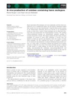
Experimental setupFigure 1
Experimental setup. a. Hip stem. b. Stinger and clamping
system. c. Impedance head. d. Shaker.
Journal of Orthopaedic Surgery and Research 2009, 4:10 />Page 4 of 10
(page number not for citation purposes)
Results
Non cemented hip stems
Thirty cases of non cemented stems were studied in vivo
and a typical evolution of the FRF graph is shown in Fig-
ures 4a–d. The Pearson's correlation coefficient (R), calcu-
lated for consecutive pairs of FRFs, is presented in Figure
4e. Stage 0 corresponds to the FRF calculated after the
stem was introduced in the femur by hand; stage 1 corre-
sponds to the FRF calculated after the first hammer blow
series, stage 2 after the second hammer blow series and so
on. The surgeon needed five stages (0 4) to completely
insert the stem in this case.
Normally, the FRF graphs shifted to the right indicating a
stiffness increase between successive insertion stages [28].
To compare the similarity of two successive FRF graphs the
Pearson's correlation coefficient was used. Due to the fact
that there is no linear dependence of one graph with
respect to the other, the two graphs are identical if the cor-
relation coefficient is 1.
In twenty six out of thirty cases (86.7%), the correlation
coefficient between the last two FRFs was above 0.99
when the surgeon stopped the insertion. In the other four
cases, when the surgeon decided to stop the insertion
because of suspected bone fragility, the final correlation
coefficient reached lower values still exceeding 0.95.
Non cemented hip stems – non-typical cases
Case 1
While testing the per-operative protocol on a human
cadaver, the stem was deliberately inserted until the femur
was fractured. The last three FRF graphs are presented in
Figures 5a and 5b.
The FRF graph slightly shifted to the left at the fifth inser-
tion stage indicating a decrease of the stability before the
sixth stage when the bone was fractured. The final FRF
graph is totally different with respect to the previous graph
indicating an important change in the stem-femur struc-
ture.
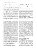
Measuring hardware (left) and surgical theatre (right*)Figure 2
Measuring hardware (left) and surgical theatre (right*). a. Portable computer. b. Vibration analyser Pimento
®
. c. Power
amplifier. *The circle indicates the place of the measuring hardware (presented in the left picture) behind the transparent wall.
A member of the surgical team holds the shaker during the insertion procedure.
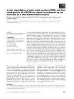
Hip stem insertionFigure 3
Hip stem insertion. a. Hip stem (almost fully inserted). b.
Stinger and clamping system. c. Shaker (held by a member of
the surgical team).
Journal of Orthopaedic Surgery and Research 2009, 4:10 />Page 5 of 10
(page number not for citation purposes)
Case 2
During a per-operative experiment, when the stem was
quasi fully inserted, the highest peak of the FRF graph
slightly shifted to the left (stage B in Figure 6a).
After a supplementary hammer blow series, the corre-
sponding FRF graph presented an abnormal shape (stage
C in Figure 6b). Inspecting the bone, a small fracture was
observed and the hammering was stopped.
Case 3
An oscillating behaviour of the FRF graph was observed
during another per-operative hip arthroplasty procedure
(stages 7, 8, and 9 in Figures 7a and 7b).
Since the stem was visibly not fully inserted, the hammer-
ing normally had to continue, but the behaviour of the
FRF, similar to the FRF evolution presented in case 2, was
indicating that the stem was blocked and, as a conse-
quence, there was a risk for fracture. The problem was
solved by pulling out the stem, adjusting the femoral
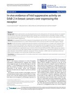
Non cemented stemFigure 4
Non cemented stem. a-d. FRF graphs corresponding to successive insertion stages. e. Pearson's correlation coefficients cal-
culated for the FRF pairs presented in Figures 4 a-d. For example, the point with the abscissa "s 0_1" and the ordinate "0.510"
represents the correlation coefficient calculated between the FRFs corresponding to the insertion stages 0 and respectively 1.
The graphs corresponding to these FRFs are presented in Figure 4a.
Journal of Orthopaedic Surgery and Research 2009, 4:10 />Page 6 of 10
(page number not for citation purposes)
canal and reinserting the prosthesis. The FRF had a normal
evolution during the reinsertion and the graphs corre-
sponding to the final two stages, labelled as stage 4a and
stage 5a, are shown in Figure 7c. The corresponding Pear-
son's correlation coefficient attained 0.998.
Partially cemented hip stems
Fifty three cases of partially cemented prostheses were
studied in vivo. In forty five cases (84.9%) an important
difference was observed between the FRF graph corre-
sponding to the non cemented stage and the FRF graph
corresponding to the cemented stage, after complete
cement hardening, in frequency and amplitude. A typical
example is shown in Figure 8a.
In the other eight cases, although some alteration could
be noticed, the FRF graph did not substantially change
after cement curing (Figure 8b).
The typical evolution of the FRF graph during the cement
curing, at 6, 10, 12, and 14 minutes after cement mixing,
is shown in Figure 9.
When the cement polymerisation sets in, the resonance
frequencies of the vibration modes associated with the
cement increase. For the presented case, the resonance fre-
quency of the vibration mode mostly influenced by the
cement curing increased from 7180 to 7680 Hz. When the
polymerisation is complete, no further changes are
observed in the FRF. The graphs corresponding to 12 and
14 minutes are nearly identical.
Discussion
During the insertion of an IMP in a femur, the changes of
boundary conditions and implant stability between dif-
ferent stages are reflected by the FRF evolution as observed
per-operatively. The higher resonance frequencies are
more sensitive to the stability change than the lower fre-
Non cemented stem in human cadaver (non-typical case 1)Figure 5
Non cemented stem in human cadaver (non-typical case 1). a. FRF graphs corresponding to the insertion stages 4 and
5. b. FRF graphs corresponding to the insertion stages 5 and 6.
Per-operative fracture (non-typical case 2)Figure 6
Per-operative fracture (non-typical case 2). a. FRF graphs corresponding to the insertion stages A and B (anomalous left
shift). b. FRF graphs corresponding to the insertion stages B and C (small fracture).
Journal of Orthopaedic Surgery and Research 2009, 4:10 />Page 7 of 10
(page number not for citation purposes)
Correction of the femoral canal (non-typical case 3)Figure 7
Correction of the femoral canal (non-typical case 3). a. FRF graphs corresponding to the insertion stages 7 and 8 (typi-
cally observed right shift). b. FRF graphs corresponding to the insertion stages 8 and 9 (anomalous left shift). c. FRF graphs cor-
responding to the two final stages (labelled 4a and 5a) of the reinsertion process, after the correction of the femoral canal.
Two typical cases of partially cemented stems completely inserted in the femurFigure 8
Two typical cases of partially cemented stems completely inserted in the femur. a. FRF graphs for two stages: with-
out cement (white) and cemented (black). An important change can be observed after cementation. b. FRF graphs for two
stages: without cement (white) and cemented (black). FRF graph slightly shifted to the right after cementation.
Journal of Orthopaedic Surgery and Research 2009, 4:10 />Page 8 of 10
(page number not for citation purposes)
quencies. This observation is in accordance with previous
finite element studies [26], and can be explained schemat-
ically as follows. The lower frequency resonances corre-
spond to vibration motions in which the deformation of
the femur (vibration mode shape) is simple, such as single
bending of the femur shaft. The higher frequency reso-
nances correspond to more intricate deformation modes
of the femur in combination with deformation modes of
the prosthesis. In the case of simple bending modes of the
femur the prosthesis stem acts just like an added mass and
its influence depends more on its position than on the fix-
ation conditions. In the case of more intricate mode
shapes the interaction between the stem and the femur
becomes more complicated and the corresponding reso-
nance frequencies become more sensitive to the interface
conditions. This explanation is completely corroborated
and further elucidated by recent advanced finite element
analyses by our group [25,29].
During the insertion of the uncemented stem the FRF
change is influenced by the stiffness of the implant-femur
system and the relative position of the two components.
The FRF graph shift to the right between successive ham-
mer blows is a normal evolution; the fixation stiffness
increase being reflected by increasing resonance frequen-
cies. The graph change in shape and position is more
important at the beginning of the insertion when the stem
displacement is important as well. At the end of the inser-
tion, when the resistance against the stem displacement
increases, the shapes of the successive graphs are very sim-
ilar and the shift is less important. When the FRF graph
does not change noticeably between two hammer blows,
the logical conclusion is that the system mechanical
parameters do not change, thus the stem cannot move
and the hammering should stop to avoid an intra-opera-
tive fracture.
For non cemented stems, the Pearson's correlation coeffi-
cient between successive FRFs can be used as a criterion for
the detection of the insertion endpoint.
Moreover, the FRF analysis can be used to detect danger-
ous situations during surgery such as stem blockage and
fracture risk. An FRF graph shift to the left indicates a
decreasing fixation, probably due to plastic deformation
of the bone, and should be a serious warning for the sur-
geon. In two cases hammering after a graph shift to the left
resulted in bone fractures (Figures 5b and 6b).
A possible fracture may have been avoided in the case of
an abnormal bone structure and a deformed endomedul-
lary canal as the FRF analysis showed an abnormality and
the surgeon was alerted to the situation in time during
insertion of the stem (Figures 7a–c).
The supplementary information obtained by vibration
analysis helps the surgical team to take the optimal deci-
sions.
The curing of bone cement in partially cemented hip stem
systems can also be monitored by vibration analysis.
In 15% cases the FRF graph did not substantially change
after cement curing. The interpretation could be that the
implant stability did not considerably change after
cementation. Probably the stems were already reasonably
well fixed in the non cemented stage. However, the shift
to the right of the FRF graph indicates an increased stabil-
ity after cementation. Comparing the Figures 8a and 8b, it
can be observed that the FRF corresponding to the com-
plete curing stages (black graphs) are very similar. Moreo-
ver, these graphs are very similar to the graphs
corresponding to the final insertion stage of the cement-
less stems (Figures 4d and 7c)
The per-operative experimental study should be com-
pleted and validated by an appropriate post-operative fol-
low-up of the patients. In an ongoing clinical study, part
of project OT/03/31, migration of the stems is followed
up by Roentgen Stereophotogrammetric Analysis (RSA)
and bone remodelling is followed up by Dual energy X-
Ray Absorptiometry (DXA). Conventional follow-up by
clinical examination, radiographs and standardised ques-
tionnaires is also part of the protocol [30,31].
Conclusion and future work
The presented per operative technique was designed to
monitor the stability and to detect the insertion endpoint
FRF graphs of a typical stem-femur system during cement curingFigure 9
FRF graphs of a typical stem-femur system during
cement curing. The evolution of the FRF at various stages
of cement curing i.e. 6, 10, 12, and 14 minutes after cement
preparation. The arrow indicates that the peak correspond-
ing to the vibrational mode mostly influenced by the cement
curing shifts to the right when the curing time increases.
Journal of Orthopaedic Surgery and Research 2009, 4:10 />Page 9 of 10
(page number not for citation purposes)
of non cemented and partially cemented hip stems, but it
can be adapted for other orthopaedic implants as well.
It does not provide direct quantitative information on the
displacements in real life loading conditions, but it is a
powerful technique for the quality and safety control of
the surgical procedure. It is a sensitive, minimally invasive
method to check whether the insertion process runs nor-
mally and results in the best possible fixation for the
patient and the prosthesis at hand, and to prevent bone
fracture.
The stability under real life loads of an optimally fitted
non cemented prosthesis such as the IMP and other pros-
thesis systems has been shown to be adequate by previous
research and clinical experience.
Nevertheless vibration analysis is currently developed
into a technique for the full mechanical characterization
of the contact between the prosthesis and the bone in
terms of contact areas, interface stresses and ultimately
stability under real loading. In a finite elements study [25]
the relation between the vibration behaviour and the spa-
tial distribution of contact areas was analyzed. In a tran-
sient dynamic analysis [29] the successive steps in the
insertion process were simulated in terms of contact areas
and interface stresses, and the vibration response in each
step was calculated by finite elements analysis.
Building on the understanding and clinical experience
built through per operative monitoring, vibration analysis
will be developed further into a technique for the non
invasive post operative assessment of prosthesis fixation,
in view of detection of loosening.
Competing interests
The authors declare that they have no competing interests.
Authors' contributions
All authors have made substantial contributions to the
conception and design of the study, analysis and interpre-
tation of data, drafting the article, and revising it critically.
Specifically, LP developed the details of the vibration
analysis protocol, operated the data acquisition equip-
ment during the peroperative measurements, processed
and analysed the FRF data (supervised by GVdP and SJ)
and drafted the figures and the initial version of the man-
uscript. GVdP and SJ conceived the principles of the vibra-
tion analysis protocol. GVdP, IJ and SVNJ drafted the
grant application from which this study was partially
funded, including the study design, and supervised the
implementation. MM was the surgeon in charge during
the THR procedures and operated the sterile part of the
vibration analysis equipment within the laminar flow
area. MM provided clinical background knowledge for the
introduction and discussion sections. Multiple critical
iterations on the initial manuscript were a joint effort of
all authors. All authors read and approved the final man-
uscript.
Appendix
The intra-operatively manufactured prosthesis
In the IMP-procedure, the stem of the prosthesis is custom
made for each individual patient, during the operation
[27]. After reaming of the femoral cavity, a 3D imprint of
the cavity is made in the form of a silicone mould. This
mould is scanned and its geometry is optimized using a
CAD-procedure. Based upon this adapted geometry, a
CNC milling machine then transforms a partially pre-
formed prosthesis stem into the final personalized shape.
Using this technique a maximum fit and fill of the femoral
cavity might be obtained allowing an optimal stability of
the femoral implant.
Between the first implantation in 1987 and the time of
writing (2009) the technique underwent some changes,
the most important of which was the application of a
hydroxyapatite layer by plasma spray coating.
Pearson's correlation coefficient
Pearson's product moment correlation coefficient, R, is a
dimensionless index that ranges from -1.0 to 1.0 inclusive
and reflects the extent of a linear relationship between two
data sets (i.e. two variables).
The equation for the correlation coefficient is:
Where x and y are the two variables and , are the cor-
responding arithmetic means [32].
Acknowledgements
This research was partially funded by a grant from the K.U. Leuven research
council (OT/03/31).
Advanced Custom Made Implants S.A./N.V. Leuven, Belgium (ACMI), are
acknowledged for providing custom made hip prostheses. Specifically, Guy
Deloge and Wim Claassen, both from ACMI, are acknowledged for the
intraoperative manufacturing.
References
1. Lucht U: The Danish Hip Arthroplasty Register. Acta Orthop
Scand 2000, 71:433-439.
2. Britton AR, Murray DW, Bulstrode CJ, McPherson K, Denham RA:
Long-term comparison of Charnley and Stanmore design
total hip replacements. J Bone Joint Surg Br 1996, 78:802-808.
3. Kavanagh BF, Ilstrup DM, Fitzgerald RH Jr: Revision total hip
arthroplasty. J Bone Joint Surg Am 1985, 67:517-526.
4. Mjöberg B: The theory of early loosening of hip prostheses.
Orthopedics 1997, 20:1169-1175.
R
xx yy
xx yy
=
−
()
−
()
∑
−
()
−
()
∑∑
22
x
y
Publish with BioMed Central and every
scientist can read your work free of charge
"BioMed Central will be the most significant development for
disseminating the results of biomedical research in our lifetime."
Sir Paul Nurse, Cancer Research UK
Your research papers will be:
available free of charge to the entire biomedical community
peer reviewed and published immediately upon acceptance
cited in PubMed and archived on PubMed Central
yours — you keep the copyright
Submit your manuscript here:
/>BioMedcentral
Journal of Orthopaedic Surgery and Research 2009, 4:10 />Page 10 of 10
(page number not for citation purposes)
5. Søballe K, Hansen ES, Rassmussen HB, Jorgensen PH, Bunger C: Tis-
sue ingrowth into titanium and hydroxyapatite-coated
implants during stable and unstable mechanical conditions. J
Orthop Res 1992, 10:285-299.
6. Meek RM, Garbuz DS, Masri BA, Greidanus NV, Duncan CP: Intra-
operative fracture of the femur in revision total hip arthro-
plasty with a diaphyseal fitting stem. J Bone Joint Surg Am 2004,
86-A:480-485.
7. Lowet G, Van Audekercke R, Perre G Van der, Geusens P, Dequeker
J, Lammens J: The relation between resonant frequencies and
torsional stiffness of long bones in vitro. Validation of a sim-
ple beam model. J Biomech 1993, 26:689-696.
8. Lowet G, Perre G Van der: Ultrasound velocity measurement in
long bones: measurement method and simulation of ultra-
sound wave propagation. J Biomech 1996, 29:1255-1262.
9. Perre G Van der, Cornelissen P: On the mechanical resonance of
a human tibia in vitro. J Biomech 1983, 16:549.
10. Perre G Van der, Van Audekercke R, Martens M, Mulier JC: Identifi-
cation of in vivo vibration modes of human tibia by modal
analysis. J Biomech Eng 1983, 105(3):244-248.
11. Nikiforidis G, Bezerianos A, Dimarogonas A, Sutherland C: Monitor-
ing of fracture healing by lateral and axial vibration analysis.
J Biomech 1990, 23:323-330.
12. Collier J, Donarski J, Worley J, Lay A: The use of externally
applied mechanical vibrations to assess both fractures and
hip prosthesis. In Micromovement in Orthopaedics Edited by: Turner-
Smith R. Oxford Univ. Press; 1993:151-163.
13. Nakatsuchi Y, Tsuchikane A, Nomura A: Assessment of fracture
healing in the tibia using the impulse response method. J
Orthop Trauma 1996, 10:50-62.
14. Perre G Van der, Lowet G: In vivo assessment of bone mechan-
ical properties by vibration and ultrasonic wave propagation
analysis. Bone 1996, 18(1 Suppl):29S-35S.
15. Meredith N, Book K, Friberg B, Jemt T, Sennerby L: Resonance fre-
quency measurements of implant stability in vivo. A cross-
sectional and longitudinal study of resonance frequency
measurements on implants in the edentulous and partially
dentate maxilla. Clin Oral Implants Res 1997, 8:226-233.
16. Burny F, Donkerwolcke M, Moulart F, Borgwardt A, Vryzakis P,
Pichon D, Collier R, Louridas S, Perre G Van der, Denayer I, Jaecques
S, Catrysse M, Puers R: STIMuLus: Smart Total Implant for the
Monitoring of Loosening [abstract]. Medical Physics 2000,
27:1406.
17. Georgiou AP, Cunningham JL: Accurate diagnosis of hip prosthe-
sis loosening using a vibrational technique. Clin Biomech (Bristol,
Avon) 2001, 16:315-323.
18. Rosenstein AD, McCoy GF, Bulstrode CJ, McLardy-Smith PD, Cun-
ningham JL, Turner Smith AR: The differentiation of loose and
secure femoral implants in total hip replacement using a
vibrational technique: an anatomical and pilot clinical study.
Proc Inst Mech Eng [H] 1989, 203:77-81.
19. Rowlands A, Duck FA, Cunningham JL: Bone vibration measure-
ment using ultrasound: Application to detection of hip pros-
thesis loosening. Med Eng Phys 2008, 30:278-284.
20. Lannocca M, Varini E, Cappello A, Cristofolini L, Bialoblocka E: Intra-
operative evaluation of cementless hip implant stability: A
prototype device based on vibration analysis. Med Eng Phys
2007, 29:886-894.
21. Jaecques S, Pastrav C, Zahariuc A, Perre G Van der: Analysis of the
fixation quality of cementless hip prostheses using a vibra-
tional technique. In Proceedings of ISMA 2004 International Confer-
ence on Noise and vibration engineering: 20–22 September 2004; Leuven
Edited by: Sas P, De Munck M. Leuven: K.U. Leuven; 2004:443-456.
22. Jaecques S, Pastrav C, Mulier M, Perre G Van der: Determination
of THR stem insertion endpoint by vibrational analysis. In
EORS – Transactions, Proceedings of 15th Annual Meeting of European
Orthopaedic Research Society – EORS 2005; Lisbon Edited by: Cabral R,
Cassiano NM, Camilo C. Lisbon: Soc Portugesa de Ortopedia e Trau-
matologia; 2005:92.
23. Pastrav C, Jaecques S, Deloge G, Mulier M, Perre G Van der: A sys-
tem for intra operative manufacturing and stability testing
of hip prostheses. In Proceedings of TEHNOMUS XIII, the 13th Inter-
national conference on New technologies and products in machine manu-
facturing technology: 6–7 May 2005; Suceava Edited by: Dumitru I.
Suceava: University of Suceava; 2005:505-510.
24. Pastrav LC, Jaecques SVN, Mulier M, Va der Perre G: Detection of
the insertion end point of cementless hip prostheses using
the comparison between successive frequency response
functions. Journal of Applied Biomaterials & Biomechanics 2008,
6:23-29.
25. Devos J, Jaecques S, Pastrav C, Perre G Van der: The influence of
contact conditions on resonance frequencies of a hip stem-
femur system: a finite element analysis. In Proceedings of the
International seminar Prediction and evaluation of total hip replacement
performance: can we plan success? 22–23 June 2007; Leuven Edited by:
Jonkers I, Jaecques S, Mulier M, Van der Perre G. Leuven: K.U. Leu-
ven; 2007:35-36.
26. Qi G, Mouchon W, Tan TE: How much can a vibrational diag-
nostic tool reveal in total hip arthroplasty loosening? Clin Bio-
mech (Bristol, Avon). 2003, 18(5):444-458.
27. Mulier JC, Mulier M, Brady LP, Steenhoudt H, Cauwe Y, Goossens M,
Elloy M: A new system to produce intraoperatively custom
femoral prosthesis from measurements taken during the
surgical procedure. Clin Orthop Relat Res 1989, 249:97-112.
28. Heylen W, Lammens S, Sas P: Modal Analysis Theory and Testing Leu-
ven: K.U. Leuven, Belgium; 1997.
29. Asiminei A, Pastrav C, Jaecques S, Mulier M, Perre G Van der: Intra-
operative manufactured hip prosthesis insertion – a finite
element simulation. In Proceedings of the International seminar Pre-
diction and evaluation of total hip replacement performance: can we plan
success? 22–23 June 2007; Leuven Edited by: Jonkers I, Jaecques S, Mul-
ier M, Van der Perre G. Leuven: K.U. Leuven; 2007:28-30.
30. Claassen W, Nijs J, Jaecques S, Perre G Van der, Mulier M: Early
periprosthetic bone remodelling around cemented and
uncemented custom-made femoral components and their
uncemented acetabular cups. In Proceedings of the International
seminar Prediction and evaluation of total hip replacement performance:
can we plan success? 22–23 June 2007; Leuven Edited by: Jonkers I, Jae-
cques S, Mulier M, Van der Perre G. Leuven: K.U. Leuven; 2007:6-8.
31. Claassen W, Nijs J, Mulier M, Jaecques S, Perre G Van der: Follow-
up of implant performance using RSA. In Proceedings of the Inter-
national seminar Prediction and evaluation of total hip replacement per-
formance: can we plan success? 22–23 June 2007; Leuven Edited by:
Jonkers I, Jaecques S, Mulier M, Van der Perre G. Leuven: K.U. Leu-
ven; 2007:2-5.
32. Spiegel MR: Theory and Problems of Statistics New York: McGraw-Hill
Publishing Co Ltd; 1972.