In-situ dehydration study of the Sr-, Cd- and Pb-exchanged natrolite
Bạn đang xem bản rút gọn của tài liệu. Xem và tải ngay bản đầy đủ của tài liệu tại đây (4.43 MB, 7 trang )
Microporous and Mesoporous Materials 346 (2022) 112277
Contents lists available at ScienceDirect
Microporous and Mesoporous Materials
journal homepage: www.elsevier.com/locate/micromeso
In-situ dehydration study of the Sr-, Cd- and Pb-exchanged natrolite
Junhyuck Im a, Jaewoo Jung b, Kiho Yang c, Donghoon Seoung d, **, Yongmoon Lee e, *
a
Decommissioning Technology Research Division, Korea Atomic Energy Research Institute (KAERI), Daejeon, 34057, South Korea
Global Ocean Research Center, Korea Institute of Ocean Science & Technology, Busan, 49111, South Korea
c
Department of Oceanography, Pusan National University, Busan, 46241, South Korea
d
Department of Earth and Environmental Sciences, Chonnam National University, Gwangju, 61186, South Korea
e
Department of Geological Sciences, Pusan National University, Busan, 46241, South Korea
b
A R T I C L E I N F O
A B S T R A C T
Keywords:
Dehydration
In-situ X-ray diffraction
Natrolite
Rietveld refinement
The removal of Sr, Cd, and Pb from nuclear and industrial waste is important as these are harmful to living
organisms and the environment. Immobilization of these ions in a zeolite framework is a simple and suitable
method. However, zeolites are easily dehydrated at high temperatures. Therefore, the environmental changes
around these adsorbed cations and water molecules in the zeolite framework must be explored for effective
immobilization and waste removal. In this study, we investigated the structural changes in fully Sr-, Cd-, and Pbexchanged natrolites (NAT) from room temperature to 350 ◦ C using in situ synchrotron X-ray powder diffraction
and Rietveld analysis. In the thermogravimetric analysis, Sr-NAT showed a gradual weight loss up to 210 ◦ C,
whereas Cd- and Pb-NAT showed a two-step weight loss in the ranges 90–280 ◦ C and 100–180 ◦ C, respectively.
Sr-, Pb-, and Cd-NAT exhibited low thermal expansions with the thermal expansion coefficients of − 3(1) × 10− 6,
− 1.0(7) × 10− 6, and 1(2) × 10− 6 K− 1, respectively, at the initial stage of increasing the temperature. During the
dehydration process, the coefficients of Sr- and Cd-NAT were − 2.7(7) × 10− 4 K− 1 up to 300 ◦ C with a 2.9%
volume contraction and − 5.3 × 10− 4 K− 1 up to 150 ◦ C with 2.7% volume contraction, respectively. At high
temperatures, structurally, the Sr2+ and Cd2+ cations had six- and seven-coordinated bonding with framework
oxygens and extra-framework species, whereas Pb2+ cations had three- and five-coordinated bonding. In
contrast, the extra-framework water molecules in Sr-NAT had three to five bonds, Cd-NAT had five, and Pb-NAT
had six. The chain rotation angle of the secondary building units (T5O10) increased in all cases, indicating that
the channel shape becomes more elliptical during dehydration. Sr- and Pb-NAT were amorphized at 350 ◦ C and
150 ◦ C, whereas Cd-NAT remained intact. We concluded that Sr- and Pb-NAT were not thermally stable owing to
the order-disorder transition of Sr2+ and high-disorder distribution of Pb2+, respectively. Our findings provide a
fundamental understanding of the structural changes and mechanism of thermal stability in natrolites containing
hazardous elements.
1. Introduction
Strontium, cadmium, and lead are common hazardous elements in
industrial waste. In particular, the radioactive 90Sr, which is formed
from β decay, is found in nuclear waste [1], whereas the heavy metals
cadmium and lead are found in industrial wastewater. If these elements
are not properly disposed of or separated, they could be harmful to
living organisms and the environment [2–5]. The elements become
untraceable if they are converted into ions in aqueous environment.
Therefore, numerous chemical methods were developed for immobi
lizing these ions [6]. For example, liquid-liquid extraction can separate
strontium cation from the aqueous dissolution of spent fuel, which is
accomplished by the formation of extractable metal–organic complexes
[1]. In addition, metal cations can be removed by chemical precipita
tion, adsorption, reverse osmosis, solvent extraction, and ion exchange
[7–10].
Zeolites are typically used as absorbents, separators, or ion ex
changers for removing pollutants owing to their high cation-exchange
capability (CEC) and high efficiencies in uptaking trace quantities of
radioactive or heavy metal cations in an ion exchange as well as low-cost
ion exchangers [11,12]. Therefore, zeolites are widely applied in various
industries [13].
* Corresponding author.
** Corresponding author.
E-mail addresses: (D. Seoung), (Y. Lee).
/>Received 8 July 2022; Received in revised form 3 October 2022; Accepted 8 October 2022
Available online 11 October 2022
1387-1811/© 2022 The Authors. Published by Elsevier Inc. This is an open access article under the CC BY license ( />
J. Im et al.
Microporous and Mesoporous Materials 346 (2022) 112277
Structurally, zeolites are composed of a three-dimensional frame
work of SiO4 or AlO4 tetrahedral structures linked by oxygen atoms.
Substituting Si4+ with Al3+ induces a negative charge in the zeolite
framework. To compensate for this charge unbalance, mono-, di-, or
trivalent extra-framework cations (EFCs) are added inside the pores or
channels and are usually coordinated with framework oxygens and
water molecules [14]. The chemical process for immobilization of
harmful cations using zeolite involves the substitution of EFCs such as
sodium, calcium, or potassium with the harmful cations. During the
cationic exchange, harmful cations will migrate to specific sites in the
channel-void system and form new coordinate bonds in the zeolite
framework [15,16]. The properties of zeolitic water molecules such as
desorption capacity are affected by the ion charges, ionic radii, and
number of metal cations in the channel. Moreover, investigation of the
thermal behavior, or stability, of zeolites by temperature is crucial and
fundamental for leaching of harmful elements since structurally altering
phenomena of zeolites can be affected by elevated temperatures. For
example, calcination and dehydration may occur cell volume contrac
tion. Also, zeolites possibly transit to a metastable phase during high
temperature reactions. Lastly, bond breaking, distributional changes,
and eventually, structural amorphization, may happen due to
mentioned thermal processes [17]. Therefore, the environmental
changes around the adsorbed cations and water molecules must be
investigated as their behavior is critical for dehydration or rehydration.
In this study, we used a common zeolite called natural natrolite
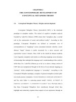
(Na2Al2Si3O10⋅2H2O). The natrolite framework is formed by the linkages
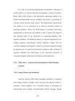
of T5O10 units among the ordered Si(Al)-tetrahedra (Fig. 1). These
linkages form elliptical channels along the c-axis. The geometrical shape
of the channel window on the ab-plane is determined by the rotation
angle (Ψ) of the T5O10 unit; the higher the angle, the more elliptical is
the shape of the channel window. The monovalent EFCs, Na+, and water
molecules are located in the middle (along the major axis) and wall
(along the minor axis) of the two-dimensional plane of the channel
(Fig. 1a). In addition, the EFCs and water molecules have six- and fourcoordinated bonding with the framework oxygens and each other,
respectively. In contrast, the divalent EFCs and water molecules of
scolecite are located at the center (along the major axis) and wall (along
the major or minor axis) of the two-dimensional plane of the channel,
respectively (Fig. 1b). The EFCs and water molecules have seven-, three-
, and five-coordinated bonding with the framework oxygens and each
other, respectively. The distribution of EFC and water molecules are
ordered in both the cases.
The analogs of natrolite, mesolite (Ca2Na2(Al2Si3O10)3⋅8H2O) and
scolecite (CaAl2Si3O10⋅3H2O), are also not solid solutions similar to
natrolite, owing to their low CEC [18–20]. In earlier work, however, we
have reported that natrolite became to be fully exchangeable for alkali,
alkaline earth, and heavy metal cations since new route was found by
using disordered phase of K-exchanged natrolite [21]. Natrolite has been
shown to excel in exchange and capture of harmful cation by tempera
ture treatment. For example, Cs-exchanged natrolite was fully dehy
drated upon heating at 100 ◦ C, and this phase was remained to be
anhydrous and non-exchangeable after quenching and even exposing to
aqueous condition.
In this study, we investigate the structural changes in Sr-, Cd- and Pbexchanged natrolites (NAT) and their thermal behaviors during
dehydration.
2. Experimental method
2.1. Sample preparation
In our previous reports, fully potassium-exchanged natrolite (K-NAT,
K16Al16Si24O80⋅14H2O) has shown enhanced cation exchange capacity
whereas natural natrolite (ideally Na16Al16Si24O80⋅16H2O) has limited
exchange rate for divalent and heavy metal cations [21,22]. In this
study, we chose K-NAT as starting material. The K-NAT was prepared
using a fully saturated KNO3 (ACS reagent grade from Sigma-Aldrich)
solution and a ground mineral natrolite (San Juan, Argentina from
OBG International) in a 100:1 wt ratio. The mixture was stirred at 80 ◦ C
by minimizing the loss of water content in a closed system. After 24 h,
the solid was separated from the solution by vacuum filtration. The dried
powder was used for the second and third exchange cycles in the same
conditions. The final product was vacuum-filtrated and air-dried. From
the Energy-dispersive X-ray spectroscopy analysis (EDS, JEOL Ltd.), we
confirmed K+ was fully exchanged. Further cation-exchange of Sr2+,
Cd2+ and Pb2+ was proceeded same solution-exchange method with
K-NAT preparation. Stirring the mixture of the powdered K-NAT and
fully saturated M(NO3)2, (M = Sr2+, Cd2+ and Pb2+), solutions in a
Fig. 1. Polyhedral representations of (a) Natrolite and (b) Scolecite viewed along [100] or [001] direction. Filled balls represent the extra-framework cations
(yellow: Na2+ and blue: Ca2+) and water molecule oxygens (red), respectively. Striped blue (sky) tetrahedra illustrate an ordered distribution of Si (Al) atoms in the
framework. Channel opening geometry is defined by chain rotation angle (Ψ, degree). Major (minor) axis is defined by long (short) distance of two framework
oxygens in 2-dimensional plane. Coordinate numbers (C.N.) of extra-framework cations and water molecules are presented. (For interpretation of the references to
colour in this figure legend, the reader is referred to the Web version of this article.)
2
J. Im et al.
Microporous and Mesoporous Materials 346 (2022) 112277
1:100 wt ratio in a closed system at 80 ◦ C for 24 h. Separating the solid
from the solution by vacuum filtration, and then repeating the above
two steps two more times. We confirmed final products (Sr-NAT,
Cd-NAT and Pb-NAT) were also fully exchanged using the Scanning
Electron Microscopy (SEM-EDS, JSM-6701F). A Field emission SEM
operating at 15 keV was used at Korea Institute of Ocean Science &
Technology, Busan, Korea. The prepared samples were totally air-dried
for a day prior to carbon coating. To determine the amount of H2O
molecules in framework, Thermalgravimetric analysis (TG) was per
formed at Pusan National University, Busan, Korea. A heating range is
from 25 to 400 ◦ C and a heating rate of 10 ◦ C/min under a nitrogen
atmosphere. Chemical analysis results were summarized in Table 1.
function proposed by Thompson et al. was used to model the observed
Bragg peaks [26], and a March-Dollase function [27] was used to ac
count preferred orientation. The structural models of the Sr-, Cd- and
Pb-NAT at room temperature and their high-temperature forms were
then established by Rietveld methods [25,28,29]. To reduce the number
of parameters, isotropic displacement factors were refined by grouping
the framework tetrahedral atoms, the framework oxygen atoms, and the
non-framework cations, respectively. Geometrical soft-restraints on the
T-O (T = Si, Al) and O–O bond distances of the tetrahedra were applied:
the distances between Si–O and Al–O were restrained to target values of
1.620 ± 0.001 Å and 1.750 ± 0.001 Å, respectively, and the O–O dis
tances to 2.646 ± 0.005 Å for the Si-tetrahedra and 2.858 ± 0.005 Å for
the Al-tetrahedra. The amounts of water molecules were calculated
using the result of Rietveld refinement with OW1, OW2, and OW3
multiplicities and occupancies. Difference Fourier syntheses have
confirmed that the channels in the dehydrated materials are free from
any meaningful residual electron densities from water molecules. In the
final stages of the refinements, all background and profile parameters,
scale factor, lattice constants, 2θ zero, preferred orientation function,
and the atomic positional and thermal displacement parameters were
simultaneously refined whereas the weight of the soft-restrain was
remained. The final refined parameters are summarized in supporting
Tables 1 and 2, and selected bond distances and angles are listed in
supporting Tables 3 and 4
2.2. In-situ dehydration experiment
In-situ high-temperature synchrotron X-ray powder diffraction ex
periments were performed at the X14A beamline at the National Syn
chrotron Light Source (NSLS) at Brookhaven National Laboratory (BNL).
The primary white beam from the bending magnet was mono
chromatized using a Si (111) crystal, and sets of parallel slits were used
to create a monochromatic X-rays with a wavelength of 0.7297(1) Å.
Powdered natrolite samples were packed into 1.0 mm quartz capillaries,
which were connected into a vacuum for the ease of dehydration. K-type
thermocouple (Omega Engineering, Inc.) was inserted into capillary to
measure temperature. The capillaries were then wrapped with a heating
coil [23]. Temperature was increased from RT to ca. 350 ◦ C by 50 ◦ C
increments. For temperature calibration, NaCl powder was loaded to
capillary and heated like abovementioned condition. Using X-ray
diffraction, unit cell volume of NaCl powder was measured by every
50 ◦ C increment up to ca. 350 ◦ C. Real temperature inside capillary is
then calibrated by matching between calculated and our measured unit
cell volume [24]. A Si-strip detector prototype consisting of a monolithic
array of 640 silicon diodes coupled to a set of BNL’s HERMES
application-specific integrated circuits (D.P. Siddons, Private commu
nications) was used to collect high-resolution powder diffraction data
(Δd/d ~ 10− 3). The Si-strip detector covered 3.2◦ in 2θ and was stepped
in 2◦ intervals over the angular range of 3–30◦ with counting times of
10s per step. The wavelength of the incident beam was determined from
a LaB6 standard (SRM 660a).
3. Result and discussion
The synchrotron powder X-ray diffraction (PXRD) patterns of Cdand Pb-NAT are indexed in the orthorhombic Fdd2 space group, whereas
Sr-NAT is in the monoclinic Cc space group at ambient conditions
(Fig. 2). The chemical structures of Sr-, Cd-, and Pb-NAT were derived
from Rietveld refinements at room temperature (RT), as shown in sup
porting Tables 1 and 2, and Supporting Figs. 3, 4, and 5. Our models of
Sr-, Cd-, and Pb-NAT are consistent with those reported in a previous
study [22]. In situ high-temperature synchrotron PXRD patterns of Sr-,
Cd-, and Pb-NAT recorded up to 350 ◦ C are shown in Fig. 2a, b, and 2c,
respectively. The reflections match with those of the RT models. During
the Thermogravimetric analysis (TGA) of Sr-NAT (Supporting Fig. 1),
gradual dehydration was observed up to ~210 ◦ C. In contrast, two
stages of dehydration were observed for Cd-NAT at 90 ◦ C and 280 ◦ C and
for Pb-NAT, at 100 ◦ C and 180 ◦ C. In the XRD patterns of Sr-NAT, there is
hardly any noticeable peak shifting when the temperature increased up
to 200 ◦ C, but from 200 ◦ C or more, most peaks in the XRD patterns
tended to shift toward high 2-theta. When the temperature reached
300 ◦ C, most of the peaks broadened. This indicates that the structure of
Sr-NAT started to collapse at 300 ◦ C and completely amorphized when
2.3. Structural analysis by rietveld refinement
Temperature-dependent changes in the unit-cell lengths and volume
were derived from a series of whole profile fitting procedures using the
GSAS suite of programs [25]. The background was fitted with a Che
byshev polynomial with ≤20 coefficients, and the pseudo-Voigt profile
Table 1
Chemical composition of the fully exchanged K-, Sr-, Cd-, and Pb-natrolites.a
Elements
K-NAT
Sr-NAT
Cd-NAT
Pb-NAT
a
b
c
d
K
Na
Al
Sr
K
Al
Cd
K
Al
Pb
K
Al
Atomic percent (%)b
Chemical Compositiond
1
2
3
4
5
%H2Oc
11.99
0.00
11.22
5.93
0.00
12.41
6.39
0.00
12.07
6.63
0.00
12.74
10.39
0.00
10.88
5.62
0.00
12.49
6.19
0.00
12.9
6.11
0.00
11.47
10.87
0.00
10.89
6.36
0.00
12.95
6.38
0.00
12.42
6.58
0.00
12.54
10.91
0.00
10.87
5.96
0.00
11.96
6.3
0.08
11.99
6.28
0.00
12.85
11.26
0.00
10.68
5.76
0.00
11.37
6.52
0.14
12.72
6.24
0.01
12.24
10.53
K16.3Al16Si24O80•13.6H2O
12.32
Sr7.6Al16Si24O80•23.2H2O
10.56
Cd8.2K0.06Al16Si24O80•20.3H2O
8.75
Pb8.2K0.03Al16Si24O80•15.8H2O
Values are normalized based on 16 aluminum atoms per unit cell.
Results from Energy dispersive X-ray Spectroscopy (EDS).
The water contents in wt%. Weight loss by Thermogravimetric analysis (TG) up to ca. 400 ◦ C.
Confirmed from EDS and TG analysis. Water contents are calculated from weight loss.
3
J. Im et al.
Microporous and Mesoporous Materials 346 (2022) 112277
Fig. 2. in-situ synchrotron X-ray powder diffraction patterns of (a) Sr-NAT, (b) Cd-NAT, and (c) Pb-NAT as a function of temperature. Selected (hkl) indices of
ambient phases are shown. Asterisk marks indicate peaks of the impurity.
the temperature reached 350 ◦ C (Fig. 2a). Fig. 2b shows the PXRD
patterns of Cd-NAT with increasing temperatures. The impurity of
Cd-NAT at RT was also confirmed as a by-product according to previous
work [22]. The patterns exhibit changes at 150 ◦ C and 300 ◦ C, which are
consistent with the TGA results that revealed two stages of weight loss
during dehydration. It is clear that most of the hkl peaks of Cd-NAT shift
abruptly toward higher 2-theta values at 150◦ , suggesting a strong lat
tice contraction (guide arrows in Fig. 1b). These peaks subsequently
shifted to lower 2θ values up to 350 ◦ C. Therefore, dehydration may
have been more dominant than thermal expansion at 150 ◦ C, and
thermal expansion may have been more dominant over dehydration up
to 350 ◦ C. Cd-NAT maintained its crystallinity better than Sr-NAT even
at 350 ◦ C, and the dehydrated phase was recovered after cooling down
to room temperature. For Pb-NAT, amorphization progressed rapidly at
150 ◦ C, indicating that it has a relatively poor thermal stability
compared to those of Sr- and Cd-NAT, and an amorphous phase was
observed without significant changes even if the temperature increased
to 350 ◦ C (Fig. 2c). In the case of Pb-NAT, all the diffracted reflections
obtained between 150 and 300 ◦ C or the various transformed phases
bearing different water molecule contents under various pressures and
temperatures could not be attributed to natrolite [30,31].
Next, temperature-dependent changes in the unit-cell parameters
and volumes of Sr-, Cd- and Pb-NAT were analyzed using the wholeprofile-fitting method (Supporting Figs. 2 and 3a). The calculated
thermal expansion coefficients of Sr-, Cd-, and Pb-NAT (Fig. 3a) is as low
as − 3(1) × 10− 6, − 1.0(7) × 10− 6, and 1(2) × 10− 6 K− 1 up to 200, 100,
Fig. 3. Temperature-induced changes of (a) unit-cell volume (Å3) and (b) the orthorhombicity, 2(b–a)/(b + a), of Sr-, Cd-, and Pb-NAT after converting space group
to Fd. Open symbol represents the recovered phase and thermal expansion coefficients are given in Fig. 3a with unit of ◦ C− 1. Estimated standard deviations are
smaller than the size of each symbol.
4
J. Im et al.
Microporous and Mesoporous Materials 346 (2022) 112277
and 100 ◦ C, respectively. Beyond the abovementioned temperature, the
coefficients of Sr-NAT and Cd-NAT were determined as − 2.7(7) × 10− 4
K− 1 up to 300 ◦ C with a 2.9% volume contraction and − 5.3 × 10− 4 K− 1
up to 150 ◦ C with a 2.7% volume contraction, respectively. For Sr- and
Cd-NAT, apparent negative volume expansion refers that dehydration is
dominant rather than thermal expansion. The volume of Cd-NAT
expanded up to 350 ◦ C with a coefficient of 4(1) × 10− 5 K− 1. Notably,
Cd-NAT had the lowest coefficient among all the samples. The thermal
expansion coefficients are listed in Supporting Table 2 and shown in
Supporting Fig. 2.
Fig. 3b shows temperature-dependent changes in the ortho
rhombicity of Sr-, Cd-, and Pb-NAT. The orthorhombicity of Cd-NAT
increases dramatically from 0.023(1) to 0.047(1) in the range
100–150 ◦ C, increasing by 104%. These drastic structural changes
originate from the temperature-dependent a-axis changes (Supporting
Fig. 2). The a-axis length of Cd-NAT decreased by 2.5% at 150 ◦ C,
whereas those of the b- and c-axes decreased only slightly. Compared to
that of Cd-NAT, the values of orthorhombicity of Sr- and Pb-NAT are
relatively constant in the range 0.017(1)–0.026(1) because a- and b-axis
lengths of Sr- and Pb-NAT decreased by a similar ratio with increasing
temperature (Supporting Fig. 2a and b).
To understand the volume changes owing to the loss of water mol
ecules during dehydration, the water contents at selected temperatures
are calculated using the Rietveld refinement (Fig. 4, supporting Tables 3
and 4). Fig. 4 shows the Rietveld refinement, structure models, chemical
compositions, stoichiometric water contents, water migration, T5O10
chain rotation angles, coordination numbers of water molecules, and
cation locations in the channels of Sr-, Cd-, and Pb-NAT at selected
temperatures. All structural models are oriented along the ab-plane to
show the changes in the extra-framework species in the elliptical
channels.
At room temperature, one cation site and three water molecule sites
in the natrolite channel of Sr-NAT are fully occupied (Fig. 4a, supporting
Table 1). The array of extra-framework species in the channel of Sr-NAT
is similar to that in scolecite (Ca2+ variant of natrolite) (Fig. 1b). The
Sr2+ cation (Sr1 site) is located in the middle of the channel, and two
water molecules (OW1 and OW2 sites) are located along the minor axis
(short axis) wall, whereas OW3 is located close to the major axis (long
axis) wall in the channel. Using the Rietveld refinement, the chemical
formula of Sr-NAT was calculated to be Sr8Al16Si24O80⋅24H2O, which
indicates that 24 water molecules are present per unit cell. The number
of water molecules is consistent with the results of the chemical analysis
(Table 1). At room temperature, the central EFC (Sr2+) in the natrolite
channel exhibits a seven-coordinated bonding with four framework
oxygens and three water molecules. The OW1 and OW2 sites have a fivecoordinated bonding with the surrounding three framework oxygens,
one EFC, and each other. The OW3 site has a three-coordinated bonding
with two framework oxygens and one EFC. Owing to the smaller number
of bonds for OW3 than those for the other water molecules, it was easily
dehydrated at 150 ◦ C, which decreased the occupancy to 66%, i.e., four
water molecules were removed (Fig. 4b).
At 300 ◦ C, the OW3 site no longer existed, and the occupancy of the
OW1 and OW2 sites also decreased to ~83%, i.e., to 13.7 water mole
cules per unit cell (Fig. 4c). In the refined structural model, the residual
water molecules remain at 300 ◦ C while TG result shows the Sr-NAT is
almost dehydrated (supporting Figs. 1 and 3d). The residual sites are
observed by Fourier density calculation in the Rietveld refinement. They
are defined as residual sites of water molecules, OW1 and OW2, due to
mismatch with framework atoms and extra-framework cations. We
expect that tight bonds of H2O-cation at 300 ◦ C interrupts to be dehy
drated. For example, H2O-cation bond length at RT, 150, and 300 ◦ C
ranges 2.67(2)–2.86(2), 2.56(3)–2.95(4), and 2.42(9)–2.53(7), respec
tively. The ordered distribution of Sr2+ up to 150 ◦ C became disordered
at two sites at 300 ◦ C, and its coordination number reduced to six similar
to that of Na+ in natural natrolite (Fig. 1a). A half occupancy becomes a
void in the EFC, resulting in less bonding with the framework or EFCwater cluster to sustain the flexible channel. These environmental
changes at 300 ◦ C lead to an unstable coordination environment in SrNAT, making it difficult to maintain the NAT type structure, compared
to that of the Sr-NAT model at room temperature (Fig. 4c).
The chemical formula of Cd-NAT at room temperature was calcu
lated to be Cd8Al16Si24O80⋅16H2O, indicating that 16 water molecules
are present per unit cell. The Cd atoms reported are two equivalent
positions in the center of the channel and has a seven-coordinated
Fig. 4. Polyhedral representations of (a) starting
material, K-NAT, (b) Sr-NAT-RT, (c) Sr-NAT-150C, (d)
Sr-NAT-300C, (e) Cd-NAT-RT, (f) Cd-NAT-150C, (g)
Cd-NAT-350C, (h) Pb-NAT-RT, and (i) Pb-NAT-100C,
viewed along [100] or [001] direction. Filled balls
represent the extra-framework cations (violet: K+,
green: Sr2+, pink: Cd2+ and grey: Pb2+) and water
molecule oxygens (red), respectively. Striped blue
(sky) tetrahedra illustrate an ordered distribution of
Si (Al) atoms in the framework. (For interpretation of
the references to colour in this figure legend, the
reader is referred to the Web version of this article.)
5
J. Im et al.
Microporous and Mesoporous Materials 346 (2022) 112277
bonding with four framework oxygens and three water molecules
(Fig. 4d). The water molecule sites, OW1 and OW2, are located along the
minor axis wall and the major axis wall, respectively; OW1 has a fivecoordinated bonding with three framework oxygens and two EFCs and
OW2 has a five-coordinated bonding with four framework oxygens and
one EFC. All extra-framework species showed disordered distribution
with an occupancy of 50%, and these atomic positions are similar to
those in natrolite (Fig. 1a). When the temperature was increased to
150 ◦ C, OW2 on the major axis was partially dehydrated, migrated along
the a-axis and subsequently merged with OW1. Owing to the environ
mental changes for OW2 as a result of dehydration, the occupancy of
OW1 increased from 50 to 79%, and Cd-NAT lost 3.4 water molecules
per unit cell at 150 ◦ C (Fig. 4e). At 350 ◦ C, the occupancy of OW1
decreased from 79 to 32% owing to the loss of five water molecules per
unit cell, whereas the Cd2+ and water molecule positions are nearly the
same as those in the model at 150 ◦ C (Fig. 4f).
Fig. 4g and h shows structures of Pb-NAT at room temperature and
100 ◦ C, respectively. The calculated chemical formula of Pb-NAT was
Pb8Al16Si24O80⋅16H2O, indicating that 16 water molecules are present
per unit cell at room temperature. Unlike the distribution of the cation
sites in Cd-NAT, two Pb2+ sites on the major axis, Pb1 and Pb2, have the
highest disorder and lowest occupancy (less than 35%) among our
models. The Pb1 site has a five-coordinated bonding with four frame
work oxygens and one water molecule, whereas Pb2 has a threecoordinated bonding with two framework oxygens and one water
molecule. The water molecule site, OW1, is located along the minor axis
wall and has a fully occupied distribution. Although two cation sites
moved at 100 ◦ C, occupancies and coordination of cations are main
tained. After the dehydration, the occupancy decreased to 58%,
comprising 9.3 water molecules per unit cell. In Pb-NAT, despite the
presence of numerous coordination bonds among the framework oxy
gens, cations, and water molecules, a higher number of water molecules
(6.7 molecules) were dehydrated and even amorphized at the low
temperature of 100 ◦ C. This can be attributed to the highly disordered
distribution of Pb2+ cation, which results in void spaces and loss of
bonding with increasing temperature. We found that dehydration
behavior.
Fig. 5 shows the changes in the chain rotation angles and remaining
number of water molecules in a unit cell with increasing temperature.
These two values usually have an inverse relationship because the T5O10
unit can be rotated to open the elliptical channels via the dehydration of
water molecules. Sr- and Cd-NAT lose water molecules according to the
slope of the equations, y = − 0.04(1)x + 25.2(6) and y = − 0.04(1)x + 17
(1), respectively. However, the rate of dehydration in Pb-NAT was more
than two times that of Sr- and Cd-NAT (y = − 0.09x + 18.2). The large
amount of water loss in Pb-NAT at 100 ◦ C is consistent with the increase
in the chain rotation angle with the highest slope of 0.04 among all our
models. Moreover, the distance between the EFC and framework oxy
gens in Pb-NAT increased from 2.42(2) to 2.87(1) Å at room tempera
ture and from 2.10(1) to 2.99(1)Å at 100 ◦ C (supporting Tables 2 and 4).
Hence, the highly disordered distribution of Pb2+ in Pb-NAT enables it to
collapse easily after partial dehydration. For Sr-NAT, the interatomic
distance between the Sr2+ site and framework oxygens increase from
2.67(1) to 2.86(1) Å at room temperature and from 2.46(3) to 2.76(3) Å
at 300 ◦ C (supporting Tables 2 and 4). Up to 150 ◦ C, the chain rotation
angle increased according to the lowest slope of 0.01 among all our
models, which indicates that the channel of Sr-NAT is well sustained by
extra-framework species during dehydration. However, the slope of the
rotation angle increased at 300 ◦ C owing to the disordered distribution
of Sr2+. At 350 ◦ C, distributional change of EFC can collapse the position
of the water molecules and the entire framework. For Cd-NAT, the
rotation angle increased with a slope of 0.03 up to 150 ◦ C, which is quite
large compared to the slopes of Pb-NAT and Sr-NAT. However, the po
sition and coordinated bonding of Cd2+ were maintained even after big
changes such as dehydration and migration of water molecules. This
reliable environment prevented the collapse of the channel above
150 ◦ C, and the rotation angle was similar to that at 300 ◦ C.
The differences of phase transition during dehydration between SrNAT and Cd-, Pb-NAT seem to be induced by their structural differ
ences at ambient condition. The Sr-NAT, monoclinic Cc symmetry,
shows one cation site with three water molecule sites in its channel with
seven hydrogen bonding with water molecules and framework oxygen
atoms. However, Cd-NAT shows orthorhombic symmetry, Fdd2, with
two cation sites and one or two water molecule sites in their channel,
Fig. 5. Changes in the chain rotation angle (ψ, degrees) and number of H2O contents per 80 framework oxygens.
6
J. Im et al.
Microporous and Mesoporous Materials 346 (2022) 112277
concomitant with seven hydrogen bonding between water molecules
and framework oxygen atoms. In case of the Pb-NAT, cation makes just
three and five bonds with other oxygen atoms. The changes of cation site
such as migration or splitting between room and high temperature make
structure more unstable. For example, distribution of cation site in the
Sr- and Cd-NAT at 300 and 350 ◦ C, respectively, is very similar. How
ever, the Sr-NAT is structurally amorphized subsequent temperature
while the Cd-NAT is still stable. In case of the Pb-NAT, distribution of
two cation sites, Pb1 and Pb2, are highly disordered and low occupied in
channel at room temperature. Also, these two sites migrate along a- and
b-axis at 100 ◦ C. Therefore, structure of the Pb-NAT easily amorphized at
comparatively lower temperature than that of the Sr-NAT.
Appendix A. Supplementary data
Supplementary data to this article can be found online at https://doi.
org/10.1016/j.micromeso.2022.112277.
References
[1] T.A. Todd, T.A. Todd, J.D. Law, R.S. Herbst, Cesium and Strontium Separation
Technologies Literature Review, United States, 2004.
[2] M. Balsamo, F. Di Natale, A. Erto, A. Lancia, F. Montagnaro, L. Santoro, Cadmium
adsorption by coal combustion ashes-based sorbents—relationship between
sorbent properties and adsorption capacity, J. Hazard Mater. 187 (2011) 371–378.
[3] B. Bayat, Comparative study of adsorption properties of Turkish fly ashes: I. The
case of nickel(II), copper(II) and zinc(II), J. Hazard Mater. 95 (2002) 251–273.
[4] K.S. Hui, C.Y.H. Chao, S.C. Kot, Removal of mixed heavy metal ions in wastewater
by zeolite 4A and residual products from recycled coal fly ash, J. Hazard Mater.
127 (2005) 89–101.
[5] L.-N. Shi, Y. Zhou, Z. Chen, M. Megharaj, R. Naidu, Simultaneous adsorption and
degradation of Zn2+ and Cu2+ from wastewaters using nanoscale zero-valent iron
impregnated with clays, Environ. Sci. Pollut. Control Ser. 20 (2013) 3639–3648.
[6] J. Devgun, Chemical separations in nuclear waste management-the state of the art
and a look to the future, Environ. Prog. 22 (2003).
[7] F. Fu, Q. Wang, Removal of heavy metal ions from wastewaters: a review,
J. Environ. Manag. 92 (2011) 407–418.
[8] W.S. Wan Ngah, M.A.K.M. Hanafiah, Removal of heavy metal ions from
wastewater by chemically modified plant wastes as adsorbents: a review,
Bioresour. Technol. 99 (2008) 3935–3948.
[9] S. Mohan, R. Gandhimathi, Removal of heavy metal ions from municipal solid
waste leachate using coal fly ash as an adsorbent, J. Hazard Mater. 169 (2009)
351–359.
[10] S. Yadav, V. Srivastava, S. Banerjee, F. Gode, Y.C. Sharma, Studies on the removal
of nickel from aqueous solutions using modified riverbed sand, Environ. Sci. Pollut.
Control Ser. 20 (2013) 558–567.
[11] M.K. Doula, Removal of Mn2+ ions from drinking water by using Clinoptilolite and
a Clinoptilolite–Fe oxide system, Water Res. 40 (2006) 3167–3176.
[12] S.E. Bailey, T.J. Olin, R.M. Bricka, D.D. Adrian, A review of potentially low-cost
sorbents for heavy metals, Water Res. 33 (1999) 2469–2479.
[13] S. Babel, T.A. Kurniawan, Low-cost adsorbents for heavy metals uptake from
contaminated water: a review, J. Hazard Mater. 97 (2003) 219–243.
[14] D.W. Breck, Zeolite Molecular Sieves: Structure, Chemistry, and Use, John Wiley
and Sons, New York, 1974.
[15] K.D. Mondale, R.M. Carland, F.F. Aplan, The comparative ion exchange capacities
of natural sedimentary and synthetic zeolites, Miner. Eng. 8 (1995) 535–548.
[16] P. Castaldi, G. Garau, P. Melis, Influence of compost from sea weeds on heavy
metal dynamics in the soil-plant system, Fresenius Environ. Bull. 13 (2004)
1322–1328.
[17] E. Olegario, C.M. Pelicano, J.C. Felizco, H. Mendoza, Thermal stability and heavy
metal (As5+, Cu2+, Ni2+, Pb2+ and Zn2+) ions uptake of the natural zeolites from
the Philippines, Mater. Res. Express 6 (2019), 085204.
[18] A. Dyer, H. Faghihian, Diffusion in heteroionic zeolites: part 1: diffusion of water in
heteroionic natrolites, Microporous Mesoporous Mater. 21 (1998) 27–38.
[19] G. Artioli, J.V. Smith, J.J. Pluth, X-ray structure refinement of mesolite, Acta
Crystallogr. C 42 (1986) 937–942.
[20] Å. Kvick, K. Ståhl, A neutron diffraction study of the bonding of zeolitic water in
scolecite at 20 Κ, Z. für Kristallogr. - Cryst. Mater. 171 (1985) 141–154.
[21] Y. Lee, D. Seoung, Natrolite may not be a "soda-stone" anymore: structural study of
fully K-, Rb-, and Cs-exchanged natrolite, Am. Mineral. 95 (2010) 1636–1641.
[22] Y. Lee, D. Seoung, Y. Lee, Natrolite is not a "soda-stone" anymore: structural study
of alkali (Li+), alkaline-earth (Ca2+, Sr2+, Ba2+) and heavy metal (Cd2+, Pb2+, Ag+)
cation-exchanged natrolites, Am. Mineral. 96 (2011) 1718–1724.
[23] K. Stahl, J. Hanson, Real-time X-ray synchrotron powder diffraction studies of the
dehydration processes in scolecite and mesolite, J. Appl. Crystallogr. 27 (1994)
543–550.
[24] Z.-H. Fang, Temperature dependence of volume thermal expansion for NaCl and
KCl crystals, Phys. B Condens. Matter 357 (2005) 433–438.
[25] B.H. Toby, EXPGUI, a graphical user interface for GSAS, J. Appl. Crystallogr. 34
(2001) 210–213.
[26] P. Thompson, D.E. Cox, J.B. Hastings, Rietveld refinement of Debye-Scherrer
synchrotron X-ray data from Al2O3, J. Appl. Crystallogr. 20 (1987) 79–83.
[27] W.A. Dollase, Correction of intensities for preferred orientation in powder
diffractometry: application of the March model, J. Appl. Crystallogr. 19 (1986)
267–272.
[28] A.C. Larson, R.B. VonDreele, GSAS; General Structure Analysis System Report
LAUR, 1986, pp. 86–748.
[29] H. Rietveld, A profile refinement method for nuclear and magnetic structures,
J. Appl. Crystallogr. 2 (1969) 65–71.
[30] J. Im, Y. Lee, D.A. Blom, T. Vogt, Y. Lee, High-pressure and high-temperature
transformation of Pb(ii)-natrolite to Pb(ii)-lawsonite, Dalton Trans. 45 (2016)
1622–1630.
[31] D. Seoung, Y. Lee, C.-C. Kao, T. Vogt, Y. Lee, Two-step pressure-induced
superhydration in small pore natrolite with divalent extra-framework cations,
Chem. Mater. 27 (2015) 3874–3880.
4. Conclusion
In this study, we demonstrated the structural behaviors of Sr-, Cd-,
and Pb-NAT during the dehydration process. At room temperature, the
structure of Sr-NAT is similar to that of scolecite, whereas those of Cdand Pb-NAT are similar to that of natrolite. For Sr-, Cd-, and Pb-NAT,
very low thermal expansion coefficients were observed before 200,
100, and 100 ◦ C, respectively. The negative thermal expansion values
for Sr- and Cr-NAT indicated a contraction of volume during dehydra
tion. The structure of Sr- and Cd-NAT collapsed beyond 300 and 100 ◦ C,
respectively, whereas that of Cd-NAT was stable up to 350 ◦ C. The chain
rotation angle and structural stability with increasing temperature
depend on the amount of water loss and distributional changes of the
EFCs in the framework, respectively. Our findings further our under
standing of the structural changes and mechanism of thermal stability in
natrolites containing hazardous elements.
CRediT authorship contribution statement
Junhyuck Im: Writing – review & editing, Writing – original draft,
Visualization. Jaewoo Jung: Writing – original draft, Software, Inves
tigation, Funding acquisition. Kiho Yang: Writing – original draft,
Methodology, Investigation, Funding acquisition. Donghoon Seoung:
Writing – review & editing, Writing – original draft, Visualization,
Methodology, Investigation, Funding acquisition. Yongmoon Lee:
Writing – review & editing, Writing – original draft, Software, Meth
odology, Investigation, Funding acquisition, Formal analysis, Data
curation, Conceptualization.
Declaration of competing interest
The authors declare that they have no known competing financial
interests or personal relationships that could have appeared to influence
the work reported in this paper.
Data availability
No data was used for the research described in the article.
Acknowledgement
This work was supported by the National Research Foundation (NRF2022R1F1A1074593, NRF-2020R1C1C1013642, NRF-2019K1A3A7A
09101574, NRF-202006710003) of the Ministry of Science and ICT of
Korean Government, and Chonnam National University Research Grant,
2017, the Korea Atomic Energy Research Institute (KAERI) [Grant No.
521240-22, South Korea]. This research was a part of the project titled
‘Selection of prospective mining area for Co-rich ferromanganese crust
in western Pacific seamounts: 3-D resource estimation and environ
mental impact evaluation’, funded by the Korean Ministry of Oceans and
Fisheries, Korea (No. 20220509). Experiments using synchrotron radi
ation were supported by X14A beamline at National Synchrotron Light
Source (NSLS) at Brookhaven National Laboratory (BNL).
7