Báo cáo khoa học: The Cockayne syndrome group B protein is a functional dimer docx
Bạn đang xem bản rút gọn của tài liệu. Xem và tải ngay bản đầy đủ của tài liệu tại đây (215.66 KB, 9 trang )
The Cockayne syndrome group B protein is a functional
dimer
Mette Christiansen
1
, Tina Thorslund
1
, Bjarne Jochimsen
2
, Vilhelm A. Bohr
3
and Tinna Stevnsner
1
1 Danish Centre for Molecular Gerontology, Department of Molecular Biology, University of Aarhus, Denmark
2 Department of Molecular Biology, University of Aarhus, Denmark
3 Laboratory of Molecular Gerontology, National Institute on Aging, National Institutes of Health, Baltimore, MD, USA
Cockayne syndrome (CS) is a segmental premature
aging syndrome with complex symptoms, including
developmental abnormalities, neurological dysfunction,
and short average lifespan. Cellular characteristics
include hypersensitivity to UV light, and failure of
RNA synthesis to recover to normal rates following
UV irradiation. Two genes have been shown to be
involved: CSA and CSB [1]. The CSB gene encodes a
protein with a predicted molecular mass of 168 kDa.
The CS group B (CSB) protein contains an acidic
domain, a glycine-rich region, and two putative nuc-
lear localization signal (NLS) sequences [2]. In addi-
tion, CSB is a member of the SWI2 ⁄ SNF2-family of
DNA-dependent ATPases that contain seven charac-
teristic motifs which are also present in DNA and
RNA helicases [3]. Helicase activity has not been dem-
onstrated for any members of the SWI2 ⁄ SNF2-family,
which is part of Superfamily 2 (SF2), but in general
they have the ability to destabilize protein–DNA inter-
actions [4]. The CSB protein displays DNA-dependent
ATPase activity and CSB is able to remodel chromatin
in vitro [5–8].
Recently, the structure of the central ATPase
domain of zebrafish Rad54 revealed that the conserved
core of this SWI2 ⁄ SNF2 protein is similar to SF2 heli-
cases [9]. This indicates that SWI2 ⁄ SNF2 proteins
translocate on DNA with a mechanism similar to heli-
cases. The integrity of the SWI2⁄ SNF2 ATPase
domain is critical for most functions of CSB in vitro
and in vivo. Mutations in motif Ia, II, V, and VI either
Keywords
Cockayne syndrome group B protein;
DNA-dependent ATPase; homodimer;
SWI2 ⁄ SNF2; transcription coupled repair
Correspondence
T. Stevnsner, Danish Centre for Molecular
Gerontology, Department of Molecular
Biology, University of Aarhus, Build. 130,
DK-8000 Aarhus C, Denmark
Tel: +45 89422657
Fax: +45 89422650
E-mail:
(Received 13 May 2005, revised 1 July
2005, accepted 4 July 2005)
doi:10.1111/j.1742-4658.2005.04844.x
Cockayne syndrome (CS) is a rare inherited human genetic disorder char-
acterized by developmental abnormalities, UV sensitivity, and premature
aging. The CS group B (CSB) protein belongs to the SNF2-family of
DNA-dependent ATPases and is implicated in transcription elongation,
transcription coupled repair, and base excision repair. It is a DNA stimula-
ted ATPase and remodels chromatin in vitro. We demonstrate for the first
time that full-length CSB positively cooperates in ATP hydrolysis as a
function of protein concentration. We have investigated the quaternary
structure of CSB using a combination of protein–protein complex trapping
experiments and gel filtration, and found that CSB forms a dimer in solu-
tion. Chromatography studies revealed that enzymatically active CSB has
an apparent molecular mass of approximately 360 kDa, consistent with
dimerization of CSB. Importantly, in vivo protein cross-linking showed the
presence of the CSB dimer in the nucleus of HeLa cells. We further show
that dimerization occurs through the central ATPase domain of the pro-
tein. These results have implications for the mechanism of action of CSB,
and suggest that other SNF2-family members might also function as
dimers.
Abbreviations
CS, Cockayne syndrome; CSB, CS group B; HA, hemaglutinin antigen; HIS, His
6
; SF1, superfamily 1; NLS, nuclear localization signal;
NTB, nucleotide binding fold; SF2, superfamily 2.
4306 FEBS Journal 272 (2005) 4306–4314 ª 2005 FEBS
abolish or drastically reduce the ATPase activity of
CSB [7,10]. CSB cDNA with point mutations in motifs
Ia, II, III, V, and VI, as opposed to wt CSB cDNA,
do not complement the deficiencies of the SV40 trans-
formed CS-B cell line, CS1AN.S3.G2 [11–13]. In con-
trast, both a deletion of the entire acidic region of 39
amino acids and a point mutation in a putative nucleo-
tide binding (NTB) motif do not interfere with the
ability of CSB to complement CSB-deficient cells
[12,14,15].
The majority of bacterial and viral DNA helicases
appear to act as oligomers, usually dimers or hexamers
[16]. Consequently, it is tempting to speculate that
members of the SWI2 ⁄ SNF2 of DNA-dependent ATP-
ases might also function as multimers. Recent results
indicate that the Swi2p ATPase subunit is present in a
single copy in the yeast SWI ⁄ SNF chromatin remodel-
ing complex [17]. In contrast, yeast Rad54, which is
involved in recombination, seems to be a monomer in
solution and a dimer ⁄ oligomer on DNA [18]. Insight
into the quaternary structure of CSB will advance the
understanding of the mechanism by which the DNA-
dependent ATPases, in general, and CSB, in particular,
functions. Furthermore, oligomerization status is
important to evaluate the stoichiometry of different
biochemical analyses. The three-dimensional structure
of CSB has not yet been elucidated, and we report here
a characterization of CSB protein structure. We find
that the CSB protein forms a dimer in vitro and in vivo,
and that this homodimerization is essential for ATP
hydrolysis of CSB. Moreover, we demonstrate that the
ATPase domain is involved in the dimerization.
Results
CSB ATP hydrolysis exhibits non-Michaelis-
Menten kinetics
In general, DNA helicases often function as oligomers
[16]. Because CSB belongs to the superfamily 2 of heli-
cases, it is of importance to investigate whether CSB
may also function as an oligomer. Initially, the dose–
response curve for ATP hydrolysis previously reported
[10] was reexamined in more detail using low levels of
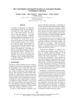
CSB protein. Figure 1A shows that product formation
is not linear with increasing concentrations of CSB
protein, suggesting positive cooperativity in ATP
hydrolysis by CSB. Thus, these results suggest that the
CSB protein, under the experimental conditions used,
functions as a multimer. Furthermore, the Hill coeffi-
cient of 2.1, which is the maximum slope from the Hill
plot (Fig. 1B), clearly indicates positive cooperativity,
suggesting that CSB acts as a dimer.
CSB displays homodimerization in solution
in vitro
To test the dimerization in further detail, we per-
formed cross-linking in solution to trap the CSB
homodimer. This is a sensitive and widely used method
for in vitro analysis of protein–protein interactions
[19,20]. We found that recombinant purified CSB at
low concentration in solution could be cross-linked
with glutaraldehyde. The cross-linked species were
identified with silver stain, and the apparent molecular
mass of 330 kDa was determined from the migration
0.4
y = 2.1x + 7.9
-1.0
-0.5
0.0
0.5
1.0
1.5
-4.5 -4.0 -3.5 -3.0
Lo
g
[ATP]
Log[V/(Vmax-V)]
-0.1
0.0
0.1
0.2
0.3
0.5
0.6
0123456
CSB (nM)
ATP hydrolysis (pmol*100/h)
A
B
Fig. 1. Effect of increasing amounts of CSB on its ATPase activity.
(A) [
32
P]ATP[cP] hydrolysis rate after incubation with 0–6 nM recom-
binant CSB, 50 l
M cold ATP and 150 ng plasmid DNA for 1 h at
30 °C. Error bars represent standard deviations of three independ-
ent experiments. (B) ATP hydrolysis rate was determined for 6 n
M
CSB incubated with increasing amounts of ATP. Graph shows a Hill
plot of a representative experiment.
M. Christiansen et al. CSB protein is a functional dimer
FEBS Journal 272 (2005) 4306–4314 ª 2005 FEBS 4307
of the molecular mass standards. Given a predicted
subunit molecular mass of 168 kDa, this corresponds
well with a homodimer of CSB (Fig. 2A). Further-
more, cross-linking also resulted in aggregation in the
slot. Interestingly, the presence of ATP, ATP[cS], co-
factor DNA, or dephosphorylation of CSB with
protein phosphatase 1 did not have any effect on the
extent of dimerization in solution (Fig. 2B and not
shown).
Gel filtration reveals enzymatic activity of the
CSB dimer
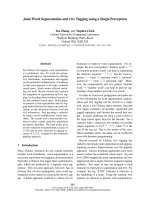
In order to characterize the quaternary structure of the
CSB protein, we carried out gel filtration. The CSB
protein eluted as a peak around fraction 24 from a
Superdex 200 column (Fig. 3) as determined by DNA-
dependent ATPase activity measured in the different
fractions. On the basis of the elution of the molecular
mass markers, this peak corresponds to a molecular
mass of approximately 360 kDa. Given a predicted
subunit molecular mass of 168 kDa, this indicates that
CSB is a dimeric protein. DNA was not present in
these fractions since the ATPase activity was only
detectable after the addition of pUC19 DNA. This
indicates that dimerization is not mediated by DNA.
Importantly, only residual ATPase activity was
observed at the monomer size (fraction 27), while sil-
ver staining of SDS ⁄ PAGE clearly showed elution of
CSB at this position (Fig. 3, compare fractions 25 and
27). This suggests that CSB is only active as an
ATPase when it is a dimer. Also, we did not detect a
peak in DNA-dependent ATPase activity at fractions
earlier than the ferritin marker (450 kDa), suggesting
that CSB does not exist as higher order oligomers in
solution.
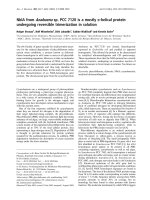
CSB exhibits homodimerization in vivo
Next, we tested whether the dimerization observed in
solution in vitro and its stimulating effects on CSB enzy-
matic activity are biologically relevant. HeLa cells were
exposed to a range of formaldehyde concentrations in
an attempt to covalently cross-link endogenous CSB.
Nuclear extracts were prepared and proteins were ana-
lyzed by western blotting using CSB-specific antibody.
Besides the CSB monomer, only a single CSB complex
was detected in western blot from the nuclear extract
after treatment of cells with 10 mm formaldehyde. This
CSB complex migrated to the position of a CSB dimer
in SDS ⁄ PAGE (Fig. 4A). Furthermore, both bands are
specific to CSB as both the monomeric and the dimeric
bands were absent in extracts from CS1AN.S3.G2 cells
which lack full-length CSB (Fig. 4A). Next, we analyzed
whether the fraction of CSB dimer compared to mono-
mer increased after UV irradiation or transcription inhi-
bition by a-amanitin, but we did not see any effect
(Fig. 4B). It remains to be determined whether other
factors, such as oxidative damage, affect the extent of
CSB dimerization in vivo.
Fig. 2. Stabilization of the CSB dimer by in vitro protein-protein
cross-linking with glutaraldehyde. (A) CSB (60 n
M) was incubated
with 0.001% (v ⁄ v) glutaraldehyde in solution for 0, 10, 20 or
40 min, and CSB was detected by 3–8% (w ⁄ v) Tris ⁄ acetate
SDS ⁄ PAGE and silver stain. (B) CSB was incubated with 0.001%
(v ⁄ v) glutaraldehyde in the presence or absence of 50 l
M ATP or
ATPcS as indicated. CSB was detected by 3–8% (w ⁄ v) Tris ⁄ acetate
SDS ⁄ PAGE and western blot with CSB specific antibody. *CSB
monomer; **CSB dimer. The size (in kDa) of a protein marker is
indicated.
CSB protein is a functional dimer M. Christiansen et al.
4308 FEBS Journal 272 (2005) 4306–4314 ª 2005 FEBS
CSB forms a homodimer through the DNA-
dependent ATPase domain
To map which part of CSB mediates homodimerization,
we carried out interaction studies of recombinant wild-
type CSB [N-terminal hemaglutinin antigen (HA) and
C-terminal His
6
(HIS) tagged] with CSB fragments
(N-terminal S- and HIS- tags and C-terminal HIS- and
HSV tags). Five tagged fragments covering the entire
region of CSB; CSB(2–341), CSB(310–520), CSB(465–
1056), CSB(953–1204), and CSB(1187–1493) were used
(Fig. 5A). The fragments were expressed in Escherichia
coli, purified, and mixed with purified wild-type CSB.
In vitro pull down experiments using S-protein-agarose
were performed and analyzed by western blot and use of
HA and HSV antibodies. The result shown in Fig. 5B
indicates that the protein homodimerizes through inter-
actions with the ATPase domain. The CSB(465–1056)
fragment, which covers the SWI ⁄ SNF-domain, interacts
tightly with the full-length CSB protein (Fig. 5B, lane
3). Approximately 10% of input full-length CSB was
pulled down by the CSB(465–1056) fragment. Import-
antly, purified wild-type CSB did not bind to S-protein-
agarose and there was little or no interaction with the
four other fragments (Fig. 5B). The fragments were all
present in similar amounts in the pull-down experiment
as shown in the lower panel of Fig. 5B.
Discussion
In this report we present evidence that CSB forms a
dimer in vitro and in vivo. Most bacterial and viral DNA
helicases appear to act as oligomers, usually dimers or
hexamers, providing the helicase with multiple DNA
binding sites [16]. Recently, the Bloom’s syndrome heli-
case was also identified as forming an oligomeric ring
structure [21]. This was the first example of oligomer
formation of a helicase of human origin. Multimeriza-
tion has previously been reported for the Saccharomyces
cerevisiae SWI2 ⁄ SNF2 family member Rad54, and only
in the presence of DNA [18]. A very recent paper des-
cribes that the CSB protein wraps DNA around its sur-
face and ATP hydrolysis leads to unwrapping. Size
analysis of scanning force microscopy pictures of DNA-
bound CSB indicated a size of approximately 270 kDa,
which lies between monomer and dimer size [22]. Here,
we demonstrate for the first time that the purified
recombinant CSB protein in fact displays biochemical
characteristics that show that the protein functions as a
dimer, and that CSB exists as a dimer in solution. In
addition, we show that endogenous CSB protein forms
a homodimer in vivo and that homodimerization occurs
via the central ATPase domain of the CSB protein.
Enzymatic evidence for dimerization
Initially, a nonlinear dose–response curve indicated
cooperativity of ATP hydrolysis and thus that CSB
was acting as an oligomer. The Hill coefficient of 2.1
suggested that at least two binding sites participate in
the catalytic activity. This is similar to results obtained
for the ATPase activity of MJ0796, an ATP-binding
cassette transporter, which forms homodimers in the
presence of ATP [23]. Trapping experiments with
0
2
4
6
8
10
12
14
16
19 20 21 22 23 24 25 26 27 28 29
fraction
% ATP hydrolysis
450 320 253 135 kDa
CSB
Fig. 3. Size-exclusion chromatography of
CSBATPase activity of fractions after elution
from Superdex 200. The elution positions of
the following markers are shown: ferritin
(450 kDa), glutamate dehydrogenase (GDH,
320 kDa), catalase (253 kDa) and lactate
dehydrogenase (LDH, 135 kDa). SDS ⁄ PAGE
(7%, w ⁄ v) and silver stain of Superdex frac-
tion 24–28 is shown in the lower panel, the
darker appearance of fraction 24 is due to
the coelution of marker protein (ferritin) in
this fraction.
M. Christiansen et al. CSB protein is a functional dimer
FEBS Journal 272 (2005) 4306–4314 ª 2005 FEBS 4309
glutaraldehyde of the CSB dimer showed that CSB
exists as a dimer in solution and indicated that the
dimer forms in the absence of DNA and ATP. In fur-
ther support of CSB acting as a multimer, it has been
reported that structural mononucleosome alterations
needed a CSB to core particle ratio of about 4 : 1 [8].
Further, CSB was shown to be present in a large
molecular mass complex of > 700 kDa in gently puri-
fied HeLa whole cell extracts [24]. The exact nature of
the complex was not determined, however, RNAPII
seemed to elute at the same size. These results were
confirmed in a more recent report, which suggested
that GFP tagged CSB resides in a high molecular mass
complex (> 800 kDa) in living cells [25]. These results
corroborate the existence of a CSB dimer, but also
suggest that the CSB dimer associates with other pro-
teins to form a larger complex in vivo. The inability to
detect other protein complexes in the current study by
formaldehyde cross-linking in vivo may indicate that
such complexes cannot be cross-linked with formalde-
hyde, or that only a small proportion of CSB protein
is part of other complexes.
Dimerization is important for CSB ATPase
activity
The quaternary structure of the CSB protein was
further analyzed by gel filtration chromatography of
recombinant purified CSB protein, and ATPase activity
was monitored in parallel to assess where active CSB
eluted. These experiments showed that the enzymatic
activity of the purified CSB protein elutes at the size of
a CSB dimer, and notably, only residual activity was
found at the monomer size. This is in contrast to results
obtained for the Bloom’s syndrome helicase (BLM)
oligomeric ring, where it was demonstrated that a
minor peak of activity eluted at the monomer size [21].
We also show that endogenous CSB exists as a
dimer in vivo in HeLa cells, thus supporting the signifi-
cance of the in vitro observations of dimerization. Only
a small fraction of the CSB protein was found to
dimerize in vivo, and concurrently we found that the
monomer only exhibited reduced ATPase activity. This
suggests that there might be an equilibrium between
monomeric, ATPase inactive, and dimeric, ATPase
active, forms of CSB, and raises the question of what
role the enzymatic inactive monomer form might play
inside a cell. Previously, we have shown that a motif II
CSB mutant deprived of ATPase activity retained
the potential to partially complement the deficiency in
incision at 8-oxoG [10,26]. Thus, it seems likely that
ATPase inactive forms of CSB may be important for
its function in the repair of oxidative damage.
Importantly, we find that homodimerization likely
occurs via the central, conserved ATPase domain.
Interestingly, it has been reported that rad50, which is
involved in double-strand break repair, dimerizes
through interaction between the Walker A and Walker
B motifs in opposing subunits [27]. These motifs are
homologous to motif I and II, respectively, in CSB
and thus supports the possibility of CSB dimerization
through the ATPase domain.
In the case of helicases, dimerization is of clear
benefit for the processivity of the helicase reaction,
such that alternating subunits can be engaged in
unwinding the DNA duplex or tethering the enzyme to
product single stranded DNA at the expense of ATP
hydrolysis. However, what role might dimerization
HCHO
-+ -+
HeLa CS1AN
250
150
**
*
100
75
p89
HCHO
-+++
250
150
**
*
p89
control
UV
α-amanitin
control
UV
α-amanitin
100
75
A
B
Fig. 4. In vivo cross-linking of the dimeric CSB complex with for-
maldehyde in HeLa cells. Western analysis with the CSB specific
antibody of (A) nuclear extracts from HeLa and CS1AN cells cross-
linked with 0 or 10 m
M formaldehyde, top panel shows analysis
with CSB specific antibody, while lower panel shows the same
western blot probed with p89 antibody and indicates equal loading.
(B) Nuclear extracts from control, UV-irradiated, or a-amanitin trea-
ted and formaldehyde (0 or 10 m
M) cross-linked HeLa cells. *CSB
monomer; **CSB dimer, size (in kDa) of a protein marker is indica-
ted, lower panel shows the same blot probed with p89 antibody.
CSB protein is a functional dimer M. Christiansen et al.
4310 FEBS Journal 272 (2005) 4306–4314 ª 2005 FEBS
have for a protein that does not act as a helicase but
as a chromatin remodeller? In this case it can be specu-
lated that the presence of multiple DNA and protein
binding sites due to dimerization of CSB in the same
manner increases the processivity of the enzyme, and
enables alternation in subunit interaction with DNA
and histones. In addition, different subunits of the
CSB dimer may interact with distinct interaction part-
ners thus creating a link between processes such as
transcription and repair. We speculate that the dimeri-
zation may play an important role in patients expres-
sing mutant forms of CSB with single amino acid
substitutions [28]. These mutations may affect the
dimerization and thus impair the activity of CSB. This,
however, needs to be investigated further.
Our in vitro experiments, using recombinant CSB
protein, indicate that dimer formation involving the
ATPase domain might be an allosteric effector for
positive cooperativity. Because we detected the CSB
dimer in vivo in the presence of other CSB-interact-
ing proteins, we propose that dimerization plays an
important role in the regulation of its activity in the
cell.
Experimental procedures
Recombinant proteins
Recombinant CSB wt protein containing an N-terminal
hemaglutinin antigen (HA) epitope and a C-terminal HIS
116
34
CSB
CSB
CSB
CSB
CSB
IV
197
65
αHA
αHSV
2-341
310-520
465-1056
953-1204
1187-1493
Mock
S-protein agarose
12 3 4 5 6
G I IA II III
VVI
NLS NLS1
A
B
Ac NTB 1493
2-341
310-520
465-1056
953-1204
1187-1493
Fig. 5. The homodimerization of CSB depends on the DNA-dependent ATPase domain. (A) Schematic representation of full-length CSB and
CSB fragments used to map the homodimerization. Full-length CSB contains an acidic domain (Ac), a glycine rich region (G), two nuclear
localization signals (NLS), a putative nucleotide binding fold (NTB), and the seven conserved DNA-dependent ATPase motifs (I, IA and II to
VI). The five CSB fragments cover amino acids 2–341, 310–520, 465–1056, 953–1204, and 1187–1493 of CSB, respectively. (B) The CSB
fragments were expressed in E. coli and purified. The CSB fragments were bound to S-protein agarose and subsequently incubated with
wild-type CSB. The beads were washed extensively and analyzed by SDS ⁄ PAGE and western. Precipitated full length HSV CSB was visual-
ized with HA antibody, while the tagged CSB fragments were visualized by antibody. Size (in kDa) of molecular mass markers is indicated.
M. Christiansen et al. CSB protein is a functional dimer
FEBS Journal 272 (2005) 4306–4314 ª 2005 FEBS 4311
tag was purified from insect cells as previously described
[10]. The cloning, expression, and purification of CSB frag-
ments will be described elsewhere. Briefly, the five CSB
fragments were amplified by PCR and cloned into the
pTriEx-4 Neo vector (Novagen, Madison, WI, USA). This
vector encodes N-terminal S- and HIS- tags and C-terminal
HIS- and HSV-tags. The fragments were over expressed in
E. coli and purified using Ni-NTA agarose (Qiagen, Valen-
cia, CA, USA).
CSB ATPase activity
The ATPase activity of CSB was determined as previ-
ously described [10]. Standard reactions (10 lL) were per-
formed with 150 ng DNA cofactor, supercoiled (> 90%)
pUC19 plasmid, and 1 lCi [
32
P]ATP[cP] (3000 Ci
mmol
)1
, Hartmann Analytic, Braunschweig, Germany) in
buffer B (20 mm Tris ⁄ HCl pH 7.5, 4 mm MgCl
2,
50 lm
ATP, 40 lgÆmL
)1
BSA, 1 mm dithiothreitol). Reactions
were incubated for 1 h at 30 °C and stopped by the addi-
tion of 5 lL 0.5 m EDTA. Samples (1 lL) were analyzed
on a polyethylenimine ⁄ cellulose thin layer chromatogra-
phy plate developed in 0.75 m KH
2
PO
4
. Plates were
exposed on screen and ATP hydrolysis was analyzed
using a Molecular Imager. For determination of the Hill
coefficient 6 nm of CSB protein was used, while the
amount of substrate was varied between 100 and 350 lm.
Less than 20% of the ATP was hydrolyzed during the
incubations.
Gel filtration
Sepharose CL 6B and Superdex 200 columns (50 mL,
Amersham Pharmacia, Piscataway, NJ, USA) were used
at 4 °C with buffer A [25 mm Hepes–KOH pH 7, 0.01%
(v ⁄ v) NP-40, 10% (v ⁄ v) glycerol, 1 mm 2-mercaptoetha-
nol, 0.1 mm phenylmethylsulfonyl fluoride, 0.3 m KCl] as
elution buffer. Samples of 100 lg homogeneous CSB pro-
tein (at an approximate concentration of 2.4 lm) were
applied. Molecular mass markers were determined by
A
440
(ferritin), NADH oxidation at A
340
(lactate dehy-
drogenase, glutamate dehydrogenase), decomposition of
H
2
O
2
at A
240
(catalase), and ATPase activity (CSB).
Selected fractions (24–28) were upconcentrated by spin-
ning on Centricons (Millipore, Billerica, MA, USA) and
analyzed by 7% (w ⁄ v) Tris ⁄ acetate SDS ⁄ PAGE and sil-
ver staining.
In vitro protein–protein cross-linking
Purified recombinant CSB (60 nm) was incubated with
0.001% glutaraldehyde and 1 mm dithiothreitol in NaCl ⁄ P
i
for 0, 10, 20, or 40 min at 37 °C. Glutaraldehyde was
quenched by adding one-tenth volumes of 1 m Tris pH 6.8,
1 m glycine. Cross-linking was monitored by 3–8% (w ⁄ v)
Tris ⁄ acetate SDS ⁄ PAGE and silver staining or western blot
using the CSB antibody. Dephosphorylation of CSB with
protein phosphatase 1 (PP1) was performed as previously
described [10].
In vivo protein–protein cross-linking
Proteins were cross-linked in vivo essentially as described by
Bakkenist and Kastan [29]. In brief, HeLa or CSB-deficient
CS1AN.S3.G2 cells were incubated with the indicated
amounts of formaldehyde in minimal essential medium (In-
vitrogen, Carlsbad, CA, USA) without serum for 10 min at
room temperature. For analysis of UV or a-amanitin influ-
ence on cross-linking, HeLa cells were irradiated with 0 or
6JÆm
)2
UV or incubated with 5 lm a-amanitin. Cells were
subsequently incubated for 4 h prior to formaldehyde
(10 mm) cross-linking. Formaldehyde was washed out using
NaCl ⁄ P
i
with 100 mm glycine. Nuclear extracts prepared
with the NE-PER extraction kit (Pierce, Rockford, IL,
USA) were analyzed by 3–8% (w ⁄ v) Tris ⁄ acetate
SDS ⁄ PAGE and western blotting using CSB and p89 anti-
body (1 : 1000, H300 and S19, respectively, Santa Cruz
Biotechnology, Santa Cruz, CA, USA).
In vitro CSB fragment pull-down
S-Protein agarose (Novagen) was equilibrated with NaCl ⁄ P
i
before incubation with 5 lg of each of the five purified
CSB fragments for 1.5 h at 4 °C. Excess fragment, and
impurities were removed by washing in NaCl ⁄ P
i
⁄ 0.1%
(v ⁄ v) Tween 20, before addition of 2 lg recombinant CSB
wt protein, in NaCl ⁄ P
i
⁄ 0.1% (v ⁄ v) Tween 20 with
2 lgÆmL
)1
bovine serum albumin, 1 : 100 protease inhibitor
cocktail set III (Calbiochem, San Diego, CA, USA),
0.1 mm phenylmethylsulfonyl fluoride, 5 mm MgCl
2
, and
5UÆmL
)1
TURBO DNase (Ambion, Austin, TX, USA).
Samples were initially incubated for 15 min at 37 °C and
then for 16 h at 4 °C. The beads were washed extensively
in NaCl ⁄ P
i
⁄ 0.1% (v ⁄ v) Tween 20 and buffer A and dis-
solved in 2 · SDS loading buffer, boiled and analyzed by
SDS ⁄ PAGE and western using HA and HSV antibody
[Y11 (1 : 2000), Santa Cruz Biotechnology, and HSV-tag
monoclonal antibody (1 : 6666), Novagen].
Acknowledgements
Ulla Birk Henriksen is acknowledged for excellent
technical assistance. Robert M. Brosh Jr. and Meltem
Muftuoglu are thanked for critical reading of the
manuscript. The project was supported by the Danish
Medical Research Council (22-03-0253). M.C. was sup-
ported by the Carlsberg Foundation.
CSB protein is a functional dimer M. Christiansen et al.
4312 FEBS Journal 272 (2005) 4306–4314 ª 2005 FEBS
References
1 Nance MA (2000) Cockayne Syndrome. In Genereviews
at Genetests-Geneclinics: Medical Genetics Information
Resource (Database Online). University of Washington,
Seattle. Available at or
2 Troelstra C, van Gool A, de Wit J, Vermeulen W,
Bootsma D & Hoeijmakers JH (1992) ERCC6, a mem-
ber of a subfamily of putative helicases, is involved in
Cockayne’s syndrome and preferential repair of active
genes. Cell 71, 939–953.
3 Eisen JA, Sweder KS & Hanawalt PC (1995) Evolution
of the SNF2 family of proteins: subfamilies with distinct
sequences and functions. Nucleic Acids Res 23, 2715–
2723.
4 Pazin MJ & Kadonaga JT (1997) SWI2 ⁄ SNF2 and rela-
ted proteins: ATP-driven motors that disrupt protein–
DNA interactions? Cell 88, 737–740.
5 Selby CP & Sancar A (1997) Human transcription-
repair coupling factor CSB ⁄ ERCC6 is a DNA-stimula-
ted ATPase but is not a helicase and does not disrupt
the ternary transcription complex of stalled RNA
polymerase II. J Biol Chem 272, 1885–1890.
6 Tantin D, Kansal A & Carey M (1997) Recruitment
of the putative transcription-repair coupling factor
CSB ⁄ ERCC6 to RNA polymerase II elongation com-
plexes. Mol Cell Biol 17, 6803–6814.
7 Citterio E, Rademakers S, van der Horst GT, van Gool
AJ, Hoeijmakers JH & Vermeulen W (1998) Biochem-
ical and biological characterization of wild-type and
ATPase-deficient Cockayne syndrome B repair protein.
J Biol Chem 273 , 11844–11851.
8 Citterio E, Van Den Boom V, Schnitzler G, Kanaar R,
Bonte E, Kingston RE, Hoeijmakers JH & Vermeulen
W (2000) ATP-dependent chromatin remodeling by
the Cockayne syndrome B DNA repair-transcription-
coupling factor. Mol Cell Biol 20, 7643–7653.
9 Thoma NH, Czyzewski BK, Alexeev AA, Mazin AV,
Kowalczykowski SC & Pavletich NP (2005) Structure
of the SWI2 ⁄ SNF2 chromatin-remodeling domain of
eukaryotic Rad54. Nat Struct Mol Biol 12, 350–356.
10 Christiansen M, Stevnsner T, Modin C, Martensen PM,
Brosh RM Jr & Bohr VA (2003) Functional conse-
quences of mutations in the conserved SF2 motifs and
post-translational phosphorylation of the CSB protein.
Nucleic Acids Res 31, 963–973.
11 Selzer RR, Nyaga S, Tuo J, May A, Muftuoglu M,
Christiansen M, Citterio E, Brosh RM Jr & Bohr VA
(2002) Differential requirement for the ATPase domain
of the Cockayne syndrome group B gene in the proces-
sing of UV-induced DNA damage and 8-oxoguanine
lesions in human cells. Nucleic Acids Res 30, 782–793.
12 Muftuoglu M, Selzer R, Tuo J, Brosh RM Jr & Bohr
VA (2002) Phenotypic consequences of mutations in the
conserved motifs of the putative helicase domain of the
human Cockayne syndrome group B gene. Gene 283,
27–40.
13 Tuo J, Muftuoglu M, Chen C, Jaruga P, Selzer RR,
Brosh RM Jr, Rodriguez H, Dizdaroglu M & Bohr VA
(2001) The Cockayne Syndrome group B gene product
is involved in general genome base excision repair of
8-hydroxyguanine in DNA. J Biol Chem 276, 45772–
45779.
14 Brosh RM Jr, Balajee AS, Selzer RR, Sunesen M, Proi-
etti De Santis L & Bohr VA (1999) The ATPase domain
but not the acidic region of Cockayne syndrome group
B gene product is essential for DNA repair. Mol Biol
Cell 10, 3583–3594.
15 Sunesen M, Selzer RR, Brosh RM Jr, Balajee AS, Ste-
vnsner T & Bohr VA (2000) Molecular characterization
of an acidic region deletion mutant of Cockayne syn-
drome group B protein. Nucleic Acids Res 28, 3151–
3159.
16 Lohman TM & Bjornson KP (1996) Mechanisms of
helicase-catalyzed DNA unwinding. Annu Rev Biochem
65, 169–214.
17 Smith CL, Horowitz-Scherer R, Flanagan JF, Wood-
cock CL & Peterson CL (2003) Structural analysis of
the yeast SWI ⁄ SNF chromatin remodeling complex.
Nat Struct Biol 10, 141–145.
18 Petukhova G, Van Komen S, Vergano S, Klein H &
Sung P (1999) Yeast Rad54 promotes Rad51-dependent
homologous DNA pairing via ATP hydrolysis-driven
change in DNA double helix conformation. J Biol Chem
274, 29453–29462.
19 Sutton MD & Walker GC (2001) umuDC-mediated
cold sensitivity is a manifestation of functions of the
UmuD(2)C complex involved in a DNA damage check-
point control. J Bacteriol 183, 1215–1224.
20 Liu X, Choudhury S & Roy R (2003) In vitro and
in vivo dimerization of human endonuclease III stimu-
lates its activity. J Biol Chem 278, 50061–50069.
21 Karow JK, Newman RH, Freemont PS & Hickson ID
(1999) Oligomeric ring structure of the Bloom’s syn-
drome helicase. Curr Biol 9, 597–600.
22 Beerens N, Hoeijmakers JH, Kanaar R, Vermeulen W
& Wyman C (2005) The CSB protein actively wraps
DNA. J Biol Chem 280, 4722–4729.
23 Moody JE, Millen L, Binns D, Hunt JF & Thomas PJ
(2002) Cooperative, ATP-dependent association of the
nucleotide binding cassettes during the catalytic cycle of
ATP-binding cassette transporters. J Biol Chem 277,
21111–21114.
24 van Gool AJ, Citterio E, Rademakers S, van Os R,
Vermeulen W, Constantinou A, Egly JM, Bootsma D &
Hoeijmakers JH (1997) The Cockayne syndrome B pro-
tein, involved in transcription-coupled DNA repair,
resides in an RNA polymerase II-containing complex.
EMBO J 16, 5955–5965.
M. Christiansen et al. CSB protein is a functional dimer
FEBS Journal 272 (2005) 4306–4314 ª 2005 FEBS 4313
25 van den Boom V, Citterio E, Hoogstraten D, Zotter A,
Egly JM, van Cappellen WA, Hoeijmakers JH, Houts-
muller AB & Vermeulen W (2004) DNA damage stabi-
lizes interaction of CSB with the transcription
elongation machinery. J Cell Biol 166, 27–36.
26 Stevnsner T, Nyaga S, de Souza-Pinto NC, van der
Horst GT, Gorgels TG, Hogue BA, Thorslund T &
Bohr VA (2002) Mitochondrial repair of 8-oxoguanine
is deficient in Cockayne syndrome group B. Oncogene
21, 8675–8682.
27 Hopfner KP, Karcher A, Shin DS, Craig L, Arthur
LM, Carney JP & Tainer JA (2000) Structural biology
of Rad50 ATPase: ATP-driven conformational control
in DNA double-strand break repair and the ABC-
ATPase superfamily. Cell 101, 789–800.
28 Mallery DL, Tanganelli B, Colella S, Steingrimsdottir
H, van Gool AJ, Troelstra C, Stefanini M & Lehmann
AR (1998) Molecular analysis of mutations in the CSB
(ERCC6) gene in patients with Cockayne syndrome.
Am J Hum Genet 62, 77–85.
29 Bakkenist CJ & Kastan MB (2003) DNA damage acti-
vates ATM through intermolecular autophosphorylation
and dimer dissociation. Nature 421, 499–506.
CSB protein is a functional dimer M. Christiansen et al.
4314 FEBS Journal 272 (2005) 4306–4314 ª 2005 FEBS