Bioactive clerodane diterpenes of giant goldenrod (Solidago gigantea Ait.) root extract
Bạn đang xem bản rút gọn của tài liệu. Xem và tải ngay bản đầy đủ của tài liệu tại đây (2.09 MB, 10 trang )
Journal of Chromatography A 1635 (2021) 461727
Contents lists available at ScienceDirect
Journal of Chromatography A
journal homepage: www.elsevier.com/locate/chroma
Bioactive clerodane diterpenes of giant goldenrod (Solidago gigantea
Ait.) root extract
Ágnes M. Móricz a,∗, Dániel Krüzselyi a, Péter G. Ott a, Zsófia Garádi b, Szabolcs Béni b,
Gertrud E. Morlock c, József Bakonyi a
a
Plant Protection Institute, Centre for Agricultural Research, Herman O. Str. 15, 1022 Budapest, Hungary
˝ Str. 26, 1085 Budapest, Hungary
Department of Pharmacognosy, Faculty of Pharmacy, Semmelweis University, Ülloi
Chair of Food Science, Institute of Nutritional Science, and TransMIT Center of Effect-Directed Analysis, Justus Liebig University Giessen, Heinrich-Buff-Ring
26-32, 35392 Giessen, Germany
b
c
a r t i c l e
i n f o
Article history:
Received 27 September 2020
Revised 13 November 2020
Accepted 16 November 2020
Available online 19 November 2020
Keywords:
High-performance thin-layer
chromatography – effect directed analysis
High-performance thin-layer
chromatography – high-resolution mass
spectrometry
Fusarium avenaceum
Giant goldenrod (Solidago gigantea Ait.)
Clerodane diterpenes
Antibacterials and antifungals
a b s t r a c t
Giant goldenrod (Solidago gigantea Ait.) root extract was screened for bioactive compounds by highperformance thin-layer chromatography (HPTLC), coupled with effect-directed analysis including antibacterial (Bacillus subtilis F1276, B. subtilis subsp. spizizenii, Aliivibrio fischeri and Xanthomonas euvesicatoria),
antifungal (Fusarium avenaceum) and enzyme inhibition (acetyl- and butyrylcholinesterases, α - and β glucosidases and α -amylase) assays. Compounds of six multipotent zones (Sg1-Sg6) were characterized
by HPTLC-heated electrospray ionization-high-resolution mass spectrometry (HRMS) and HPTLC-Direct
Analysis in Real Time-HRMS. Apart from zone Sg3, containing three compounds, a single characteristic
compound was detectable in each bioactive zone. The bioassay-guided isolation using preparative-scale
flash chromatography and high-performance liquid chromatography provided eight compounds that were
identified by NMR spectroscopy as clerodane diterpenes. All isolates possessed inhibiting activity against
at least one of the tested microorganisms.
© 2020 The Author(s). Published by Elsevier B.V.
This is an open access article under the CC BY license ( />
1. Introduction
A continuously higher qualitative and quantitative supply of
agricultural raw materials can meet the increasing demand of
the food and feed industry. To improve plant production, appropriate pest management is needed, using effective agrochemicals
that are more and more difficult to find due to the emerging
(multi)resistance in pathogens against the generally used agents
[1,2]. Therefore, many research projects are aimed at discovering effective agrochemicals with new chemical base structure and
possibly with low toxicity and fast biodegradability. In the last
decade synthetic approaches have not resulted in new antibacterial agents, and the target-based drug discovery also brought disappointment [3]. Purposeful tracking, characterization and isolation of bioactive compounds from natural sources can be achieved
by bioassay-guided processes, comprising extraction, fractionation
and purification steps, all associated with biomonitoring [4,5]. The
high-throughput, relatively cheap effect-directed analysis (EDA) is
∗
Corresponding author.
E-mail address: (Á.M. Móricz).
enabled by high-performance thin-layer chromatography (HPTLC)
combined with bioactivity assays [4,6,7]. EDA is a useful tool to
point to individual bioactive compounds (according to the selected
assay) separated from a complex matrix, e.g., plant extract. The
characterization of potent compounds can easily be achieved by
HPTLC-mass spectrometry (MS) using various ionization interfaces
[8].
The genus Fusarium contains more than twenty species that
are among the most important filamentous fungal pathogens on
crops, causing economic losses via significant yield reductions and
mycotoxin contaminations by this harmful secondary metabolites
[9]. In Europe, Fusarium avenaceum is one of the dominant Fusarium species, causing diseases including head blight of cereals, root
rot of legumes and dry rot of potato [10,11]. The pathogen produces several harmful mycotoxins, such as moniliformin, beauvericin and enniatins [12]. Several Fusarium species have been introduced for TLC-bioautography. Among them, TLC-direct bioautography, in which diffusion of bioactive substances through agar layer
is eliminated, F. culmorum [13], F. sambucinum [14], F. oxysporum
[15,16], F. lateritium [17], F. virguliforme [17], F. solani [15] and F.
proliferatum [15] have been exploited. As an inoculum, their spore
(conidium) suspensions were used for the detection of the ger-
/>0021-9673/© 2020 The Author(s). Published by Elsevier B.V. This is an open access article under the CC BY license ( />
Á.M. Móricz, D. Krüzselyi, P.G. Ott et al.
Journal of Chromatography A 1635 (2021) 461727
mination and/or mycelial growth inhibition by separated bioactive compounds. The inhibition zone was indicated by the lack of
visible fungal hyphae, however, vital dyes [15,16] or iodine vapor
[18] significantly improved the detectability. Conidial suspension of
F. avenaceum has been used for dot blot test [18], which is similar to direct bioautography, but the TLC adsorbent is only used to
hold the sample, not to separate its components. Spore germination and hyphal growth are distinct biological processes, thus, to
assess inhibition of the fungal growth independently of germination, a hyphal segments (practically, their suspension) are required.
The use of mycelial growth inhibitors are useful in plant protection
as they prevent local spread of the fungal pathogen. So far, neither
an HPTLC adsorbent nor a mycelial suspension have been applied
for direct bioautography of Fusarium species.
Solidago gigantea (giant goldenrod) is native to North America.
About 250 years ago, it was introduced to Europe as an ornamental
and has become an exceptionally successful invasive and competitive species in most European countries with an abundant biomass
[19]. It is widespread in whole Europe and a serious invader of
abandoned fields, forest edges and river banks [20]. Goldenrod is
also a medicinal plant and listed in the European Pharmacopoeia
as Solidaginis herba (the whole or cut dried flowering aerial part
of either S. gigantea Ait. and/or S. canadensis L.) used to treat disorders of the urinary tract, prostate and kidney. The goldenrod extract was shown to display favourable antibacterial [21], antifungal
[22], insecticidal [23] and anti-obesity [24] activities that can be
attributed to its essential oil [25], phenolics [26], saponins [27] and
diterpenes [28].
Recently, S. gigantea root extract was reported to have antihyperglycaemic (α - and β -glucosidase and α -amylase inhibitory)
and cholinesterase inhibitory effects in a screening of five goldenrod species [29]. The present study targeted the detailed characterization and bioprofiling of the giant goldenrod root extract using
HPTLC-EDA. The discovered active compounds against enzymes,
bacteria (Bacillus subtilis, Aliivibrio fischeri and Xanthomonas euvesicatoria) and fungus (F. avenaceum) were characterized by HPTLCheated electrospray ionization (HESI)-high resolution (HR)MS and
HPTLC-direct analysis in real time (DART)-HRMS. The bioassayguided isolated compounds were identified by NMR and their antimicrobial activity was confirmed by HPTLC-antimicrobial tests.
dapest, Hungary. Gram-negative, naturally luminescent marine
bacterium Aliivibrio fischeri (DSM 7151) were obtained from Leibniz Institute DSMZ, German Collection of Microorganisms and
Cell Cultures, Berlin, Germany. The Hungarian paprika pathogen
Xanthomonas euvesicatoria was obtained by János Szarka, Primordium Kft., Budapest, Hungary. Fusarium avenaceum strain IMI
319947 was from CABI-IMI Culture Collection, Egham, UK. 3[4,5-Dimethylthiazol-2-yl]-2,5-diphenyltetrazoliumbromide (MTT),
2,3,5-triphenyl-tetrazolium chloride (TTC) and 2-(4-iodophenyl)-3(4-nitrophenyl)-5-phenyl-2H-tetrazolium chloride (INT) were from
Carl Roth and Sigma, respectively. Vegetable juice (V8) was bought
at the local market. Tryptone was from Microtrade (Budapest, Hungary), yeast extract form Scharlau (Barcelona, Spain) and benomyl
(Fundazol 50WP) from Chinoin ZRT. (Budapest, Hungary).
2.2. Sample preparation
Roots of Solidago gigantea Ait. were collected in February 2017
(young shoots), August 2017 (full flowering) and July 2019 (full
flowering) in the Great Plain, Hungary (N 46° 41 52.1" E 19° 03
3.6" Alt. 90 m). The fresh root was gently washed with water,
chopped, dried at room temperature and ground (Bosch MKM60 0 0,
Stuttgart, Germany). Powdered samples were macerated in ethanol
(150 mg/mL) for 24 h. The filtered crude extract (17 mg dry
weight/mL) was used for HPTLC and isolation. Isolated compounds
were dissolved in ethanol (2.5 mg/mL).
2.3. HPTLC method
Root extracts (1–2 μL/band for antimicrobial tests and 5
μL/band for enzyme tests) and isolated compounds (0.2–0.5
μL/band for antimicrobial tests and 2 μL/band for reagent) were
applied as 6-mm bands with 8-10 mm track distance onto the
HPTLC plate (Automated TLC Sampler ATS4 or ATS3, CAMAG, Muttenz, Switzerland) at 8 mm distance from the bottom. HPTLC separation was carried out with n-hexane – isopropyl acetate – acetone 16:3:1, V/V/V (MP1) [29] or n-hexane – isopropyl acetate –
acetic acid 40:9:1, V/V/V (MP2) up to a migration distance of 70
mm (Twin Trough Chamber TTC, CAMAG). Plates were dried in
a cold stream of air (5 min). Residues of acetic acid were eliminated by a 20-min drying (Automatic Developing Chamber ADC2,
CAMAG) or by potassium hydroxide in the opposite TTC trough
for 2 h [30]. The excess of potassium hydroxide was evaporated
by a stream of cold air for 15 min. The plate was cut (blade or
smartCUT Plate Cutter, CAMAG) into segments for various bioactivity assays or derivatization with vanillin sulphuric acid reagent
(40 mg vanillin, 10 mL ethanol and 200 μL concentrated sulphuric
acid, heated at 110 °C for 5 min and documented at UV 365 nm
and white light illumination in transmittance mode). The chromatograms were detected by a UV lamp and digital camera (Cybershot DSC-HX60, Sony, Neu-Isenburg, Germany) or TLC Visualizer
Documentation System or TLC Scanner 4 (both CAMAG).
2. Materials and methods
2.1. Materials
HPTLC or TLC silica gel 60 F254 plates or foils and MS-grade
methanol were supplied by Merck (Darmstadt, Germany). Formic
acid, vanillin, potassium hydroxide, calcium carbonate, sodium
chloride and analytical grade solvents used for layer and flash
chromatography were purchased from Reanal (Budapest, Hungary),
Th. Geyer (Renningen, Germany) or Sigma-Aldrich (Steinheim,
Germany). Gradient grade methanol, acetonitrile (Molar Chemicals, Budapest, Hungary) and pure water produced by a Millipore Direct-Q3 UV system (Merck) were used for HPLC. Gentamicin, methanol-d4 (99.8%), acetylcholinesterase lyophilisate (from
Electrophorus electricus, AChE), butyrylcholinesterase (from horse
serum, BChE), Fast Blue Salt B (95%), α -glucosidase solution
(from Saccharomyces cerevisiae), 2-naphthyl-β -D-glucopyranoside,
α -amylase (from pig pancreas) and 2-chloro-p-nitrophenyl-α D-maltotrioside (CNP-G3) were from Sigma. 2-Naphthyl-α -Dglucopyranoside was from Fluorochem (Karlsruhe, Germany).
α -Naphthyl acetate was from Panreac (Barcelona, Spain). β Glucosidase (from almond) was purchased from Carl Roth (Karlsruhe, Germany). Gram-positive Bacillus subtilis subsp. spizizenii
soil bacterium (DSM 618) was from Merck and B. subtilis (strain
F1276) from József Farkas, Central Food Research Institute, Bu-
2.4. HPTLC-EDA
Bacterial cell suspensions were prepared and the antibacterial
effect was detected, as described in previous methods using B. subtilis F1276 [31], B. subtilis subsp. spizizenii [32], A. fischeri [33,34]
and X. euvesicatoria [31]. Briefly, the developed, neutralized plates
were immersed into the cell suspensions for 6 s. The dark antibacterial zones in the bioautograms of luminescent A. fischeri were
instantly documented (BioLuminizer, CAMAG or iBrightTM FL10 0 0
Imaging System, Thermo Fisher Scientific, Budapest, Hungary). In
the cases of non-luminescent bacteria, bioautograms were visualized after a 2-h incubation (100% humidity at appropriate temperature) by staining with an aqueous MTT solution (1 mg/mL) fol2
Á.M. Móricz, D. Krüzselyi, P.G. Ott et al.
Journal of Chromatography A 1635 (2021) 461727
lowed by a 0.5-h incubation. Colorless (bright) antibacterial zones
were obtained against a bluish background.
The reported HPTLC-AChE/BChE [35], HPTLC-α /β -glucosidase
and HPTLC-α -amylase [29] inhibition assays were applied. For the
α -amylase assay, the plate was immersed into the substrate solution (1.4 mg/mL CNP-G3 in ethanol), dried for 2 min in a stream
of cold air, immersed in the buffered α -amylase enzyme solution, incubated at 37 °C for 15 min and dried. For the other enzyme assays, each plate was dipped into the respective buffered
enzyme solution and incubated for 15-20 min at 37 °C. The
AChE/BChE and α /β -glucosidase autograms were immersed in a
solution of the substrate (α -naphthyl acetate and 2-naphthyl-α /β D-glucopyranoside, respectively, followed by a 10-min incubation)
and the chromogenic reagent (Fast Blue Salt B) and dried. Documentation was performed at white light illumination in the reflectance mode. Colorless (bright) zones indicated the active compounds against a yellow (α -amylase) or violet (AChE/BChE and
α /β -glucosidase) background. The detailed enzyme inhibition assay methods are presented in the supplementary data.
The F. avenaceum strain was grown on V8 agar medium (80 mL
water, 20 mL Campbell’s V8 vegetable juice, 100 μL 1 M potassium hydroxide to adjust to pH 6.5, 0.4 m/m% calcium carbonate,
1.8 g agar) at 20 °C in the dark. Lysogenic broth (LB, 20 mL; 10
g/L tryptone, 5 g/L yeast extract and 10 g/L sodium chloride) was
inoculated in a 100 mL Erlenmeyer flask with the fungal culture
grown on the V8 agar plate and shaken at 120 rpm at 21 °C for 3
days in the dark. The mycelium was washed with LB to eliminate
the conidia and cut to small pieces with a sterile rotor-stator type
homogenizer (Model TR-10, Tekmar, Cincinnati, OH) for 2 min two
times. The mycelium suspension was diluted to OD600 0.4 - 0.8.
Developed HPTLC plates were dipped into the mycelium suspension for 6 s and incubated in a vapor chamber at 21 °C for 48 - 72
h. The lack of visible white fungal hyphae indicated the inhibition
zones. By spraying aqueous INT solution (1 mg/mL; cca 2 mL/plate)
on the bioautograms followed by a 1-h incubation, the contrast
was enhanced by revealing bright inhibition zones against a violet background. An aqueous solution of benomyl (1 μL/band; 25
mg/mL, active ingredient of Fundazol 50WP) was applied as positive control (at the edge of the developed plate before immersion
in the mycelium suspension).
To evaluate the effects on B. subtilis subsp. spizizenii and AChE,
the (bio)autograms were scanned in the fluorescence mode at 546
and 533 nm using the mercury and wolfram lamp (TLC Scanner 4,
CAMAG), respectively. Images were processed with ImageJ software
(NIH, Bethesda, MA, USA).
lary temperature 270 °C, sheath and auxiliary gases at 20 and 10
arbitrary units, respectively). Tandem mass spectra were acquired
as parallel reaction monitoring with mass isolation of the target
molecule (fragmentation/collision energy of 40 - 60 eV, resolution
of 17,500, automatic gain control target of 2 × 10⁵ , maximum injection time of 100 ms and isolation window of m/z 2 for m/z 50). Operation and data processing were performed with Xcalibur 3.0.63
software (Thermo).
A DART interface (IonSense, Saugus, MA, USA) modified for
scanning of HPTLC plates [36] was coupled to the mentioned
HRMS system. The ion source was operated with an initial needle
voltage of 4 kV and grid voltage of 50 V in the positive ionization
mode using helium (99.999%) at a flow rate of 3.0 L min−1 and
temperature of 500 °C. Spectra were recorded in the full scan (m/z
100 − 750) at a resolution of 280,0 0 0, automatic gain control target of 5 × 10⁴ and maximum injection time of 50 ms. The DART
scanning speed was 0.2 mm s−1 . The extracted ion current (EIC)
chromatograms were processed with a Gauss smoothing function
width of 11 points.
2.6. Preparative column chromatography
The extract (10 mL, dried and re-suspended in n-hexane) was
fractionated with Combiflash NextGen 300 (Teledyne Isco, Lincoln,
NE, USA) flash chromatography system. Separation was performed
on a RediSep Rf Gold silica gel column (20-40 μm, 12 g; Teledyne Isco) at a flow rate of 30 mL/min with a gradient of nhexane (A) and acetone (B): 0-0.5 min, 0% B; 0.5-8.5 min 0-30%
B. The chromatogram was monitored by absorbance measurement
at 215 nm. Compounds in the collected active fractions were further fractionated and purified. HPLC separations (conditions are in
Table 1) were carried out using an LC–MS-2020 system (Shimadzu,
Kyoto, Japan), including binary gradient solvent pump, vacuum degasser, thermostated autosampler, column oven, photodiode detector and electrospray ionization (ESI)-MS system. Instrument control
and data acquisition were performed with the LabSolutions 5.42v
program (Shimadzu). First, analytical methods were developed that
were scaled up using a semi-preparative column. The analytical
separation of the fractions (5 μL) on a Gemini C18 column (250
mm x 4.6 mm ID, 5 μm particle size, Phenomenex, Torrance, CA,
USA) at 35 °C with a step-wise gradient was detected by MS (nitrogen as nebulizer gas, flow rate 1.5 L/min, drying gas (nitrogen)
flow rate 15 L/min, interface temperature 350 °C, heatblock temperature 400 °C, desolvation line temperature 250 °C and detector voltage 4.5 kV). Full scan mass spectra were recorded in the
positive and negative ionization mode in the range of m/z 150 –
900 with a scan speed of 5000 amu/s. The semi-preparative separation of the fractions (100 μL) on a Gemini C18 column (250 mm
x 10 mm ID, 10 μm particle size, Phenomenex) was detected in
the UV. Compound Sg6 (90 μL) was purified on a Kinetex pentafluorophenyl (PFP) column (100 mm x 4.6 mm ID, 2.6 μm particle
size, Phenomenex) at 35 °C. The fractionation/purification was repeated up to 10-times. The combined fractions were investigated
by HPTLC-assays, dried with a rotary evaporator (Rotavapor R-134,
Büchi, Flawil, Switzerland) at 40 °C and transferred to NMR spectroscopy.
2.5. HPTLC-HRMS and HRMS-MS
Prewashed plates (with methanol−water 4:1, V/V dried at 100
°C for 20 min) were used and separated zones of interest were
marked. For HPTLC-HESI-HRMS, the quaternary pump (Ultimate
LPG-3400 XRS, Dionex Softron, Germering, Germany) guided the
methanol at a flow rate of 0.1 mL/min through the oval elution
head (4 mm x 2 mm) of the PlateExpress interface (Advion, Ithaca,
NY, USA) to the HESI-II installed at the hybrid quadrupole-orbitrap
mass spectrometer (Q Exactive Plus, Thermo Fisher Scientific, Bremen, Germany). Spray voltage was 3.5 kV, capillary temperature
was 270 °C, and nitrogen as sheath and auxiliary gas (20 and 10
arbitrary units, respectively) was produced by an SF2 compressor
(Atlas Copco Kompressoren und Drucklufttechnik, Essen, Germany).
Full scan HRMS spectra were recorded in the negative and positive
ionization mode in the range of m/z 50 - 750 with a resolution
of 280,0 0 0, automatic gain control target of 3 × 10⁶ and maximum
injection time of 100 ms. Isolated compounds and flash chromatographic fractions were directly injected by flow injection analysis
(FIA) in the mentioned HRMS system (spray voltage 3.5 kV, capil-
2.7. NMR
NMR spectra were recorded in deuterated methanol (Methanold4 , 99.8 atom % D, containing 0.05% tetramethylsilane) on a Bruker
Avance III HD 60 0 (60 0/150 MHz) instrument equipped with a
Prodigy cryo-probehead at 295 K. The pulse programs were taken
from the Bruker software library (TopSpin 3.5). Chemical shifts (δ )
and coupling constants (J) are given in ppm and in Hz, respectively. 1 H chemical shifts are given in ppm relative to tetramethyl3
Á.M. Móricz, D. Krüzselyi, P.G. Ott et al.
Journal of Chromatography A 1635 (2021) 461727
Table 1
Detailed description of preparative and analytical scale HPLC methods
Flow rate (mL/min)
0.8; 3.5 enter 0.7; 3.5
enter 0.7; 4 enter 0.8
Method
Extract
Column
Eluent A
Eluent B
Gradient
1
flash fractions
Gemini C18, 250 mm x
10 mm, 10 μm
Gemini C18, 250 mm x
4.6 mm, 5 μm
5% aqueous
methanol
acetonitrile
0-14 min 75% B 14-20 min
100% B 20-26 min 75% B
4 0.8
Sg1, Sg2,
Sg3a+Sg3b,
Sg4+Sg3c, Sg5
2
Sg3a + Sg3b
methanol
Sg3a, Sg3b
Sg4 + Sg3c
3.5 0.7
Sg4, Sg3c
4
Sg5
40.8
Sg5
5
flash fraction
13
0-18 min 75% B 18-21 min
100% B 21-25 min 75% B
0-18 min 80-88% B 18-21 min
100% B 21-25 min 80% B
0-14 min 75% B 14-20 min
100% B 20-26 min 75% B
0-12.5 min 65-77% B
12.5-15.5 min 100% B
15.5-19.5 min 65% B
3.50.7
3
5% aqueous
methanol
5% aqueous
methanol
5% aqueous
methanol
5% aqueous
methanol
0.6
Sg6
Kinetex PFP, 100 mm x
4.6 mm, 2.6 μm
methanol
acetonitrile + 0.1%
formic acid
methanol
Collected peaks
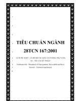
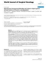
Fig. 1. HPTLC chromatograms and autograms of the S. gigantea root extract, developed with n-hexane – isopropyl acetate – acetone 16:3:1 (V/V/V, MP1) and detected at UV
254 nm (a), UV 365 nm (b), after derivatization with vanillin sulphuric acid reagent at UV 365 nm (c) and white light illumination (d; also e-g and i-k) AChE (e), BChE (f),
B. subtilis subsp. spizizenii (g), A. fischeri (h, greyscale image of the bioluminescence), α -glucosidase (i), β -glucosidase (j) and α -amylase (k).
58) and Sg6 (at hRF 37) showed α -glucosidase, β -glucosidase and
α -amylase inhibition. The application zone up to hRF 9 was also
effective in α -glucosidase and α -amylase assays. Cholinesterase inhibitors are of therapeutic interest in the Alzheimer disease, the
most common cause of dementia and one of the major public
health issues. Compounds in the zones Sg1 (hRF 9), Sg4 (hRF 51),
Sg5 and Sg6 inhibited AChE and BChE (Fig. 1e and f). However,
with MP2 and neutralization, BChE inhibitory zones of Sg1 (hRF
17) and Sg4 (hRF 55) were lacking (Fig. 2f). Multipotent compounds
are generally not favorable for pharmaceutical use, however, these
compounds can become lead compounds toward the development
of more specific, thus more useful derivatives.
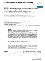
Antibacterial agents are widely required for the fight against infectious plant, animal and human diseases caused by pathogenic
bacterial and fungal strains. In the antibacterial assays, the Grampositive B. subtilis subsp. spizizenii soil bacterium, the Gramnegative, naturally luminescent marine A. fischeri and the Gramnegative paprika pathogen X. euvesicatoria were employed. In the
B. subtilis assay, Sg1 and compounds at an even lower hRF were
only active in the MP2 separation (Figs. 1g and 2g). All marked
zones were active against A. fischeri (Figs. 1h and 2h) and X. euvesicatoria (Fig. 2i), except for Sg3 at the given amount applied.
Additionally, Sg6 showed activity against A. fischeri only on acidfree bioautograms. The positions of the bioactive zones were con-
silane (δ =0.00 ppm). 13 C chemical shifts are given in ppm relative to the NMR solvent (δ =49.00 ppm). The complete 1 H and 13 C
assignments were deduced using conventional 1D (1 H NMR, 13 C
NMR, DeptQ) and 2D (1 H-1 H COSY, 1 H-13 C edHSQC 1 H-13 C HMBC
and 1 H-1 H NOESY) measurements.
3. Results and discussion
3.1. HPTLC-EDA
Two methods were investigated for the separation of the giant goldenrod root compounds (Figs. 1 and 2) on HPTLC plates
silica gel 60 with either n-hexane – isopropyl acetate – acetone
16:3:1 V/V/V (MP1, [29]) or n-hexane – isopropyl acetate – acetic
acid 40:9:1 V/V/V (MP2). Both separations detected at white light
illumination after derivatization with the universal vanillin sulphuric acid reagent, provided the six distinguishable colored zones
Sg1-Sg6 that were hardly detectable without the derivatization
(Figs. 1a, 1b, 2a and 2b). MP2 resulted in higher hRF values, which
was obvious for Sg5 and Sg6, indicating a potential acidic character. MP2 chromatograms required a neutralization step before EDA.
Compounds with antidiabetic (anti-hyperglycaemic) effect could be
used for the treatment of type 2 diabetes and obesity that are the
most common and increasing chronic diseases in the world. In the
HPTLC-EDA screening with MP1 (Fig. 1e-k), the zones Sg5 (at hRF
4
Á.M. Móricz, D. Krüzselyi, P.G. Ott et al.
Journal of Chromatography A 1635 (2021) 461727
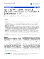
Fig. 2. HPTLC chromatograms and autograms of the S. gigantea root extract, developed with n-hexane – isopropyl acetate – acetic acid 40:9:1 (V/V/V, MP2) and detected at
UV 254 nm (a), UV 365 nm (b), after derivatization with vanillin sulphuric acid reagent at UV 365 nm (c) and white light illumination (d; also e-g and i), AChE (e), BChE (f),
B. subtilis subsp. spizizenii (g), A. fischeri (h, greyscale image of the bioluminescence) and X. euvesicatoria (i) assays.
5-days-old was found to be less sensitive (Fig. S3). In agreement to
our previous study, no characteristic inhibition was observed using
older microbial (bacterial) cells in the stationary and death phases
[37]. The staining procedure enabled the reduction of the incubation time to 2 days. The incubation time could not be shortened by increasing the OD600 of the mycelial inoculum from 0.2 to 0.8
- whithout a loss in discernability of the inhibition zones. In the
final protocol, the mycelium suspension at an OD600 of 0.4 was incubated for 2 days on the HPTLC plate and then detected by INT
staining, followed by a 1-h incubation and documentation under
white light illumination in the reflectance mode (Fig. S4).
3.2. HPTLC-MS
The bioactive compound zones Sg1-Sg6 were separated with
MP1 and characterized by HPTLC-HESI-HRMS and HPTLC-DARTHRMS (Table 2, Fig. S5). HPTLC-HESI+ -HRMS signals were obtained
for all six compound zones, but the signal intensities were very
low for Sg1 at m/z 341.2088 [M+Na]+ (C20 H30 O3 Na+ ) and Sg2 at
m/z 355.1879 [M+Na]+ (C20 H28 O4 Na+ ). However, it still allowed
the assignment of their molecular formulae. The recorded mass
signals for the Sg3 zone at m/z 337.1776 [M+Na]+ (C20 H26 O3 Na+ )
and at very low intensity m/z 325.2140 [M+Na]+ (C20 H30 O2 Na+ )
indicated the coelution of at least two compounds. As discussed
later, even three compounds Sg3a-c were identified in this zone.
An intense mass signal was recorded at m/z 339.1931 [M+Na]+
(C20 H28 O3 Na+ ) for Sg4. For Sg5 and Sg6, prominent mass signals
were obtained in both ionization mode at m/z 339.1931 [M+Na]+
(C20 H28 O3 Na+ ) and m/z 437.2304 [M+Na]+ (C25 H34 O5 Na+ ) as well
as at m/z 315.1956 [M-H]− and m/z 413.2304 [M-H]− , respectively. These zone assignments were successfully confirmed by
separation with MP2 and HPTLC-ESI-MS (Fig. S6). In the positive
and negative ionization mode, also the sodium-methanol-adducts
[M+CH3 OH+Na]+ and sodium-acetate-adducts of the respective
compounds were observed, which was caused by a too low drying gas flow.
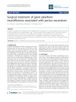
With exception of Sg1, all other compound zones gave mass signals by HPTLC-DART-HRMS (Table 2, Figs. 4 and S7). In the positive ionization mode, the protonated molecule [M+H]+ was detected for Sg3 and Sg5. Further, ammonium adducts [M+NH4 ]+
and dehydroxylated molecular ions [M-OH]+ were prominent. In
the negative ionization mode, the deprotonated molecules were
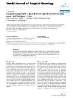
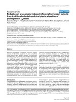
Fig. 3. HPTLC-F. avenaceum bioautogram of giant goldenrod root extract (E, 2 and 3
μL/band) and positive control benomyl (B, 25 μg/band) developed with n-hexane –
isopropyl acetate – acetone 16:3:1 (V/V/V, MP1) and detected at white light illumination after the bioassay (a) and subsequent staining with INT (b).
firmed by densitometry or videodensitometry of the MP2 bioautograms (Fig. S1). The results of the enzyme assays are in agreement
with our previous observations [29], however, the influence of an
acidic development and its neutralization on the outcome needs
further study.
An HPTLC-antifungal assay using plant pathogenic fungus F. avenaceum was newly developed. F. avenaceum grew well on V8 solid
agar medium and the surface of a 5 cm petri dish was totally occupied by its white hyphae within 2-3 days after inoculation by
an agar block (5 mm x 5 mm) in the center. Similarly, a dense
mycelium suspension was obtained within 3 days by shaking the
liquid inoculum (LB medium) at 21 °C. After cutting the mycelium
to small fragments, the suspension was initially diluted to reach
an OD600 of 0.4. Dipping a plate into this suspension, an appropriate mycelium growth was optically reached after a 3-days incubation in a vapor chamber at 21 °C (Fig. 3), indicating as mycelium
growth inhibition the antifungal activity of zones Sg1, Sg2, Sg3, Sg4
and Sg5. For detection, the three different tetrazolium dyes INT,
MTT and TTC were investigated, whereby the fastest coloration was
achieved with INT, which colored the hyphae violet (Figs. 3 and
S2). Compared to the 3-days-old mycelium suspension, the 1-dayold inoculum did not grow as fast on the HPTLC plate, and the
5
Á.M. Móricz, D. Krüzselyi, P.G. Ott et al.
Journal of Chromatography A 1635 (2021) 461727
detected for the Sg5 and Sg6 zones, and also the dimer for Sg5
and a C5 H7 O2 − fragment for Sg6.
ysis (Fig. S8c, Table 2). The HPLC method 1 (Table 1) was found
to be suitable for compound isolation in the Sg1, Sg2 and Sg5
zones, whereby the latter zone was tailing. Acid-free mobile phases
were selected to avoid acid-analyte reactions, which might impair
the bioactivity result. However, in the case of Sg5, the collected
peak was further purified with an acidic conditioning in the HPLC
method 4. The Sg6 compound zone was eluted from the C18 column during the washing step. Thus, its retention was achieved on
a PFP column (HPLC method 5). The HPTLC separation revealed
two co-eluting compounds in each of the HPLC peaks at 10-min
(Sg3a and Sg3b) and 12.5-min retention times (Sg3c and Sg4) (Fig.
3.3. Isolation of the active compounds
The root extract (10 mL) was fractionated by flash chromatography on a silica gel column, resulting in nineteen fractions (fr1fr19), which were studied by HPTLC-Vis after derivatization with
the vanillin sulphuric acid reagent (Fig. S8a and b). The six fractions containing the previously characterized bioactive compounds
(fr10-fr14 and fr17) were subjected to RP-HPLC-DAD-ESI-MS anal-
Table 2
Chemical structures, molecular formulae and assignments (using orthogonal techniques) of the eight discovered bioactive compounds in S. gigantea root extract.
(continued on next page)
6
Á.M. Móricz, D. Krüzselyi, P.G. Ott et al.
Journal of Chromatography A 1635 (2021) 461727
Table 2 (continued)
Fig. 4. EIC chromatograms of scanning HPTLC-DART-MS in the positive ionization mode (a) and respective HPTLC chromatogram of the bioactive giant goldenrod root
components separated with n-hexane – isopropyl acetate – acetone 16:3:1 (V/V/V, MP1) detected at white light illumination after derivatization with vanillin sulphuric acid
reagent (b).
7
Á.M. Móricz, D. Krüzselyi, P.G. Ott et al.
Journal of Chromatography A 1635 (2021) 461727
Fig. 5. Confirmation of purity and bioactivity: HPTLC chromatogram and autograms of the bioactive compounds Sg1-Sg6 isolated from giant goldenrod root extract (E)
separated with n-hexane – isopropyl acetate – acetone 16:3:1 (V/V/V, MP1) and detected at white light illumination (a, b and d) after derivatization with the vanillin–
sulphuric acid reagent (a), B. subtilis F1276 (b), A. fischeri (c, greyscale image of the bioluminescence) and F. avenaceum (d) assays.
S8c). For their separation, HPLC methods 2 and 3 were developed,
respectively. Eight compounds were isolated using a preparative
C18 column (Fig. S9) and an analytical PFP column (Fig. S10). These
were assigned as Sg1 (1.1 mg), Sg2 (1.6 mg), Sg3a (0.9 mg), Sg3b
(1.0 mg), Sg3c (1.5 mg), Sg4 (2.1 mg), Sg5 (2.2 mg) and Sg6 (6.2
mg) according to their respective HPTLC zones.
lished to inhibit Staphylococcus aureus [42]. The activity of three of
our isolated compounds has already been studied mainly against
insects. Sg5 was inactive, Sg1 caused the retardation of follicle development in mosquito ovaries [28] and displayed moderate toxicity against brine shrimp (Artemia salina) [49]. In contrast to Sg1,
Sg3b was found as anti-feedant and repellent agent in a study of
mealworms (Tenebrio molitor) [50]. Antiplasmodial (antimalarial)
and antileishmanial (inhibition of the intracellular parasite Leishmania donovani) effects of Sg3b were also observed, whereby it
did not show cytotoxicity against Vero cells (African green monkey
kidney fibroblast) [51]. Except for the new antifungal profiling, the
HPTLC-bioprofiling of the root extract has been previously reported
by us, however, it lacked in identification of the active compounds
by HRMS and NMR. To the best of our knowledge, this is the first
report about the antimicrobial profile of the eight isolated S. gigantea root diterpenoids. Further investigations are intended to evaluate their possible use as safe and effective pesticides, especially
fungicides.
3.4. Identification and characterization of the isolated compounds
The isolates were analyzed by FIA-HESI-HRMS/MS to successfully confirm that their identity was not changed during the isolation procedure, including fractionation and purification. Fragmentation of the respective sodium adduct of the compounds was not
observed except for Sg6 (Table 2), and thus, also the respectively
less intense protonated or deprotonated molecules were selected.
The intense deprotonated molecules of Sg5 and Sg6 were easily
fragmented.
The HPTLC-Vis analysis of the isolates (separated with MP1,
Fig. 5a, and MP2, Fig. S11 and detected after derivatization with
the vanillin sulphuric acid reagent) showed that only a single compound was present in all previously assigned zones, except for
three compounds (Sg3a-c) detected in Sg3. All isolates were active against B. subtilis, whereby Sg3b had only a weak activity
(Fig. 5b). Most isolated compounds strongly affected A. fischeri,
whereby Sg3b showed a mild effect, and Sg3c and Sg6 no response
at all (Fig. 5c). All compounds except Sg3b suppressed the growth
of F. avenaceum mycelium with varying degrees of effectiveness
(Fig. 5d). Although for the Sg3 and Sg6 zones of the root extract, no
activity against B. subtilis and F. avenaceum, respectively, was evident, their respective isolates did have an antimicrobial effect. This
was explained by the fact that their concentrations in the root extract were too low to reach the minimum inhibitory concentration.
The eight isolated compounds were most probably responsible for
the enzyme inhibiting activity, but this was not confirmed. The purity of the eight isolates was adequate, as confirmed by NMR analysis (Table S1, Figs. S12-S59). The eight compounds were identified as the furan-containing clerodane diterpenoids: a glycol (Sg1,
known also as kingidiol, white solid), an epoxy-hemiacetal (Sg2,
oil), a dialdehyde (Sg3a, white solid), the clerodane lactone (Sg3b,
oil, also known as hautriwaic lactone), an alcohol (Sg3c, oil), a
hemiacetal (Sg4, oil), the solidagoic acid A (Sg5, white solid) and
the solidagoic acid B (Sg6, white solid), which have been described
as components of S. gigantea roots [38-40] (Table 2). NMR data in
literature supported the identification of the isolated compounds
[28,38,39,41,42]. Antimicrobial [43-46], cytotoxic [45-47] and antiinflammatory effect [48] of several clerodane diterpenes isolated
from plants have been described. Among Solidago species, clerodane diterpenes of European goldenrod (S. virgaurea) were pub-
Conclusions
The newly developed HPTLC-F. avenaceum assay was demonstrated for the first time using mycelium suspension. It allowed
a cost-effective screening of complex plant extracts for antifungal
(mycelium growth inhibiting) compounds in S. gigantea root extract. Further bioprofiling of the extract against various bacterial
strains and enzymes by HPTLC-EDA pointed to multi-potent compounds. The comparison of the use of a neutral versus acidic mobile phase demonstrated that the latter can influence the bioassay
result. The state of the art combination of HPTLC-bioactivity assays, HPTLC-HESI-HRMS, HPTLC-DART-HRMS, preparative-scale column chromatography (flash chromatography and HPLC) and NMR
provided eight multi-potent clerodane diterpenoids in goldenrod.
It is the first report on the antifungal, antibacterial and enzyme
inhibiting activity of the multipotent isolates, which showed potential as lead compounds especially for various infectious plant
diseases.
Declaration of Competing Interest
The authors declare that they have no known competing financial interests or personal relationships that could have appeared to
influence the work reported in this paper.
Acknowledgment
Á.M. Móricz thanks the OECD for the scholarship JA0 0 092484
that allowed her to stay at JLU Giessen. Instrumentation was par8
Á.M. Móricz, D. Krüzselyi, P.G. Ott et al.
Journal of Chromatography A 1635 (2021) 461727
tially funded by the Deutsche Forschungsgemeinschaft (DFG, German Research Foundation) - INST 162/471-1 FUGG; INST 162/536-1
FUGG. This work was also funded by the National Research, Development and Innovation Office of Hungary (NKFIH K128921) and
partially supported by the János Bolyai Research Scholarship of the
Hungarian Academy of Sciences and Bolyai+ New National Excellence Program (ÚNKP-19-4-SE-53) of the Ministry of Human Capacities. The authors thank Salim Hage, Tim Häbe, Imanuel Yüce
and Tamara Schreiner for their support in the performance of the
ChE assay, DART- and HESI-HRMS experiments, respectively.
[20] G. Jakobs, E. Weber, P.J. Edwards, Introduced plants of the invasive Solidago gigantea (Asteraceae) are larger and grow denser than conspecifics in the native
range, Divers. Distrib. 10 (2004) 11–19, doi:10.1111/j.1472-4642.20 04.0 0 052.x.
[21] Barbara Kołodziej, Antibacterial and antimutagenic activity of extracts aboveground parts of three Solidago species: Solidago virgaurea L., Solidago canadensis L. and Solidago gigantea Ait, J. Med. Plants Res. 5 (2011), doi:10.5897/
JMPR11.1098.
[22] D. Webster, P. Taschereau, R.J. Belland, C. Sand, R.P. Rennie, Antifungal activity
of medicinal plant extracts; preliminary screening studies, J. Ethnopharmacol
115 (2008) 140–146, doi:10.1016/j.jep.2007.09.014.
[23] G. Benelli, R. Pavela, K. Cianfaglione, D.U. Nagy, A. Canale, F. Maggi, Evaluation of two invasive plant invaders in Europe (Solidago canadensis and Solidago
gigantea) as possible sources of botanical insecticides, J. Pest Sci. 92 (2019)
805–821 (2004), doi:10.1007/s10340- 018- 1034- 5.
[24] Z. Wang, J.H. Kim, Y.S. Jang, C.H. Kim, J.-Y. Lee, S.S. Lim, Anti-obesity effect of
Solidago virgaurea var. gigantea extract through regulation of adipogenesis and
lipogenesis pathways in high-fat diet-induced obese mice (C57BL/6N), Food
Nutr. Res. 61 (2017) 1273479, doi:10.1080/16546628.2016.1273479.
[25] D. Kalemba, B. Thiem, Constituents of the essential oils of four micropropagatedSolidago species, Flavour Fragr. J. 19 (2004) 40–43, doi:10.1002/ffj.1271.
[26] J. Radusiene, M. Marska, L. Ivanauskas, V. Jakstas, B. Karpaviciene, Assessment
of phenolic compound accumulation in two widespread goldenrods, Ind. Crops
Prod. 63 (2015) 158–166, doi:10.1016/j.indcrop.2014.10.015.
[27] G. Reznicek, M. Freiler, M. Schader, U. Schmidt, Determination of the content
and the composition of the main saponins from Solidago gigantea Ait. using
high-performance liquid chromatography, J. Chromatogr. A 755 (1996) 133–
137, doi:10.1016/S0 021-9673(96)0 0571-7.
[28] S.-H. Lee, H.-W. Oh, Y. Fang, S.-B. An, D.-S. Park, H.-H. Song, S.-R. Oh, S.-Y. Kim,
S. Kim, N. Kim, A.S. Raikhel, Y.H. Je, S.W. Shin, Identification of plant compounds that disrupt the insect juvenile hormone receptor complex, Proc. Natl.
Acad. Sci. 112 (2015) 1733–1738, doi:10.1073/pnas.1424386112.
[29] Á.M. Móricz, M. Jamshidi-Aidji, D. Krüzselyi, A. Darcsi, A. Böszörményi, P. Csontos, S. Béni, P.G. Ott, G.E. Morlock, Distinction and valorization of 30 root
extracts of five goldenrod (Solidago) species, J. Chromatogr. A 1611 (2020)
460602, doi:10.1016/j.chroma.2019.460602.
[30] Á.M. Móricz, P.G. Ott, I. Yüce, A. Darcsi, S. Béni, G.E. Morlock, Effect-directed
analysis via hyphenated high-performance thin-layer chromatography for bioanalytical profiling of sunflower leaves, J. Chromatogr. A 1533 (2018) 213–220,
doi:10.1016/j.chroma.2017.12.034.
[31] Á.M. Móricz, T.T. Häbe, A. Böszörményi, P.G. Ott, G.E. Morlock, Tracking and
identification of antibacterial components in the essential oil of Tanacetum
vulgare L. by the combination of high-performance thin-layer chromatography with direct bioautography and mass spectrometry, J. Chromatogr. A 1422
(2015) 310–317, doi:10.1016/j.chroma.2015.10.010.
[32] M. Jamshidi-Aidji, G.E. Morlock, Bioprofiling of unknown antibiotics in herbal
extracts: Development of a streamlined direct bioautography using Bacillus
subtilis linked to mass spectrometry, J. Chromatogr. A 1420 (2015) 110–118,
doi:10.1016/j.chroma.2015.09.061.
[33] S. Krüger, O. Urmann, G.E. Morlock, Development of a planar chromatographic
method for quantitation of anthocyanes in pomace, feed, juice and wine, J.
Chromatogr. A 1289 (2013) 105–118, doi:10.1016/j.chroma.2013.03.005.
[34] Á.M. Móricz, T.T. Häbe, P.G. Ott, G.E. Morlock, Comparison of high-performance
thin-layer with overpressured layer chromatography combined with direct
bioautography and direct analysis in real time mass spectrometry for tansy
root, J. Chromatogr. A 1603 (2019) 355–360, doi:10.1016/j.chroma.2019.03.068.
[35] S. Hage, G.E. Morlock, Bioprofiling of Salicaceae bud extracts through highperformance thin-layer chromatography hyphenated to biochemical, microbiological and chemical detections, J. Chromatogr. A 1490 (2017) 201–211,
doi:10.1016/j.chroma.2017.02.019.
[36] T.T. Häbe, G.E. Morlock, Improved desorption/ionization and ion transmission
in surface scanning by direct analysis in real time mass spectrometry, Rapid
Commun. Mass Spectrom. 30 (2016) 321–332, doi:10.1002/rcm.7434.
[37] Á. Móricz, N. Adányi, E. Horváth, P. Ott, E. Tyihák, Applicability of the BioArena
system to investigation of the mechanisms of biological effects, J. Planar Chromatogr. – Mod. TLC 21 (2008) 417–422, doi:10.1556/JPC.21.2008.6.4.
[38] M.S. Henderson, R. McCrindle, D. McMaster, Constituents of Solidago species.
Part V. Non-acidic diterpenoids from Solidago gigantea var. serotina, Can. J.
Chem. 51 (1973) 1346–1358, doi:10.1139/v73-201.
[39] T. Anthonsen, M.S. Henderson, A. Martin, R.D.H. Murray, R. McCrindle, D. McMaster, Constituents of Solidago species. Part IV. Solidagoic acids A and B,
diterpenoids from Solidago gigantea var. serotina, Can. J. Chem. 51 (1973) 1332–
1345, doi:10.1139/v73-200.
[40] T. Anthonsen, M.S. Henderson, A. Martin, R. McCrindle, R.D.H. Murray, Furancontaining diterpenoids from Solidago serotina Ait, Acta Chem. Scand. 22
(1968) 351–352, doi:10.3891/acta.chem.scand.22-0351.
[41] T.G. Payne, P.R. Jefferies, The chemistry of Dodonaea spp - IV, Tetrahedron 29
(1973) 2575–2583, doi:10.1016/0040-4020(73)80176-0.
[42] C.M. Starks, R.B. Williams, M.G. Goering, M. O’Neil-Johnson, V.L. Norman, J.F. Hu, E. Garo, G.W. Hough, S.M. Rice, G.R. Eldridge, Antibacterial clerodane
diterpenes from goldenrod (Solidago virgaurea), Phytochemistry 71 (2010) 104–
109, doi:10.1016/j.phytochem.2009.09.032.
[43] V.K. Gupta, N. Tiwari, P. Gupta, S. Verma, A. Pal, S.K. Srivastava, M.P. Darokar,
A clerodane diterpene from Polyalthia longifolia as a modifying agent of the resistance of methicillin resistant Staphylococcus aureus, Phytomedicine 23 (2016)
654–661, doi:10.1016/j.phymed.2016.03.001.
Supplementary materials
Supplementary material associated with this article can be
found, in the online version, at doi:10.1016/j.chroma.2020.461727.
References
[1] B.A. Lorsbach, T.C. Sparks, Innovations in agrochemical discovery and the role
of metabolism, bioavailability and formulations, Pest Manag. Sci. 73 (2017)
655–657, doi:10.1002/ps.4533.
[2] M. Hahn, The rising threat of fungicide resistance in plant pathogenic
fungi: Botrytis as a case study, J. Chem. Biol. 7 (2014) 133–141, doi:10.1007/
s12154- 014- 0113- 1.
[3] P.S. Hoffman, Antibacterial Discovery: 21st Century Challenges, Antibiotics 9
(2020) 213, doi:10.3390/antibiotics9050213.
[4] Á.M. Móricz, P.G. Ott, T.T. Häbe, A. Darcsi, A. Böszörményi, Á. Alberti,
D. Krüzselyi, P. Csontos, S. Béni, G.E. Morlock, Effect-directed discovery of
bioactive compounds followed by highly targeted characterization, isolation
and identification, exemplarily shown for Solidago virgaurea, Anal. Chem. 88
(2016) 8202–8209, doi:10.1021/acs.analchem.6b02007.
[5] W. Jonker, M.H. Lamoree, C.J. Houtman, J. Kool, Methodologies for effectdirected analysis: environmental applications, food analysis, and drug discovery, in: Anal. Biomol. Interact. by Mass Spectrom, Wiley-VCH Verlag
GmbH & Co. KGaA, Weinheim, Germany, 2015, pp. 109–163, doi:10.1002/
9783527673391.ch4.
[6] G. Morlock, W. Schwack, Hyphenations in planar chromatography, J. Chromatogr. A 1217 (2010) 6600–6609, doi:10.1016/j.chroma.2010.04.058.
[7] G.E. Morlock, Chromatography combined with bioassays and other hyphenations – the direct link to the compound indicating the effect, in: 2014: pp.
101–121. 10.1021/bk-2014-1185.ch005.
[8] G. Morlock, W. Schwack, Coupling of planar chromatography to mass spectrometry, TrAC Trends Anal. Chem. 29 (2010) 1157–1171, doi:10.1016/j.trac.
2010.07.010.
[9] J. Yu, R.H. Proctor, D.W. Brown, K. Abe, K. Gomi, M. Machida, F. Hasegawa,
W.C. Nierman, D. Bhatnagar, T.E. Cleveland, Genomics of economically significant Aspergillus and Fusarium species, in: 2004: pp. 249–283. 10.1016/S18745334(04)80013-3.
[10] S. Vogelgsang, M. Sulyok, A. Hecker, E. Jenny, R. Krska, R. Schuhmacher, H.R. Forrer, Toxigenicity and pathogenicity of Fusarium poae and Fusarium avenaceum on wheat, Eur. J. Plant Pathol. 122 (2008) 265–276, doi:10.1007/
s10658- 008- 9279- 0.
[11] A.T. Pollard, P.A. Okubara, Real-time PCR quantification of Fusarium avenaceum
in soil and seeds, J. Microbiol. Methods 157 (2019) 21–30, doi:10.1016/j.mimet.
2018.12.009.
[12] S. Uhlig, A.C. Gutleb, U. Thrane, A. Flåøyen, Identification of cytotoxic principles from Fusarium avenaceum using bioassay-guided fractionation, Toxicon 46
(2005) 150–159, doi:10.1016/j.toxicon.20 05.03.0 05.
[13] A.L. Homans, A. Fuchs, Direct bioautography on thin-layer chromatograms as
a method for detecting fungitoxic substances, J. Chromatogr. A 51 (1970) 327–
329, doi:10.1016/S0021-9673(01)96877-3.
[14] K.D. Burkhead, D.A. Schisler, P.J. Slininger, Pyrrolnitrin Production by biological
control agent Pseudomonas cepacia B37w in culture and in colonized wounds
of potatoes, Appl. Environ. Microbiol. 60 (1994) 2031–2039, doi:10.1128/AEM.
60.6.2031-2039.1994.
[15] N. Kasanah, L.L. Farr, A. Gholipour, D.E. Wedge, M.T. Hamann, Metabolism
and resistance of Fusarium spp. to the manzamine alkaloids via a putative
retro Pictet-Spengler reaction and utility of the rational design of antimalarial and antifungal agents, Mar. Biotechnol. 16 (2014) 412–422, doi:10.1007/
s10126-014-9557-0.
[16] R.E. Beale, D. Pitt, The antifungal properties of Minimedusa polyspora, Mycol.
Res. 99 (1995) 337–342, doi:10.1016/S0953-7562(09)80910-6.
[17] G.L. Gallardo, N.I. Peña, G.M. Cabrera, Neric acid derivatives produced by the
honey bee fungal entomopathogen Ascosphaera apis, Phytochem. Lett. 1 (2008)
155–158, doi:10.1016/j.phytol.20 08.07.0 08.
[18] F. Hadacek, H. Greger, Testing of antifungal natural products: methodologies,
comparability of results and assay choice, Phytochem. Anal. 11 (20 0 0) 137–147,
doi:10.1002/(SICI)1099-1565(200005/06)11:3 137:AID-PCA514 3.0.CO;2-I.
[19] M. Szymura, T.H. Szymura, Interactions between alien goldenrods (Solidago and
Euthamia species) and comparison with native species in Central Europe, Flora
- Morphol. Distrib. Funct. Ecol. Plants. 218 (2016) 51–61, doi:10.1016/j.flora.
2015.11.009.
9
Á.M. Móricz, D. Krüzselyi, P.G. Ott et al.
Journal of Chromatography A 1635 (2021) 461727
[44] A. Bisio, A.M. Schito, S.N. Ebrahimi, M. Hamburger, G. Mele, G. Piatti, G. Romussi, F. Dal Piaz, N. De Tommasi, Antibacterial compounds from Salvia
adenophora Fernald (Lamiaceae), Phytochemistry 110 (2015) 120–132, doi:10.
1016/j.phytochem.2014.10.033.
[45] P. Chawengrum, J. Boonsombat, P. Kittakoop, C. Mahidol, S. Ruchirawat,
S. Thongnest, Cytotoxic and antimicrobial labdane and clerodane diterpenoids
from Kaempferia elegans and Kaempferia pulchra, Phytochem. Lett. 24 (2018)
140–144, doi:10.1016/j.phytol.2018.02.009.
[46] A.L. Pfeifer Barbosa, A. Wenzel-Storjohann, J.D. Barbosa, C. Zidorn, C. Peifer,
D. Tasdemir, S.S. ầiỗek, Antimicrobial and cytotoxic effects of the Copaifera
reticulata oleoresin and its main diterpene acids, J. Ethnopharmacol. 233 (2019)
94–100, doi:10.1016/j.jep.2018.11.029.
[47] P.M.P. Ferreira, G.C.G. Militão, D.J.B. Lima, N.D. de, J. Costa, K.da C. Machado,
A.G. dos Santos, A.J. Cavalheiro, V.da S. Bolzani, D.H.S. Silva, C. Pessoa, Morphological and biochemical alterations activated by antitumor clerodane diterpenes, Chem. Biol. Interact. 222 (2014) 112–125, doi:10.1016/j.cbi.2014.10.015.
[48] Y. Wang, J. Lin, Q. Wang, K. Shang, D.-B. Pu, R.-H. Zhang, X.-L. Li, X.-C. Dai, X.J. Zhang, W.-L. Xiao, Clerodane diterpenoids with potential anti-inflammatory
activity from the leaves and twigs of Callicarpa cathayana, Chin. J. Nat. Med. 17
(2019) 953–962, doi:10.1016/S1875-5364(19)30118-9.
[49] C. Labbe, M. Castillo, M. Hernandez, Diterpenoids from Baccharis lejía, Phytochemistry 30 (1991) 1607–1611, doi:10.1016/0031- 9422(91)84217- G.
[50] M.E. Sosa, C.E. Tonn, O.S. Giordano, Insect antifeedant activity of clerodane
diterpenoids, J. Nat. Prod. 57 (1994) 1262–1265, doi:10.1021/np50111a012.
[51] A.A.da S. Filho, D.O. Resende, M.J. Fukui, F.F. Santos, P.M. Pauletti, W.R. Cunha,
M.L.A. Silva, L.E. Gregório, J.K. Bastos, N.P.D. Nanayakkara, In vitro antileishmanial, antiplasmodial and cytotoxic activities of phenolics and triterpenoids
from Baccharis dracunculifolia D. C. (Asteraceae), Fitoterapia 80 (2009) 478–482,
doi:10.1016/j.fitote.20 09.06.0 07.
10