Báo cáo khoa học: Sp1 and Sp3 are involved in up-regulation of human deoxyribonuclease II transcription during differentiation of HL-60 cells pptx
Bạn đang xem bản rút gọn của tài liệu. Xem và tải ngay bản đầy đủ của tài liệu tại đây (251.49 KB, 8 trang )
Sp1 and Sp3 are involved in up-regulation of human
deoxyribonuclease II transcription during differentiation of HL-60 cells
San-Fang Chou
1
, Hui-Ling Chen
2
* and Shao-Chun Lu
1
*
1
Department of Biochemistry and Molecular Biology, College of Medicine, National Taiwan University, Taipei, Taiwan;
2
Hepatitis Research Center, National Taiwan University Hospital, Taipei, Taiwan
Expression of DNase II in macrophages is potentially cru-
cially important in the removal of unwanted DNA. We have
previously shown that DNase II expression is up-regulated
at the transcriptional level during the phorbol 12-myristate-
13-acetate (PMA)-induced differentiation of HL-60 and
THP-1 cells. In this study, we investigated the cis-regulatory
elements and transcription factors involved in this process in
HL-60 cells. cis-Regulatory elements in the DNase II pro-
moter were located by 5¢ deletion and site-directed muta-
genesis of promoter-luciferase constructs and transient
transfection of HL-60 cells. Furthermore, the binding pro-
teins were identified by electrophoretic mobility shift assay
(EMSA) in the presence of specific antibodies. In the
DNase II promoter, 249 base pairs upstream of the
transcription start site were essential for maximal promoter
activity in both untreated and PMA-treated HL-60 cells and,
within this region, three Sp1 and Sp3 binding sites were
identified as essential for transcriptional regulation and
PMA induction. Western blot analysis showed that PMA
treatment resulted in increased levels of Sp1 and Sp3 pro-
teins. Furthermore, cotransfection analysis in Drosophila
SL2 cells showed that Sp1 was more potent than Sp3 in
activating the DNase II promoter. We therefore conclude
that Sp1 and/or Sp3 are involved in the up-regulation of
DNase II expression during the differentiation of HL-60
cells.
Keywords: DNase II; Sp1; Sp3; HL-60; PMA.
Deoxyribonuclease II (DNase II; EC.3.1.22.1) is a well
known lysosomal acid endonuclease that hydrolyses DNA,
producing 3¢-phosphoryl oligonucleotides [1,2]. DNase II
activity and mRNA are detected in most human tissues, the
highest levels being found in the adrenal gland, thyroid
gland, lymph nodes, and pituitary gland [3]. DNase II
activity is higher in macrophages than in various nonmacro-
phage cell lines and is increased during the differentiation
of HL-60 cells and peripheral blood monocytes to macro-
phages [4]. Using a single radial enzyme diffusion method,
Yasuda et al. [5,6] found that DNase II activity in the
Japanese population can be classified into low-activity
(DNASE2 L) and high-activity (DNASE2 H), resulting
from a genetic polymorphism in the DNase II gene
promoter region. However, no association has been found
between DNase II activity and disease.
Recently, research on DNase II has focused on its role in
apoptosis. Barry & Eastman [7] demonstrated that DNase II
mediates the digestion of internucleosomal DNA in apop-
totic cells. Torriglia et al. [8] have shown that DNase II is
involved in the degradation of fiber cell DNA during lens
cell differentiation. Furthermore, McIlroy et al. [9] sugges-
ted that DNase II is responsible for DNA fragmentation in
apoptotic cells after they are engulfed by phagocytic cells.
Mice with targeted disruption of the DNase II gene die at
birth because of severe anemia [10] and/or asphyxiation [11];
after examination of the DNase II-null embryos, it was
suggested that macrophage DNase II is required for
degradation of nuclear DNA expelled during erythrocyte
maturation [10] and for the digestion of DNA in apoptotic
cells [11] during fetal development. These results suggest
that macrophage DNase II plays a pivotal role in the
removal of Ôunwanted DNAÕ.
We previously reportedanincreaseinacid nuclease activity
and DNase II mRNA levels during the myelomonocytic
differentiation of HL-60 and THP-1 cells and demonstrated
that the increase in DNase II mRNA levels was mainly due
to transcriptional activation of the gene [4]. In the present
study, our aim was to identify cis-regulatory element(s) and
transcription factor(s) that mediate the transcriptional acti-
vation of the human DNase II gene in HL-60 cells. Using
transient transfection and electrophoretic mobility shift
assay (EMSA), we demonstrated that binding of Sp1 and/
or Sp3 to three GC-boxes within the proximal region of the
DNase II promoter is critical for DNase II transcription in
phorbol 12-myristate-13-acetate (PMA)-treated HL-60 cells.
Materials and methods
Cell culture
The human acute promyelocytic leukemia cell line, HL-60,
obtained from the ATCC (Manassas, VA, USA), was
Correspondence to S C. Lu, Department of Biochemistry and
Molecular Biology, College of Medicine, National Taiwan University,
no. 1, Sec. 1, Jen-Ai Road, Taipei, Taiwan 100.
Fax: + 886 2 2391 5295, Tel.: + 886 2 2312 3456 ext 8224,
E-mail:
Abbreviations: DNase II, deoxyribonuclease II; EMSA, electropho-
retic mobility shift assay; PMA, phorbol 12-myristate-13-acetate.
Enzyme: deoxyribonuclease II (DNase II; EC.3.1.22.1).
*These authors contributed equally to this work.
(Received 12 December 2002, revised 20 February 2003,
accepted 3 March 2003)
Eur. J. Biochem. 270, 1855–1862 (2003) Ó FEBS 2003 doi:10.1046/j.1432-1033.2003.03551.x
grown and induced to differentiate by PMA treatment as
described previously [4]. Schneider’s Drosophila cell line 2
(SL2) cells (generously supplied by Y S. Chang, Graduate
Institute of Basic Science, Chang-Gung University School
of Medicine, Taiwan) were maintained in Schneider’s
Insect Medium (Gibco, BRL) supplemented with 10%
fetal bovine serum, 50 lgÆmL
)1
of streptomycin, and
50 lgÆmL
)1
of penicillin at 25 °C with atmospheric CO
2
.
Plasmid construction
A DNase II promoter-luciferase chimeric gene contain-
ing nucleotides )1875 to +72 of the DNase II gene
(pDNaseII()1875/+72)-Luc) was constructed as described
previously [4]. To construct pDNaseII()934/+72)-Luc and
pDNaseII()249/+72)-Luc, pDNaseII()1875/+72)-Luc
was digested with SacIandXmaI to remove nucleotides
)1875 to )935 or )1875 to ) 250, respectively, and the
remaining DNA fragments were ligated using T4 DNA
ligase. The fragments )149 to +72, )68 to +72, and )32 to
+72 of the DNase II 5¢ flanking sequences were obtained
by PCR from pDNaseII()1875/+72)-Luc using specific
primers (Table 1), then the PCR products were cloned into
the MluI/XhoI sites of the pGL3-basic vector (Promega) to
produce pDNaseII()149/+72)-Luc, pDNaseII()68/+72)-
Luc, and pDNaseII()32/+72)-Luc. In order to mutate the
three GC boxes starting at nucleotides )135, )72 and )45,
mutated oligonucleotides were synthesized (Table 1) and
used to generate mutants of GC-I, GC-II, and/or GC-III on
pDNaseII()249/+72)-Luc by an overlap extension method
[11]. All clones were verified by restriction enzyme mapping
and sequencing. The Sp1 (pPacSp1) and Sp3 (pPacUSp3)
expression plasmids and their maternal plasmid, pPac0,
were kindly provided by G. Suske (Philipps-Universitat,
Marburg, Germany) [13].
Transfection of HL-60 and SL2 cells
HL-60 cells were transfected using the DEAE-dextran
procedure as previously described [4]. Briefly, cells (2 · 10
7
)
were collected by centrifugation, resuspended in 1 mL of
25 m
M
Tris/HCl buffer, pH 7.4, 5 m
M
KCl, 0.7 m
M
CaCl
2
,137m
M
NaCl, 0.6 m
M
Na
2
HPO4, 0.5 m
M
MgCl
2
,
containing 5 lgoftestplasmidDNA,5lg of phRL-TK
DNA (Promega), and 50 lgÆmL
)1
of DEAE-dextran
(Sigma), and incubated at room temperature for 15 min.
The cells were centrifuged and the pellet was washed, and
resuspended in RPMI 1640 medium supplemented with
20% fetal bovine serum, then divided and cultured in the
presence or absence of 30 n
M
PMA (Sigma) for another
48 h before being lysed by addition of 100 lL of Passive
Lysis Buffer (Dual-Luciferase Reporter Assay System,
Promega). Cell lysates from three dishes transfected with
the same construct were pooled. Photinus and Renilla
luciferase activities in the lysates were assayed using
the Dual-Luciferase Reporter Assay System as described
previously [4]. The light intensity produced by Photinus
luciferase (test plasmid) was normalized to that produced
by Renilla luciferase (control plasmid). Promoter activity
was expressed relative to that of cells transfected with
pGL3-b (relative value ¼ 1). At least three independent
experiments in duplicate were performed using each
construct.
SL2 cells were transfected using FuGENE 6 (Roach,
Indianapolis, IN, USA) according to the manufacturer’s
instructions. Briefly, 10, 50, 100, or 150 ng of expression
vector (pPacSp1 or pPacUSp3) was mixed with 50 ng of
pDNaseII()249/+72)-Luc, and the total amount of DNA
adjusted to 200 ng with pPac0. The DNA was mixed with
0.6 lLofFuGENE6in100lL of serum-free Schneider’s
Insect Medium (Gibco, BRL) and incubated at room
temperature for 5 min. The DNA/FuGENE 6 mixture was
then added to 24-well plates, each well containing 5 · 10
5
SL2 cells. Forty-eight hours after transfection, the cells were
washed twice with NaCl/P
i
, then the luciferase activity was
measured using the Luciferase Assay System (Promega).
Luciferase activity was normalized to total cellular protein.
Transfections were performed in duplicate and repeated two
to four times to ensure reproducibility and to monitor
transfection efficiency.
Table 1. Sequences of the oligonucleotides used. mt, mutated.
Location Sequence Note
Oligonucleotides used for reporter constructs
a
Forward primers:
)149 to )129: 5¢-CGG
ACGCGTCGTGGGCGTGGTCTGGGC-3¢ pDNaseII()149/+72)
)68 to ) 44: 5¢-AGGA
ACGCGTACCCTCGTGATGTCCCCG-3¢ pDNaseII()68/+72)
)32 to ) 11: 5¢-CAG
ACGCGTTTAGGGAAGTGAAAGGCGCCA-3¢ pDNaseII()32/+72)
Reverse primers:
+72 to +51 5¢-
CTCGAGCTGCTATGGGGCTGAGATCC-3¢
Oligonucleotides used for mutagenesis and EMSA
b
)151 to )129 5¢-CCCGTCGTGGGCGTGGTCTGGGC-3¢ GC-I
)151 to )129 5¢-CCCGTCGTGG
TATTGGTCTGGGC-3¢ mtGC-I
)89 to ) 64 5¢-CGCGTCTCGGGGGAGTAGTCTGTACC-3¢ GC-II
)89 to ) 64 5¢-CGCGTCTCGG
TTTAGTAGTCTGTACC-3¢ mtGC-II
)61 to ) 36 5¢-CGTGATGTCCCCGCCCCGGTTCCCAG-3¢ GC-III
)61 to ) 36 5¢-CGTGATGTCCC
AAACCCGGTTCCCAG-3¢ mtGC-III
a
The underlined ACGCGT and CTCGAG are MluI and XhoI restriction sites, respectively, created to facilitate cloning.
b
Mutated bases
are underlined.
1856 S F. Chou et al. (Eur. J. Biochem. 270) Ó FEBS 2003
Nuclear extract preparation
Nuclear extracts were prepared as described by Garban
et al. [14], with some modifications. Briefly, cells were
treatedwith30n
M
PMA for 60 h and collected by
centrifugation, washed twice with ice-cold phosphate-buf-
fered saline, and resuspended in 20 volumes of hypotonic
lysis buffer (10 m
M
Hepes/KOH, pH 7.9, 10 m
M
KCl,
1.5 m
M
MgCl
2
,0.5m
M
dithiothreitol, 0.1% NP-40, and
0.2 m
M
phenylmethanesulfonyl fluoride). After incubation
of the mixture on ice for 15 min, nuclei were pelleted by
centrifugation at 500 g for 5 min at 4 °C, washed once with
hypotonic lysis buffer, and pelleted again, then nuclear
proteins were extracted by incubation of the nuclei for
15 min at 4 °C with intermittent vortexing in 20 m
M
Hepes/KOH, pH 7.9, 25% glycerol, 420 m
M
NaCl,
1.5 m
M
MgCl
2
,0.2m
M
EDTA, 0.5 m
M
dithiothreitol,
0.2 m
M
phenylmethanesulfonyl fluoride, and 1 · protease
inhibitor cocktail (Roche); cell debris was removed by
centrifugation at 12 900 g for 10 min at 4 °C. The Bradford
method (DC Protein Assay, Bio-Rad) was used to measure
the protein concentration in the extract, which was then
stored in aliquots at )80 °C.
Electrophoretic mobility shift assays
The oligonucleotides and complementary oligonucleotides
used in the EMSA (Table 1) were custom synthesized. The
complementary primers were annealed to each other to
produce a double-stranded DNA fragment, which was then
32
P-labeled using Taq DNA polymerase (Invitrogen) and
[a-
32
P]dCTP (NEN Life Science Products, Boston, MA,
USA). Binding reactions were performed by incubating
5 lg of nuclear extract and 600 fmol of
32
P-labeled double-
stranded oligonucleotide, with or without competitor, for
30 min at room temperature in a final volume of 20 lLof
binding buffer (20 m
M
Hepes, pH 7.9, 60 m
M
KCl, 6 m
M
MgCl
2
,0.5m
M
EDTA, 10% glycerol, 1 m
M
dithiothreitol,
0.1 lgÆlL
)1
of poly dI-dC, 160 lgÆmL
)1
of BSA, 0.008%
NP-40, and protease inhibitor). Competitors [either a 10- or
50-fold excess of unlabeled wild-type or mutant probe or a
0.6- to threefold excess of Sp1 consensus oligonucleotides
(Promega)] were added to the mixture immediately after the
labeled probe. For the supershift assay, the nuclear extract
was incubated for 1 h on ice with rabbit polyclonal anti-Sp1
or anti-Sp3 IgG (both from Santa Cruz Biotechnology,
Santa Cruz, CA, USA) or mouse monoclonal antibody to
the sterol response element binding protein-1 (SREBP-1;
ATCC). The probe was then added and the mixture was
incubated for a further 30 min at room temperature and
immediately loaded onto a 5% nondenaturing polyacryl-
amide gel containing 0.5 · Tris/borate/EDTA (45 m
M
Tris,
45 m
M
boric acid, 1 m
M
EDTA, pH 8.3) buffer. Electro-
phoresis was carried out at 4 °C at 250 V. Gels were
vacuum heat-dried and analyzed on a PhosphorImager
(Molecular Dynamics, Sunnyvale, CA, USA).
Western blot analysis
Nuclear proteins (20 lg of protein per lane) were separ-
ated by SDS/PAGE on 10% gels and transferred to a
poly (vinylidene difluoride) membrane, which was blocked
overnight at 4 °C with blocking buffer (10 m
M
Tris/HCl,
pH 8.0, 0.15
M
NaCl, 0.1% Tween 20, and 5% fat-free
milk). The blots were then incubated for 1 h at room
temperature with 0.5 lgÆmL
)1
of rabbit polyclonal anti-Sp1
or anti-Sp3 IgG (both from Santa Cruz Biotechnology) and
for 40 min at room temperature with peroxidase-conjugated
anti-(rabbit IgG) IgG (Amersham-Pharmacia Biotech), and
bound antibody was detected using an improved chemi-
luminescence detection system (NEN).
Statistical analysis
Data were analyzed using
STATISCA
for
WINDOWS
v4.5
(StatSoft, Tulsa, OK). Differences between mean values
were evaluated using the Duncan’s multiple range test and
were considered significant at P <0.05.
Results
Dissection of the 5¢ flanking sequence of the human
DNase II gene
To define the regulatory sequences required for transcrip-
tion of the DNase II gene, HL-60 cells were cotransfected
with a series of 5¢-deleted DNase II-Luc constructs and
phRL-TK, a control plasmid containing the gene coding for
Renilla luciferase driven by the TK promoter. After
transfection, the cells were divided and cultured for 48 h
in RPMI 1640 supplemented with 20% fetal bovine serum
inthepresenceorabsenceof30n
M
PMA.
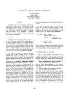
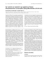
As shown in Fig. 1, in non-PMA-treated cells, deletion of
nucleotides )1875 to )249 had no significant effect on
luciferase activity (P > 0.05); however, deletion to nucleo-
tide )149 resulted in a significant lower luciferase activity
compared to that seen with pDNaseII()1875/+72)-Luc
(P < 0.05). Further deletion to nucleotide )68ledtoa
further 82% drop in luciferase activity compared to that
seen with pDNaseII()149/+72)-Luc (P < 0.01). Deletion
to nucleotide )32 resulted in complete loss of luciferase
activity.
Fig. 1. Promoter activity of human DNase II-luciferase hybrid genes in
HL-60 cells. Schematic representations of the 5¢-deleted promoter-
luciferase constructs. The hybrid genes were constructed as described
in the Materials and methods. All of the constructs were cotransfected
with phRL-TK (internal control) into HL-60 cells using the DEAE-
dextran method. Luciferase activity was normalized to Renilla luci-
ferase activity and is shown as a relative activity compared to that for
pGL3-b. The values are the means ± SD of at least three independent
experiments.
Ó FEBS 2003 Transcriptional regulation of human DNase II by Sp1 and Sp3 (Eur. J. Biochem. 270) 1857
In contrast, in PMA-treated cells, luciferase activities
were significantly higher than in untreated control cells and
gradual deletion of 5¢ sequences from nucleotides )1875 to
)249 resulted in a gradual increase in luciferase activity
(Fig. 1). Maximal activity, seen with pDNaseII()249/+72)-
Luc, was 152% that seen with pDNaseII()1875/+72)-Luc
(P < 0.05). On further deletion to nucleotide )149, luci-
ferase activity fell to 46% of the maximal activity
(P < 0.01), and deletion to nucleotide )68 resulted in a
substantial reduction to only 2% of the maximal activity.
Deletion to nucleotide )32 again resulted in complete loss of
luciferase activity.
These results show that the region from nucleotide )249
to nucleotide )32 is required for maximal expression of
DNase II in HL-60 cells, both in the presence and absence
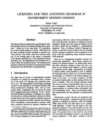
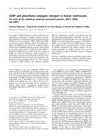
of PMA. Sequence analysis of nucleotides )249 to +72
using the
MATINSPECTOR
program [15] revealed three GC
boxes, referred to as GC-I, GC-II, and GC-III (Fig. 2),
starting at nucleotides )135, )72, and )45 relative to the
start of transcription.
Examination of GC boxes by
in vitro
mutagenesis
and transfection
To define the contribution of these three GC boxes to
DNase II expression in HL-60 cells, they were mutated,
individually or in combination, by an overlap extension
method using pDNaseII()249/+72)-Luc as template, then
the GC mutant constructs were transiently transfected into
HL-60 cells, which were then cultured in the absence or
presence of 30 n
M
PMA and their luciferase activity
compared to that of cells transfected with the wild-type
construct, pDNaseII()249/+72)-Luc (relative luciferase
activity ¼ 100).
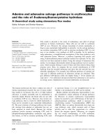
In non-PMA-treated cells (Fig. 3, upper panel), single
mutation of GC-I, GC-II, or GC-III resulted, respectively,
in a significant reduction of 48, 70, or 36% in luciferase
activity (P < 0.05), while mutation of all three GC boxes
ledtoafallof96%(P < 0.01). In PMA-treated cells
(Fig. 3, lower panel), mutation of GC-I, GC-II, or GC-III
resulted in respective decreases in luciferase activity of 83,
63, or 53% (P < 0.05), and mutation of all three GC boxes
resulted in complete loss of promoter activity (P < 0.01).
These results show that all three GC boxes are required for
maximal activity of the DNase II promoter in both control
and PMA-treated HL-60 cells.
Electrophoretic mobility shift assays
To explore protein binding to these GC boxes, protein-
DNA complex formation was examined in vitro using the
electrophoretic mobility shift assay (EMSA). When nuclear
extracts from control HL-60 cells were incubated with
32
P-labeled probe I, three weak DNA–protein complexes
(C1, C2, and C3) were detected (Fig. 4A, lane 2). Significant
increases in these complexes and the presence of two
additional complexes were detected when the same probe
was incubated with nuclear extracts from PMA-treated cells
(Fig. 4A, lane 3). The intensity of these complexes was
markedly decreased in the presence of a 10- or 50-fold molar
excess of unlabeled probe I (lanes 4 and 5), but not in the
presence of unlabeled GC-I mutated probe I (lanes 6 and 7).
The formation of complexes C1, C2, and C3 was partially
blocked by a 0.6- or threefold excess of GC consensus
oligonucleotide (Promega) (lanes 8 and 9). In order to verify
the involvement of Sp proteins, the nuclear extracts were
incubated with anti-Sp1 or anti-Sp3 Ig before addition of
Fig. 2. DNA sequence of the human DNase II
promoter region. The GC-rich sequences,
referred to as GC-I, GC-II, and GC-III, are
marked above the sequence. The dashed lines
under the sequence indicate the probes used in
the EMSA. The numbers show the distance
from the transcription start site (+1) [5]. The
initiation codon is boxed.
Fig. 3. Transient expression analysis of the three GC boxes in the
proximal region of the human DNase II promoter. HL-60 cells were
transfected with wild-type or GC mutants of pDNaseII()249/+72)-
Luc, then were either left untreated (–PMA) or treated with PMA
(+PMA) as described in the Materials and methods. The different
mutants are shown on the left, the GC box mutated being indicated by
a cross. The luciferase activity of the mutant constructs is expressed
relative to that of the wild-type construct (relative value ¼ 100). The
values are the mean ± SD of at least three independent experiments.
1858 S F. Chou et al. (Eur. J. Biochem. 270) Ó FEBS 2003
the labeled probe. When anti-Sp1 IgG was used, complex
C1 disappeared and a new complex, SC1, with a higher
molecular mass was formed (lane 11), and, when anti-Sp3
IgG was used, bands C2 and C3 disappeared and bands
SC3a and SC3b appeared (lane 12). Coaddition of the two
antibodies resulted in the loss of bands C1, C2, and C3 (lane
13). In contrast, the use of a control monoclonal antibody
against SREBP did not affect the formation of any of the
complexes (lane 14).
EMSA experiments using labeled probe II (Fig. 4B) or
probe III (Fig. 4C) gave results similar to those shown in
Fig. 4A, the main differences being that only three
complexes were identified in both control using probe II
or probe III and that the GC consensus oligonucleotide
eliminated the formation of most of the complexes identified
using probe II or probe III (Fig. 4B,C, lanes 8 and 9), but
only partially competed for the DNA–protein complexes
formed with probe I (Fig. 4A, lanes 8 and 9). These results
suggest that both Sp1 and Sp3 are able to bind to the GC
boxes and that binding of Sp1 forms complex C1 and
binding of Sp3 forms complexes C2 and C3.
PMA treatment increases Sp1 and Sp3 protein
expression in HL-60 cells
Western blotting was used to estimate levels of Sp1 and Sp3
in nuclear extracts from control and PMA-treated HL-60
cells. Using anti-Sp1 IgG, two protein bands with approxi-
mate molecular masses of 105 and 95 kDa were detected.
The intensity of the 105 kDa band was significantly
increased in the PMA treated cells than in the control cells,
whereas that of the 95 kDa band was not changed
(Fig. 5A). Using anti-Sp3 IgG, three proteins with approxi-
mate molecular masses of 110, 70, and 60 kDa were seen,
the levels of which were again greatly increased by PMA
treatment (Fig. 5B).
Overexpression of Sp1 results in increased DNase II
promoter activity in SL2 cells
Although the EMSA showed more binding of Sp1 and
Sp3 to GC boxes in PMA-treated cells compared to
untreated cells (Fig. 4), it was not known whether binding
of Sp1 and/or Sp3 functionally transactivated the
DNase II promoter. To determine whether this was the
case, Drosophila SL2 cells were cotransfected with Sp1 or
Sp3 expression plasmid (pPacSp1 or pPacUSp3, respect-
ively) and either the wild-type pDNaseII()249/+72)-Luc
construct or the same construct mutated in all three
GC boxes. As shown in Fig. 6A, using wild-type
pDNaseII()249/+72)-Luc, a dose-dependent increase in
luciferase activity was seen in the presence of increasing
amounts of the pPacSp1 plasmid, and a similar, but much
smaller, effect was seen using pPacUSp3. In contrast,
when pDNaseII()249/+72)-Luc mutated in all three GC
boxes was used (Fig. 6B), pPacSp1 or pPacUSp3 had very
little effect on luciferase activity. These results show that
Fig. 4. Electrophoretic mobility shift assays using probes containing the
GC boxes. EMSAs were carried out on nuclear extracts from control
(lane 2) or PMA-treated (lanes 3–14) HL-60 cells as described in the
Materials and methods using probe I (A), II (B), or III (C) (shown in
Fig. 2). Competitions were performed using a 10-fold (10·) or 50-fold
(50·) molar excess of unlabeled wild-type or mutant oligonucleotide
competitors or a 0.6-fold (0.6·) or threefold (3·)excessofaGCcon-
sensus oligonucleotide. Supershift assays were performed using anti-
Sp1 and/or anti-Sp3 IgG (lanes 11–13). Anti-(SREBP-1) IgG (lane 14)
was used as a negative control. The positions of DNA–protein com-
plexes (C) and DNA–protein–antibody complexes (SC) are indicated.
Fig. 5. Western blot analysis of Sp1 and Sp3 in nuclear extracts of
HL-60 cells. Twenty micrograms of nuclear extracts from untreated (–)
or PMA-treated (+) HL-60 cells was separated on a 10% SDS-
polyacrylamide gel and immunoblotted using polyclonal anti-Sp1 (A)
or anti-Sp3 (B) IgG as described in the Materials and methods.
Ó FEBS 2003 Transcriptional regulation of human DNase II by Sp1 and Sp3 (Eur. J. Biochem. 270) 1859
Sp1 and/or Sp3 transactivated the DNase II promoter
through the GC boxes.
Discussion
We have previously shown that DNase II promoter activity
increases following chronic exposure of HL-60 cells to
PMA, accounting for the observed increase in DNase II
mRNA and protein levels and activity [4]. In this study, we
showed that 249 bp upstream of the transcription start site
were essential for maximal promoter activity in both
untreated HL-60 cells and HL-60 cells treated with PMA
for 48 h (Fig. 1). Within this region, three GC boxes were
located starting at nucleotides )135, )72, and )45. Muta-
tion of any one of these GC boxes resulted in decreased
promoter activity in both untreated and PMA-treated
HL-60 cells, while mutation of all three led to complete loss
of promoter activity (Fig. 3), suggesting a critical transcrip-
tional role of these GC boxes in HL-60 cells.
When analyzing genetic polymorphism of a high
(DNASE2*H) and a low (DNASE2*L) DNase II activity
allele in man, Yasuda et al. [6] found that DNASE2*H has
a G residue at nucleotide )75 of the DNase II promoter,
whereas DNASE2*L has an A residue, and a transient
transfection assay showed that the DNASE2*H promoter
has fivefold higher transcriptional activity than the DNA-
SE2*L promoter in HepG2 cells. Nucleotide )75 is located
within GC-II, which we found to be critical for DNase II
promoter activity in HL-60 cells. Yasuda et al. [6] also
showed that deletion of nucleotides )151 to )137, contain-
ing GC-I, results in a drastic decrease in promoter activity in
HepG2 and TCO-1 cells. In experiments in which we
transfected HepG2 cells with wild-type and GC-mutated
pDNaseII()934/+72)-Luc, the promoter activity of the
GC-I or GC-II mutated form was 42 or 24%, respectively,
that of the wild-type construct (data not shown). These
results suggest that GC-I is also essential for basal promoter
activity of the DNase II gene in HepG2 cells.
Figure 4 shows that the binding of Sp1 and Sp3 to the
GC boxes was increased in PMA-treated cells. This result
could be attributed, at least partly, to significantly increased
levels of Sp1 and Sp3 proteins in PMA-treated cells (Fig. 5).
Up-regulation of Sp1 protein levels by PMA has been
demonstrated in THP-1 cells [16], but Sp3 protein levels
were not evaluated. In Drosophila SL2 cells, cotransfection
of an Sp1 or Sp3 expression plasmid with wild-type
pDNaseII()249/+72)-Luc resulted in an Sp1/Sp3 dose-
dependent increase in DNase II promoter, this effect being
lost when all three GC boxes were mutated (Fig. 6). Taken
together, these results suggest that the PMA-induced
expression of Sp1 and Sp3 is involved in the PMA-mediated
up-regulation of DNase II expression. In addition to an
increase in protein levels, Sp1 may regulate gene expression
by changing DNA binding affinity or transcriptional
activity. Several reports have shown that phosphorylation
or glycosylation of Sp1 regulates its binding and transcrip-
tional activities [17–19]. Using anti-Sp1 IgG, two protein
bands, with approximate molecular masses of 95 and
105 kDa, were detected on Western blots of nuclear extracts
(Fig. 5). The intensity of the 105 kDa band, presumably the
phosphorylated form of Sp1 [20], was significantly increased
in PMA-treated cells, whereas that of the 95 kDa band was
not altered. It is possible that increased levels of the 105 kDa
Sp1 contribute to the increased Sp1 binding to GC boxes
and DNase II promoter activity. Other mechanisms, such
as interactions with other factors, may also be involved in
increasing the DNA binding and transcriptional activities of
Sp1. As shown in Fig. 4A, two DNA–protein complexes
other than C1, C2, and C3 were detected in PMA-treated
cells, the formation of which was not affected by addition of
anti-Sp1 or anti-Sp3 IgG, indicating they contain proteins
other than Sp1 or Sp3. It is not clear whether these
unknown factors interact with Sp1 or Sp3, facilitating their
binding to GC-I and enhancing their transcriptional acti-
vity. On the basis of these results, we cannot rule out the
possibility that other factors binding to probe I may interact
with Sp1 or Sp3, and promote their DNA binding and
transcription activity.
Fig. 6. Cotransfection of Drosophila SL2 cells with the human DNase II
promoter-Luc chimeric gene and an Sp1 or Sp3 expression plasmid.
(A) Drosophila SL2 cells were transfected with 50 ng of wild-type
pDNaseII()249/+72)-Luc and increasing amounts (10–150 ng) of
Sp1 (pPacSp1) or Sp3 (pPacUSp3) expression plasmid. (B) SL2 cells
were cotransfected with 50 ng wild-type or GC-mutated pDN-
aseII()249/+72)-Luc and 10 ng of pPacSp1 or pPacUSp3. Luciferase
activity was normalized to the protein concentration of the cell lysate
and expressed relative to that of cells transfected with wild type or
GC-mutated pDNaseII()249/+72)-Luc and pPac0. The values pre-
sented are the mean ± SD of at least three independent experiments
performed in duplicate.
1860 S F. Chou et al. (Eur. J. Biochem. 270) Ó FEBS 2003
Early studies indicated that Sp1 is responsible for
recruiting TATA-binding protein [21] and guiding tran-
scriptional initiation [22] at promoters without a TATA
box. Recent studies showed that Sp1 is implicated in the
transcriptional activation that occurs following a number of
different stimuli. Biggs et al. [23] showed that it is involved
in the PMA-induced expression of the WAF/CIP1 gene in
U937 cells, while Sakamoto and Taniguchi [24] demonstra-
ted that Sp1 binding to the PMA-response element mediates
the PMA-induced up-regulation of the interferon-c receptor
gene in THP-1 cells. Schmitz et al. [16,25] showed that Sp1
acts in concert with AP2 to mediate the PMA-induced
transcription of lysosomal acid lipase and acid sphingo-
myelinase in THP-1 cells. In this study, we show that Sp1 is
involved in PMA-induced expression of DNase II in HL-60
cells. Although, Sp3 has been reported to repress the
promoters of the genes coding for uteroglobin [26], the
thrombin receptor [27], and HTLV-III [28] by competitively
binding to Sp1 binding sites. In this study, transfection of
Drosophila SL2 cells with an Sp1 or Sp3 expression plasmid
showed that Sp1 is a strong activator, and Sp3 a weak
activator, of the DNase II promoter (Fig. 6). In HL-60
cells, PMA treatment also resulted in increased levels of Sp3
protein, the greatest increase being seen in the levels of the
110 kDa protein (Fig. 5). These Sp3 proteins with different
molecular masses are presumably derived from 5¢ and
internal initiation sites [29]. Noti [30], using an antisense
strategy to knock out endogenous Sp3 in HL-60 cells,
demonstrated that it is involved in the activation of the CD
11c and CD 11b promoters. The contribution of Sp1 and
Sp3 to DNase II promoter activation during HL-60 cell
differentiation requires further investigation.
In summary, we have demonstrated that DNase II
transcription increases during the PMA-initiated differenti-
ation of HL-60 cells. Three GC boxes, found within the
249 bp upstream of the DNase II promoter, are essential
for both basal and PMA-mediated induction of DNase II
transcription. These sites bind Sp1 and Sp3, and protein
levels and binding of Sp1 and Sp3 are increased in PMA-
treated cells. These findings indicate that Sp1 and Sp3 play a
pivotal role in the transcriptional activation of DNase II in
HL-60 cells during PMA-induced differentiation.
Acknowledgements
We are greatly indebted to Dr Guntram Suske (Philipps-Universitat,
Marburg, Germany) for providing the pPacSp3, pPacUsp3, pPacSp1
and pPac0 plasmids. We also thank Dr Yu-Sun Chang (Graduate
Institute of Basic Science, Chang-Gung University School of Medicine,
Taiwan) for providing Drosophila SL2 cells. This work was supported
by research grants 89M012 from the National Taiwan University
Hospital, and NSC90-2320-B-002-117 from the National Science
Council of Taiwan.
References
1. Bernardi, G. (1971) Spleen Acid Deoxyribonuclease. In The
Enzymes (Boyer, P.D., ed.), pp. 271–287. Academic Press, New
York.
2. Liao, T.H. (1985) The subunit structure and active site sequence of
porcine spleen deoxyribonuclease. J. Biol. Chem. 260, 10708–
10713.
3. Yasuda, T., Takeshita, H., Iida, R., Nakajima, T., Hosomi, O.,
Nakashima, Y. & Kishi, K. (1998) Molecular cloning of the
cDNA encoding human deoxyribonuclease II. J. Biol. Chem. 273,
2610–2616.
4.Chou,S.F.,Chen,H.L.&Lu,S.C.(2002)Up-regulationof
human deoxyribonuclease II gene expression during myelo-
monocytic differentiation of HL-60 and THP-1 cells. Biochem.
Biophys. Res. Commun. 296, 48–53.
5. Yasuda, T., Nadano, D., Sawazaki, K. & Kishi, K. (1992) Genetic
polymorphism of human deoxyribonuclease II (DNase II): low
activity levels in urine and leukocytes are due to an autosomal
recessive allele. Ann. Hum. Genet. 56, 1–10.
6. Yasuda, T., Takeshita, H., Nakazato, E., Nakajima, T., Naka-
shima, Y., Mori, S., Mogi, K. & Kishi, K. (2000) The molecular
basis for genetic polymorphism of human deoxyribonuclease II
(DNase II): a single nucleotide substitution in the promoter region
of human DNase II changes the promoter activity. FEBS Lett.
467, 231–234.
7. Barry, M.A. & Eastman, A. (1993) Identification of deoxy-
ribonuclease II as an endonuclease involved in apoptosis. Arch.
Biochem. Biophys. 300, 440–450.
8. Torriglia, A., Chaudun, E., Chany-Fournier, F., Jeanny, J.C.,
Courtois, Y. & Counis, M.F. (1995) Involvement of DNase II in
nuclear degeneration during lens cell differentiation. J. Biol. Chem.
270, 28579–28585.
9. McIlroy, D., Tanaka, M., Sakahira, H., Fukuyama, H., Suzuki,
M., Yamamura, K., Ohsawa, Y., Uchiyama, Y. & Nagata, S.
(2000) An auxiliary mode of apoptotic DNA fragmentation pro-
vided by phagocytes. Genes Dev. 14, 549–558.
10. Kawane, K., Fukuyama, H., Kondoh, G., Takeda, J., Ohsawa,
Y., Uchiyama, Y. & Nagata, S. (2001) Requirement of DNase II
for definitive erythropoiesis in the mouse fetal liver. Science 292,
1546–1549.
11. Krieser, R.J., MacLea, K.S., Longnecker, D.S., Fields, J.L.,
Fiering, S. & Eastman, A. (2002) Deoxyribonuclease II alpha is
required during the phagocytic phase of apoptosis and its loss
causes perinatal lethality. Cell Death Differ. 9, 956–962.
12. Ho, S.N., Hunt, H.D., Horton, R.M., Pullen, J.K. & Pease, L.R.
(1989) Site-directed mutagenesis by overlap extension using the
polymerase chain reaction. Gene 77, 51–59.
13. Hagen, G., Muller, S., Beato, M. & Suske, G. (1994) Sp1-mediated
transcriptional activation is repressed by Sp3. EMBO J. 13, 3843–
3851.
14. Garban, H.J. & Bonavida, B. (2001) Nitric oxide inhibits the
transcription repressor Yin-Yang 1 binding activity at the silencer
region of the Fas promoter: a pivotal role for nitric oxide in the
up-regulation of Fas gene expression in human tumor cells.
J. Immunol. 167, 75–81.
15. Quandt, K., Frech, K., Karas, H., Wingender, E. & Werner, T.
(1995) MatInd and MatInspector: new fast and versatile tools for
detection of consensus matches in nucleotide sequence data.
Nucleic Acids Res. 23, 4878–4884.
16. Langmann, T., Buechler, C., Ries, S., Schaeffler, A., Aslanidis, C.,
Schuierer, M., Weiler, M., Sandhoff, K., de Jong, P.J. & Schmitz,
G. (1999) Transcription factors Sp1 and AP-2 mediate induction
of acid sphingomyelinase during monocytic differentiation.
J. Lipid Res. 40, 870–880.
17. Jackson, S.P. & Tjian, R. (1988) O-glycosylation of eukaryotic
transcription factors: implications for mechanisms of transcrip-
tional regulation. Cell 55, 125–133.
18. Schaufele, F., West, B.L. & Reudelhuber, T.L. (1990) Overlapping
Pit-1 and Sp1 binding sites are both essential to full rat growth
hormone gene promoter activity despite mutually exclusive Pit-1
and Sp1 binding. J. Biol. Chem. 265, 17189–17196.
Ó FEBS 2003 Transcriptional regulation of human DNase II by Sp1 and Sp3 (Eur. J. Biochem. 270) 1861
19. Ge, Y., Matherly, L.H. & Taub, J.W. (2001) Transcriptional
regulation of cell-specific expression of the human cystathionine
beta-synthase gene by differential binding of Sp1/Sp3 to the -1b
promoter. J. Biol. Chem. 276, 43570–43579.
20. Jackson, S.P., MacDonald, J.J., Lees-Miller, S. & Tjian, R. (1990)
GC box binding induces phosphorylation of Sp1 by a DNA-
dependent protein kinase. Cell 63, 155–165.
21. Dynan, W.S. & Tjian, R. (1983) The promoter-specific tran-
scription factor Sp1 binds to upstream sequences in the SV40 early
promoter. Cell 35, 79–87.
22. Kollmar, R., Sukow, K.A., Sponagle, S.K. & Farnham, P.J.
(1994) Start site selection at the TATA-less carbamoyl-phosphate
synthase (glutamine-hydrolyzing)/aspartate carbamoyltransferase/
dihydroorotase promoter. J. Biol. Chem. 269, 2252–2257.
23. Biggs, J.R., Kudlow, J.E. & Kraft, A.S. (1996) The role of the
transcription factor Sp1 in regulating the expression of the WAF1/
CIP1 gene in U937 leukemic cells. J. Biol. Chem. 271, 901–906.
24. Sakamoto, S. & Taniguchi, T. (2001) Identification of a phorbol
ester-responsive element in the interferon-gamma receptor 1 chain
gene. J. Biol. Chem. 276, 37237–37241.
25. Ries, S., Buchler, C., Langmann, T., Fehringer, P., Aslanidis, C. &
Schmitz, G. (1998) Transcriptional regulation of lysosomal acid
lipase in differentiating monocytes is mediated by transcription
factors Sp1 and AP-2. J. Lipid Res. 39, 2125–2134.
26. Dennig, J., Hagen, G., Beato, M. & Suske, G. (1995) Members of
the Sp transcription factor family control transcription from the
uteroglobin promoter. J. Biol. Chem. 270, 12737–12744.
27. Wu, Y., Ruef, J., Rao, G.N., Patterson, C. & Runge, M.S. (1998)
Differential transcriptional regulation of the human thrombin
receptor gene by the Sp family of transcription factors in human
endothelial cells. Biochem. J. 330, 1469–1474.
28. Majello, B., De Luca, P., Hagen, G., Suske, G. & Lania, L. (1994)
Different members of the Sp1 multigene family exert opposite
transcriptional regulation of the long terminal repeat of HIV-1.
Nucleic Acids Res. 22, 4914–4921.
29. Kennett, S.B., Udvadia, A.J. & Horowitz, J.M. (1997) Sp3
encodes multiple proteins that differ in their capacity to stimulate
or repress transcription. Nucleic Acids Res. 25, 3110–3117.
30. Noti, J.D. (1997) Sp3 mediates transcriptional activation of
the leukocyte integrin genes CD11C and CD11B and cooper-
ates with c-Jun to activate CD11C. J. Biol. Chem. 272, 24038–
24045.
1862 S F. Chou et al. (Eur. J. Biochem. 270) Ó FEBS 2003