Báo cáo khoa học: Concepts and tools to exploit the potential of bacterial inclusion bodies in protein science and biotechnology pdf
Bạn đang xem bản rút gọn của tài liệu. Xem và tải ngay bản đầy đủ của tài liệu tại đây (279.95 KB, 11 trang )
MINIREVIEW
Concepts and tools to exploit the potential of bacterial
inclusion bodies in protein science and biotechnology
Pietro Gatti-Lafranconi
1,
*, Antonino Natalello
2,
*, Diletta Ami
2
, Silvia Maria Doglia
2
and Marina Lotti
2
1 Department of Biochemistry, University of Cambridge, UK
2 Department of Biotechnology and Biosciences, State University of Milano-Bicocca, Italy
Protein aggregation in the bacterial
cytoplasm: regulation, override and
effects
It is estimated that the global macromolecule concen-
tration in the Escherichia coli cytoplasm is around
200–400 gÆL
)1
and that macromolecules occupy 20–
30% of the total cytoplasmic volume [1,2]. Individual
proteins are represented at relatively low concentration
(nm to lm) but in the cytoplasm this translates into
the distance between any two molecules having the
same dimensions as proteins themselves [3]. Crowding
increases non-specific, attractive and electrostatic inter-
actions and modifies diffusion rates, with detrimental
effects on the behaviour of all macromolecules [4]. In
these conditions, folding becomes a kinetic race against
aggregation: although the native state is thermodynam-
ically favoured [5], aggregation can trap folding inter-
mediates into non-native folding landscapes that,
in the absence of further control mechanisms, would
Keywords
aggregation; amyloid-like structures;
biocatalysis; electron and optical
microscopies; fourier transform infrared
spectroscopy; inclusion bodies; IB structural
properties; native-like conformation;
recombinant proteins; stress response
Correspondence
S. M. Doglia, M. Lotti, Department of
Biotechnology and Biosciences, State
University of Milano-Bicocca, Piazza della
Scienza 2, 20126 Milano, Italy
Fax: +39 02 64483565
Tel: +39 02 64483459
E-mail: ;
*These authors contributed equally to this
work
(Received 28 January 2011, revised 20
March 2011, accepted 5 April 2011)
doi:10.1111/j.1742-4658.2011.08163.x
Cells have evolved complex and overlapping mechanisms to protect their
proteins from aggregation. However, several reasons can cause the failure
of such defences, among them mutations, stress conditions and high rates
of protein synthesis, all common consequences of heterologous protein pro-
duction. As a result, in the bacterial cytoplasm several recombinant pro-
teins aggregate as insoluble inclusion bodies. The recent discovery that
aggregated proteins can retain native-like conformation and biological
activity has opened the way for a dramatic change in the means by which
intracellular aggregation is approached and exploited. This paper summa-
rizes recent studies towards the direct use of inclusion bodies in biotechnol-
ogy and for the detection of bottlenecks in the folding pathways of specific
proteins. We also review the major biophysical methods available for
revealing fine structural details of aggregated proteins and which informa-
tion can be obtained through these techniques.
Abbreviations
DAAO,
D-amino acid oxidase; GFP, green fluorescent protein; IB, inclusion body; TF, trigger factor.
2408 FEBS Journal 278 (2011) 2408–2418 ª 2011 The Authors Journal compilation ª 2011 FEBS
irreversibly lead to the formation of aggregates (for an
excellent review on protein folding in the cytoplasm
see [6] and references therein).
As translation is a relatively slow process (it can
take up to 75 s to synthesize a protein 300 amino acids
long) and proteins larger than around 100 amino acids
fold slowly [7], cells developed a series of mechanisms
to avoid the exposure of aggregation-prone proteins to
the cytoplasm. As first line of defence, around 40
amino acids of the nascent polypeptide can be accom-
modated inside the ribosome exit tunnel and it has
been demonstrated that secondary (mainly helical)
structure formation is possible inside the tunnel [8].
Outside the ribosome, de novo folding of a growing
chain is facilitated by a number of chaperones: the
trigger factor (TF), the DnaK, DnaJ, GrpE system
and the GroEL–GroES pair. A comprehensive review
of the folding process transcends the aim of this paper
and can be found in [9,10] and references therein. The
folding machinery allows most proteins to efficiently
reach their native state but, even in non-stress condi-
tions, some molecules fail to do so. When the folding
machinery fails, cells deal with unfolded proteins
through alternative mechanisms. Holding chaperones
(IbpA ⁄ B, Hsp31 and Hsp33) temporarily bind misfold-
ed peptides on their surfaces and present them to
DnaK ⁄ J or GroEL ⁄ ES. AAA+ proteases act on
formed aggregates triggering the degradation of mis-
folded proteins while ClpB releases them from inclu-
sion bodies (IBs) and presents unfolded polypeptides
to the (re)folding machinery. Altogether, under physio-
logical conditions, this quality control system can
sense, react to, control and reduce to negligible levels
the amount of partially unfolded and aggregated pro-
teins in the E. coli cytoplasm (Fig. 1A).
Stress conditions, however, cause the impairment of
the cellular quality control system, thus inducing mis-
folded proteins to accumulate in the cytoplasm as
insoluble aggregates, or IBs (Fig. 1B). In the case of
E. coli and other bacteria used as microbial cell facto-
ries, main stress conditions are ageing, rate of protein
synthesis, mutations and aberrant protein biogenesis,
environmental (usually heat or oxidative) stress and
heterologous protein production.
Ageing is mostly known to induce protein aggrega-
tion-related diseases in higher eukaryotes but there is
evidence for age-dependent protein aggregation also in
bacterial cells [11] and mechanisms to neutralize it
have been characterized in E. coli [12]. If IBs are pres-
ent in a cell, as they tend to aggregate at one extremity
of the bacterium, cell division will produce an IB-free
cell (healthier, young and with higher growth rate) and
an IB-containing one that will grow more slowly [13].
Half of the bacterial progeny will thus have better fit-
ness: ageing is not avoided at single cell but at popula-
tion level.
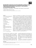
Fig. 1. Protein biosynthesis and aggregation under normal and
stress conditions. (A) Under normal conditions, nascent polypep-
tides either can fold autonomously or require the help of folding
chaperones. Aberrant protein products due to translation errors and
misfolding are handled by the quality control system, composed of
refolding chaperones and proteases. The system is energetically
demanding (most processes are ATP-dependent) but drives the
equilibrium towards the native, folded state [10]. (B) Under most
stress conditions equilibrium is shifted toward the formation of
aberrant products (red lines). This is naturally counteracted by cellu-
lar optimization strategies already present at the source (DNA, pro-
tein sequences and regulation of expression levels) or induced
upon exposure to stress conditions (upregulation of the quality con-
trol machinery). Heterologous protein overproduction, however, can
further affect this delicate balance by competing for available
resources (ribosomes, chaperones but also ATP).
P. Gatti-Lafranconi et al. Potential of bacterial inclusion bodies
FEBS Journal 278 (2011) 2408–2418 ª 2011 The Authors Journal compilation ª 2011 FEBS 2409
Rate and ‘quality’ of protein synthesis can favour
misfolding over folding. Intuitively, an increase in the
concentration of nascent polypeptides makes the fold-
ing process more severe and this is indeed naturally
counteracted by the increase in chaperone concentra-
tion in exponentially growing cells [2]. Rate can, how-
ever, be increased above tolerable limits by mutations
and overexpression, as discussed in the next para-
graphs. Also, ‘quality’ of protein synthesis is affected
by environmental stress due to an increase in the rate
of translational errors, amino acid misincorporation,
premature chain truncation and incomplete modifica-
tions. Such aberrant molecules accumulate in the cyto-
plasm and increase protein aggregation [14].
In general, mutations are retained only if folding
propensity remains above a critical point, indepen-
dently of the advantage that they would provide to
the host [15]. Mutations can affect aggregation, how-
ever, even if protein activity is not compromised, even
if no amino acid replacement is introduced: the DNA
sequence itself determines the rate of synthesis. There
is indeed evidence for genomic-level optimization of
protein folding at both DNA and protein sequence
levels. The distribution of codons in mRNAs has been
found to be unbalanced: as the first 30–50 codons
have low translation efficiency, translation has a slow
start, reducing ribosome clashing, translation stalling
and eventually favouring folding [16]. At protein level,
regions in the primary sequence that are intrinsically
aggregation-prone correlate with those having low
folding propensity (and with chaperone dependence)
[17]. The localization of ‘fast’ codons around those
regions [18] is believed to kinetically promote folding
and the burial of aggregation-prone patches in the
core of the native structure. Although DNA and pro-
tein sequences evolved to optimize translation and
folding efficiency, even a single silent mutation can
induce IB formation, while amino acid replacements
that alter the chemical properties of the polypeptide
will easily result in increased aggregation propensity.
During recombinant protein production, heterologous
proteins will not have their sequence optimized for
expression in E. coli and therefore suffer from poor
folding efficiency even if expression levels are kept
low.
In microbial cell factories, overproduced proteins
can represent up to 90% of the total protein content
and cause the failure of the quality control system that
will result in the accumulation of misfolded proteins
first and eventually lead to the formation of IBs. This
process is highly protein-dependent, driven by DNA
and protein sequences, as discussed above, but can
also be affected by specific folding requirements (i.e.
disulfide bonds) or transcend the folding capability of
E. coli. Other causes of aggregation are heat or oxida-
tive stresses, environmental conditions that cells are
likely to face in natural environments and biotechno-
logical applications. Growth above optimal tempera-
ture eventually results in massive protein unfolding
while reactive oxygen species cause fragmentation and
chemical modification of side chains. Both these events
raise the aggregation propensity of proteins in the
cytoplasm, either by increasing hydrophobic patch
exposure or by altering protein chemical properties
that can result in crosslinking and misfolding.
Recombinant protein production might
induce aggregation and elicit stress
responses
Heterologous protein production is by itself cause of
toxicity for cells, independently of the nature of the
recombinant protein. Energy depletion is the most
immediate result and is due to both the overproduced
protein and the upregulation of those involved in stress
responses. If degradation of the heterologous protein
occurs, even higher energy consumption will result in
little product accumulation at the expenses of biomass
and growth rate. Also, aminoacilated-tRNA depletion
triggers the stringent response [19,20] that causes the
downregulation of the protein and amino acid biosyn-
thesis machinery. In a condition of limited resources
for protein biosynthesis, competition is won by the
recombinant mRNA, causing a decrease in housekeep-
ing mechanisms (i.e. DNA and protein synthesis), rear-
rangements in cellular catabolic rates and slower, if
any, growth rate [21] (Fig. 1B). The DNA damage-
induced SOS response is also reported to be activated
and, although there is no agreement about how protein
overproduction triggers this response, it is likely that
elevated transcription rates of plasmid-encoded genes
causes DNA suffering in cells [22].
While these effects occur ubiquitously, overproduced
proteins have been reported to specifically trigger dif-
ferent cellular responses depending on their properties,
particularly for what concerns aggregation propensity.
Reports on the upregulation of the quality control sys-
tem upon the accumulation of misfolded proteins in
the cytoplasm suggest that this mechanism shares simi-
lar features with the heat-shock response, which causes
the upregulation of genes controlled by the transcrip-
tion factor r
32
. r
32
regulates the expression of genes
coding for known heat-shock proteins (which include
chaperones and proteases) and its own activity depends
on the same chaperones that it regulates [23,24]. It is
believed that, under non-stress conditions, chaperones
Potential of bacterial inclusion bodies P. Gatti-Lafranconi et al.
2410 FEBS Journal 278 (2011) 2408–2418 ª 2011 The Authors Journal compilation ª 2011 FEBS
act as anti-sigma factors, inhibiting r
32
activity
through an induced conformational change [24,25].
When the number of misfolded proteins increases in
the cell, chaperones are saturated and the equilibrium
shifts toward the free version of r
32
, leading to induc-
tion of the stress response.
Nevertheless, the nature and variability of the
recombinant protein stress response suggests a
far more complex and adjustable ‘heat-shock-like’
mechanism [26]. The normal heat-shock response is
transient, fading away shortly after cells are released
from stress, but increased synthesis rates of DnaK,
GroEL chaperones and Lon (the main heat-shock pro-
tease) have been found to last for the whole length of
overproduction. The extent and kinetics of the heat-
shock-like response vary among different production
systems and are influenced by the nature of the protein
synthesized: while energy metabolism, SOS response,
nutrient uptake and the core of the heat-shock
response undergo comparable changes, different
recombinant proteins have distinct impacts on intracel-
lular stress control and growth rates [27–29]. The small
heat-shock proteins IbpA and IbpB, for example, are
upregulated exclusively when proteins accumulate as
IBs, inhibit IB degradation and reduce the stress
response, thus favouring growth [30,31]. A membrane
and a membrane-bound recombinant protein have
opposite effects on growth rate but activate the same
stress-response pattern both at cytoplasm and envelope
level [32]. Conversely, in the cytoplasm recombinant
proteins with different aggregation profiles increase the
abundance of the same set of envelope proteins while
membrane composition and permeability specifically
react to the aggregation state of the recombinant pro-
tein. It has been suggested that the cell membrane
might react with exquisite sensitivity not only to aggre-
gation but even to the complexity of the aggregates
(whether soluble aggregates or large insoluble IBs) and
that membrane lipids may act as a second stress sensor
responsive to the aggregation state of the recombinant
protein [33,34].
The bright side of IBs: from
recombinant protein reservoir to tools
for basic investigation and direct
application in biotechnology
Before the last decade, the properties of protein aggre-
gates knew little glory while most studies pursued
either solubility improvement or denaturation ⁄ renatur-
ation of purified IBs. Within the first line, the most
successful techniques are fusion with solubility tags,
use of molecular and chemical chaperones and modu-
lation of the expression conditions to reduce the rate
of protein biosynthesis [9,35–37], whereas in the second
major efforts are devoted to optimizing the refolding
process so as to regain highest biological activity
(reviewed in [38]). Only during the last decade has a
deeper knowledge of the structural and functional
properties of IBs drawn researchers’ attention to the
possibility to control the conformation of aggregated
proteins, paving the way for the use of IBs in a series
of studies and applications that were difficult to envis-
age only a few years ago.
Such developments require that IBs can be charac-
terized in fine detail, their structure and aggregation
process monitored and controlled. Having structural
information in hand would enable these methods to be
applied in an informed fashion and thus allow a fine
modulation of the aggregation process. In the next sec-
tion we describe and illustrate with some examples the
major tools available for the structural analysis of pro-
teins within aggregates and of aggregates within cells.
Synergic to the latter goal are computational methods
allowing the identification of aggregation-prone
regions within protein primary sequences (reviewed by
Hamodrakas in this issue).
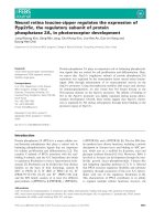
Fig. 2. Methods for the characterization of IBs. (A) Scheme of IB
formation and structural properties. Folding intermediates form sol-
uble aggregates that merge in one or two IBs per cell. The polypep-
tides embedded in IBs can retain native-like structure and activity.
Moreover, IBs can acquire amyloid-like features. Possible applica-
tions related to the peculiar IB structural properties are indicated.
(B) Principal methods of investigation of IB formation and character-
ization.
P. Gatti-Lafranconi et al. Potential of bacterial inclusion bodies
FEBS Journal 278 (2011) 2408–2418 ª 2011 The Authors Journal compilation ª 2011 FEBS 2411
Structural properties of IBs: a review
of the methods
We provide in the following an updated view about
the principal biophysical methods available for the
characterization of proteins aggregated in IBs and
summarize the information generated by their applica-
tion (Fig. 2).
The aggregation of recombinant proteins can be
monitored in vivo by fluorescence spectroscopy and
microscopy if the target protein is fused to a fluores-
cent partner such as the green fluorescent protein
(GFP) or its variants [39]. Using this approach it was
determined that multiple, small and soluble aggregates
form at early stages of the process while, at later times,
these assemblies merge into one or two large aggre-
gates localized at the poles of the cells [39,40]. In vivo
aggregation can also be monitored in real time label-
ling the target protein with the tetra-Cys sequence tag
(Cys-Cys-X-X-Cys-Cys) that specifically binds a fluo-
rescein analogue containing two arsenoxides (FIAsH).
In this approach, the tetra-Cys motif is introduced by
mutagenesis into the protein sequence at a specific
position where its accessibility and binding to FIAsH
will depend on the folding state of the protein. In this
way, FIAsH fluorescence reports on protein stability
and aggregation within cells [41]. Other applications of
fluorescence-based analysis rely on proteins within IBs
retaining native-like structure and activity. For exam-
ple, it was shown that in IBs formed by a GFP-fusion
protein fluorescence emission was higher in the core of
the aggregates than in their external shell [42]. This
observation ruled out the possibility that the biological
activity retained by IBs depends on native-like proteins
passively trapped in the aggregate and instead attrib-
uted this distribution to the specific mechanisms of
protein deposition and removal, and further suggested
that aggregated proteins can complete their folding
and activation process once deposited in IBs [42]. Pro-
tein–protein interactions within IBs have also been
studied using higher resolution fluorescence approaches
such as the Fo
¨
rster resonance energy transfer (FRET)
in which interacting proteins are labelled by two differ-
ent fluorescent probes [43]. Higher FRET efficiency
was obtained when the two probes were fused to the
same peptide rather than to different ones, suggesting
that the process of aggregation is highly protein-spe-
cific [44]. The spatial resolution of optical microscop-
ies, including fluorescence microscopy, is of the order
of 0.1 lm (in the image X, Y plane) due to the diffrac-
tion limit of the employed light. Even in laser scanning
confocal microscopy, the highest resolution of about
0.5 lm is obtained in the Z direction [45].
Electron and atomic force microscopies reach a na-
nometric – and even subnanometric – resolution but
they rely on a more invasive approach to the sample.
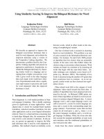
In transmission electron microscopy, thin sections of
fixed cells show IBs as spherical or ellipsoidal electron
dense structures [46,47] and purified IBs appear as
spherical, ellipsoidal or cylindrical particles of 0.5–
1.8 lm characterized by a smooth and porous surface
in both scanning and transmission electron microscopy
(Fig. 3) [46,48]. The porous structure of IBs, also con-
firmed by sedimentation techniques [49], is of relevance
in view of a direct application of active aggregates in
biocatalysis: thanks to the porous and hydrated IB
structure, substrates and products can diffuse inside
and outside making IBs useful depositories of highly
purified enzymes. Electron microscopy was also
applied to studying the shape and surface to volume
ratio of protein aggregates used as biomaterials in
applications where these features are of relevance [50].
Furthermore, both electron microscopy and, in partic-
ular, atomic force microscopy image the surface mor-
phology of the sample at nanometric resolution [51]
and allowed amyloid-like fibrils to be detected in
freshly purified IBs of the human bone morphogenetic
protein-2 (fragment 13–74) [52] and of the prion of the
filamentous fungus Podospora anserine HET-s (frag-
ment 218–289) [53]. Fibrillar structures became more
evident after IB incubation at 37 °C for 12 h [52] or in
the presence of proteinase K [44,54].
The structural properties of IBs at molecular level
have been investigated at a resolution ranging from
protein backbone conformations to single residues by
several optical spectroscopies, such as FTIR, Raman,
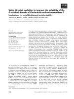
Fig. 3. Transmission electron micrograph of IBs within E. coli cells.
The picture shows IBs formed by GFP fused to an aggregation-
prone domain and the immunolocalization of GFP. Courtesy of Ele-
na Garcı
´
a-Fruito
´
s and Antonio Villaverde.
Potential of bacterial inclusion bodies P. Gatti-Lafranconi et al.
2412 FEBS Journal 278 (2011) 2408–2418 ª 2011 The Authors Journal compilation ª 2011 FEBS
CD and fluorescence, as well as by NMR and X-ray
diffraction.
FTIR spectroscopy allows the study of protein sec-
ondary structures and aggregation through the analysis
of the amide I band, occurring in the 1700–1600 cm
)1
absorption region, which is due to the CO stretching
vibration of the peptide bond (reviewed in [55] and ref-
erences therein). Absorption of the different secondary
structures of the proteins overlaps in this spectral
range and can be resolved by resolution enhancement
approaches, such as the second derivative analysis of
the spectra. In this way, the secondary structure com-
ponents appear as negative peaks in the derivative
spectrum and each peak can be assigned according to
its wavenumber. For instance, in water a-helices and
random coils absorb between 1660 and 1648 cm
)1
,
intramolecular b-sheets between 1640 and 1623 cm
)1
and around 1686 cm
)1
, whereas intermolecular b-sheet
absorption in protein aggregates is found between
1630 and 1620 cm
)1
and around 1695 cm
)1
. FTIR
(micro)spectroscopy allows protein secondary struc-
tures and aggregation to be studied also within com-
plex biological systems, i.e. whole intact cells [56–58],
tissues [59] and whole organisms [60]. Moreover,
changes in the intensity of the aggregate spectral com-
ponent around 1625 cm
)1
have been used to follow the
kinetics of IB formation within a growing culture of
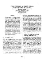
E. coli. To exemplify this approach, Fig. 4A reports
the second derivative spectrum of E. coli cells during
production of a recombinant lipase. Six hours after
induction at 37 °C the protein is mainly deposited in
aggregates, as can easily be determined based on the
appearance of a shoulder at 1627 cm
)1
that has no
counterpart in the control cells and is attributed to
intermolecular b-sheet structures in protein aggregates.
Subtraction of the spectrum of control cells allowed
the spectral component (1627 cm
)1
) unique to aggre-
gates to be resolved in more detail (Fig. 4B) and the
kinetics of IB formation at different temperatures,
namely at 37 and 27 °C, the latter compatible with the
partitioning of the recombinant protein between solu-
ble and insoluble proteins, to be monitored and com-
pared [57]. Spectra of IBs (Fig. 4C) purified from cells
revealed that the intermolecular b-sheet component of
protein aggregates, peaked at 1627 cm
)1
, was higher at
the higher temperature, while proteins embedded in
IBs formed at 27 °C retained more native-like a-helical
content (1656 cm
)1
). These results suggest FTIR
(micro)spectroscopy as a technique of choice also in
the study of the influence of the physiology of expres-
sion (i.e. temperature, induction, formation of disulfide
bonds) on the kinetics of aggregation and on the struc-
ture of aggregated proteins [57,61].
Another vibrational technique that can be employed
to characterize the structural properties of IBs is
Raman (micro)spectroscopy, where the inelastic scat-
tering of laser light from the sample is detected. Pio-
neering work of Przybycien et al. detected in IBs
formed by recombinant b-lactamase an increased level
of b-sheet structures and the retention of native-like a-
helix content [62]. This technique can be considered
complementary to FTIR spectroscopy, since the two
methods detect different vibrational modes of the sam-
ple. Raman spectroscopy is more sensitive to the
amino acid side chain response [63] while – as dis-
cussed above – FTIR is more sensitive to the backbone
amide I vibrations. We believe that Raman
(micro)spectroscopy could offer advantages still
Fig. 4. FTIR analysis of the aggregation of a recombinant protein in
E. coli. (A) Second derivatives of the FTIR absorption spectra of
E. coli cells synthesizing a recombinant lipase from Pseudomonas
fragi (PFL) at 37 °C after 6 h from induction (continuous line) and of
the control cells (dashed line). (B) Second derivative of the differ-
ence spectrum between cells producing the recombinant protein
and control cells reported in (A) (continuous line). In this subtracted
spectrum, the band at 1627 cm
)1
due to intermolecular b-sheets in
aggregates is well resolved allowing the kinetics of IB formation
within intact cells to be monitored. The same analysis performed at
27 °C is shown (dotted-dashed line). (C) Second derivative absorp-
tion spectra of IBs extracted after 10 h from induction at 27 °C
(dotted-dashed line) and 37 °C (continuous line).
P. Gatti-Lafranconi et al. Potential of bacterial inclusion bodies
FEBS Journal 278 (2011) 2408–2418 ª 2011 The Authors Journal compilation ª 2011 FEBS 2413
unexplored in IB studies, since relevant information on
disulfide bond formation and on solvent accessibility
of specific amino acid side chains can be obtained [63].
The presence of b-sheet structures in extracted IBs
can also be detected by far UV CD [52,54], even if it is
not easy to discriminate between intramolecular and
intermolecular b-sheets. The use of this spectroscopic
technique for the study of IB aggregates is often
limited by the intrinsic insolubility of the samples,
responsible for a high level of light scattering distur-
bances and signal loss.
The characteristic presence of b-sheet structures
within extracted IBs has also been confirmed by X-ray
diffraction. Spectra typically display two circular
reflections around 4.7 A
˚
and 10.2 A
˚
, respectively,
assigned to the spacing between strands within a
b-sheet and between b-sheets. The circular shape of
these reflections has been suggested to arise from not
strongly aligned b-sheets within IBs [52,64].
NMR spectroscopy has been widely applied in pro-
tein science, since it enables detailed structural infor-
mation at the specific residue level up to the three-
dimensional structure of the protein to be obtained. In
particular, solid state NMR rotational-echo double-
resonance (REDOR) has been applied to IBs, both
extracted and within intact cells [65]. In this approach,
the backbone carbonyl and nitrogen are labelled (
13
CO
and
15
N) for each amino acid, since its
13
CO chemical
shift allows information to be obtained on local con-
formation. In this way, Curtis-Fiske et al. were able to
identify native a-helices of the N-terminal 185 residues
of the functional domain of the HA2 subunit of the
influenza virus hemagglutinin protein and to detect
conformational heterogeneity of the protein within IBs
[65]. NMR spectroscopy has also been applied to
localize b-sheet structures in protein aggregates, mainly
by hydrogen ⁄ deuterium (H ⁄ D) exchange experiments
that allow residue-specific backbone amides protected
from solvent exchange because they are involved in
hydrogen bonds to be detected. The assignment of sol-
vent-protected residues to b-sheet structures can be
obtained also by other spectroscopic techniques such
as CD and X-ray diffraction [52]. It is noteworthy that
NMR-based approaches, such as solid-state NMR
13
C–
13
C proton-driven spin diffusion and liquid-state
NMR H ⁄ D exchange experiments, offer the unique
possibility of comparing at the residue-specific level
protein aggregates of different types, such as IBs, amy-
loid fibrils and thermal aggregates [53,64]. The out-
comes of these NMR experiments could therefore
allow the aggregate residue-specific structural proper-
ties to be correlated with their functional features, such
as enzymatic activity or cellular toxicity.
Exploitation of IBs in biotechnology
and in protein science
It is widely recognized that proteins can aggregate in
IBs in different folding states that can eventually coex-
ist within the same aggregates. The conformation
acquired within aggregates is dependent on the nature
of the protein itself [66] but can also be controlled
through the genetic background of the host cells
and ⁄ or manipulation of the experimental conditions.
This novel and in a way revolutionary knowledge has
important consequences in the rationale of handling
and studying IBs. The development of methods to con-
trol and monitor the process of aggregation allows for
the production of aggregated proteins endowed with
residual structure and biological activity that can find
direct use in biotechnology. In addition, a detailed
analysis of the mode of building and of the structure
of aggregates can be useful to dissect pathways and
bottlenecks in the folding of specific proteins, for
example those containing disulfide bonds or requiring
cofactors, multidomain proteins, fusion proteins. In
the following we summarize recent progress in this
field, whereas the use of IBs in the study of amyloid
aggregation is developed in the accompanying review
paper by Garcı
´
a-Fruito
´
s and colleagues.
Two very relevant accomplishments towards IB
exploitation in biotechnology are based on the ability
to enrich aggregates in native-like structured proteins
making them suitable for direct use in biocatalysis
and ⁄ or as a source of relatively pure proteins that can
be released through mild solubilization. Given that
aggregation often cannot be fully avoided – or is even
considered an advantage – the same experimental
‘tricks’ developed to improve the solubility of recombi-
nant proteins (reviewed in [35]) can be applied to pro-
duce IBs mostly composed of native-like, although not
soluble, recombinant proteins.
The list of recombinant proteins that precipitate in
IBs in a conformation permissive for biological activity
has progressively grown since researchers started to
measure this parameter and includes, among others,
b-galactosidase [67], endoglucanase [68], GFP [69],
a bacterial lipase [57], oxidases [70], kinases [71]
phosphorylases [72] aldolases [73], transglutaminases
[74] and the colony stimulating factor [75]. This knowl-
edge soon generated the idea of directly using IBs in
biocatalysis, thus avoiding the cumbersome step of
resolubilization. Since recovery of IBs from cell
extracts can be quite easily achieved, this method
could be of broad scope, provided aggregated proteins
retain enough biological activity. Unfortunately, so far
the comparison of the specific activity of soluble and
Potential of bacterial inclusion bodies P. Gatti-Lafranconi et al.
2414 FEBS Journal 278 (2011) 2408–2418 ª 2011 The Authors Journal compilation ª 2011 FEBS
aggregated proteins has been performed only sporadi-
cally although the competitiveness of IB catalysis
depends on the balance between a possible reduction
of specific activity and the advantages produced by
avoiding solubilization steps. Data available show that
depending on the protein and the production protocol
the biological activity of aggregates can vary from 11%
[69] to nearly 100% [68] of the soluble counterpart.
IBs embedding native-like proteins are also proposed
as a source of pure recombinant proteins that can be
easily released upon mild treatments that avoid chemi-
cal disruption of cells and denaturation of the aggre-
gates. Protein–protein interactions are in fact weaker
and ‘relaxed’ IBs can be dissolved in mild detergent at
low concentration. Since proteins have not been dena-
tured during solubilization, there is no need to intro-
duce refolding steps, which is of great advantage since
solubilization ⁄ refolding is often a critical step in the
production of recombinant proteins. Interestingly the
approach has been successfully tested with proteins not
related in their structure, among them the granulocyte
colony stimulating factor, GFP and a truncated form
of the tumour necrosis factor [76].
An innovative evolution towards IB-based catalysis
exploits the idea of forcing otherwise soluble proteins
to aggregate in IBs. This method is proposed as an
alternative to the better known procedures of enzyme
insolubilization via immobilization on carriers or via
aggregation by crosslinking (reviewed in [77]). The pro-
tein of interest is fused to an aggregation-prone moiety
promoting the aggregation of the chimeric polypeptide.
The cellulose-binding module, a very poorly soluble
protein, was used to induce intracellular deposition of
the recombinant d-amino acid oxidase (DAAO) from
Trigonopsis variabilis, an enzyme used in the synthesis
of 7-amino cephalosporanic acid [70]. The observation
that DAAO IBs retained specific activity close to that
of the soluble enzyme and were resistant under condi-
tions that inactivate free DAAO substantiated the fea-
sibility of this approach, which was then applied also
to a maltodextrin phosphorylase [72], a polyphosphate
kinase [71] and a sialic acid aldolase [73]. Clearly,
fusion with the cellulose binding domain did not inter-
fere with the correct folding of the partner protein that
aggregated in a form endowed with biological activity
(in the case of DAAO this means also ability to bind
the cofactor).
In the same conceptual frame – making soluble pro-
teins insoluble – other authors have developed a self-
assembly complex in which IBs are formed through
in vivo aggregation of polyhydroxybutyrate synthase
PhaC carrying at its N-terminus a negatively charged
coil [78]. Aggregates of this protein expose on their
surface charged regions that can bind active soluble
enzymes tagged at their C-terminus with a positively
charged coil.
In both cases, examples available are still too few to
be generalized in a broad scope experimental
approach. However, the importance of IBs as direct or
indirect immobilization carriers might increase when,
for instance, different enzymes ⁄ proteins can participate
in the same aggregate to build a multifunctional aggre-
gated catalyst.
Finally, but not less important, it should be con-
sidered that pathways of protein folding are reflected
in the formation of IBs and in their structure. Study-
ing protein aggregates can therefore provide a first
glimpse about the occurrence of folding-limiting
steps. The finding that aggregates of several different
proteins, for example INF-a-2b [56], a bacterial
lipase [57], a mutant of the Ab42 Alzheimer peptide
[79] and GFP [69] can be endowed with substantial
amounts of native structure led to the conclusion
that the process of intracellular aggregation can
involve proteins in a continuum of conformational
states. This idea is well substantiated by the demon-
stration that different conformations of the same
polypeptide coexist in IBs [80]. However, the struc-
ture of aggregated TEM1-b-lactamase inside IBs
could not be affected by any of the usual means
[81]. In this particular case it was therefore con-
cluded that TEM aggregation is only controlled by
the amino acid sequence and not by the kinetics of
folding, since changing the rate of biosynthesis did
not result in structural changes in the aggregates.
This result was interpreted as evidence about the
existence of a single specific folding step critical for
the protein undergoing either aggregation or native
folding.
Analysis of the modulation of aggregation in bacte-
ria was also of support in clarifying critical steps of
oxidative folding of bovine b-lactoglobulin. b-Lacto-
globulin carries five cysteine residues, four of which
link in disulfide bridges, raising questions about the
role (if any) of the free thiol during in vivo folding.
Upon overproduction in E. coli cells optimized for the
intracellular formation of disulfide bonds, it was
observed that a mutant protein deprived of the
unpaired Cys was more prone to aggregation than the
wild type, pointing to a contribution of the free thiol
in the pathway leading to the formation of native
bonds [61].
The number of proteins studied up to now is still
too limited to try to generalize which structural,
sequence and kinetic properties might dictate the fine
detail of aggregation. However, structural analysis of
P. Gatti-Lafranconi et al. Potential of bacterial inclusion bodies
FEBS Journal 278 (2011) 2408–2418 ª 2011 The Authors Journal compilation ª 2011 FEBS 2415
IBs produced in different conditions can be considered
as an easy tool to detect the presence of critical
folding intermediates to be characterized with other
techniques.
To conclude, we believe that a truly successful
understanding and exploitation of IBs requires an
advanced understanding of cellular and protein mech-
anisms leading to aggregation as well as powerful
biophysical detection methods. Reported examples
highlight the potential of these approaches in creating
new generation protein depositories and biocatalysts.
Acknowledgements
S. M. D. and M. L. acknowledge support by FAR
(Fondo di Ateneo per la Ricerca) of the University of
Milano-Bicocca. P. G. -L. is the recipient of a Marie
Curie Intra-European F ellowship. A. N. and D. A. a ckno-
wledge postdoctoral fellowships of the University of
Milano-Bicocca.
References
1 Ellis RJ & Minton AP (2003) Cell biology: join the
crowd. Nature 425, 27–28.
2 Vendeville A, Lariviere D & Fourmentin E (2011) An
inventory of the bacterial macromolecular components
and their spatial organization. FEMS Microbiol Rev 35,
395–414.
3 Ando T & Skolnick J (2010) Crowding and hydrody-
namic interactions likely dominate in vivo macromolecu-
lar motion. Proc Natl Acad Sci USA 107, 18457–18462.
4 McGuffee SR & Elcock AH (2010) Diffusion, crowding
and protein stability in a dynamic molecular model of
the bacterial cytoplasm. PLoS Comput Biol 6, e1000694.
5 Cheung MS, Klimov D & Thirumalai D (2005) Molecu-
lar crowding enhances native state stability and refold-
ing rates of globular proteins. Proc Natl Acad Sci USA
102, 4753–4758.
6 Gershenson A & Gierasch LM (2011) Protein folding in
the cell: challenges and progress. Curr Opin Struct Biol
21, 32–41.
7 Jahn TR & Radford SE (2005) The yin and yang of
protein folding. FEBS J 272, 5962–5970.
8 Kramer G, Boehringer D, Ban N & Bukau B (2009)
The ribosome as a platform for co-translational pro-
cessing, folding and targeting of newly synthesized pro-
teins. Nat Struct Mol Biol 16, 589–597.
9 Baneyx F & Mujacic M (2004) Recombinant protein
folding and misfolding in Escherichia coli. Nat Biotech-
nol 22, 1399–1408.
10 Hartl FU & Hayer-Hartl M (2009) Converging concepts
of protein folding in vitro and in vivo. Nat Struct Mol
Biol 16, 574–581.
11 Maisonneuve E, Ezraty B & Dukan S (2008) Protein
aggregates: an aging factor involved in cell death.
J Bacteriol 190, 6070–6075.
12 Lindner AB, Madden R, Demarez A, Stewart EJ &
Taddei F (2008) Asymmetric segregation of protein
aggregates is associated with cellular aging and rejuve-
nation. Proc Natl Acad Sci USA 105, 3076–3081.
13 Tyedmers J, Mogk A & Bukau B (2010) Cellular strate-
gies for controlling protein aggregation. Nat Rev Mol
Cell Biol 11, 777–788.
14 Kurland C & Gallant J (1996) Errors of heterologous
protein expression. Curr Opin Biotechnol 7, 489–493.
15 Tokuriki N & Tawfik DS (2009) Stability effects of
mutations and protein evolvability. Curr Opin Struct
Biol 19, 596–604.
16 Tuller T, Carmi A, Vestsigian K, Navon S, Dorfan Y,
Zaborske J, Pan T, Dahan O, Furman I & Pilpel Y
(2010) An evolutionarily conserved mechanism for con-
trolling the efficiency of protein translation. Cell 141,
344–354.
17 Tartaglia GG & Vendruscolo M (2010) Proteome-level
interplay between folding and aggregation propensities
of proteins. J Mol Biol 402, 919–928.
18 Lee Y, Zhou T, Tartaglia GG, Vendruscolo M & Wilke
CO (2010) Translationally optimal codons associate
with aggregation-prone sites in proteins. Proteomics 10,
4163–4171.
19 Potrykus K & Cashel M (2008) (p)ppGpp: still magical?
Annu Rev Microbiol 62, 35–51.
20 Gallant JA (1979) Stringent control in E coli. Annu Rev
Genet 13, 393–415.
21 Hoffmann F & Rinas U (2004) Stress induced by
recombinant protein production in Escherichia coli. Adv
Biochem Eng Biotechnol 89, 73–92.
22 Wegrzyn G & Wegrzyn A (2002) Stress responses and
replication of plasmids in bacterial cells. Microb Cell
Fact 1,2.
23 Yura T & Nakahigashi K (1999) Regulation of the
heat-shock response. Curr Opin Microbiol 2, 153–158.
24 Guisbert E, Yura T, Rhodius VA & Gross CA (2008)
Convergence of molecular, modeling, and systems
approaches for an understanding of the Escherichia coli
heat shock response. Microbiol Mol Biol Rev 72, 545–554.
25 Baneyx F & Nannenga BL (2010) Chaperones: a story
of thrift unfolds. Nat Chem Biol 6, 880–881.
26 Harcum SW & Haddadin FT (2006) Global transcrip-
tome response of recombinant Escherichia coli to heat-
shock and dual heat-shock recombinant protein induc-
tion. J Ind Microbiol Biotechnol 33, 801–814.
27 Durrschmid K, Reischer H, Schmidt-Heck W, Hrebicek
T, Guthke R, Rizzi A & Bayer K (2008) Monitoring of
transcriptome and proteome profiles to investigate the
cellular response of E. coli towards recombinant
protein expression under defined chemostat conditions.
J Biotechnol 135, 34–44.
Potential of bacterial inclusion bodies P. Gatti-Lafranconi et al.
2416 FEBS Journal 278 (2011) 2408–2418 ª 2011 The Authors Journal compilation ª 2011 FEBS
28 Gill RT, Valdes JJ & Bentley WE (2000) A comparative
study of global stress gene regulation in response to
overexpression of recombinant proteins in Escherichia
coli. Metab Eng 2, 178–189.
29 Smith HE (2007) The transcriptional response of
Escherichia coli to recombinant protein insolubility.
J Struct Funct Genomics 8, 27–35.
30 Hoffmann F & Rinas U (2000) Kinetics of heat-shock
response and inclusion body formation during tempera-
ture-induced production of basic fibroblast growth fac-
tor in high-cell-density cultures of recombinant
Escherichia coli. Biotechnol Prog 16, 1000–1007.
31 Lethanh H, Neubauer P & Hoffmann F (2005) The
small heat-shock proteins IbpA and IbpB reduce the
stress load of recombinant Escherichia coli and delay
degradation of inclusion bodies. Microb Cell Fact 4,6.
32 Xu LY & Link AJ (2009) Stress responses to heterolo-
gous membrane protein expression in Escherichia coli.
Biotechnol Lett 31, 1775–1782.
33 Villa R, Lotti M & Gatti-Lafranconi P (2009)
Components of the E. coli envelope are affected by and
can react to protein over-production in the cytoplasm.
Microb Cell Fact 8, 32.
34 Ami D, Natalello A, Schultz T, Gatti-Lafranconi P, Lotti
M, Doglia SM & de Marco A (2009) Effects of recombi-
nant protein misfolding and aggregation on bacterial
membranes. Biochim Biophys Acta 1794, 263–269.
35 Sorensen HP & Mortensen KK (2005) Soluble expres-
sion of recombinant proteins in the cytoplasm of
Escherichia coli. Microb Cell Fact 4,1.
36 de Marco A, Deuerling E, Mogk A, Tomoyasu T &
Bukau B (2007) Chaperone-based procedure to increase
yields of soluble recombinant proteins produced in
E. coli. BMC Biotechnol 7, 32.
37 de Marco A, Vigh L, Diamant S & Goloubinoff P
(2005) Native folding of aggregation-prone recombinant
proteins in Escherichia coli by osmolytes, plasmid- or
benzyl alcohol-overexpressed molecular chaperones. Cell
Stress Chaperones 10, 329–339.
38 Burgess RR (2009) Refolding solubilized inclusion body
proteins. Methods Enzymol 463, 259–282.
39 Rokney A, Shagan M, Kessel M, Smith Y, Rosenshine
I & Oppenheim AB (2009) E. coli transports aggregated
proteins to the poles by a specific and energy-dependent
process. J Mol Biol 392, 589–601.
40 Winkler J, Seybert A, Konig L, Pruggnaller S, Hasel-
mann U, Sourjik V, Weiss M, Frangakis AS, Mogk A
& Bukau B (2010) Quantitative and spatio-temporal
features of protein aggregation in Escherichia coli and
consequences on protein quality control and cellular
ageing. EMBO J 29, 910–923.
41 Ignatova Z & Gierasch LM (2004) Monitoring protein
stability and aggregation in vivo by real-time fluorescent
labeling. Proc Natl Acad Sci USA 101, 523–528.
42 Garcia-Fruitos E, Aris A & Villaverde A (2007) Locali-
zation of functional polypeptides in bacterial inclusion
bodies. Appl Environ Microbiol 73 , 289–294.
43 Lakowicz JR (2006) Principles of Fluorescence Spectros-
copy, 3rd edn. Springer, New York, NY.
44 Morell M, Bravo R, Espargaro A, Sisquella X, Aviles
FX, Fernandez-Busquets X & Ventura S (2008) Inclu-
sion bodies: specificity in their aggregation process and
amyloid-like structure. Biochim Biophys Acta 1783,
1815–1825.
45 Wilson T (1990) Confocal Microscopy. Academic Press,
London.
46 Bowden GA, Paredes AM & Georgiou G (1991) Struc-
ture and morphology of protein inclusion-bodies in Esc-
herichia coli. Biotechnol 9, 725–730.
47 Rinas U, Boone TC & Bailey JE (1993) Characteriza-
tion of inclusion-bodies in recombinant Escherichia coli
producing high-levels of porcine somatotropin. J Bio-
technol 28, 313–320.
48 Carrio MM, Cubarsi R & Villaverde A (2000) Fine
architecture of bacterial inclusion bodies. FEBS Lett
471, 7–11.
49 Taylor G, Hoare M, Gray DR & Marston FAO (1986)
Size and density of protein inclusion-bodies. Biotechnol
4, 553–557.
50 Garcia-Fruitos E, Seras-Franzoso J, Vazquez E &
Villaverde A (2010) Tunable geometry of bacterial
inclusion bodies as substrate materials for tissue engi-
neering. Nanotechnology 21, 205101.
51 Muller DJ & Dufrene YF (2008) Atomic force micros-
copy as a multifunctional molecular toolbox in nano-
biotechnology. Nat Nanotechnol 3, 261–269.
52 Wang L, Maji SK, Sawaya MR, Eisenberg D & Riek R
(2008) Bacterial inclusion bodies contain amyloid-like
structure. PLoS Biol 6, 1791–1801.
53 Wasmer C, Benkemoun L, Sabate R, Steinmetz MO,
Coulary-Salin B, Wang L, Riek R, Saupe SJ & Meier
BH (2009) Solid-state NMR spectroscopy reveals that
E. coli inclusion bodies of HET-s(218-289) are amy-
loids. Angew Chem Int Ed 48, 4858–4860.
54 Sabate R, Espargaro A, Saupe SJ & Ventura S(2009)
Characterization of the amyloid bacterial inclusion
bodies of the HET-s fungal prion. Microbial Cell
Fact 8, 56.
55 Doglia SM, Ami D, Natalello A, Gatti-Lafranconi P &
Lotti M (2008) Fourier transform infrared spectroscopy
analysis of the conformational quality of recombinant
proteins within inclusion bodies. Biotechnol J 3, 193–201.
56 Ami D, Natalello A, Taylor G, Tonon G & Doglia SM
(2006) Structural analysis of protein inclusion bodies by
Fourier transform infrared microspectroscopy. Biochim
Biophys Acta 1764, 793–799.
57 Ami D, Natalello A, Gatti-Lafranconi P, Lotti M &
Doglia SM (2005) Kinetics of inclusion body formation
P. Gatti-Lafranconi et al. Potential of bacterial inclusion bodies
FEBS Journal 278 (2011) 2408–2418 ª 2011 The Authors Journal compilation ª 2011 FEBS 2417
studied in intact cells by FT-IR spectroscopy. FEBS
Lett 579, 3433–3436.
58 Ami D, Bonecchi L, Cali S, Orsini G, Tonon G & Do-
glia SM (2003) FT-IR study of heterologous protein
expression in recombinant Escherichia coli strains.
Biochim Biophys Acta 1624, 6–10.
59 Choo LP, Wetzel DL, Halliday WC, Jackson M,
LeVine SM & Mantsch HH (1996) In situ characteriza-
tion of beta-amyloid in Alzheimer’s diseased tissue by
synchrotron Fourier transform infrared microspectros-
copy. Biophys J 71, 1672–1679.
60 Diomede L, Cassata G, Fiordaliso F, Salio M, Ami D,
Natalello A, Doglia SM, De Luigi A & Salmona M
(2010) Tetracycline and its analogues protect
Caenorhabditis elegans from beta amyloid-induced
toxicity by targeting oligomers. Neurobiol Dis 40,
424–431.
61 Invernizzi G, Annoni E, Natalello A, Doglia SM &
Lotti M (2008) In vivo aggregation of bovine beta-lacto-
globulin is affected by Cys at position 121. Protein Expr
Purif 62, 111–115.
62 Przybycien TM, Dunn JP, Valax P & Georgiou G
(1994) Secondary structure characterization of beta-lac-
tamase inclusion-bodies. Protein Eng 7, 131–136.
63 Wen ZQ (2007) Raman spectroscopy of protein phar-
maceuticals. J Pharm Sci 96, 2861–2878.
64 Wang L, Schubert D, Sawaya MR, Eisenberg D & Riek
R (2010) Multidimensional structure–activity
relationship of a protein in its aggregated states.
Angewandte Chemie Intl Edn 49, 3904–3908.
65 Curtis-Fisk J, Spencer RM & Weliky DP (2008) Native
conformation at specific residues in recombinant
inclusion body protein in whole cells determined with
solid-state NMR spectroscopy. J Am Chem Soc 130,
12568–12569.
66 de Groot NS & Ventura S (2010) Protein aggrega-
tion profile of the bacterial cytosol. PLoS ONE 5,
e9383.
67 Worrall DM & Goss NH (1989) The formation of bio-
logically active beta-galactosidase inclusion bodies in
Escherichia coli. Aust J Biotechnol 3, 28–32.
68 Tokatlidis K, Dhurjati P, Millet J, Beguin P &
Aubert JP (1991) High activity of inclusion bodies
formed in Escherichia coli overproducing Clostridium
thermocellum endoglucanase D. FEBS Lett 282,
205–208.
69 Vera A, Gonzalez-Montalban N, Aris A & Villaverde A
(2007) The conformational quality of insoluble recombi-
nant proteins is enhanced at low growth temperatures.
Biotechnol Bioeng 96, 1101–1106.
70 Nahalka J & Nidetzky B (2007) Fusion to a pull-down
domain: a novel approach of producing Trigonopsis
variabilis D-amino acid oxidase as insoluble enzyme
aggregates. Biotechnol Bioeng 97, 454–461.
71 Nahalka J & Patoprsty V (2009) Enzymatic synthesis of
sialylation substrates powered by a novel polyphosphate
kinase (PPK3). Org Biomol Chem 7, 1778–1780.
72 Nahalka J (2008) Physiological aggregation of maltod-
extrin phosphorylase from Pyrococcus furiosus
and its
application in a process of batch starch degradation to
alpha-d-glucose-1-phosphate. J Ind Microbiol Biotechnol
35, 219–223.
73 Nahalka J, Vikartovska A & Hrabarova E (2008) A
crosslinked inclusion body process for sialic acid synthe-
sis. J Biotechnol 134, 146–153.
74 Carvajal P, Gibert J, Campos N, Lopera O, Barbera E,
Torne JM & Santos M (2011) Activity of maize trans-
glutaminase overexpressed in Escherichia coli inclusion
bodies: an alternative to protein refolding. Biotechnol
Prog 27, 232–240.
75 Jevsevar S, Gaberc-Porekar V, Fonda I, Podobnik B,
Grdadolnik J & Menart V (2005) Production of non-
classical inclusion bodies from which correctly folded
protein can be extracted. Biotechnol Prog 21, 632–639.
76 Peternel S, Grdadolnik J, Gaberc-Porekar V & Komel
R (2008) Engineering inclusion bodies for non denatur-
ing extraction of functional proteins. Microb Cell Fact
7, 34.
77 Roessl U, Nahalka J & Nidetzky B (2010) Carrier-free
immobilized enzymes for biocatalysis. Biotechnol Lett
32, 341–350.
78 Steinmann B, Christmann A, Heiseler T, Fritz J & Kol-
mar H (2010) In vivo enzyme immobilization by inclusion
body display. Appl Environ Microbiol 76, 5563–5569.
79 de Groot NS, Aviles FX, Vendrell J & Ventura S
(2006) Mutagenesis of the central hydrophobic cluster
in Abeta42 Alzheimer’s peptide. Side-chain properties
correlate with aggregation propensities. FEBS J 273,
658–668.
80 Schrodel A & de Marco A (2005) Characterization of
the aggregates formed during recombinant protein
expression in bacteria. BMC Biochem 6, 10.
81 Margreiter G, Schwanninger M, Bayer K & Obinger C
(2008) Impact of different cultivation and induction
regimes on the structure of cytosolic IBs of TEM1-beta-
lactamase. Biotechnol J 3, 1245–1255.
Potential of bacterial inclusion bodies P. Gatti-Lafranconi et al.
2418 FEBS Journal 278 (2011) 2408–2418 ª 2011 The Authors Journal compilation ª 2011 FEBS