Báo cáo khoa học: A steady-state competition model describes the modulating effects of thrombomodulin on thrombin inhibition by plasminogen activator inhibitor-1 in the absence and presence of vitronectin ppt
Bạn đang xem bản rút gọn của tài liệu. Xem và tải ngay bản đầy đủ của tài liệu tại đây (440.93 KB, 10 trang )
A steady-state competition model describes the modulating effects
of thrombomodulin on thrombin inhibition by plasminogen activator
inhibitor-1 in the absence and presence of vitronectin
Rob J. Dekker, Hans Pannekoek and Anton J. G. Horrevoets
Department of Biochemistry, Academic Medical Center, University of Amsterdam, the Netherlands
Thrombomodulin (TM) slows down the interaction rate
between thrombin and plasminogen activator inhibitor 1
(PAI-1). We now show that the 12-fold reduced inhibition
rate in the presence of TM does not result from an altered
distribution between PAI-1 cleavage and irreversible com-
plex formation. Surface plasmon resonance (SPR) revealed
an over 200-fold reduced affinity of TM for thrombin-
VR1
tPA
as compared to thrombin, demonstrating the
importance of the VR1 loop in the interaction of thrombin
with both TM and PAI-1. Furthermore, in contrast to ATIII,
PAI-1 was not able to bind the thrombin/TM complex
demonstrating complete competitive binding between PAI-1
and TM. Kinetic modeling on the inhibitory effect of TM
confirms a mechanism that involves complete steric blocking
of the thrombin/PAI-1 interaction. Also, it accurately
decribes the biphasic inhibition profile resulting from the
substantial reduction of the extremely fast rate of reversible
Michaelis complex formation, which is essential for efficient
inhibition of thrombin by PAI-1. Vitronectin (VN) is shown
to partially relieve TM inhibitory action only by vastly
increasing the initial rate of interaction between free
thrombin and PAI-1. In addition, SPR established that
solution-phase PAI-1/VN complexes and non-native VN
(extracellular matrix form) bind TM directly via the chon-
droitin sulphate moiety of TM. Collectively, these results
show that VR1 is a subsite of exosite 1 on thrombin’s surface,
which regulates exclusive binding of either PAI-1 or TM.
This competition will be physiologically significant in con-
trolling the mitogenic activity of thrombin during vascular
disease.
Keywords: serine protease; serpin; suicide substrate mecha-
nism; competitive inhibitor; kinetic modeling.
Classically, the serine protease thrombin is known for its
dual role in hemostasis, exhibiting coagulant as well as
anticoagulant properties. Reversible binding of thrombin to
the endothelial cell surface cofactor thrombomodulin (TM)
endows thrombin with potent anticoagulant properties [1,2].
The thrombin/TM complex is no longer able to bind and
cleave fibrinogen and various other substrates and inhibitors
but becomes a potent activator of protein C. The catalytic
activity of thrombin can be inhibited by a number of serine
protease inhibitors (serpins), including antithrombin III
(ATIII), heparin cofactor II, and PAI-1. Inactivation of
thrombin by PAI-1, however, is a very inefficient process
with a second-order rate constant (k
i
)of10
3
M
)1
Æs
)1
,which
can be increased up to 250-fold by the cofactors vitronectin
(VN) and heparin [3,4].
The VR1- or 37-loop of thrombin has been implicated in
a number of intriguing interactions. First, substitution of the
VR1-loop of thrombin by that of t-PA, yielding thrombin-
VR1
tPA
, increases the bimolecular rate constant of inhibi-
tion by PAI-1 at least 1000-fold to 10
6
M
)1
Æs
)1
[5]. Recently,
we reported that this alteration results from an increased
rate of a unimolecular catalytic step [6]. It has been
unambiguously evidenced that VR1 is essential for the
interaction of both t-PA [7] and thrombin [5,6] with PAI-1.
Second, binding kinetics and structural studies have esta-
blished that the epidermal growth factor domains 4–6
(EGF4–6) of TM bind thrombin electrostatically at exosite
1 [8], but are also involved in hydrophobic contacts with
VR1-loop residues [9,10]. As a result, a marked influence of
TM binding on the interaction of PAI-1 and thrombin can
be envisioned. The hirudin-derived decapeptide hirugen,
however, also interacts with thrombin by utilizing exosite I
[1,2,11], although it did not prevent binding of PAI-1 but
altered a catalytic step of the reaction between thrombin and
PAI-1 [6].
TM acts as a positive effector on other thrombin
interactions, e.g. with ATIII, protein C inhibitor, thrombin-
activatable fibrinolysis inhibitor (TAFI) and protein C
Correspondence to A. J. G. Horrevoets, Academic Medical Center,
Department of Biochemistry, Room K1-161, Meibergdreef 15,
1105 AZ Amsterdam, the Netherlands.
Fax: + 31 20 6915519, Tel.: + 31 20 5665153,
E-mail:
Abbreviations: VR1, variable region-1 (also 37-loop); t-PA, tissue-type
plasminogen activator; u-PA, urokinase-type plasminogen activator;
VR1
tPA
, VR1 loop of t-PA; serpin, serine protease inhibitor; PAI-1,
plasminogen activator inhibitor type 1; RCL, reactive center loop;
PPACK, Phe-Pro-Arg-chloromethylketone; VN, vitronectin;
TM, thrombomodulin; rl-TM, rabbit-lung thrombomodulin;
solulin, soluble human recombinant thrombomodulin; SPR, surface
plasmon resonance; ATIII, antithrombin III; k
i
, second-order rate
constant of inhibition; r, partition ratio; k
on
, association rate constant;
k
off
, dissociation rate constant; K
d
, thermodynamic equilibrium
dissociation constant.
(Received 15 October 2002, revised 24 December 2002,
accepted 3 March 2003)
Eur. J. Biochem. 270, 1942–1951 (2003) Ó FEBS 2003 doi:10.1046/j.1432-1033.2003.03552.x
[11–13]. However, previous work from our laboratory has
demonstrated that thrombin inhibition by PAI-1/VN com-
plexes is impaired in the presence of TM [4]. These findings
have later been studied in more detail, though the mecha-
nism of TM interference in this interaction has not yet been
elucidated, nor is any evidence available on the possible
physiologic role [14,15]. Interestingly, using immunohisto-
chemistry, TM antigen was demonstrated on vascular
smooth muscle cells (SMC), monocytes, and macrophages
in atherosclerotic lesions of the human and rabbit aorta [14].
Also, due to the colocalization of thrombin, PAI-1 and VN
in the vessel wall, increasing attention is being paid to the
mitogenic effect of thrombin, and its control by PAI-1/VN
in the pathogenesis of vascular disease [16,17]. Together, the
presence of PAI-1, VN and TM in the vessel wall, including
the unique property of thrombin to inactivate PAI-1,
suggests a novel role of TM in controlling the behavior of
vascular cells.
Here, we report that binding of TM and PAI-1 to
thrombin is mutually exclusive, both in the presence and
absence of VN. Furthermore, the data presented here is in
agreement with a mechanism in which the rate of thrombin
inhibition by PAI-1 is dependent on the rate of dissociation
of thrombin from TM, explaining the observed biphasic
inhibition profiles. These findings are in marked contrast to
the binding of all other thrombin-binding components, and
comprise yet another level of specificity switching of
thrombin that is controlled by TM.
Materials and methods
Materials
The chromogenic substrate H-
D
-Phe-Pip-Arg-p-nitroaniline
(where Pip is l-pipecolic acid; S2238) was obtained from
Chromogenix (Mo
¨
lndal, Sweden). All additional chemicals
were obtained from Sigma (St Louis, MO, USA). Poly-
sorbate-20 (Surfactant P20), and all additional BIAcore
materials were obtained from BIAcore AB (Uppsala,
Sweden).
Proteins
Ovalbumin (grade V) was obtained from Sigma. Rabbit-
lung TM (rl-TM) was purchased from American Diagnos-
tica Inc. (Lot #970117A, Veenendaal, the Netherlands).
Recombinant soluble human TM (solulin) was a gift of
J. Morser (Berlex Biosciences, Richmond, CA, USA).
Active PAI-1 was generously provided by T. M. Reilly
(Dupont de Nemours, Wilmington, DE, USA). Human
a-thrombin purified from plasma was a gift of G. Tans
(University of Maastricht, the Netherlands). Construction,
expression, and activation of recombinant prothrombin
variants were described [6]. VN was a kind gift of K. T.
Preissner (Justus Liebig University, Giessen, Germany).
Antithrombin III was obtained from the Sanquin Founda-
tion (CLB, Amsterdam, the Netherlands).
Determination of the PAI-1 inhibition rates
To prevent protein adsorption, all experiments were
performed in Eppendorf tubes or in wells of a microtiter
plate (Nunc Maxisorp; Gibco-BRL, Gaithersburg, MD,
USA) that had been pretreated for 1 h at 37 °Cwith1%
(w/v) polyethylene glycol 20 000 and subsequently washed
with distilled water. Prior to all experiments, PAI-1 dilutions
were titrated on a calibrated t-PA standard. The decrease of
thrombin amidolytic activity during the inhibition by PAI-1
was determined after incubating 15 n
M
thrombin with
1.5 l
M
PAI-1 at 37 °C in HBSO buffer (20 m
M
Hepes,
pH 7.4, 150 m
M
NaCl, and 0.5 mgÆmL
)1
ovalbumin). At
specific time intervals, aliquots of 5 lL were withdrawn and
the reaction was quenched by diluting 45-fold in HBSO
buffer containing 0.65 m
M
of S2238 chromogenic substrate.
Residual thrombin amidolytic activity in these aliquots was
measured at 37 °C by continuously recording the absorb-
ance at 405 nm in a Titertek Twinreader (Flow Laborat-
ories, Irvine, UK). Plots of residual activity (relative to
thrombin activity in the absence of PAI-1) vs. time were
constructed and analyzed as described [6]. The effect of
increasing concentrations of solulin on the inhibition of
thrombin and thrombin-VR1
tPA
by PAI-1 was determined.
To that end, a solution of 15 n
M
human a-thrombin was
prewarmed in NaCl/P
i
/Tween buffer [NaCl/P
i
with 0.01%
(v/v) Tween 80] for 5 min at 37 °C, in the presence of
increasing concentrations of solulin (0–800 n
M
in NaCl/P
i
/
Tween buffer) or rl-TM (0–100 n
M
in 20 m
M
Tris buffer,
pH 7.4, with 100 m
M
NaCl). Subsequently, the inhibition
reaction was started by the addition of PAI-1 to a final
concentration of 1.5 l
M
. At various time intervals, the
residual thrombin amidolytic activity was determined as
described above. Alternatively, the inhibition of 2 n
M
thrombin-VR1
tPA
by 10 n
M
PAI-1 was determined in the
absence or presence of 800 n
M
solulin. Therefore, 13 lL
aliquots were quenched by diluting ninefold in HBSO
buffer, containing 0.9 m
M
of S2238 chromogenic substrate.
Surface plasmon resonance (SPR) binding studies
Reversible binding of various components was studied using
SPR in a BIAcore 2000 system (BIAcore AB, Uppsala,
Sweden). Binding experiments were performed using CM5
Sensor Chips (BIAcore AB) at 25.0 °C. All recorded
sensorgrams were corrected for refractive index variations,
using an empty flow cell. Thrombin-S195A and thrombin-
S195A-VR1
tPA
were immobilized on a sensor chip as
described for thrombin [18]. Thrombin was immobilized at
40 ngÆlL
)1
in 10 m
M
sodium acetate buffer (pH 6.0), rl-TM
was immobilized at 15 ngÆlL
)1
in 10 m
M
sodium formate
buffer (pH 3.6), and vitronectin was immobilized at
120 ngÆlL
)1
in 10 m
M
sodium acetate buffer (pH 4.8),
resulting in approximately 4000, 2000, and 17 000 immobi-
lized resonance units, respectively. In all SPR experiments,
HBS buffer [20 m
M
Hepes, pH 7.4, 150 m
M
NaCl, 2 m
M
CaCl
2
, 0.005% (v/v) P20] was used ata20-lLÆmin
)1
flow rate.
Binding of thrombin and thrombin-VR1
tPA
to immobi-
lized rl-TM was monitored by applying either thrombin
(0.5–20 n
M
), or thrombin-VR1
tPA
(10–100 n
M
)inHBS
buffer, at 20 lLÆmin
)1
. Association and dissociation rate
constants were determined from the SPR sensor grams by
global nonlinear regression using the
BIAEVALUATION
soft-
ware (BIAcore AB). Binding of various analytes to rl-TM/
thrombin complexes was studied as follows: 10 lLof
200 n
M
human recombinant a-thrombin in HBS buffer was
Ó FEBS 2003 Thrombomodulin sterically blocks the thrombin/PAI-1 interaction (Eur. J. Biochem. 270) 1943
injected on a sensorchip with immobilized rl-TM, directly
followed by a 40-lL injection (using the coinject option) of
the respective proteins or HBS buffer alone. Hereafter,
dissociation of thrombin and bound analyte from TM was
continuously monitored in HBS buffer.
Direct binding to rl-TM of 200 n
M
PAI-1, 200 n
M
latent
PAI-1, 300 n
M
VN, or 200 n
M
active or latent PAI-1
preincubated with 75–300 n
M
VN was studied by injecting
60 lL of the respective proteins in HBS buffer. Latent PAI-1
was obtained by incubating 200 n
M
PAI-1 at 37 °Cforat
least 20 h.
Direct binding of rl-TM and solulin to VN was studied by
injecting 40 lL200n
M
rl-TM, or 1 l
M
solulin, in the
absence or presence of heparin (0–1000 UÆmL
)1
). Alternat-
ively, previous to the TM injections, 40 lL 500 n
M
PAI-1
solution was injected to form PAI-1/VN complexes on the
chip surface. Hereafter, during the slow dissociation of
PAI-1/VN, rl-TM or solulin was injected as described above.
Kinetic modeling
The procedure of numerical integration of the rate equa-
tions derived from the mechanism shown below has been
described elsewhere, including the rate constants of the
suicide-substrate mechanism (k
1
to k
3
)thatwereused[6].
Briefly, at various combinations of k
on
and k
off
for the
thrombin/TM interaction, the total thrombin amidolytic
activity was calculated at various time intervals, i.e. the sum
of actual free thrombin, thrombin/TM complex, and free
thrombin resulting from completion of all thrombin/PAI-1
intermediates after quenching of the reaction. The throm-
bin/TM complex has a similar amidolytic activity towards
S2238 as thrombin (data not shown). For all combinations
of k
on
and k
off
, the calculated total thrombin activity was
compared to the experimental activity decrease shown in
Fig. 1A.
Results
TM is an effective inhibitor of the thrombin interaction
with PAI-1 in the absence and presence of VN
Earlier studies both from our group and others have shown
that the rate of thrombin inhibition by PAI-1 is significantly
reduced in the presence of TM [4,15]. In this study, we
performed quantitative measurements of this inhibition.
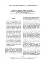
Fig. 1. Effect of TM and VN on the rates of thrombin inhibition by PAI-1. Residual thrombin amidolytic activity was measured at various time
intervals and used to calculate the half times (t
1/2
) of PAI-1 inhibition. (A) Residual activity was monitored during the inhibition of 15 n
M
human
thrombin by 1.5 l
M
PAI-1, in the absence (d)orpresenceof30n
M
(s), 50 n
M
(j), 100 n
M
(h), 400 n
M
(m)or800n
M
(n) TM (solulin).
(B) Residual thrombin activity was monitored as in (A), but in the presence of 0 (d), 10 (s), 20 (j), 50 (h)or100(m) nM rl-TM. (C) Residual
activity decrease was monitored during the inhibition of 2 n
M
thrombin-VR1
tPA
by 15 n
M
PAI-1 in the absence (d) or presence of 100 n
M
rl-TM
(s). (D) Thrombin activity decrease that was observed during the inhibition of 15 n
M
thrombin by 100 n
M
PAI-1/VN complexes, in the absence (d)
or presence of 100 n
M
rl-TM (s).
1944 R. J. Dekker et al. (Eur. J. Biochem. 270) Ó FEBS 2003
Furthermore, we attempted to elucidate the mechanism of
interference by TM on thrombin inhibition by PAI-1. The
rate of thrombin inhibition by 1.5 l
M
PAI-1 was measured
in the presence of increasing concentrations (0–800 n
M
)of
solulin. Solulin lacks the transmembrane domain and does
not contain chondroitin sulphate, which might have an
additional heparin-like effect on the thrombin/PAI-1 inter-
action [19]. In this way, only protein–protein interactions
between thrombin and TM are considered. At the highest
concentration of TM (800 n
M
solulin), thrombin is inhibited
by PAI-1 with a half-time of the reaction (t
1/2
)thatis12.5-
fold longer than in the absence of TM, i.e. 6810 and 545 s,
respectively (Fig. 1A). Because of the lower affinity of
solulin for thrombin (due to the lack of the chondroitin
sulphates) [20], high concentrations of the TM preparation
were necessary to obtain the observed effect. Hence, rabbit-
lung TM (rl-TM), which has a higher affinity for thrombin
due to its chondroitin sulphate moiety, was also used to
study the effect on the thrombin/PAI-1 interaction. The
inhibitory effect of only 100 n
M
rl-TM on the rate of
inhibition of human plasma thrombin by PAI-1 was more
substantial than that of the highest concentration of solulin
(800 n
M
) (Fig. 1B). Next, the effect of TM on the inhibition
of the substitution variant thrombin-VR1
tPA
by PAI-1 was
studied. In marked contrast to thrombin, only a 1.7-fold
inhibitory effect of 100 n
M
rl-TM is observed on the
inhibition of 2 n
M
thrombin-VR1
tPA
by 15 n
M
PAI-1,
measured as difference of the half-time (t
1/2
)ofthereaction
(Fig. 1C). Opposed to the accelerating effect of TM on
thrombin inhibition by ATIII and protein C inhibitor, these
findings indicate that TM considerably reduces the rate of
thrombin inhibition by PAI-1.
The poor rate of inhibition of thrombin by PAI-1 alone
(k
i
10
3
M
)1
Æs
)1
) can be substantially increased by the
cofactor VN [3]. Complexed to VN, PAI-1 inhibits throm-
bin at a rate that is at least two orders of magnitude higher
compared to PAI-1 alone (k
i
10
5
M
)1
Æs
)1
). Therefore, the
inhibitory effect of TM on the inhibition of 15 n
M
thrombin
by preincubated PAI-1/VN complexes (100 n
M
PAI and
150 n
M
VN) was determined (Fig. 1D). The presence of
100 n
M
rl-TM in this reaction decreased the inhibition rate
by 14-fold (t
1/2
¼ 940 and 67 s, respectively). Still, even in
the presence of rl-TM, VN accelerates the rate of thrombin
inhibition by PAI-1 36-fold [Fig. 1B (m)vs.1D(s)].
TM binding to thrombin-VR1
tPA
is substantially
reduced
The minor effect of TM on the rate of thrombin-VR1
tPA
inhibition by PAI-1 suggests that the binding between
thrombin and TM, involving the VR1 loop of thrombin, is
affected in the substitution variant. Indeed, the rate of
protein C activation by thrombin-VR1
tPA
,whichiscom-
parable to that of thrombin, was not affected by TM,
whereas TM substantially increased the rate of protein C
activation by thrombin, as expected (data not shown). The
affinity of TM for thrombin-VR1
tPA
was determined using
Surface Plasmon Resonance (SPR, data not shown).
Binding of thrombin-VR1
tPA
to immobilized rl-TM was
significantly reduced (K
d
¼ 121 ± 23 n
M
) compared to
thrombin (K
d
0.5 n
M
) [2,11]. Thus, the minor effect of
TM on the thrombin-VR1
tPA
–PAI-1 interaction appears to
be the result of the decreased ability of TM to bind
thrombin-VR1
tPA
. Moreover, these results demonstrate
that, as for PAI-1, the VR1 loop of thrombin is an essential
interaction site for TM.
The stoichiometry of the suicide-substrate
mechanism is not influenced by TM
The kinetics of the inhibition of thrombin by PAI-1 can be
described by the so-called Ôsuicide-substrateÕ mechanism as
previously elaborated by our group [6,21]. In this mecha-
nism, each productive encounter of serpin and protease can
either lead to formation of the enzyme/inhibitor complex or
can result in cleavage of the inhibitor and release of active
enzyme. A decreased overall inhibition rate can thus be the
result of a shift of the rate constants of the branched part of
mechanism, i.e. increased cleavage at the expense of
complex formation. Therefore, the products of the reaction
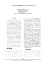
were analyzed by SDS/PAGE (Fig. 2). We found no
evidence of increased cleavage indicating that TM does
not alter the product distribution of the suicide-substrate
reaction between thrombin and PAI-1. These findings leave
a role for TM open in altering the initial binding step
between thrombin and PAI-1 or in changing the ability of
thrombin to catalyze subsequent steps that are common
to both branches in the mechanism, i.e. steric hindrance
vs. allosteric modulation, respectively.
PAI-1 and TM compete for an overlapping binding
site on thrombin
At this point, the mechanism of the inhibitory effect of TM
on the interaction between PAI-1 and thrombin remains to
be elucidated. To that end, binding of PAI-1 to immobilized
thrombin/TM complexes was studied in real-time using
SPR. The high rate constant of initial thrombin/PAI-1
complex formation (k
1
10
6
m
)1
Æs
)1
) [6] would result in a
significant increase in surface-bound mass if formation of
Fig. 2. TM does not alter the distribution of the cleavage and substrate
pathway. Analysis by SDS/PAGE of the products of the reaction of
600 n
M
thrombin with 3.5 l
M
PAI-1 in the presence of 0 (lanes 2–3),
430 (lanes 1, 4–5), 630 (lanes 6–7), and 885 (lanes 8–9) nM TM (sol-
ulin). After 0 min (lane 1), 3 min (lanes 2, 4, 6 and 8) or 16 h (lanes 3, 5,
7 and 9) the samples were immediately quenched by adding sample
buffer, subjected to 10% (w/v) SDS/PAGE and stained with Coo-
massie Brilliant Blue. Indicated are free thrombin (T), intact PAI-1 (P),
cleaved PAI-1 (P*), SDS-stable thrombin/PAI-1 complex (T-P), and
thrombomodulin (TM), as described in the legend of Fig. 4. Note that
after 16 h incubation, the thrombin–PAI-1 complexes formed in the
presence of TM (lanes 5, 7 and 9) have mostly been degraded to lower
molecular mass species by the remaining free, active thrombin as noted
before [21].
Ó FEBS 2003 Thrombomodulin sterically blocks the thrombin/PAI-1 interaction (Eur. J. Biochem. 270) 1945
ternary thrombin/PAI-1/TM complexes occurs. First,
thrombin/TM complexes were formed by applying a
solution of 200 n
M
thrombin on rl-TM that was immobi-
lized on a sensor chip. A high affinity interaction between
thrombin and TM was observed, consistent with a disso-
ciation constant of the thrombin/TM complex in the
subnanomolar range [2,11]. Binding was mass transport-
limited, precluding exact determination of the rate constants
for this interaction under these conditions. Alternatively, an
estimate of k
off
can be given using the half time of throm-
bin/TM dissociation, i.e. t
1/2
¼ 470 s and k
off
10
)3
Æs
)1
.
Second, immediately following the thrombin injection,
either a solution of 800 n
M
PAI-1, 800 n
M
latent PAI-1
or buffer was applied to study the formation of ternary
TM/thrombin/PAI-1 complexes on the chip surface. How-
ever, thrombin slowly and continuously dissociated from
the immobilized TM at the same rate as in the absence of
PAI-1 and no increase in surface-bound mass was observed
as a result of PAI-1 binding to TM/thrombin complexes
(Fig. 3A). Also, no significant difference in binding between
the active and latent form of PAI-1 was observed, the latter
rendered unable to bind thrombin. The absence of ternary
complex formation implies that TM has a steric effect on the
interaction between thrombin and PAI-1. An allosteric
effect of TM is not consistent with these results as the
formation of ternary complexes, that would be more slowly
converted to stable protease/serpin complexes due to
TM-induced allosteric changes in thrombin, is not observed,
even at the high concentrations of PAI-1 used.
As a positive control, identical experiments were per-
formed with ATIII that inhibits thrombin at a rate
comparable to PAI-1, and in contrast to PAI-1 is known
to inhibit the thrombin/TM complex even slightly more
efficient than thrombin alone [11]. Injection of ATIII after
formation of thrombin/TM complexes on the chip surface,
resulted in a considerably increased dissociation of throm-
bin from TM, depending on the ATIII concentration that
was used (Fig. 3A). These findings are in agreement with
fast binding of ATIII to thrombin/TM followed by rapid
dissociation of the thrombin/ATIII complex from TM,
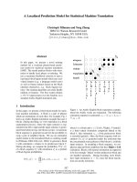
Fig. 3. TM and PAI-1 competitively bind thrombin. Binding of various proteins to immobilized rl-TM was studied using SPR (A–C). Plots show the
increase in surface-associated mass (D Resonance Units) measured in real-time, resulting from binding to rl-TM that was immobilized on the sensor
chip surface. (A) At 0 s 10 lL200n
M
recombinant thrombin was injected directly followed (at 30 s) by 40 lL 800 n
M
active PAI-1 (––), 800 n
M
latent PAI-1 (- - -), buffer (ÆÆÆ), 600 n
M
(-Æ-) or 6 l
M
(-ÆÆ) ATIII. Hereafter, dissociation was continuously monitored by injecting buffer alone (beyond
150 s). (B) Again, 10 lL 200 n
M
rII
A
was injected directly followed by 40 lL800n
M
active PAI-1 (ÆÆÆ), 300 n
M
VN (—), 200 n
M
latent PAI-1
preincubated with 300 n
M
VN (- - -), or 200 n
M
active PAI-1 preincubated with 75–300 n
M
VN (–– labeled 75–300). (C) 60 lL of the following
solutions was directly applied (at 0 s) to the immobilized rl-TM in the absence of thrombin, and dissociation was monitored under continuous
buffer flow (180 s and beyond): 800 n
M
active PAI-1 (ÆÆÆ), 300 n
M
VN (- - -), 200 n
M
latent PAI-1 preincubated with 300 n
M
VN (––), or 200 n
M
active PAI-1 preincubated with 300 n
M
VN (-Æ-). (D) Alternatively, direct binding of rl-TM to VN was studied by immobilizing VN on a sensor chip.
Next, at 0 s 40 lL200n
M
rl-TM was directly injected in the absence (––) or presence of 200 (- - -), 500 (-Æ-) or 1000 (ÆÆÆ)UÆmL
)1
heparin.
Subsequently, dissociation was monitored under continuous buffer flow (beyond 120 s).
1946 R. J. Dekker et al. (Eur. J. Biochem. 270) Ó FEBS 2003
resulting in a net decrease in TM-associated mass on the
chip surface. Moreover, these data are consistent with
the previously described strong reduction of the affinity of
the thrombin/ATIII complex for TM [11,22].
The PAI-1/VN complex directly binds to TM
Recently, we have shown that the PAI-1/vitronectin com-
plex can be treated as an entity with different kinetic
properties than PAI-1 alone [21]. The finding that the PAI-1/
VN complex still inhibits the thrombin/TM complex at a
36-fold higher rate than thrombin alone, suggests that a
possibly different binding mode of PAI-1/VN to thrombin
might allow the formation of quaternary TM/thrombin/
PAI-1/VN complexes. Therefore, binding of PAI-1/VN to
TM/thrombin complexes was tested using SPR. Increasing
concentrations of VN (0–300 n
M
) were preincubated with
200 n
M
PAI-1 and binding to preformed thrombin/rl-TM
complexes was monitored (Fig. 3B). In contrast to PAI-1
and ATIII, binding of PAI-1/VN to the chip surface was
observed without an apparent increased dissociation of
TM-bound thrombin. Interestingly, neither active PAI-1 or
VN alone, nor latent PAI-1 preincubated with VN, had a
significant effect on the thrombin–TM dissociation rate.
However, direct binding of active PAI-1/VN complexes to
rl-TM was observed making a possible interaction between
PAI-1/VN and TM-bound thrombin unlikely (Fig. 3C).
Again, the observed binding was specific for active PAI-1
when preincubated with VN, as no binding was observed
for latent PAI-1 in the presence of VN. Additional binding
studies demonstrate that rl-TM (glycosylated), but not
solulin (not glycosylated) binds directly to immobilized VN,
even in the absence of PAI-1 (data not shown). Therefore,
binding of VN to rl-TM occurs either directly to immobi-
lized VN or to VN in solution exclusively when bound to
active PAI-1. The latter is in agreement with the inability of
latent PAI-1 to bind VN [23].
Finally, the lack of the chondroitin sulphate moiety on
solulin, in conjunction with its inability to bind PAI-1/VN
complexes, suggests the involvement of the chondroitin
sulphate of rl-TM in the binding of PAI-1/VN. Indeed,
both in the absence and presence of PAI-1, the inter-
action of rl-TM with immobilized VN could be com-
peted by including increasing concentrations of heparin
(200–1000 UÆmL
)1
) in the SPR experiments (Fig. 3D).
In conclusion, these results suggest a mechanism in
which TM sterically blocks both PAI-1 and PAI-1/VN
complexes in the association with thrombin. In addition,
upon binding PAI-1, VN is able to bind the chondroitin
sulphate moiety of rl-TM independent of TM-bound
thrombin.
Kinetic modeling using TM as a competitive inhibitor
correctly predicts the inhibitory effect of TM
We decided to model the kinetics of the modulating effect of
TM on the thrombin/PAI-1 reaction to supply a mecha-
nistic basis for the multicomponent reactions. Solulin
kinetic and binding data were used throughout the initial
modeling, as the equilibrium binding of rl-TM to thrombin
displayed a sigmoidal character due to the cooperative
effects of both protein/protein and protein/glycosamino-
glycan interactions. Complete sterical blocking of the
thrombin/PAI-1 interaction would suggest the ability of
TM to completely prevent the inhibition of thrombin by
PAI-1 at high TM concentrations. However, this is not
observed experimentally at TM concentrations that are
several fold higher than K
d
(rl-TM 200-fold; solulin >
10-fold). An indication that could explain this apparent
discrepancy is our previous description of the high reversible
association rate of thrombin and PAI-1 [6]. The reaction
scheme in Fig. 4 implies that at infinite TM concentration
all thrombin is in complex with TM, and the inhibition of
thrombin activity by PAI-1 is thus dependent on the
Fig. 4. Competitive mechanism of TM inhibition of the thrombin/PAI)1 interaction. The inhibitor PAI-1 (P) forms a reversible Michaelis-type
complex (TP) with thrombin (T), characterized by the bimolecular association rate constant k
1
and the dissociation rate constant k
)1
. Subsequently,
an intermediate irreversible complex (TP¢) is formed with rate constant k
2
, that can convert with a rate constant k
3
into the SDS-stable complex
(T-P), or it can react according to a substrate mechanism, resulting in the release of free enzyme together with cleaved, inactive inhibitor (P*) with
the rate constant rÆk
3
. The partition ratio (r) represents the number of catalytic turnovers per inactivation event, where 1 + r is the apparent
stoichiometry. The rate of PAI-1 conversion to its latent form (P
L
) is described by the rate constant k
L
. Alternatively, thrombin binds to TM
forming a reversible complex (T-TM) described by the association and dissociation rate constants k
on
and k
off
, respectively.
Ó FEBS 2003 Thrombomodulin sterically blocks the thrombin/PAI-1 interaction (Eur. J. Biochem. 270) 1947
dissociation rate of thrombin/TM complexes. Upon disso-
ciation of the thrombin/TM complex, there will be compe-
tition between PAI-1 and TM for (re)associating with
thrombin. Competition is dependent on the second-order
rate constants for the thrombin/PAI-1 and thrombin/TM
interaction, including the actual concentration of TM and
PAI-1 throughout the course of the reaction. Therefore, an
infinite concentration of TM would completely compete
PAI-1 binding to thrombin. To test this concept, numerical
integration of the rate equations that describe the mechan-
isminFig.4wasperformedtoobtaintheoreticalinsight
into the effect of the second-order rate constants and
concentrations of TM and PAI-1 on the rate of thrombin/
PAI-1 complex formation. For various combinations of k
on
and k
off
for the thrombin/TM interaction, the concentration
of all reactants and intermediates was calculated through-
out the course of the reaction. The model fits the experi-
mental data best when k
on
¼ 3 · 10
4
M
)1
Æs
)1
and
k
off
¼ 1 · 10
)3
Æs
)1
(data not shown). The K
d
for solulin
( 30 n
M
) that was derived from these values is in good
agreement with literature [24]. The total thrombin amido-
lytic activity that is thus predicted was compared to the
experimentally observed decrease in thrombin activity
(Fig. 5A). The modeling adequately predicts the effect of
TM on the thrombin/PAI-1 inhibition kinetics. The
extremely fast formation of the initial thrombin/PAI-1
Michaelis-type complex, which is predicted during the
presteady state phase of the reaction, is dependent on the
starting concentration of free thrombin molecules (which
itself is dependent on the total TM concentration) (Fig. 5B).
Consequently, the maximal concentration of this reversible
intermediate determines the rate of formation of the first
irreversible intermediate TP¢ and thus establishes the overall
rate of thrombin inhibition, i.e. the rate at which TP
disappears in time at steady state after the rapid initial
increase. The pronounced biphasic character of the inhibi-
tion profiles in Fig. 1A–C is explained by the presteady state
and steady state phases of the reaction that are predicted by
the modeling. At presteady state free thrombin is quickly
captured in the reversible TP complex, which accounts for
the rapid decrease of thrombin activity that is observed
during the first minutes. The second phase in the inhibition
profiles describes the steady state phase of the reaction
where thrombin is slowly released by TM and inhibited by
PAI-1. The reduced affinity of TM for thrombin-VR1
tPA
results in a higher free thrombin concentration at the start of
the reaction and thus a more prominent biphasic inhibition
profile with a longer initial phase (Fig. 1C). According to
Fig. 5B the difference in the maximal concentration of TP,
which is reached in the absence or presence of 800 n
M
TM,
is approximately 12-fold. This value is in agreement with the
inhibitory effect of TM that is observed experimentally.
Finally, a similar model describing an allosteric inhibitory
Fig. 5. A computer-simulated competitive model correctly predicts the
effect of TM on the thrombin/PAI-1 inhibition kinetics. Computer-aided
numerical integration was performed, using the method of Runge-
Kutta, to predict the concentration of all reactants and intermediates
described in Fig. 4 during the full time course of the inhibition reac-
tion. The rate constants k
on
and k
off
were fitted to the experimental
data from Fig. 1A. All other rate constants have been described in a
previous study [6]. (A) The time-dependent decrease of residual
thrombin activity predicted by the model fits closely to the experi-
mental data. Lines represent the residual thrombin activity that was
calculated using the same set of rate constants for all TM concentra-
tions. The lower panel shows the residuals of the fit expressed as the
difference between the experimental and calculated values. (B) Shows
the calculated change in concentration of the thrombin–PAI-1
reversible Michaelis complex ([TP]), as predicted throughout the
course of the reaction modeled in panel A. The maximal amount of TP
complexes that is formed during the initial phase of the reaction
(< 10 s) is reduced in a TM concentration-dependent fashion. This
decrease is related to the free thrombin concentration at the start of the
inhibition reaction that is determined by the TM concentration and the
K
d
of the thrombin–TM complex. Symbols are identical to Fig. 1A.
Lines represent theTM concentration thatwas used, i.e.0 (––),30 (– – –),
50 (- - -), 100 (-Æ-), 400 (-ÆÆ) and 800 (ÆÆÆ)n
M
.
1948 R. J. Dekker et al. (Eur. J. Biochem. 270) Ó FEBS 2003
effect of TM, by allowing thrombin/PAI-1/TM complex
formation, did not fit the experimentally observed TM-
dependent decrease of the thrombin/PAI-1 interaction rate
(results not shown).
Discussion
TM functions as a powerful procoagulant to anticoagulant
specificity switch upon binding to thrombin with high
affinity [1,2]. The binding of Na
+
to thrombin constitutes a
second switch that potently modulates the coagulant vs.
anticoagulant functions of thrombin [25]. The allosteric
effect that results from the binding of Na
+
makes thrombin
a significantly more efficient procoagulant [26]. However, in
both its Na
+
-bound and Na
+
-free form thrombin has a
high specificity for protein C in the presence of TM. The
data presented in this study demonstrates that the VR1 loop
of thrombin, being in close vicinity of the compact
functional epitope for TM [27], is the subsite of exosite-1
that regulates exclusive binding of either PAI-1 or TM.
Previous studies have unambiguously demonstrated that the
VR1 loop is responsible for the specific interaction of PAI-1
with t-PA [7] and thrombin [5,6,21]. The dominant contri-
bution of the t-PA VR1 loop residues to inhibition by PAI-1
strongly suggests binding of PAI-1 to VR1 residues in t-PA
and thrombin-VR1
tPA
. In contrast, the interaction of
thrombin with ATIII is primarily determined by subsites
located in the Western-exit of thrombin (hydrophobic
binding pocket/N-terminal subsites), which is located
distant from the VR1 loop on the opposite side of the
active-center [9,28,29]. Interestingly, PAI-1 appears to be the
only serpin that utilizes the VR1 loop, in contrast to ATIII
and protein C inhibitor (PCI). In agreement with this
concept, the thrombin/TM complex can be efficiently
inhibited by ATIII and PCI [11,12]. Moreover, TM acts
as a stimulator of thrombin inhibition by these serpins, in
line with the cofactor effect of TM on protein C activation.
The exclusive function of the VR1 loop is now further
supported by the results obtained with the exosite-binding
proteins TM and hirugen (this study and [6]). The possibility
of ternary complex formation between thrombin (–VR1
tPA
),
PAI-1 and hirugen demonstrates that PAI-1 does not bind
the part of the anion binding exosite-1 of thrombin that is
utilized by hirugen as well as by TM [22,29,30]. However, as
demonstrated in this study, binding of TM does sterically
hinder PAI-1 binding to thrombin (–VR1
tPA
). Therefore,
the binding of PAI-1 to thrombin either involves a small
part of the VR1 loop that is physically blocked by TM, or
the bulkiness of TM bound to exosite-1 decreases the
accessibility of the VR1 loop for PAI-1 [9,10,27]. Previous
Ala scanning mutagenesis studies have demonstrated that
the VR1 residues Phe34, Lys36, Pro37 and Gln38 are
involved in the binding of TM to thrombin [27,31]. These
residues therefore comprise the most likely overlapping
binding site for TM and PAI-1 on thrombin, as they are also
of substantial importance to the inhibition of thrombin by
PAI-1 [5,6,21]. In addition, the reduced affinity of TM for
thrombin-VR1
tPA
is in agreement with the significant
contribution of this part of the VR1 loop to the binding
of TM by thrombin. Binding of the carboxy-terminal part
of the reactive center loop of PAI-1 in the small cleft formed
by the 60-loop and VR1 loop can be envisioned. The
kinetics of the interaction of thrombin and TM were shown
to be governed by electrostatic interactions, explaining
the fast association rates [8]. This does not explain, however,
the high affinity binding of TM to thrombin as shown by the
slow dissociation rates observed in this study (k
off
10
)3
Æs
)1
; Fig. 3). Structural arguments were put for-
ward that a major hydrophobic interaction in this strong
hydrophilic environment governs the specificity and tight-
ness of TM binding to thrombin [10]. Hydrophobic residues
of TM are buried in a surface hydrophobic pocket that is
partly formed by VR1 and exosite-1. This would explain the
significantly reduced binding of TM to thrombin-VR1
tPA
as
compared to thrombin, despite the fact that according to the
structure the lower, highly charged rim of exosite-1 is
unchanged [6]. This hydrophobic interaction, involving
Phe34 in the VR1 loop, would then exclude the interaction
of PAI-1 with the VR1 if thrombin is bound to TM.
The kinetic model of TM/PAI-1 competition for throm-
bin described here has the following features. Previous
studies from our laboratory have shown that initial
Michaelis complex formation is not the rate-limiting step
in the thrombin/PAI-1 reaction, but rather a unimolecular
step in the mechanism (k
2
in Fig. 4). These findings imply
that with the high PAI-1 concentrations that were used to
rapidly inhibit thrombin, a major fraction of the thrombin
molecules is quickly forming a reversible Michaelis complex
with PAI-1 during the initial phase of the reaction.
Subsequently, the formation of stable thrombin/PAI-1
complexes is dependent on the (rate-limiting) efficiency of
successive catalytic events in the inhibition pathway (i.e. k
2
and k
3
in Fig. 4). When the amidolytic activity of thrombin
is assayed during the course of the reaction, quenching of
the reaction mixture leads to dissociation of the majority of
initial Michaelis complexes, and thus releases active throm-
bin. In the presence of a theoretical infinite concentration of
TM, all thrombin would be in complex with TM at the start
of the reaction. When PAI-1 is added, competition between
TM and PAI-1 for thrombin will occur after dissociation of
each thrombin/TM complex, which was thus far at equi-
librium. Consequently, the concentrations of PAI-1 and
TM, including their rate constants of association with
thrombin, will determine the maximum rate at which
the thrombin/TM complex will be irreversibly inhibited by
PAI-1 (Fig. 5). Under the experimental conditions used in
this study, relatively low TM concentrations (< 1 l
M
)are
sufficient to have most thrombin in complex with TM. At
the high PAI-1 concentration (> 1 l
M
)thatwasused,
PAI-1 will compete efficiently with TM as k
1
> k
on
and
[PAI-1] > [TM]. Therefore, even though TM significantly
slows down the thrombin/PAI)1 interaction, eventually
thrombin will be completely inhibited by PAI-1. The
inhibitory effect of TM on the thrombin/PAI-1 interaction
is partly masked in the presence of the cofactor VN when
assayed kinetically ([15] and this study). The data presented
in this study demonstrate that only PAI-1/VN complexes
and immobilized VN directly bind to TM, most likely via its
glycosaminoglycan sugar moiety. In contrast, free VN or
PAI-1 does not bind to TM. In agreement with this finding,
VN is known to expose a high-affinity heparin binding-site
only after it is converted to its non-native (active) confor-
mation, i.e. when bound to PAI-1 or when immobilized on a
surface [32]. Therefore, the interaction between VN and TM
Ó FEBS 2003 Thrombomodulin sterically blocks the thrombin/PAI-1 interaction (Eur. J. Biochem. 270) 1949
has probably no effect on the rate of thrombin inhibition by
PAI-1 in the presence of TM. A slightly larger inhibitory
effect of TM on the rate of thrombin inhibition by PAI-1/
VN was observed compared to inhibition by PAI-1 alone,
i.e. 14 vs. 12-fold, respectively. In the presence of TM,
however, thrombin is still inhibited 36-fold faster by the
PAI-1/VN complex compared to PAI-1 alone. The vastly
increased association rate between thrombin and PAI-1/VN
would result in an immediate capturing of any free
thrombin that is at equilibrium with the thrombin/TM
species by competing more efficiently for reassociating
with TM.
The physiologic relevance of TM interference in the
thrombin/PAI-1 interaction can possibly be found in the
(atherosclerotic) vessel wall, where all these proteins and
cofactors are present [17], including TM on vascular SMC
[14]. Both thrombin and PAI-1 can substantially influence
migration and proliferation of vascular SMC, the latter
process via the protease-activated receptors, of which
PAR1 was found to be expressed by SMC in vivo [33]. In
this respect, the interplay between thrombin and PAI-1 in
thevesselwallhastwofaces.First,PAI-1isabletoinhibit
the mitogenic potential of thrombin. On the other hand,
cleavage and inactivation of PAI-1 by thrombin controls
the urokinase-type plasminogen activator (u-PA)-mediated
migratory effect of PAI-1 on SMC. The suicide-substrate
mechanism stoichiometry is rather ÔunfavorableÕ for the
thrombin/PAI-1 protease/serpin pair, especially in the
presence of VN, being six inactivated (cleaved) PAI-1
molecules for each thrombin molecule that is inhibited
(r ¼ 5) [21]. Probably the main physiologic consequence of
this interaction is an inactivation of the PAI-1 pool in the
vascular wall by thrombin, making it no longer available
for interaction with u-PA and VN, which can explain part
of the effect of thrombin on the proliferation and
migration of vascular SMC [4,34]. In the context of the
vessel wall, TM might therefore function as a regulator of
PAI-1 inactivation by thrombin in the presence of the
abundant matrix protein VN. The presence of TM on the
surface of SMC might be important in focusing its
modulatory potential to the cell surface. In this respect,
physiologic significance can be attributed to the binding of
VN in its unfolded conformation (i.e. as adhered matrix
protein or in solution complexed to PAI-1) to the
chondroitin sulphate moiety of TM as was observed in
this study. Neointimal vascular SMC can thus focus TM
to sites where VN is present, e.g. at the leading edge of
migration, and prevent local inactivation of PAI-1 by
thrombin. The ability of PAI-1 to compete with the SMC
surface-exposed integrin a
v
b
3
and u-PA receptor for
binding VN therefore suggests a possible migratory role
of TM in neointimal hyperplasia. This concept is in
agreement with the expression of TM by neointimal
vascular SMC that was found in vivo [14,34].
In conclusion, this study provides a mechanistic concept,
elucidating a multicomponent system of proteases, serpins
and cofactors. Again, TM acts as a molecular switch by
excluding an interaction between thrombin and PAI-1
thereby protecting the serpin from inactivation. Further-
more, these findings propose a possible novel role for TM
expressed by vascular SMC in the pathogenesis of vascular
disease.
Acknowledgements
This work was supported by the Netherlands Heart Foundation, the
Hague, by grant NHS 96.094 and the Molecular Cardiology Program
grant M 93.007.
References
1. Esmon, C.T. (1987) The regulation of natural anticoagulant
pathways. Science 235, 1348–1352.
2. Esmon, C.T. (1995) Thrombomodulin as a model of molecular
mechanisms that modulate protease specificity and function at the
vessel surface. FASEB J. 9, 946–955.
3. Ehrlich, H.J., Klein-Gebbink, R., Keijer, J., Linders, M., Preiss-
ner, K.T. & Pannekoek, H. (1990) Alteration of serpin specificity
by a protein cofactor: Vitronectin endows plasminogen activator
inhibitor 1 with thrombin inhibitory properties. J. Biol. Chem. 265,
13029–13035.
4. Ehrlich, H.J., Klein-Gebbink, R., Preissner, K.T., Keijer, J.,
Esmon, N., Mertens, N. & Pannekoek, H. (1991) Thrombin
neutralizes plasminogen activator inhibitor 1 (PAI-1) that is
complexed with vitronectin in the endothelial cell matrix. J. Cell.
Biol. 115, 1773–1781.
5. Horrevoets, A.J.G., Tans, G., Smilde, A.E., van Zonneveld, A J.
& Pannekoek, H. (1993) Thrombin-variable region 1 (VR1).
Evidence for the dominant contribution of VR1 of serine proteases
to their interaction with plasminogen activator inhibitor 1. J. Biol.
Chem. 268, 779–782.
6. Dekker, R.J., Eichinger, A., Stoop, A.A., Bode, W., Pannekoek,
H. & Horrevoets, A.J. (1999) The variable region-1 from tissue
type plasminogen activator confers specificity for plasminogen
activator inhibitor-1 to thrombin by facilitating catalysis: release
of a kinetic block by a heterologous protein surface loop. J. Mol.
Biol. 293, 613–627.
7. Madison, E.L., Goldsmith, E.J., Gerard, R.D., Gething, M J.H.,
Sambrook, J.F. & Bassel-Duby, R.S. (1990) Amino acid residues
that affect interaction of tissue-type plasminogen activator with
plasminogen activator inhibitor 1. Proc. Natl Acad. Sci. USA 87,
3530–3533.
8. Baerga-Ortiz, A., Rezaie, A.R. & Komives, E.A. (2000) Electro-
static dependence of the thrombin–thrombomodulin interaction.
J. Mol. Biol. 296, 651–658.
9. Mathews, I.I., Padmanabhan, K.P. & Tulinsky, A. (1994) Struc-
ture of a nonadecapeptide of the fifth EGF domain of thrombo-
modulin complexed with thrombin. Biochemistry 33, 13547–
13552.
10. Fuentes-Prior,P.,Iwanaga,Y.,Huber,R.,Pagila,R.,Rumennik,
G.,Seto,M.,Morser,J.,Light,D.R.&Bode,W.(2000)Structural
basis for the anticoagulant activity of the thrombin-thrombo-
modulin complex. Nature 404, 518–525.
11. Hofsteenge, J., Taguchi, H. & Stone, S.R. (1986) Effect of
thrombomodulin on the kinetics of the interaction of thrombin
with substrates and inhibitors. Biochem. J. 237, 243–251.
12. Rezaie, A.R., Cooper, S.T., Church, F.C. & Esmon, C.T. (1995)
Protein C inhibitor is a potent inhibitor of the thrombin-throm-
bomodulin complex. J. Biol. Chem. 270, 25336–25339.
13. Bajzar, L., Morser, J. & Nesheim, M. (1996) TAFI, or plasma
procarboxypeptidase B, couples the coagulation and fibrinolytic
cascades through the thrombin-thrombomodulin complex. J. Biol.
Chem. 271, 16603–16608.
14.Tohda,G.,Oida,K.,Okada,Y.,Kosaka,S.,Okada,E.,
Takahashi, S., Ishii, H. & Miyamori, I. (1998) Expression of
thrombomodulin in atherosclerotic lesions and mitogenic activity
of recombinant thrombomodulin in vascular smooth muscle cells.
Arterioscler. Thromb. Vasc. Biol. 18, 1861–1869.
1950 R. J. Dekker et al. (Eur. J. Biochem. 270) Ó FEBS 2003
15. Rezaie, A.R. (1999) Role of exosites 1 and 2 in thrombin reaction
with plasminogen activator inhibitor-1 in the absence and presence
of cofactors. Biochemistry 38, 14592–14599.
16. Smith, E.B., Crosbie, L. & Carey, S. (1996) Prothrombin-related
antigens in human aortic intima Semin. Thromb. Hemost. 22,
347–350.
17. Stoop, A.A., Lupu, F. & Pannekoek, H. (2000) Colocalization of
thrombin, PAI-1, and vitronectin in the atherosclerotic vessel wall:
a potential regulatory mechanism of thrombin activity by PAI-1/
vitronectin complexes. Arterioscler. Thromb. Vasc. Biol. 20,
1143–1149.
18. van Meijer, M., Stoop, A.A., Smilde, A., Preissner, K.T., van
Zonneveld, A J. & Pannekoek, H. (1997) The composition of
complexes between plasminogen activator inhibitor 1, vitronectin
and either thrombin or tissue-type plasminogen activator.
Thromb. Haemost. 77, 516–521.
19. Parkinson, J.F., Grinnell, B.W., Moore, R.E., Hoskins, J., Vlahos,
C.J. & Bang, N.U. (1990) Stable expression of a secretable deletion
mutant of recombinant human thrombomodulin in mammalian
cells. J. Biol. Chem. 265, 12602–12610.
20. Clarke, J.H., Light, D.R., Blasko, E., Parkinson, J.F., Nagashima,
M., McLean, K., Vilander, L., Andrews, W.H., Morser, J. &
Glaser, C.B. (1993) The short loop between epidermal growth
factor-like domains 4 and 5 is critical for human thrombomodulin
function. J. Biol. Chem. 268, 6309–6315.
21. van Meijer, M., Smilde, A.E., Tans, G., Nesheim, M.E., Panne-
koek, H. & Horrevoets, A.J. (1997) The suicide substrate reaction
between plasminogen activator inhibitor 1 and thrombin is regu-
lated by the cofactors vitronectin and heparin. Blood 90, 1874–
1882.
22. Bock, P.E., Olson, S.T. & Bjo
¨
rk, I. (1997) Inactivation of
thrombin by antithrombin is accompanied by inactivation of
regulatory exosite I. J. Biol. Chem. 272, 19837–19845.
23. Seiffert, D. & Loskutoff, D.J. (1991) Kinetic analysis of the
interaction between type 1 plasminogen activator inhibitor and
vitronectin and evidence that the bovine inhibitor binds to a
thrombin-derived amino-terminal fragment of bovine vitronectin.
Biochim. Biophys. Acta 1078, 23–30.
24. Light, D.R., Glaser, C.B., Betts, M., Blasko, E., Campbell, E.,
Clarke,J.H.,McCaman,M.,McLean,K.,Nagashima,M.,
Parkinson,J.F.,Rumennik,G.,Young,T.&Morser,J.(1999)
The interaction of thrombomodulin with Ca
2+
. Eur. J. Biochem.
262, 522–533.
25. Dang, Q.D. & Di Cera, E. (1996) Residue 225 determines the
Na(+)-induced allosteric regulation of catalytic activity in serine
proteases. Proc. Natl Acad. Sci. USA 93, 10653–10656.
26. Pineda, A.O., Savvides, S.N., Waksman, G. & Di Cera, E. (2002)
Crystal structure of the anticoagulant slow form of thrombin.
J. Biol. Chem. 277, 40177–40180.
27. Pineda, A.O., Cantwell, A.M., Bush, L.A., Rose, T. & Di Cera, E.
(2002) The thrombin epitope recognizing thrombomodulin is a
highly cooperative hot spot in exosite I. J. Biol. Chem. 277, 32015–
32019.
28. Tsiang, M., Jain, A.K. & Gibbs, C.S. (1997) Functional require-
ments for inhibition of thrombin by antithrombin III in the
presence and absence of heparin. J. Biol. Chem. 272, 12024–
12029.
29. Skrzypczak-Jankun, E., Carperos, V.E., Ravichandran, K.G.,
Tulinsky, A., Westbrook, M. & Maraganore, J.M. (1991) Struc-
ture of the hirugen and hirulog 1 complexes of a-thrombin. J. Mol.
Biol. 221, 1379–1393.
30. Liu, L.W., Vu, T.K., Esmon, C.T. & Coughlin, S.R. (1991) The
region of the thrombin receptor resembling hirudin binds to
thrombin and alters enzyme specificity. J. Biol. Chem. 266, 16977–
16980.
31. Hall, S.W., Nagashima, M., Zhao, L., Morser, J. & Leung, L.L.
(1999) Thrombin interacts with thrombomodulin, protein C, and
thrombin-activatable fibrinolysis inhibitor via specific and distinct
domains. J. Biol. Chem. 274, 25510–25516.
32. Stockmann, A., Hess, S., Declerck, P., Timpl, R. & Preissner, K.T.
(1993) Multimeric vitronectin. Identification and characterization
of conformation–dependent self association of the adhesive pro-
tein. J. Biol. Chem. 268, 22874–22882.
33. Nelken, N.A., Soifer, S.J., O’Keefe, J., Vu, T.K., Charo, I.F. &
Coughlin, S.R. (1992) Thrombin receptor expression in
normal and atherosclerotic human arteries. J. Clin. Invest. 90,
1614–1621.
34. Kanse, S.M., Kost, C., Wilhelm, O.G., Andreasen, P.A. & Pre-
issner, K.T. (1996) The urokinase receptor is a major vitronectin-
binding protein on endothelial cells. Exp. Cell. Res. 224, 344–353.
Ó FEBS 2003 Thrombomodulin sterically blocks the thrombin/PAI-1 interaction (Eur. J. Biochem. 270) 1951