Báo cáo khoa học: Limited proteolysis of Escherichia coli cytidine 5¢-triphosphate synthase. Identification of residues required for CTP formation and GTP-dependent activation of glutamine hydrolysis potx
Bạn đang xem bản rút gọn của tài liệu. Xem và tải ngay bản đầy đủ của tài liệu tại đây (681.58 KB, 12 trang )
Limited proteolysis of
Escherichia coli
cytidine 5¢-triphosphate
synthase. Identification of residues required for CTP formation
and GTP-dependent activation of glutamine hydrolysis
Dave Simard, Kerry A. Hewitt, Faylene Lunn, Akshai Iyengar and Stephen L. Bearne
Department of Biochemistry and Molecular Biology, Dalhousie University, Halifax, Nova Scotia, Canada
Cytidine 5¢-triphosphate synthase catalyses the ATP-
dependent formation of CTP from UTP using either
ammonia or
L
-glutamine as the source of nitrogen. When
glutamine is the substrate, GTP is required as an allosteric
effector to promote catalysis. Limited trypsin-catalysed
proteolysis, Edman degradation, and site-directed muta-
genesis were used to identify peptide bonds C-terminal to
three basic residues (Lys187, Arg429, and Lys432) of
Escherichia coli CTP synthase that were highly susceptible to
proteolysis. Lys187 is located at the CTP/UTP-binding site
within the synthase domain, and cleavage at this site
destroyed all synthase activity. Nucleotides protected the
enzyme against proteolysis at Lys187 (CTP > ATP >
UTP > GTP). The K187A mutant was resistant to pro-
teolysis at this site, could not catalyse CTP formation, and
exhibited low glutaminase activity that was enhanced slightly
by GTP. K187A was able to form tetramers in the presence
of UTP and ATP. Arg429 and Lys432 appear to reside in an
exposed loop in the glutamine amide transfer (GAT)
domain. Trypsin-catalyzed proteolysis occurred at Arg429
and Lys432 with a ratio of 2.6 : 1, and nucleotides did not
protect these sites from cleavage. The R429A and R429A/
K432A mutants exhibited reduced rates of trypsin-catalyzed
proteolysis in the GAT domain and wild-type ability to
catalyse NH
3
-dependent CTP formation. For these mutants,
the values of k
cat
/K
m
and k
cat
for glutamine-dependent CTP
formation were reduced % 20-fold and % 10-fold, respect-
ively, relative to wild-type enzyme; however, the value of K
m
for glutamine was not significantly altered. Activation of the
glutaminase activity of R429A by GTP was reduced 6-fold at
saturating concentrations of GTP and the GTP binding
affinity was reduced 10-fold. This suggests that Arg429 plays
a role in both GTP-dependent activation and GTP binding.
Keywords: activation; amidotransferase; CTP synthase;
glutaminase; proteolysis; site-directed mutagenesis.
CTP synthase [CTPS; EC 6.3.4.2; UTP:ammonia ligase
(ADP-forming)] catalyses the ATP-dependent formation of
CTP from UTP using either
L
-glutamine or NH
3
as the
nitrogen source (Scheme 1) [1,2]. This glutamine amido-
transferase is a single polypeptide chain containing 545
amino acids and consisting of two domains. The C-terminal
glutamine amide transfer (GAT) domain catalyses the
hydrolysis of glutamine, and the nascent NH
3
derived from
glutamine hydrolysis is transferred to the N-terminal
synthase domain where the amination of UTP is catalysed
[3,4]. CTPS belongs to the Triad family of glutamine
amidotransferases [5,6] which utilizes a Cys-His-Glu triad to
catalyse glutamine hydrolysis and also includes anthranilate
synthase, carbamoyl phosphate synthase, formylglycin-
amidine synthase, GMP synthase, imidazole glycerol phos-
phate synthase, and aminodeoxychorismate synthase.
CTPS catalyses the final step in the de novo synthesis of
cytosine nucleotides. Because CTP has a central role in the
biosynthesis of nucleic acids [7] and membrane phospho-
lipids [8], CTPS is a recognized target for the development
of antineoplastic agents [7,9], antiviral agents [9,10], and
antiprotozoal agents [11–13]. Recently, CTP synthase
inhibition has been shown to potentiate the cytotoxic effects
of the anticancer drug 1-b-
D
-arabinofuranosylcytosine [14]
and anti-HIV therapies [15].
CTPS from E. coli is the most thoroughly characterized
CTPS with respect to its physical and kinetic properties, and
is regulated in a complex fashion [1]. GTP is required as a
positive allosteric effector to increase the efficiency (k
cat
/K
m
)
of glutamine-dependent CTP synthesis 45-fold but has a
negligible effect on the reaction when NH
3
is the substrate
[16,17]. In addition, the enzyme is inhibited by the product
CTP [18], exhibits negative cooperativity for glutamine [19],
and displays positive cooperativity for ATP and UTP
Scheme 1. CTP-forming reactions catalysed by CTPS.
Correspondence to S. L. Bearne, Department of Biochemistry and
Molecular Biology, Dalhousie University, Halifax, Nova Scotia,
Canada B3H 1X5. Tel.: +1 902 494 1974, Fax: + 1 902 494 1355,
E-mail:
Abbreviations: CTPS, CTP synthase; GAT, glutamine amide transfer;
GF-HPLC, gel-filtration-HPLC; PVDF, poly(vinylidene difluoride).
Enzymes: CTP synthase (EC 6.4.3.2).
(Received 28 February 2003, revised 17 March 2003,
accepted 21 March 2003)
Eur. J. Biochem. 270, 2195–2206 (2003) Ó FEBS 2003 doi:10.1046/j.1432-1033.2003.03588.x
[18–20]. ATP and UTP act synergistically to promote
tetramerization of the enzyme to its active form [20].
The structure of CTPS has not yet been determined and
hence little is known about the enzyme’s tertiary structure.
However, analysis of crystal structures of the Triad
amidotransferases GMP synthase and carbamoyl phos-
phate synthase reveal that the structures of the GAT
domains are probably closely related among all Triad
enzymes [21,22]. Site-directed mutagenesis studies and
sequence comparisons have revealed structural and cata-
lytic roles of several amino acid residues within the GAT
domain of CTPS, including residues of the catalytic triad
(Cys379, His515, and Glu517) [3], residues comprising the
oxyanion hole (Gly351, Gly377, Gly381, and possibly
adjacent hydrophobic residues) [23], and residues between
Ala346 and Tyr355 that appear to play an important
structural role [4]. Recently, Willemoe
¨
s reported that
Thr431 and Arg433 in the GAT domain of Lactococcus
lactis CTPS play a role in GTP-dependent activation of
glutamine hydrolysis [24].
Our knowledge about the synthase domain is much more
limited. Analyses of mutant CTP synthases from Chlamydia
trachomatis [25], hamster [26], and yeast [27] have revealed
that mutations which render cells resistant to both the
cytotoxic effects of cyclopentenylcytosine and feedback
inhibition by CTP occur between residues 116 and 229
(E. coli numbering), with many of the mutations clustering
between residues 146 through 158. Hence, this region of the
synthase domain is believed to form part of the CTP-
binding site. Competitive inhibition experiments have
suggested that for E. coli CTPS, this site may also be the
UTP-binding site [18]. The locations of the ATP- and GTP-
binding sites have not yet been identified. Recent studies
from our laboratory have revealed that residues Asp107 and
Leu109 in the synthase domain of E. coli CTPS facilitate
efficient coupling of glutamine hydrolysis to CTP synthesis
[28].
To learn more about the structure of CTPS, we inves-
tigated controlled proteolysis of the enzyme. Using limited
trypsin-catalysed proteolysis and site-directed mutagenesis,
we have identified peptide bonds C-terminal to three basic
residues of E. coli CTPS that are highly susceptible to
proteolysis. One residue, Lys187, is located at the CTP/
UTP-binding site within the synthase domain and is
essential for catalysis but not for enzyme tetramerization.
The other two residues, Arg429 and Lys432 appear to reside
in an exposed loop that is important for both GTP binding
and GTP-dependent allosteric activation of glutamine
hydrolysis.
Materials and methods
Materials
HisÆBind resin and thrombin cleavage capture kits were
from Novagen; broad range protein markers were from
New England Biolabs; Pfu Turbo DNA polymerase was
from Stratagene Inc.; nucleotides, a-chymotrypsin from
bovine pancreas (54 UÆmg
)1
), Pronase from Streptomyces
griseus (4.7 UÆmg
)1
), protease V8 from Staphylococcus
aureus (1000 UÆmg
)1
), thermolysin from Bacillus thermo-
proteolyticus rokko (55 UÆmg
)1
), and TPCK-treated trypsin
from bovine pancreas (10 900 UÆmg
)1
), and all other
chemicals were from Sigma-Aldrich Canada Ltd. Oligo-
nucleotide primers for DNA sequencing and site-directed
mutagenesis were commercially synthesized by ID Labor-
atories (London, ON, Canada). QIAprep spin plasmid
miniprep kit (Qiagen Inc.) was used for the preparation of
plasmids for mutagenesis and transformation. DNA
sequencing was conducted at the Dalhousie University–
NRC Institute for Marine Biosciences Joint Laboratory
(Halifax, NS, Canada) and the Robarts Research Institute
(London, ON, Canada), while the N-terminal amino acid
sequencing was carried out at the Eastern Que
´
bec Proteo-
mics Core Facility (Ste-Foy, QC, Canada). Predictions of
secondary structure were conducted using the programs 3-
D
PSSM
[29],
GOR
4[30],
HNN
[31],
J
-
PRED
[32],
PREDATOR
[33],
PSIPRED
[34], and
SSPRO
[35]. Sequence alignments were
conducted using
CLUSTALW
[36].
Enzyme expression and purification
Wild-type and mutant forms of recombinant E. coli CTPS
were expressed in and purified from E. coli strain
BL21(DE3) cells transformed with either mutant or wild-
type plasmid pET15b-CTPS1 as described previously [16].
This construct encodes the CTPS gene product with an
N-terminal hexahistidine tag. Thrombin-catalysed cleavage
of the histidine tag from soluble enzymes (new N-terminus,
GSHMLEM
1
…) was carried out as described previously
[16]. The resulting enzyme was dialysed into Hepes buffer
(70 m
M
, pH 8.0) containing EDTA (0.5 m
M
)andMgCl
2
(10 m
M
). The results of purification and cleavage pro-
cedures were routinely monitored using SDS/PAGE. The
amino acid residues in the recombinant wild-type and
mutant enzymes are numbered according to the sequence of
the wild-type E. coli enzyme starting with M
1
as position
one.
Mutagenesis
The plasmid pET15b-CTPS1 [16] was used as the template
for site-directed mutagenesis. Site-directed mutagenesis was
conducted using the Quikchange Site-Directed Mutagenesis
Kit (Stratagene Inc.) and following the manufacturer’s
protocol. The synthetic deoxyoligonucleotide forward (F)
and reverse (R) primers used to construct the mutants were:
5¢-GCGTCTGGTGAAGTC
GCAACCAAACCGACT
CAG-3¢ (F, K187A), 5¢-GCTGAGTCGGTTTGGTT
GCGACTTCACCAGACGC-3¢ (R, K187A), 5¢-CGG
CAACGTTGAAGTT
GCTAGCGAGAAGAGCG-3¢ (F,
R429A), 5¢-CGCTCTTCTCGCTA
GCAACTTCAACGT
TGCCG-3¢ (R,R429A),5¢-GCAACGTTGAAGTT
GCTAGCGAGGCGAGCGATCTCG-3¢ (F, R429A/
K432A), 5¢-CGAGATCGCTC
GCCTCGCTAGCAACT
TCAACGTTGC-3¢ (R, R429A/K432A), where the posi-
tions of the mismatches are underlined. Potential mutant
plasmids were isolated and used to transform competent
DH5a cells. These cells were used for plasmid maintenance
and for all sequencing reactions. The entire mutant genes
were sequenced to verify that no other alterations of the
nucleotide sequence had been introduced. Competent
E. coli strain BL21(DE3) cells were used as the host for
target gene expression.
2196 D. Simard et al. (Eur. J. Biochem. 270) Ó FEBS 2003
Enzyme assay and protein determinations
CTPS activity was determined at 37 °C using a continuous
spectrophotometric assay by following the rate of increase
in absorbance at 291 nm resulting from the conversion of
UTP to CTP (De ¼ 1338Æ
M
)1
Æcm
)1
) [18]. The standard
assay mixture consisted of Hepes buffer (70 m
M
,pH8.0)
containing EDTA (0.5 m
M
)andMgCl
2
(10 m
M
), CTPS,
and saturating concentrations of UTP (1 m
M
)andATP
(1 m
M
) in a total volume of 1 mL. Enzyme and nucleotides
were preincubated together for 2 min at 37 °C followed by
addition of substrate (NH
4
Cl or glutamine) to initiate the
reaction. Total NH
4
Cl concentrations used in the assays
were 5, 10, 20, 30, 50, 60, 80, and 100 m
M
,andCTPS
concentrations were % 3.0 lgÆmL
)1
(wild-type),
3.0 lgÆmL
)1
(R429A), and 4.0 lgÆmL
)1
(R429A/K432A).
For glutamine assays, concentrations of glutamine
were 0.1, 0.2, 0.3, 0.5, 1.0, 2.0, 3.0, and 6.0 m
M
and
CTPS concentrations were % 4.0 lgÆmL
)1
(wild-type),
1.4 mgÆmL
)1
(R429A), and 0.9 mgÆmL
)1
(R429A/
K432A). The concentration of GTP was maintained at
0.25 m
M
for all assays when glutamine was used as the
substrate. In addition, the ionic strength was maintained at
0.25
M
in all spectrophotometric assays by the addition of
KCl. The apparent activation constant (K
A
) for R429A
CTPS (0.4 mgÆmL
)1
) with respect to GTP was determined
for glutamine-dependent CTP formation as described
previously [28].
All kinetic parameters were determined in triplicate and
average values are reported. Initial rate kinetic data was fit
to Eqn (1) by nonlinear regression analysis using the
program
ENZYMEKINETICS
v1.5 (1996) from Trinity Soft-
ware (Plymouth, NH). In Eqn (1), v
i
is the initial velocity,
V
max
(¼ k
cat
[E]
T
) is the maximal velocity at saturating
substrate concentrations, [S] is the substrate concentration
(glutamine or NH
3
), and K
m
is the Michaelis constant for
the substrate. Values of K
m
for NH
3
were calculated using
the concentration of NH
3
presentatpH8.0{pK
a
(NH
4
+
) ¼ 9.24 [37]}. Values of k
cat
were calculated for
CTPS variants with the hexahistidine tag removed using
the molecular masses (Da) of 61 029 (wild-type), 60 944
(R429A), and 60 887 (R429A/K432A). The reported
errors are standard deviations. Except where noted
otherwise, protein concentrations were determined using
the Bio-Rad Protein Assay (Bio-Rad Laboratories Ltd.)
with BSA standards.
m
i
¼
V
max
½S
K
m
þ½S
ð1Þ
Glutaminase activity
Values of k
cat
for the hydrolysis of glutamine, at fixed
saturating concentrations of glutamine (6 m
M
), UTP
(1 m
M
), and ATP (1 m
M
) were determined as described
previously [38]. Data describing the dependence of the
apparent k
cat
values on the concentration of GTP were
fitted to Eqn (2) for hyperbolic nonessential activation
kinetics where K
A
is the apparent activation constant, k
o
is
the turnover number in the absence of GTP, and k
act
is
the turnover number at saturating concentrations of GTP
[39].
k
apparent
cat
¼
k
o
þ k
act
GTP
½
K
A
1 þ
GTP
½
K
A
ð2Þ
Limited proteolysis
Initially, wild-type CTPS was subjected to limited proteo-
lysis by several endopeptidases including trypsin, chymo-
trypsin, pronase, thermolysin and V8 protease. Proteolysis
was conducted in Hepes buffer (70 m
M
, pH 8.0) containing
EDTA (0.5 m
M
)andMgCl
2
(10 m
M
)at37°Cusinga
CTPS/protease ratio (lgprotein)of60:1inatotalvolume
of 1 mL. Limited proteolysis using pronase and thermolysin
was also conducted in potassium phosphate buffer (50 m
M
,
pH 7.2) containing EDTA (1 m
M
)andMgCl
2
(10 m
M
).
EDTA was omitted from the buffers for all thermolysin-
catalysed reactions. Proteolysis experiments were conducted
in the absence and presence of ATP (10 m
M
)andUTP
(10 m
M
). During the proteolysis reactions, aliquots (25 lL)
were removed from the reaction mixture over the course of
1h,andtransferredtogelloadingbuffer[25lL; Tris/HCl
(170 m
M
, pH 6.8) containing dithiothreitol (120 m
M
), SDS
(5.4%, w/v), Bromophenol blue (0.03%, w/v) and glycerol
(27.2%, w/v)] to terminate the reaction. The samples were
then boiled for 5 min and the proteolytic fragments
were separated using SDS/PAGE (12% gels). Fragments
were visualized by staining with Coomassie blue R-250 and
subsequent de-staining in a solution of methanol/H
2
O/
acetic acid (45 : 45 : 10). Detailed studies were subsequently
conducted using trypsin as trypsin-catalysed proteolysis
gave different fragments depending on whether the nucleo-
tides were absent or present.
Limited trypsin-catalysed proteolysis of both wild-type
and mutant CTP synthases was analysed by monitoring
CTPS activity (vide infra) at specific time points and by
SDS/PAGE. Trypsin proteolysis reactions (1 mL total
volume) contained either wild-type or mutant CTPS
(0.20 mgÆmL
)1
), and were conducted for 1 h in Hepes
buffer (70 m
M
, pH 8.0) containing EDTA (0.5 m
M
)and
MgCl
2
(10 m
M
)at37°C. Reactions were initiated by
addition of trypsin (0.1 lgÆmL
)1
) and aliquots (100 lL)
were removed every 10 min over 1 h and assayed for
activity using NH
3
as the substrate. The cleavage fragments
produced from trypsin-catalysed proteolysis were analysed
using SDS/PAGE (12% and 20% gels). Reactions were
conducted as described above, with the exception that at 10,
20, 30, and 60 min, aliquots (20 lL) were removed and
transferred to gel loading buffer (15 lL) to terminate the
reaction. A zero time point was obtained using 3 lLof
theenzymestocksolution(% 1.5 mgÆmL
)1
)usedforthe
reaction. The ability of various ligands to protect both wild-
type and mutant CTP synthases from proteolysis was
examined using ATP (10 m
M
), UTP (10 m
M
), ATP and
UTP together (10 m
M
each), CTP (0.1, 0.5, and 1.0 m
M
),
GTP (2.5 m
M
)and
L
-glutamine (10 m
M
).
Inactivation assays
Aliquots from two separate proteolysis reactions were
assayed using both NH
4
Cl (100 m
M
) and glutamine
Ó FEBS 2003 Limited proteolysis of CTP synthase (Eur. J. Biochem. 270) 2197
(10 m
M
) as substrates, as described above. Inactivation of
CTPS activity followed first-order kinetics, and the apparent
first-order rate constants for inactivation were calculated
from plots of the percent activity remaining as a logarithmic
function of the time of incubation. The ability of various
ligands to protect both wild-type and mutant CTPS from
proteolysis was examined using ATP (0.5, 1.0, 2.0, and
10 m
M
), UTP (0.5, 1.0, 2.0, and 10 m
M
), ATP and UTP
together (10 m
M
each), GTP (0.25 m
M
), and
L
-glutamine
(10 m
M
) at the concentrations indicated.
N-Terminal sequence analysis
Approximately 190 lg total protein containing CTPS and
the various trypsin-catalysed cleavage fragments, produced
after a 90-min proteolysis reaction, were separated using
SDS/PAGE (12% or 20% gels) and subsequently trans-
ferred to a poly(vinylidene difluoride) (PVDF) membrane
[Immun-Blot 0.2 lm (Bio-Rad Laboratories Ltd) for
fragments with molecular masses of 25, 28 and 53 kDa;
Immobilon-Psq0.2 lm PVDF (Millipore Ltd) for the
fragment with a molecular mass of 10 kDa] as described
by Wilson and Yuan [40]. Electroblotting was conducted
in CAPS buffer (0.01
M
, pH 11.0) containing methanol
(10%). Whole enzyme and cleavage fragments were located
on the PVDF membrane by staining with Coomassie blue
followed by destaining in 50% methanol. Sections (50 mm
2
)
of the PVDF membrane with adsorbed protein were
submitted for N-terminal amino acid sequence analysis.
CD spectra
CD spectra were obtained using a JASCO J-810 spectro-
polarimeter and were recorded for both the wild-type and
mutant enzymes (K187A, R429A, R429A/K432A) over the
range 190–260 nm in the absence of nucleotides. A marked
decrease in buffer transparency was observed below
190 nm and therefore all spectra were truncated at this
wavelength. The resulting CD spectra obtained from
enzyme solutions (0.2 mgÆmL
)1
) in Bis-Tris propane buffer
(10 m
M
, pH 8.0) containing MgSO
4
(10 m
M
)wereanalysed
for percent a-helix and b-sheet structure using CDNN CD
Spectra Deconvolution v. 2.1 developed by G. Bo
¨
hm [41].
Protein concentrations were determined spectrophotomet-
rically at 280 nm using an extinction coefficient equal to
38 030Æ
M
)1
Æcm
)1
for the wild-type, K187A, R429A, and
R429A/K432A CTP synthases.
Tetramerization of CTPS
The ability of the K187A CTPS to form tetramers was
evaluated using gel-filtration-HPLC (GF-HPLC) with
native tryptophan fluorescence detection. Wild-type and
mutant CTP synthases, and standard proteins were eluted
under isocratic conditions using Hepes buffer (pH 8.0,
0.07
M
) containing MgCl
2
(10 m
M
)andEDTA(0.5m
M
)at
a flow rate of 1.0 mLÆmin
)1
on a BioSep–SEC-S 3000
column (7.80 · 300 mm; Phenomenex, Torrance, CA). A
Waters 510 pump and 680 controller were used for solvent
delivery. Injections were made using a Rheodyne 7725i
sample injector fitted with a 20-lL injection loop. The eluted
proteins were detected by native protein fluorescence
(excitation and emission wavelengths of 285 nm and
335 nm, respectively) using a Waters 474 scanning fluores-
cence detector. GF-HPLC of both wild-type and mutant
enzymes was conducted in the absence and presence of ATP
(1 m
M
)andUTP(1m
M
), and the retention times were
compared with those observed for the wild-type enzyme.
The column was standardized using the following proteins
(0.5 mgÆmL
)1
): bovine thyroglobulin (669 kDa), b-amylase
(200 kDa), BSA (66 kDa), and carbonic anhydrase
(29 kDa). Chromatograms were analysed using
PEAKSIM-
PLE
software from Mandel Scientific (Guelph, ON, Canada).
The retention time of bovine thyroglobulin was used to
estimate the column void volume (V
o
).
Results
Limited proteolysis of CTPS
CTPS from E. coli was subjected to controlled proteolysis
by five endopeptidases (pronase, a-chymotrypsin, V8 pro-
tease, thermolysin, and trypsin) in the absence or presence of
the nucleotides ATP and/or UTP (data not shown). These
preliminary experiments revealed that only treatment of
CTPS with trypsin produced a limited number of cleavage
fragments over the course of 1 h, of which the formation of
some fragments was suppressed in the presence of ATP and/
or UTP. Thus, trypsin was used in the present study to
investigate the accessibility of regions in CTPS to proteolytic
cleavage in the presence and absence of various ligands.
Limited trypsin-catalysed cleavage of wild-type CTPS
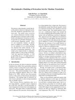
Limited trypsin-catalysed proteolysis of wild-type CTPS
produced different cleavage fragments depending on whe-
ther ATP and/or UTP were absent or present in the reaction
mixture (Fig. 1). In the absence of nucleotides, trypsin-
catalysed cleavage of CTPS (63 kDa) produced four
fragments with molecular masses corresponding to 10, 25,
28, and 53 kDa. However, in the presence of either ATP or
UTP (data not shown), ATP and UTP together, or CTP,
only fragments with molecular masses of 10 and 53 kDa
were produced indicating that these nucleotides protected
CTPS from cleavage at the site which produced the 25- and
28-kDa fragments. Neither glutamine nor GTP protected
CTPS from limited trypsin-catalysed digestion.
The sites of trypsin-catalysed cleavage yielding each of the
fragments were identified using Edman degradation to
obtain the N-terminal amino acid sequence of each
fragment (Table 1), and the known nucleotide sequence
encoding the enzyme [3]. The N-terminal sequence of the
25- and 53-kDa fragments were the same as that of
the whole recombinant wild-type protein indicating that the
cleavage site was located at the C-terminus of each of these
two fragments. The N-terminal sequence of the 28-kDa
fragment indicated that one of the cleavage sites was
Lys187. N-terminal analysis of the 10-kDa fragment
produced two sequences indicating that cleavage occurred
at Arg429 and Lys432 with a ratio of 2.6 : 1, respectively.
The cleavage pattern observed in the absence of any
protecting ligands, and the sites identified using N-terminal
analysis are summarized in Fig. 2. The molecular mass of
each polypeptide fragment, calculated using the known
2198 D. Simard et al. (Eur. J. Biochem. 270) Ó FEBS 2003
amino acid sequence [3], is in excellent agreement with the
values deduced from SDS/PAGE calibration (Table 1).
Limited trypsin-catalysed cleavage of mutant CTP
synthases
To confirm that the cleavage sites identified using
N-terminal analysis were indeed correct, site-directed mut-
agenesis was used to construct two single mutants (K187A
and R429A) and one double mutant (R429A/K432A).
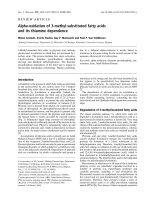
Limited trypsin-catalysed proteolysis of K187A CTPS in
the absence of nucleotides produced only the 10- and
53-kDa fragments (Fig. 3A), a cleavage pattern which was
identical to that observed for wild-type CTPS in the
presence of ATP and UTP (Fig. 1B). Proteolysis of
R429A in the absence of nucleotides gave fragments with
molecular masses of approximately 10, 25, 38, and 53 kDa
(Fig. 3B). The 38-kDa fragment accumulated because rapid
cleavage at Arg429 no longer occurred while cleavage at
Fig. 1. SDS/PAGE analysis of trypsin-catalysed cleavage of recom-
binant wild-type CTPS in the absence and presence of ligands. For each
gel: lane 1 contains molecular mass standards and lanes 2–6 contain
wild-type CTPS treated with trypsin for 0, 10, 20, 30, and 60 min,
respectively. In the absence of any ligands (A), the wild-type protein is
rapidly cleaved to yield a 10-kDa (not shown except in E) and a
53-kDa fragment, the latter which is subsequently cleaved to yield two
fragments with molecular masses of %25 and % 28 kDa. In the pres-
ence of ATP and UTP (10 m
M
each) (B), and CTP (2.5 m
M
)(C),only
production of the 10- and 53-kDa cleavage fragments is observed. In
the presence of GTP (2.5 m
M
)(D)and
L
-glutamine (10 m
M
)(E),
fragments with molecular masses of 10, 25, 28, and 53 kDa are pro-
duced.Allgelsare12%exceptfor(E)whichis20%.
Table 1. N-terminal amino acid sequences of trypsin-catalysed cleavage
fragments.
Molecular mass
a
(kDa) N-terminal sequence
Cleavage site
identified
b
63 (61) GSHMLEM1 … None
53 (48) GSHML None
28 (27) T188KPTQHSVKE Lys187
25 (21) GSHML None
10 (13.1) S430EKSDLGGTM (major)
c
Arg429
(12.8) S433DLGGTMRL (minor)
c
Lys432
a
Apparent molecular mass for full-length recombinant wild-type
E. coli CTPS and fragments determined from SDS/PAGE calib-
ration are given. The corresponding molecular masses calculated
using the known amino acid sequence are given in parentheses.
b
The
amino acid listed is that which provides the carbonyl function to the
scissile peptide bond. The numbers correspond to the numbering for
wild-type E. coli CTPS.
c
The ratio of the major peptide to minor
peptide was 2.6 : 1 and was determined by integration of the HPLC
chromatogram peaks corresponding to the phenylthiohydantoin
derivatives of the N-terminal serines.
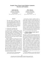
Fig. 2. Fragments generated by limited trypsin-
catalysed proteolysis of wild-type CTPS.
Peptide bond cleavage occurs C-terminal to
Lys187 in the synthase domain, and Arg429
and Lys432 in the GAT domain. The CTP/
UTP-binding site and residues comprising the
catalytic triad (Cys379, His515, and Glu517)
are also shown.
Ó FEBS 2003 Limited proteolysis of CTP synthase (Eur. J. Biochem. 270) 2199
Lys187 divided the protein into the 25- and 38-kDa
fragments. The 10- and 53-kDa fragments were formed in
much lower amounts than observed with wild-type CTPS
because of slow cleavage at Lys432. In the presence of ATP
and UTP, cleavage of R429A at Lys187 was suppressed and
only the 10- and 53 kDa fragments were formed because of
slow cleavage at Lys432 (Fig. 3C). Limited proteolysis of the
double mutant (R429A/K432A) in the absence of nucleo-
tides produced only two fragments with molecular masses
corresponding to 25 and 38 kDa consistent with cleavage
occurring only at Lys187 (Fig. 3D). In the presence of ATP
and UTP, proteolysis of R429A/K432A CTPS was com-
pletely suppressed (Fig. 3E).
Inactivation and protection studies
Treatment of wild-type and mutant CTP synthases with
trypsin produced time-dependent loss of both NH
3
-depend-
ent activity and glutamine-dependent activity (Fig. 4),
which followed first-order kinetics up to at least 90% of
the reaction. The observed first-order inactivation rate
constants for CTPS activity assayed using either NH
3
or
glutamine as the substrate are given in Table 2. In the
absence of ligands, the observed first-order rate constant for
trypsin-catalysed proteolysis of CTPS was slightly greater
when glutamine-dependent CTP formation was measured
than when NH
3
-dependent CTP formation was measured.
A reduction in the observed first-order rate constants for the
inactivation of the NH
3
-dependent activity was observed for
increasing concentrations of ATP, UTP, and CTP consis-
tent with each of these nucleotides providing protection
from trypsin-catalysed cleavage. Interestingly, in the
Fig. 3. SDS/PAGE analysis of trypsin-catalysed cleavage of mutant
CTP synthases in the absence and presence of ligands. (A) Lane 1,
molecular mass standards; lane 2, trypsin (at 7000 times the concen-
tration used in the proteolysis reactions); lanes 3–7 contain K187A
CTPS treated with trypsin for 0, 10, 20, 30, and 60 min, respectively.
The wild-type protein is rapidly cleaved to yield a 10- (not shown) and
a 53-kDa fragment, the latter which is not cleaved to yield the 25- and
28-kDafragments.ForeachgelshowninBthroughE,lane1contains
molecular mass standards and lanes 2–6 contain mutant CTPS treated
with trypsin for 0, 10, 20, 30, and 60 min, respectively. Limited pro-
teolysis of R429A CTPS (B) in the absence of nucleotides produced
fragments with molecular masses of 10, 25, 38, and 53 kDa. However,
in the presence of ATP and UTP (10 m
M
each) (C), only the 10- and
53-kDa fragments are produced. Limited proteolysis of R429A/
K432A CTPS (D) in the absence of nucleotides produces fragments
with molecular masses of 25 and 38 kDa. However, in the presence of
ATP and UTP (10 m
M
each) (E), no cleavage fragments were pro-
duced over the course of 1 h indicating that proteolysis was greatly
suppressed. All gels are 12%.
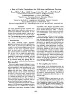
Fig. 4. Time-dependent inactivation of wild-type CTPS by trypsin. (A)
Inactivation of CTPS-catalysed NH
3
-dependent CTP formation in the
absence of ligands (s) and in the presence of ATP (10 m
M
, n), UTP
(10 m
M
, h),andATPandUTPcombined(10m
M
each, ,). Panel B
shows the inactivation of CTPS-catalysed glutamine-dependent CTP
formation in the absence of nucleotides (s) and in the presence of UTP
(10 m
M
, n), ATP (10 m
M
, h),andATPandUTPcombined(10m
M
each, ,). In both panels, the activity of the enzyme in the absence of
trypsinisalsoshown(d).
2200 D. Simard et al. (Eur. J. Biochem. 270) Ó FEBS 2003
presence of ATP and UTP, the rate constant for inactiva-
tion of glutamine-dependent activity was greater than that
observed for inactivation of the NH
3
-dependent activity.
This observation is consistent with the cleavage sites in the
GAT domain not being protected by these nucleotides and
the resulting 53-kDa fragment still possessing NH
3
-depend-
ent activity. Although all CTPS ligands tested (ATP, UTP,
CTP, GTP, and glutamine) protected CTPS from inactiva-
tion to some degree, the most effective protection was
afforded by CTP.
The observed first-order inactivation rate constants for
the NH
3
-dependent activity of the R429A and R429A/
K432A CTP synthases were less than that observed for
wild-type CTPS. Apparently, reduced cleavage within the
GAT domain results in less rapid cleavage within in the
synthase domain (i.e. at Lys187) and hence a lower value for
the rate constant for the loss of NH
3
-dependent activity.
Inactivation of the K187A enzyme could not be studied
because this enzyme was inactive (vide infra).
CD
The secondary structural content of wild-type CTPS and
the three mutant enzymes was analysed using CD
spectroscopy. Fig. 5 shows that the secondary structure
content of all the mutant proteins is similar to that of the
wild-type enzyme, except that the a-helix content of the
K187A and R429A/K432A mutants is slightly reduced
while the content of antiparallel b-sheet structure is slightly
increased, relative to wild-type CTPS. Although no signi-
ficant gross perturbations in secondary structure are
evident in the mutant proteins, the possibility that the
mutations cause a localized perturbation of secondary
structure or conformational change cannot be ruled out.
Mutant enzyme kinetics
The kinetic parameters k
cat
and K
m
for CTP formation were
determined with respect to NH
3
and glutamine for each of
the mutant enzymes except for K187A CTPS which was
inactive (Table 3). Direct examination of the conversion of
glutamine to glutamate (glutaminase activity) revealed that
K187A CTPS was able to catalyse the hydrolysis of
glutamine, but had a value of k
cat
thatwashalfofthat
observed for wild-type CTPS in the absence of GTP. When
the concentration of GTP was increased to 1 m
M
,thevalue
of k
act
was increased fivefold, compared to 50-fold for wild-
type CTPS (Fig. 6).
R429A and R429A/K432A CTP synthases displayed
similar kinetic properties. Each mutant had close to wild-
type NH
3
-dependent activity; however, glutamine-depend-
ent CTP formation was impaired. Interestingly, K
m
for
glutamine only increased 1.3- to 1.7-fold indicating that the
mutations had little effect on glutamine binding. However,
k
cat
was reduced % 15-fold for each mutant so that the
catalytic efficiency (k
cat
/K
m
) of glutamine-dependent CTP
formation was decreased 25- and 19-fold for the R429A and
R429A/K432A CTP synthases, respectively.
The kinetics of R429A CTPS were investigated in detail
to determine if the impaired glutamine-dependent CTP
formation was caused by an inability of GTP to activate
glutamine hydrolysis. In the presence of ATP and UTP
(1 m
M
each) and saturating glutamine (6 m
M
), GTP
(0.25 m
M
) caused a 2.5-fold increase in k
cat
for glutamine-
dependent CTP formation catalysed by R429A CTPS
compared to a 30-fold increase for wild-type CTPS (data
not shown). Concentrations of GTP above 0.25 m
M
(up to
1m
M
) did not enhance the observed rate of CTP formation.
Direct examination of the glutaminase activity revealed
that k
cat
was reduced approximately 10-fold for the R429A
and R429A/K432A enzymes relative to wild-type CTPS
with the concentration of GTP equal to 0.25 m
M
(Table 3).
More detailed analysis of the glutaminase activity of R429A
CTPS (Fig. 6) revealed that GTP binding and k
act
were
reduced approximately 10-fold and sixfold, relative to wild-
type CTPS. Thus mutation of Arg429 to alanine impairs
both GTP binding and allosteric activation of glutamine
hydrolysis.
Comparison of the k
cat
values for the glutaminase activity
and glutamine-dependent CTP formation catalysed by
R429A CTPS reveals that ammonia is produced from
glutamine hydrolysis at a rate that is slightly higher than the
rate at which CTP is formed. This observation suggests that
there may be a partial uncoupling of the glutaminase and
synthase reactions, however, the k
cat
values for the corres-
ponding reactions catalysed by the R429A/K432A mutant
are experimentally equal.
Oligomerization of CTPS
To determine if the K187A mutant was inactive because
it was unable to form tetramers, we investigated the
Table 2. Observed rate constants for inactivation of recombinant wild-
type and mutant CTP synthases
a
. ND, Not determined.
Protecting
ligand
Concentration
(m
M
)
k
obs
(· 10
)2
min
)1
)
NH
3
as
substrate
L
-glutamine
as substrate
None 0 7 (± 2) 9.0 (± 0.3)
None (R429A) 0 3.9 (± 0.3) ND
None
(R429A/K432A)
0 1.9 (± 0.4) ND
ATP 0.5 6 (± 2) ND
1.0 1.7 (± 0.5) ND
2.0 0.31 (± 0.04) ND
10.0 0.4 (± 0.1) 1.9 (± 0.2)
UTP 0.5 5.6 (± 0.9) ND
1.0 6 (± 1) ND
2.0 4 (± 1) ND
10.0 0.7 (± 0.1) 3.9 (± 0.9)
ATP and UTP 10 + 10 0.15 (± 0.07) 0.8 (± 0.2)
CTP 0.1 3 (± 1) ND
0.5 0.26 (± 0.09) ND
1.0 0.16 (± 0.03) ND
GTP 0.25 1.7 (± 0.5) ND
L
-glutamine 10.0 1.5 (± 0.3) ND
Control
b
0 0.075 (± 0.009) 0.07 (± 0.04)
a
All k
obs
values are for inactivation of wild-type CTPS except
where indicated otherwise.
b
Control refers to the inactivation of
CTPS that is observed during incubation of the enzyme at 37 °C
for 1 h in the absence of added trypsin.
Ó FEBS 2003 Limited proteolysis of CTP synthase (Eur. J. Biochem. 270) 2201
Fig. 5. CD analysis of wild-type and mutant
CTP synthases. (A) Spectra for wild-type,
K187A, R429A, and R429A/K432A CTP
synthases are shown. Each spectrum is the
average of three scans for each CTPS variant.
(B)Therelativeamountofeachtypeofsec-
ondary structure is indicated for each CTPS
variant. Error bars represent the standard
deviation of the mean for three independent
trials.
Table 3. Kinetic parameters for wild-type and mutant CTP synthases. ND, not determined.
Reaction (substrate) Kinetic parameter
a
CTPS variants
Wild-type K187A R429A R429A/K432A
CTP formation K
m
(m
M
) 1.70 ± 0.08 1.60 ± 0.08 1.82 ± 0.04
(NH
3
) k
cat
(s
)1
) 7.8 ± 0.1 NA
b
7.3 ± 0.3 6.5 ± 0.8
k
cat
/K
m
(m
M
)1
Æs
)1
) 4.5 ± 0.2 4.6 ± 0.3 3.6 ± 0.5
CTP formation K
m
(m
M
) 0.24 ± 0.04 0.40 ± 0.03 0.32 ± 0.08
(
L
-glutamine) k
cat
(s
)1
) 5.4 ± 0.8 NA 0.36 ± 0.04 0.39 ± 0.04
k
cat
/K
m
(m
M
)1
s
)1
) 22.6 ± 4.5 0.9 ± 0.1 1.2 ± 0.4
L
-glutamate
formation (fixed
L
-glutamine with
varying [GTP])
k
cat
(s
)1
) [GTP] ¼ 0.25 m
M
5.01 ± 0.18 0.13 ± 0.03 0.48 ± 0.03 0.49 ± 0.07
k
o
(s
)1
) 0.14 ± 0.03 0.07 ± 0.02 0.14 ± 0.04 ND
k
act
(s
)1
) 7.1 ± 0.3 –
c
1.19 ± 0.04 ND
K
A
(m
M
) 0.032 ± 0.006 –
c
0.38 ± 0.04 ND
a
Assay conditions are as described in Materials and methods. [ATP] ¼ [UTP] ¼ 1m
M
.
b
No activity was observed (i.e. < 0.5% wild-type
CTPS activity).
c
Values could not be determined accurately because of the low activity.
2202 D. Simard et al. (Eur. J. Biochem. 270) Ó FEBS 2003
ability of this mutant to form tetramers in the presence of
nucleotides using GF-HPLC. The observed molecular
masses for wild-type CTPS in the absence of nucleotides
and in the presence of ATP and UTP were 123 and
251 kDa, respectively. These values are similar to the
predicted values of 122 and 245 kDa, based on the amino
acid sequence of the recombinant mutant protein, and are
consistent with wild-type CTPS existing primarily as
dimers in the absence of ATP and UTP, and with a
shifting of the equilibrium to favour the tetrameric species
in the presence of ATP and UTP [42]. The K187A
mutant had an apparent molecular mass of 178 kDa in
the absence of nucleotides. This value is slightly higher
than that observed for the wild-type enzyme and corres-
ponds to the enzyme existing as % 30% tetramer as
calculated using Eqn (3) [20], where X is the fraction of
the enzyme in the tetramer form and the molecular
masses of the dimer (121 944 Da) and the tetramer
(243 888 Da) are those predicted based on the monomer
molecularmassof60972DaforrecombinantK187A
lacking the histidine tag. In the presence of ATP and
UTP, the observed molecular weight for K187A was
259 kDa. Thus it appeared that K187A CTPS was
capable of forming tetramers in the presence of saturating
concentrations of UTP and ATP, similar to the wild-type
enzyme.
molecular mass ¼
ð243 888Þ
2
X þð121 944Þ
2
ð1 À XÞ
ð243 888ÞX þð121 944Þð1 À XÞ
ð3Þ
Discussion
Limited proteolysis has been used to delineate the structural
organization of several amidotransferases including aspa-
ragine synthase [43], carbamoyl phosphate synthase
[44–50], anthranilate synthase [51], and glucosamine-6-
phosphate synthase [52]. This methodology has been
particularly useful for identifying both ligand-binding sites
and, in the case with glucosamine-6-phosphate synthase, an
exposed ÔhingeÕ region that, when cleaved by a-chymotryp-
sin, led to separation of the enzyme into its GAT and
synthase domains. Our interest in delineating structural
aspects of E. coli CTPS led us to examine the susceptibility
of CTPS to controlled proteolysis. In preliminary experi-
ments with endopeptidases of different specificity, we
identified trypsin as the enzyme of choice. Trypsin-catalysed
cleavage of wild-type CTPS generated four fragments in
the absence of ATP and UTP, but only two fragments in
the presence of these nucleotides. Determination of the
N-terminal sequence of these fragments, in conjunction
with the known nucleotide sequence of the E. coli pyrG
gene [3], permitted us to identify three principal cleavage
sites: Lys187 in the synthase domain, and Arg429 and
Lys432 in the GAT domain. A summary of the fragmen-
tation pattern arising from trypsin-catalysed cleavage at
these sites is presented in Fig. 2.
Lys187 resides in a region of the synthase domain that is
highly conserved among CTP synthases from different
organisms. This region, between residues 116 and 229, has
been suggested to comprise the CTP/UTP-binding site
[18,25–27]. Our observation that both CTP and UTP afford
effective protection to CTPS from trypsin-catalysed clea-
vage at Lys187 also supports the notion that this residue is
located in the CTP/UTP-binding site. Interestingly, ATP
also provides protection against cleavage and does so better
than UTP. Such protection could arise because: (a) ATP
binds at an adjacent site and sterically blocks access of
trypsin to Lys187; (b) ATP-induced tetramerization yields a
quaternary structure in which the Lys187 site is not
accessible to trypsin; or (c) ATP induces a conformational
change in CTPS to yield a conformation in which Lys187
is no longer exposed to bulk solvent.
Replacement of Lys187 by an alanine residue yielded a
protein that was resistant to limited trypsin-catalysed
proteolysis in the synthase domain, supporting our conclu-
sion that cleavage occurred C-terminal to this residue. The
K187A mutant could not catalyse the formation of CTP,
however, it retained the ability to form tetramers in the
presence of nucleotides, and exhibited a very low level of
GTP-dependent glutaminase activity which was enhanced
slightly by GTP. Interestingly, in the absence of nucleotides,
K187A existed as % 30% tetramer suggesting that neutrali-
zation of positive charge at residue 187 might play a role in
promoting enzyme tetramerization. Indeed, hydrophobic
interactions between dimers of E. coli CTPS have been
suggested to play a role in the formation of tetramers [53].
Predictions of the secondary structure of the highly
conserved region of amino acid sequence between residues
185 and 192 suggest that Lys187 constitutes part of a
conserved loop. It is not clear whether nucleotides protect
this putative loop from proteolytic cleavage because
nucleotide binding directly blocks access of trypsin to the
cleavage site or, because nucleotide binding causes a change
in the enzyme’s conformation or quaternary structure (i.e.
tetramerization) that subsequently conceals the cleavage site
from trypsin.
Fig. 6. Glutaminase activity for mutant CTP synthases. The values of
k
apparent
cat
for the hydrolysis of glutamine by K187A (d) and R429A (s)
CTP synthases are shown. Inset: values of k
apparent
cat
for the hydrolysis of
glutamine by wild-type CTPS. The curves shown are from a fit of the
data to Eqn (2) and the values of k
o
, k
act
,andK
A
are given in Table 3.
Ó FEBS 2003 Limited proteolysis of CTP synthase (Eur. J. Biochem. 270) 2203
Finally, we note that studies on the chemical modification
of E. coli CTPS with thiourea dioxide led Roberston et al.
[54] to conclude that lysine residues were important for
catalysis. To our knowledge, the present study represents
the first identification of a catalytically essential lysine
residue in E. coli CTPS involved in either amido-/NH
3
transfer or UTP phosphorylation.
Arg429 and Lys432 reside in a region of amino acid
sequence within the GAT domain that is partially conserved
only among CTP synthases from some sources (Fig. 7). In
accord with our expectations, nucleotides offered no
protection against cleavage at these sites but replacement
of these residues by alanine (i.e. R429A and R429A/K432A)
yielded mutant enzymes that were more resistant to
proteolytic cleavage in the GAT domain. These mutant
enzymes displayed wild-type activity with respect to
NH
3
-dependent CTP formation and wild-type affinity for
glutamine, but glutamine-dependent CTP formation was
markedly impaired. Interestingly, although these mutations
in the GAT domain did not impair the enzyme’s ability to
utilize NH
3
as a substrate (i.e. the activity associated with
the synthase domain [16]), they did cause the rate of loss of
NH
3
-dependent activity during limited trypsin-catalysed
proteolysis to be less than would have been predicted based
on the inactivation rate constant observed for wild-type
CTPS (Table 2). This is consistent with previous reports
that suggested interactions between the GAT and synthase
domains within the tertiary structure of the enzyme
[17,28,55]. The existence of such interactions is also
supported by our observation that mutation of Lys187 to
alanine in the synthase domain severely impairs the
glutaminase activity in the GAT domain.
Our observations that R429A CTPS binds GTP with
reduced affinity (% 10-fold) and, at saturating concentra-
tions of GTP, the apparent k
cat
value for glutamine-
dependent CTP formation is reduced sixfold suggest that
Arg429 plays a role in both binding GTP and the
mechanism for allosteric activation of glutamine hydrolysis.
Secondary structure predictions suggest that Arg429 and
Lys432 are located within a region where a b-strand
undergoes a transition into a loop structure. The ability of
trypsin to catalyse cleavage adjacent to these residues
suggests that this loop is exposed to bulk solvent. Despite
the fact that this region is not highly conserved between
organisms, it does appear to be required for E. coli CTPS to
catalyse glutamine turnover. Arg429 and Lys432 lie close to
a conserved sequence motif [GG(TS)(ML)RLG] within the
GAT domain (shaded residues 436–442 in Fig. 7) that was
recently identified by Willemoe
¨
s [24]. Using site-directed
mutagenesis experiments on CTPS from L. lactis,
Fig. 7. Sequence comparison of a portion of the C-terminus (GAT domain) of CTP synthases. For the protein sequences shown, invariant residues (*),
conservative substitutions (:), and semiconservative substitutions (.) are indicated. The two residues (Arg429 and Lys432) identified as cleavage sites
during limited trypsin proteolysis and mutated in the present study are indicated (›). These residues reside in a region of the primary structure that is
not conserved among different organisms. The conserved sequence motif (GG[TS][ML]RLG) identified by Willemoe
¨
s [24] is shaded. The proteins
included in the alignment are as follows (accession numbers in parentheses): Girardia intestinalis (AAB41453.1), Synechococcus (Q54775), Spiro-
plasma citri (P52200), Synechocystis (P74208), Bacillus subtilis (P13242), Mycobacterium leprae (S72961), Mycobacterium bovis (AAB48045.1),
Methanococcus jannaschii (Q58574), Chlamydia trachomatis (Q59321), Haemophilus influenzae (P44341), Neisseria meningitidis (CAB84970.1),
Nitrosomonas europaea (AAC33441.1), Azospirillum brasilense (P28595), Campylobacter jejuni (CAB72520.1), Heliobacter pylori (O25116), Borrelia
burgdorferi (O51522), Cricetulus griseus (P50547), Mus musculus (P70698), Homo sapiens (NP_001896.1), Arabidopsis thaliana (AAC78703.1),
Saccharomyces cerevisiae H (URA-8, P38627), Saccharomyces cerevisiae G (URA-7, P28274), Plasmodium falciparum (AAC36385.1), Lactococcus
lactis (CAA09021.2), and Escherichia coli (AAA69290.1). Numbering shown is for the E. coli sequence.
2204 D. Simard et al. (Eur. J. Biochem. 270) Ó FEBS 2003
Willemoe
¨
s demonstrated that Thr431 and Arg433 (Thr438
and Arg440 in E. coli CTPS) within this motif play a role in
GTP-dependent activation of glutamine hydrolysis but
concluded that these residues were not involved in GTP
binding [24]. However, our observation that Arg429 is
important for GTP binding is consistent with Willemoe
¨
s’
observation that R433A L. lactis CTPS exhibited a 10–17-
fold increase in GTP-binding affinity [24] and support the
notion that the conserved sequence motif and adjacent
residues may also be important for GTP binding.
Acknowledgements
The authors thank the Canadian Institutes of Health Research for an
operating grant (S. L. B.), the Nova Scotia Health Research Founda-
tion and Cancer Care Nova Scotia for graduate fellowships (A. I.), and
Cancer Care Nova Scotia for research training grants (K. H. & D. S).
The authors also express thanks to Prof. M. Dobson and J. Chew for
technical advice and assistance with electroblotting.
References
1. Koshland, D.E. Jr & Levitzki, A. (1974) CTP Synthetase and
related enzymes. In The Enzymes (Boyer, P.D., ed.), pp. 539–559.
Academic Press, New York.
2. Long, C. & Koshland, D.E. Jr (1978) Cytidine triphosphate syn-
thetase. Methods Enzymol. 51, 79–83.
3. Weng, M., Makaroff, C.A. & Zalkin, H. (1986) Nucleotide
sequence of Escherichia coli pyrG encoding CTP synthetase.
J. Biol. Chem. 261, 5568–5574.
4. Weng, M.L. & Zalkin, H. (1987) Structural role for a conserved
region in the CTP synthetase glutamine amide transfer domain.
J. Bacteriol. 169, 3023–3028.
5. Zalkin, H. (1993) The amidotransferases. Adv. Enzymol. Relat.
Areas Mol. Biol. 66, 203–309.
6. Zalkin, H. & Smith, J.L. (1998) Enzymes utilizing glutamine as an
amide donor. Adv. Enzymol. Relat. Areas Mol. Biol. 72, 87–144.
7. Hatse, S., De Clercq, E. & Balzarini, J. (1999) Role of anti-
metabolites of purine and pyrimidine nucleotide metabolism in
tumor cell differentiation. Biochem. Pharmacol. 58, 539–555.
8. Kennedy, E.P. (1986) The biosynthetsis of phospholipids. In
Lipids and Membranes: Past, Present and Future (Op den Kamp,
J.A.F., Roelofsen, B. & Wirtz, K.W.A., eds), pp. 171–206. Else-
vier. Scientific Publishers, Amsterdam.
9. Kensler, T.W. & Cooney, D.A. (1989) Inhibitors of the de novo
pyrimidine pathway. In Design of Enzyme Inhibitors as Drugs
(Sandler, M. & Smith, H.J., eds), pp. 379–401. Oxford University
Press, New York.
10. De Clercq, E. (1993) Antiviral agents: characteristic activity
spectrum depending on the molecular target with which they
interact. Adv. Virus. Res. 42, 1–55.
11. Hendriks, E.F., O’Sullivan, W.J. & Stewart, T.S. (1998) Molecular
cloning and characterization of the Plasmodium falciparum cyti-
dine triphosphate synthetase gene. Biochim. Biophys. Acta 1399,
213–218.
12. Hofer,A.,Steverding,D.,Chabes,A.,Brun,R.&Thelander,L.
(2001) Trypanosoma brucei CTP synthetase: a target for the
treatment of African sleeping sickness. Proc. Natl Acad. Sci. USA
98, 6412–6416.
13. Lim, R.L., O’Sullivan, W.J. & Stewart, T.S. (1996) Isolation,
characterization and expression of the gene encoding cytidine
triphosphate synthetase from Giardia intestinalis. Mol. Biochem.
Parasitol. 78, 249–257.
14. Verschuur, A.C., Van Gennip, A.H., Leen, R., Voute, P.A.,
Brinkman, J. & Van Kuilenburg, A.B. (2002) Cyclopentenyl
cytosine increases the phosphorylation and incorporation into
DNA of 1-b-
D
-arabinofuranosyl cytosine in a human T-lym-
phoblastic cell line. Int. J. Cancer 98, 616–623.
15. Gao, W.Y., Johns, D.G. & Mitsuya, H. (2000) Potentiation of the
anti-HIV activity of zalcitabine and lamivudine by a CTP synthase
inhibitor, 3-deazauridine. Nucleosides Nucleotides Nucl. Acids 19,
371–377.
16. Bearne, S.L., Hekmat, O. & Macdonnell, J.E. (2001) Inhibition of
Escherichia coli CTP synthase by glutamate gamma-semialdehyde
and the role of the allosteric effector GTP in glutamine hydrolysis.
Biochem. J. 356, 223–232.
17. Levitzki,A.&Koshland,D.E.Jr(1972)Roleofanallosteric
effector. Guanosine triphosphate activation in cytosine triphos-
phate synthetase. Biochemistry 11, 241–246.
18. Long, C.W. & Pardee, A.B. (1967) Cytidine triphosphate syn-
thetase of Escherichia coli B. I. Purification and kinetics. J. Biol.
Chem. 242, 4715–4721.
19. Levitzki, A. & Koshland, D.E. Jr (1969) Negative cooperativity
in regulatory enzymes. Proc.NatlAcad.Sci.USA62, 1121–
1128.
20. Levitzki, A. & Koshland, D.E. Jr (1972) Ligand-induced dimer-
to-tetramer transformation in cytosine triphosphate synthetase.
Biochemistry 11, 247–253.
21. Tesmer, J.J., Klem, T.J., Deras, M.L., Davisson, V.J. & Smith,
J.L. (1996) The crystal structure of GMP synthetase reveals a
novel catalytic triad and is a structural paradigm for two enzyme
families. Nat. Struct. Biol. 3, 74–86.
22. Thoden, J.B., Holden, H.M., Wesenberg, G., Raushel, F.M. &
Rayment, I. (1997) Structure of carbamoyl phosphate synthetase:
a journey of 96 A
˚
from substrate to product. Biochemistry 36,
6305–6316.
23. Chittur, S.V., Klem, T.J., Shafer, C.M. & Davisson, V.J. (2001)
Mechanism for acivicin inactivation of triad glutamine amido-
transferases. Biochemistry 40, 876–887.
24. Willemoe
¨
s, M. (2003) Thr-431 and Arg-433 are part of a con-
served sequence motif of the glutamine amidotransferase domain
of CTP synthases and are involved in GTP activation of the
Lactococcus lactis enzyme. J. Biol. Chem. 278, 9407–9411.
25. Wylie, J.L., Wang, L.L., Tipples, G. & McClarty, G. (1996) A
single point mutation in CTP synthetase of Chlamydia trachomatis
confers resistance to cyclopentenyl cytosine. J. Biol. Chem. 271,
15393–15400.
26. Whelan, J., Phear, G., Yamauchi, M. & Meuth, M. (1993) Clus-
tered base substitutions in CTP synthetase conferring drug
resistance in Chinese hamster ovary cells. Nature Genet. 3, 317–
322.
27. Ostrander,D.B.,O’Brien,D.J.,Gorman,J.A.&Carman,G.M.
(1998) Effect of CTP synthetase regulation by CTP on phospho-
lipid synthesis in Saccharomyces cerevisiae. J. Biol. Chem. 273,
18992–19001.
28. Iyengar, A. & Bearne, S.L. (2003) Aspartate 107 and leucine 109
facilitate efficient coupling of glutamine hydrolysis to CTP
synthesis by E. coli CTP synthase. Biochem. J. 369, 497–507.
29. Kelley, L.A., MacCallum, R.M. & Sternberg, M.J. (2000)
Enhanced genome annotation using structural profiles in the
program 3D-PSSM. J. Mol. Biol. 299, 499–520.
30. Garnier, J., Gibrat, J.F. & Robson, B. (1996) GOR method for
predicting protein secondary structure from amino acid sequence.
Methods Enzymol. 266, 540–553.
31. Guermeur, Y. (1997) Combinaison de Classifieurs Statistiques:
application a la Pre
´
diction de la Structure Secondaire des
Prote
´
ines, PhD Thesis, Universite
´
Pierre et Marie Curie, Paris,
France.
32. Cuff, J.A. & Barton, G.J. (2000) Application of enhanced multiple
sequence alignment profiles to improve protein secondary struc-
ture prediction. In Proteins. 40, 502–511.
Ó FEBS 2003 Limited proteolysis of CTP synthase (Eur. J. Biochem. 270) 2205
33. Frishman, D. & Argos, P. (1996) Incorporation of non–local
interactions in protein secondary structure prediction from the
aminoacidsequence.Protein Eng. 9, 133–142.
34. McGuffin, L.J., Bryson, K. & Jones, D.T. (2000) The
PSIPRED protein structure prediction server. Bioinformatics 16,
404–405.
35. Baldi,P.,Brunak,S.,Frasconi,P.,Soda,G.&Pollastri,G.(1999)
Exploiting the past and the future in protein secondary structure
prediction. Bioinformatics 15, 937–946.
36. Higgins, D., Thompson, J., Gibson, T., Thompson, J.D., Higgins,
D.G. & G. (1994) CLUSTAL W: improving the sensitivity of
progressive multiple sequence alignment through sequence
weighting, position-specific gap penalties and weight matrix
choice. Nucl. Acids Res. 22, 4673–4680.
37. Jencks, W.P. & Regenstein, J. (1968) Ionization constants
of acids and bases. In Handbook of Biochemistry (Sober,
H.A., ed.), pp. J150–189. The Chemical Rubber Co, Cleveland,
Ohio.
38. Iyengar, A. & Bearne, S.L. (2002) An assay for cytidine 5¢-
triphosphate synthetase glutaminase activity using high perfor-
mance liquid chromatography. Anal. Biochem. 308, 396–400.
39. Cornish-Bowden, A. (1995) Fundamentals of Enzyme Kinetics.
Portland Press Ltd, London.
40. Wilson, K.J. & Yuan, P.M. (1989) Protein and peptide mod-
ification. In Protein Sequencing: a Practical Approach (Findlay,
J.B.C. & Geisow, M.J., eds), pp. 13. Oxford University Press,
New York.
41. Bo
¨
hm, G., Muhr, R. & Jaenicke, R. (1992) Quantitative analysis
of protein far UV circular dichroism spectra by neural networks.
Protein Eng. 5, 191–195.
42. Robertson, J.G. (1995) Determination of subunit dissociation
constants in native and inactivated CTP synthetase by sedimen-
tation equilibrium. Biochemistry 34, 7533–7541.
43. Hongo, S. & Sato, T. (1983) Some molecular properties of
asparagine synthetase from rat liver. Biochim. Biophys. Acta 742,
484–489.
44. Carrey, E.A. (1986) Nucleotide ligands protect the inter-domain
regions of the multifunctional polypeptide CAD against limited
proteolysis, and also stabilize the thermolabile part-reactions of
the carbamoyl-phosphate synthase II domains within the CAD
polypeptide. Biochem. J. 236, 327–335.
45. Guadalajara, A., Grisolia, S. & Rubio, V. (1987) Limited pro-
teolysis reveals low-affinity binding of N-acetyl-L-glutamate to
rat-liver carbamoyl-phosphate synthetase (ammonia). Eur. J.
Biochem. 165, 163–169.
46. Marshall, M. & Fahien, L.A. (1988) Proteolysis as a probe of
ligand-associated conformational changes in rat carbamyl phos-
phate synthetase I. Arch. Biochem. Biophys. 262, 455–470.
47. Evans, D.R. & Balon, M.A. (1988) Controlled proteolysis of
ammonia-dependent carbamoyl-phosphate synthetase I from
Syrian hamster liver. Biochim. Biophys. Acta 953, 185–196.
48. Kim, H., Kelly, R.E. & Evans, D.R. (1992) The structural orga-
nization of the hamster multifunctional protein CAD. Controlled
proteolysis, domains, and linkers. J. Biol. Chem. 267, 7177–7184.
49. Mareya, S.M. & Raushel, F.M. (1995) Mapping the structural
domains of E. coli carbamoyl phosphate synthetase using limited
proteolysis. Bioorg. Med. Chem. 3, 525–532.
50. Guy, H.I., Bouvier, A. & Evans, D.R. (1997) The smallest car-
bamoyl-phosphate synthetase. A single catalytic subdomain cat-
alyzes all three partial reactions. J. Biol. Chem. 272, 29255–29262.
51. Walker, M.S. & DeMoss, J.A. (1986) Organization of the func-
tional domains of anthranilate synthase from Neurospora crassa.
Limited proteolysis studies. J. Biol. Chem. 261, 16073–16077.
52. Denisot, M.A., Le Goffic, F. & Badet, B. (1991) Glucosamine-6-
phosphate synthase from Escherichia coli yields two proteins upon
limited proteolysis: identification of the glutamine amidohydrolase
and 2R ketose/aldose isomerase-bearing domains based on their
biochemical properties. Arch. Biochem. Biophys. 288, 225–230.
53. Anderson, P.M. (1983) CTP synthetase from Escherichia coli:an
improved purification procedure and characterization of hysteretic
and enzyme concentration effects on kinetic properties. Biochem-
istry 22, 3285–3292.
54. Robertson, J.G., Sparvero, L.J. & Villafranca, J.J. (1992)
Inactivation and covalent modification of CTP synthetase by
thiourea dioxide. Protein Sci. 1, 1298–1307.
55. Lewis, D.A. & Villafranca, J.J. (1989) Investigation of the
mechanism of CTP synthetase using rapid quench and isotope
partitioning methods. Biochemistry 28, 8454–8459.
2206 D. Simard et al. (Eur. J. Biochem. 270) Ó FEBS 2003