Báo cáo khoa học: Expression, localization and potential physiological significance of alcohol dehydrogenase in the gastrointestinal tract pdf
Bạn đang xem bản rút gọn của tài liệu. Xem và tải ngay bản đầy đủ của tài liệu tại đây (598.57 KB, 11 trang )
Expression, localization and potential physiological significance
of alcohol dehydrogenase in the gastrointestinal tract
Julia Vaglenova
1,
*,‡, Susana E. Martı
´
nez
1,
†,‡, Sergio Porte
´
1
, Gregg Duester
2
, Jaume Farre
´
s
1
and Xavier Pare
´
s
1
1
Department of Biochemistry and Molecular Biology, Universitat Auto
`
noma de Barcelona, Spain;
2
OncoDevelopmental Biology
Program, The Burnham Institute, La Jolla, CA, USA
ADH1 and ADH4 are the major alcohol dehydrogenases
(ADH) in ethanol and retinol oxidation. ADH activity and
protein expression were investigated in rat gastrointestinal
tissue homogenates by enzymatic and Western blot analyses.
In addition, sections of adult rat gastrointestinal tract were
examined by in situ hybridization and immunohistochem-
istry. ADH1 and ADH4 were detected along the whole tract,
changing their localization and relative content as a function
of the area studied. While ADH4 was more abundant in
the upper (esophagus and stomach) and lower (colorectal)
regions, ADH1 was predominant in the intestine but also
present in stomach. Both enzymes were detected in mucosa
but, in general, ADH4 was found in outer cell layers, lining
the lumen, while ADH1 was detected in the inner cell layers.
Of interest were the sharp discontinuities in the expression
found in the pyloric region (ADH1) and the gastroduodenal
junction (ADH4), reflecting functional changes. The precise
localization of ADH in the gut reveals the cell types where
active alcohol oxidation occurs during ethanol ingestion,
providing a molecular basis for the gastrointestinal alcohol
pathology. Localization of ADH, acting as retinol dehydro-
genase/retinal reductase, also indicates sites of active retinoid
metabolism in the gut, essential for mucosa function and
vitamin A absorption.
Keywords: ethanol; immunohistochemistry; in situ hybridi-
zation; retinol; retinoic acid.
The major pathway for the elimination of ethanol is
through its oxidation to acetaldehyde that occurs mostly in
liver [1], though ethanol metabolism is also significant in
other tissues [2]. Alcohol dehydrogenase (ADH) is the main
enzyme responsible for the first step in ethanol elimination
[3]. ADH is expressed in several molecular forms, grouped
in five enzymatic classes [4], and four of them have been well
characterized at the protein level in mammals [5,6]. In the
rat, ADH1 has a low K
m
for ethanol and it is responsible for
the hepatic ethanol metabolism [7]. ADH2 and ADH3 are
not active at moderate concentrations of ethanol [7,8].
ADH4 has high K
m
and k
cat
values for ethanol [9], and it is
found in gastrointestinal mucosa, blood vessels, central
nervous system and many epithelia, but it is absent in
normal liver [2,10,11]. Moreover, these ADH forms have
retinol dehydrogenase activity [12–17], and recent genetic
studies in knockout mice have demonstrated that ADH1,
ADH3, and ADH4 participate in the retinoic acid (RA)
synthesis pathway [16,18,19].
Previous studies have shown that the rat ADH system is
comprised of single isozyme representatives of each class,
making it a simpler system to study, compared to the
human ADH [5,6]. In spite of several reports on the
localization of ADH in rodent [2,7,20–22] and human
[23–30] gastrointestinal tissues, these works are only partial.
This paper presents a complete analysis of the whole
gastrointestinal tract in the rat: ADH activity levels were
measured by spectrophotometric assays, ADH expression
pattern by electrophoretic and Western blot analyses, and
the localization of ADH (at mRNA and protein levels) in
the distinct cell layers of each gastrointestinal region by
in situ hybridization (ISH) and immunohistochemistry
(IHC)
1
. Our results demonstrate that ADH1 and ADH4
coexist throughout the gastrointestinal tract and provide
new data to understand the physiological role of ADH
classes in the gastrointestinal tract and the etiopathogeny
related to alcohol abuse.
Experimental procedures
Animals
Adult Sprague–Dawley rats (n ¼ 5; male, 200–250 g) were
used. Animal protocols were approved by the Ethical
Committee of the Universitat Auto
`
noma de Barcelona.
After decapitation, gastrointestinal organs were removed
and processed rapidly as described below.
Correspondence to X. Pare
´
s, Department of Biochemistry and
Molecular Biology, Faculty of Sciences, Universitat Auto
`
noma de
Barcelona, E-08193 Bellaterra, Barcelona, Spain.
Fax: + 34 93 5811264, Tel.: + 34 93 5813026,
E-mail:
Abbreviations: ADH, alcohol dehydrogenase; ALDH, aldehyde
dehydrogenase; IHC, immunohistochemistry; ISH, in situ
hybridization; RA, retinoic acid.
Note: àThese authors made equal contributions to this study.
*Present address: Department of Pharmacal Sciences, 401 Pharmacy
Bldg., Auburn University, Auburn, AL 36849, USA.
Present address:BiologyDepartment,BostonCollege,321Higgins
Hall, 140 Commonwealth Ave., Chesnut Hill, MA 02467, USA.
(Received 27 February 2003, revised 21 April 2003,
accepted 28 April 2003)
Eur. J. Biochem. 270, 2652–2662 (2003) Ó FEBS 2003 doi:10.1046/j.1432-1033.2003.03642.x
ADH activity assay and starch gel electrophoresis
Tissues from gastrointestinal tract were dissected carefully
and subsequently washed in ice-cold homogenization
buffer (50 m
M
sodium phosphate, pH 7.6, 0.5 m
M
dithio-
threitol). The specimens were cut into small fractions
and homogenized at 4 °C. Crude homogenates were
centrifuged (24 000 g,4°C, 30 min) and supernatants
were used for activity assay or analysis by starch gel
electrophoresis [2]. After electrophoresis, gels were stained
for ADH activity using 100 m
M
2-buten-1-ol as a
substrate. Also, ADH activity of homogenates was
monitored at 340 nm in a UV-VIS spectrophotometer
(Cary 400Bio; Varian), in 0.1
M
NaCl/P
i
, pH 7.5, 2.4 m
M
NAD
+
,at25°C, using 10 m
M
ethanol or 1
M
ethanol
as a substrate. At 10 m
M
ethanol, we determined the
contribution of ADH1 (K
m
¼ 1.4 m
M
, k
cat
¼ 40 min
)1
)
[7]. At 1
M
ethanol, the observed activity was mainly due
to ADH4 (K
m
¼ 2.4
M
, k
cat
¼ 2600 min
)1
)[9].Atthis
ethanol concentration, ADH1 shows substrate inhibition
[31] and the contribution of ADH3 is still negligible
because of its extremely low activity at pH 7.5 [7]. One
activity unit corresponds to the reduction of 1 lmol
NAD per min. Protein concentrations were estimated by
the method of Bradford [32] using bovine serum albumin
as standard.
In situ
hybridization analysis (ISH)
Generation of ADH1 and ADH4 specific sense and
antisense riboprobes was performed as reported previously
[11]. The gastrointestinal tract was removed and divided
into regions corresponding to the various tissues. After
dissection, digestive samples were immediately rinsed in
NaCl/P
i
(0.1
M
sodium phosphate buffer, pH 7.4, 0.15
M
NaCl) and immersed in 4% (w/v) paraformaldehyde
in NaCl/P
i
for 12 h. The paraffin-embedded tissues were
sliced into serial 8-lm sections using a Leica microtome
and attached to coated microscope slides. Insituhybrid-
ization and subsequent immunochemical chromogenic
detection of digoxigenin-labeled hybrids was performed
as previously described [11]. The hybridization signal
corresponding to each probe appeared highly specific, as
demonstrated by the negative controls performed with the
sense RNA probes.
Protein immunoblotting and Western blot analysis
Homogenates were prepared from fresh adult rat tissue
as reported previously [11], except that 1 m
M
phenyl-
methanesulfonyl fluoride, 1 lgÆmL
)1
leupeptin, and
1 lgÆmL
)1
pepstatin were added as protease inhibitors.
Protein blots were incubated with affinity-purified rabbit
antiserum raised against mouse ADH4 (1 : 500) [21].
Immunodetection was carried out using goat anti-(rabbit
IgG)-alkaline phosphatase conjugate (Bio-Rad) for 1 h at
room temperature. Alkaline phosphatase activity was
then visualized by incubation with 0.1
M
Tris/HCl,
pH 9.5, containing 5-bromo-4-chloro-3-indolylphosphate
and nitroblue tetrazolium as substrates according to the
instructions of the Alkaline Phosphatase Conjugate
Substrate kit (Bio-Rad).
Immunohistochemistry
Rat gastrointestinal tissues were fixed, processed routinely,
and embedded in paraffin as described for ISH. Localiza-
tion of ADH4 was investigated using affinity-purified
antibodies specific for ADH4 [21] diluted to 1 : 500 on
serial 5-lm tissue sections. Slides were treated with xylene
and hydrated through a graded series of decreasing ethanol
concentrations. Endogenous peroxidase activity was
blocked with 1% (v/v) hydrogen peroxide for 15 min.
After rinsing in Tris/HCl-buffered saline, slides were
blocked with 2% (v/v) of normal serum, and the primary
antibody was applied for 1 h. Biotinylated goat anti-(rabbit
IgG) Ig (Dakopatts) was used as a secondary antibody and
was visualized by avidin–biotin complex (Strept–ABCom-
plex–HRP; Dakopatts; dilution 1 : 400 in blocking solu-
tion) with peroxidase detection using the Vectastain
Universal Elite ABC kit (Vector Laboratories, Inc.,
Burlingame, CA, USA). 3,3¢-Diaminobenzidine tetra-
hydrochloride (DAB; Sigma-Aldrich) was used as a
chromogen (50 mg DAB in 100 mL 0.05
M
TBS, pH 7.4,
with 33.3 lLH
2
O
2
, prepared prior to use). Tissues were
then rinsed in Tris/HCl, dehydrated and mounted using a
xylene-based medium (ENTELLANÒ neu; Merck). Adja-
cent slides were stained with Harris hematoxylin (Vecta-
stain), dehydrated through a graded series of increasing
ethanol concentrations, followed by two xylene washes,
and cover-slipped with ENTELLANÒ neu (Merck). Both
the omission of anti-ADH4 IgG and the preadsorption of
anti-ADH4 IgG with excess of purified recombinant
ADH4 abolished the positive reaction in the control
sections, demonstrating the specificity of the staining.
Control experiments had showed that anti-ADH4 IgG
immunoreacted with recombinant purified rat ADH4 but
did not cross-react with any other ADH classes.
Image analysis
Following ISH and IHC techniques, digestive tract sections
were examined under a Leica DMRD fluorescense micro-
scope with a Hamamatsu C5310 CCD or a Leica DC200
camera. Image acquisition was carried out with
IMAGE
PROPLUS
software and imported into Adobe
PHOTOSHOP
v5.5 (Adobe). Color images were transformed into black
and white images using a grey-scale function, and brightness
and contrast were adjusted. All sections were examined
concurrently and compared to published pictures and
schemes [33].
Results
ADH expression in rat gastrointestinal homogenates
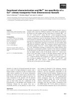
Homogenates from gastrointestinal tissues (tongue, eso-
phagus, stomach, duodenum, jejunum, ileum, caecum,
colon and rectum) were analyzed for the presence of
ADH at activity and protein levels by using starch gel
electrophoresis, spectrophotometric measurements, and
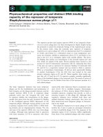
immunoblotting (Fig. 1). Both ADH1 and ADH4 were
detected throughout the entire gastrointestinal tract but
with a differential tissue distribution. ADH1 was detected
mainly in duodenum and the colorectal region, while ADH4
Ó FEBS 2003 Alcohol dehydrogenase in the gastrointestinal tract (Eur. J. Biochem. 270) 2653
was highly expressed in the upper (mainly esophagus and
stomach) and colorectal regions. ADH3 was detected in all
tissues examined. No large differences were found in activity
or in the tissue distribution of the ADH forms between
different animals (Fig. 1).
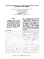
Localization of ADH in tongue and esophagus
Immunohistochemistry (IHC) of rat tongue showed ADH4
in the mucosa. The signal was detected in the papillae,
specifically in the stratified squamous epithelium (Fig. 2B,
C,E). ADH4 was also detected in the endothelium of
microvessels (Fig. 2C). Insituhybridization (ISH) analysis
of esophagus revealed that ADH1 mRNA was only
localized in the base line of the stratified squamous
epithelium (data not shown). In contrast, ADH4 mRNA
was present at high level in all cell layers of stratified
squamous epithelium (Fig. 2G). No specific signal was
detected in the lamina propia and muscularis mucosae.In
good agreement, ADH4 immunostaining was detected in
the stratified squamous epithelium (Fig. 2I). Interestingly, a
strong ADH4 protein signal was observed in the keratinized
layer of epithelium, where the ADH4 mRNA was not
detected.
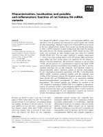
Localization of ADH in stomach and the gastroduodenal
junction
ADH1 and ADH4 mRNAs were both expressed in the
gastric mucosa from cardiac to pyloric stomach but each
form was confined to distinct layers and cell types. In the
stomach body, ADH1 was localized in the medium and
basal layers of the mucosa, and muscularis mucosae but not
in mucus-secreting cells (Fig. 3A). However, towards the
pyloric region, ADH1 gradually appeared in the mucus-
secreting epithelium as well (cf. Fig. 3B,C,D). In contrast,
ADH4 mRNA was detected in the mucus-secreting cells,
in some of the inner cell layers, and in muscularis mucosae
Fig. 1. Detection of ADH1 and ADH4 in tis-
sue homogenates of the gastrointestinal tract.
(A) Starch gel electrophoresis stained for
activity using 100 m
M
2-buten-1-ol as a sub-
strate. (B) Graphic representation of ADH1
(black bars) and ADH4 (grey bars) activity
levels. Activity assays were performed with
0.1
M
sodium phosphate, pH 7.5, at 25 °C,
with 10 m
M
(ADH1) or 1
M
(ADH4) ethanol
as a substrate and 2.4 m
M
NAD
+
as a coen-
zyme. Values are expressed as the arithmetical
mean ± SD of measures from four different
animals, each determination run in duplicate.
(C)Immunoblotanalysisoftissueextracts
(30 lg) using affinity-purified rabbit anti-
(mouseADH4)IgG.Lanes:T,tongue;
E, esophagus, S, stomach; D, duodenum;
J,jejunum;I,ileum;C,caecum;Cl,colon,
R, rectum. Liver (L) was used as a control.
2654 J. Vaglenova et al. (Eur. J. Biochem. 270) Ó FEBS 2003
throughout the cardiac, fundic, and pyloric regions
(Fig. 3E,I,J). A strongly positive and specific signal was
found in the epithelial cells lining the surface of gastric pits
of the gastric body (Fig. 3F). Detection by IHC confirmed
expression of ADH4 in the mucus-secreting cells of pylorus
(Fig. 3G). Therefore, in the surface epithelium, ADH1 and
ADH4 only overlapped in the gastric region close to the
pylorus. The endothelium lining small blood vessels within
the gastric mucosa and submucosa also showed ADH4
expression (data not shown).
Fig. 2. Localization of ADH4 in tongue and esophagus by ISH and IHC analyses. Hematoxylin-stained section of filiform (A) and fungiform (D)
tongue papillae. Immunodetection of ADH4 protein in stratified epithelium of tongue mucosa (B, C and E). Control section incubated with anti-
ADH4 IgG preadsorbed with 15 lg recombinant mouse ADH4 (F). Detection of ADH4 mRNA in stratified squamous epithelium in sections of
esophagus hybridized with antisense riboprobe (G). Control section of esophagus hybridized with ADH4 sense riboprobe (H). Immunodetection of
ADH4 protein in queratinized stratified epithelium of esophageal mucosa (I) and control section of rat esophagus incubated with anti-ADH4 IgG
preadsorbed with 15 lg recombinant mouse ADH4 (J). C, circumvallate papilla; E, squamous stratified epithelium; FI, filiform papilla; LP, lamina
propia; MM, muscularis mucosae. Calibration bars: A–F (shown in F), G–H (shown in H) and I–J (shown in J), 50 lm.
Ó FEBS 2003 Alcohol dehydrogenase in the gastrointestinal tract (Eur. J. Biochem. 270) 2655
Fig. 3. Localization of ADH1 and ADH4 in stomach body, pyloric region and the gastroduodenal junction by ISH and IHC analyses. Detection of ADH1 mRNA in the gastric mucosa of stomach body (A) and
region close to pylorus (B–D). ADH4 mRNA (E,F,I,J) and protein (G) in the cells lining gastric pits of mucosa in the stomach body (E,F) and the pyloric region (G,I,J), but absent in duodenal mucosa (I).
Control section incubated with anti-ADH4 IgG preadsorbed with 15 lg recombinant mouse ADH4 protein (H). B, Brunner glands; GP, gastric pits; L, lumen; MM, muscularis mucosae; SM, submucosa; V,
villi. Calibration bars: A,E (shown in E) and I, 400 lm;B–D(showninD)andJ,200lm;F,G,H(showninG),50lm.
2656 J. Vaglenova et al. (Eur. J. Biochem. 270) Ó FEBS 2003
Localization of ADH in small intestine
In duodenum, ISH showed abundance of ADH1 mRNA
in the absorptive mucosa and muscularis mucosae layer
(data not shown), following the same pattern observed in
the lower pyloric region (Fig. 3D). While ADH4 was
abundant in stomach mucosa outer cell layers (Fig. 3E), it
was absent in the duodenum external mucosa after a sharp
transition at the gastroduodenal junction (Fig. 3I,J).
ADH4 was only detected in muscularis mucosae. In the
jejunum and ileum, both ADH1 and ADH4 mRNAs were
detected throughout the epithelium in intestinal villi and
crypts of Lieberku
¨
hn (Fig. 4A,B). By IHC, ADH4 was
prominent in the epithelial cells lining intestinal villi, in
contrast to crypts of Lieberku
¨
hn, which stained weakly
(Fig. 4C). Connective tissue, lamina propia, and muscularis
mucosae were not stained.
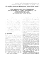
Localization of ADH in the colorectal region
Analysis of colorectal sections showed that ADH1 mRNA
was localized primarily in the cells of the lower part of the
crypts of Lieberku
¨
hn (Fig. 5A,B). In contrast, ADH4, at
mRNA and protein levels, was detected uniformly along
the crypts of Lieberku
¨
hn and in the surface brush-border
epithelium (Fig. 5D,E,G,H). ADH4 immunostaining was
Fig. 4. Localization of ADH1 and ADH4 in jejunum and ileum. ADH1 (A) and ADH4 (B) mRNA detection in jejunal mucosa. Immunodetection of
ADH4 protein in ileal mucosa (C). Omission of anti-ADH4 IgG in an adjacent control section of ileum (D). CL, crypt of Lieberku
¨
hn; LP, lamina
propia; MM, muscularis mucosae; SM, submucosa; V, villi; v, vessel. Calibration bars (shown in D): A,B, 200 lm; C,D, 50 lm.
Ó FEBS 2003 Alcohol dehydrogenase in the gastrointestinal tract (Eur. J. Biochem. 270) 2657
absent in the submucosa, lamina propia and muscularis
mucosae.
Discussion
Although several works had provided information on the
ADH distribution in rodent digestive organs [2,7,20,21,34],
the present report represents the most thorough study on
the localization of the ethanol-metabolizing ADHs in the
digestive tract tissues of adult rat. In previous reports,
ADH1 was, in general, undetected in upper digestive
organs, including stomach while ADH4 was not found in
several intestinal regions [21,34]. Notably, here we demon-
strate that ADH1 and ADH4 are expressed throughout the
Fig. 5. Localization of ADH1 and ADH4 in the colorectal region. ADH1 (A,B) and ADH4 (D,E) mRNA detection in the longitudinal (A,D) and
transversal (B,E) section of crypts of Lieberku
¨
hn. Control sections hybridized with ADH1 (C) and ADH4 (F) sense riboprobe. Immunodetection of
ADH4 protein in longitudinal (G) and transversal (H) section of the Lieberku
¨
hn glands of colorectal mucosa. Control section incubated with anti-
ADH4 IgG preadsorbed with 15 lg recombinant mouse ADH4 protein (I). CL, crypt of Lieberku
¨
hn; G, goblet cell; LP, lamina propia; MM,
muscularis mucosae; SB, striated border of enterocytes. Calibration bars: A–F (shown in F) and G–I (shown in I), 50 lm.
2658 J. Vaglenova et al. (Eur. J. Biochem. 270) Ó FEBS 2003
rat gastrointestinal tract. Each ADH form, however, is
confined to specific regions and cell populations. Thus,
ADH1 is localized predominantly in the intestinal area
whereas ADH4 is prominent in the most external parts
(esophagus, stomach and colorectum) of the digestive
system. In each tissue, except for the duodenum, ADH1 is
confined to the inner cell layers of the mucosa, while ADH4
is localized in the outer cell layers exposed to the lumen.
Interestingly, duodenum is the only region where ADH4 is
absent from the external cell layers of the mucosa. The
precise colocalization of mRNA, protein and activity
demonstrates that these enzymes are present in the same
regions where their mRNA is found. However, the restric-
tion of the ADH1 and ADH4 expression to a relatively
small number of cell types in specific regions could explain
the previous difficulty of demonstrating their presence in
various digestive organs [2,7,20,24,35]. Also, it should be
considered that there may exist some rat/mouse species
differences in ADH localization along the gastrointestinal
tract that account for the slightly different ADH localization
reported here for rat as compared to that previously
reported for mouse [21].
Although ADH1 and ADH4 are found in all digestive
tube organs, discontinuity exists regarding the cellular layers
where the enzymes are expressed. Thus, while ADH1 is not
expressed in the gastric pits of most of the stomach mucosa,
it is of interest the progressive increase in expression in this
external area as the mucosa reaches the pyloric region
(Fig. 3B,C,D). Even more impressive is the sharp disap-
pearance of ADH4 expression from the mucosa outer cell
layers in the gastroduodenal junction (Fig. 3J). The sudden
change in functional requirements in the transition between
stomach and duodenum is therefore also reflected by
marked differences in the expression levels of the ADH
enzymes.
Comparison of the present data from rat with the partial
information available from human [24–25,29,30, and
S. Porte
´
,S.E.Martı
´
nez,J.Farre
´
sandX.Pare
´
s, unpublished
results
2
], indicates that the general pattern of ADH distribu-
tion in the gastrointestinal tract is similar in the two species.
The present results along with previous in vitro studies
on the substrate specificity of ADH1 and ADH4
[2,7,9,12–14] provide the basis to hypothesize some
physiological functions for these enzymes in the gastro-
intestinal tract. However, precaution should be taken
when extrapolating conclusions to human because of
different ADH4 K
m
values for ethanol between rat and
human (2.4
M
vs. 37 m
M
, respectively) [9] and differences
in diet, intestinal flora, etc.
Role of gastrointestinal ADH in retinoid metabolism
The expression of ADH1 and ADH4 in certain cell layers of
gastrointestinal tissues, and its colocalization with the
biochemical apparatus associated with RA responsiveness
and metabolism [36–42], support the contribution of ADH1
and ADH4 (both exhibiting retinol dehydrogenase activity
[12–15,17]) to RA generation in adult gastrointestinal tract.
ADH1 and ADH4 displayed some nonoverlapping locali-
zation which might reflect distinct roles, as has been
suggested by studies with knockout animals [43]. ADH4,
located in the most external tissues and cell layers with a
high epithelial cell turnover, is well suited to fulfill a function
in RA synthesis. In this sense, esophageal, gastric and
colorectal mucosa show NAD
+
-dependent RA formation
from all-trans-retinol, that is disturbed by inhibitors of
ADH and aldehyde dehydrogenase (ALDH) [44,45]. On the
other hand, b-carotene absorbed by intestinal enterocytes is
converted to retinal which is subsequently reduced to retinol
for transport and storage [46]. Thus, ADH1 and ADH4
(k
cat
/K
m
for retinal ¼ 500 m
M
)1
Æmin
)1
[13] and
1750 m
M
)1
Æmin
)1
[14], respectively) could be also involved
in the step to generate retinol that would be immediately
esterified in vivo. This could shift the reaction equilibrium
towards retinal reduction, even in the absence of a favorable
NAD/NADH ratio. Interestingly, we have shown that
ADH4 is not present in duodenal enterocytes, where most
b-carotene cleavage occurs [46]. Therefore, ADH1 would be
the main ADH for the physiological retinal reduction in
duodenum, although microsomal retinal reductases may
also contribute to this function [47,48]. ADH4, specialized
in retinal generation from retinol in specific tissues [21],
could not be necessary in duodenal enterocytes where retinal
is directly formed from b-carotene.
Role of gastrointestinal ADH in alcohol metabolism
and pathology
Substrate specificity predicts that both ADH1 and ADH4
participate in the elimination of ingested alcohols and
aldehydes, ethanol generated by intestinal microbial flora,
and products of lipid peroxidation [12,13]. ADH4, located in
the upper part of the gastrointestinal tract and the luminal
part of the mucosa, would be in contact with the highest
concentrations of ingested alcohols and aldehydes, and in
areas subjected to high levels of oxidative stress. Therefore,
ADH4 could act as a first metabolic barrier. Likewise,
ADH1, that is positioned more internally along the tract and
within the mucosa, could act as a second metabolic barrier.
The localization of ADH4 suggests its contribution to
the first-pass metabolism [49–53], mostly at high ethanol
concentration (K
m
¼ 2.4
M
) [9]. In addition, we have
demonstrated here that ADH1 is also present in the upper
digestive tract and therefore it may have a role as well in the
first-pass metabolism, mostly at low ethanol concentrations
(K
m
¼ 1.4 m
M
) [7]. In the lower gastrointestinal tract,
colonic flora is the major source of endogenous ethanol in
mammals that is produced constantly [54–56]. The main
function of the high amount of ADH1 in colon might be
the elimination of this endogenous ethanol.
The presence of ADH throughout the gut can be related
to alcohol pathology. Thus, ethanol and acetaldehyde have
been associated with epithelial hyperegeneration of the
mucosa and cancer [57–59]. On the other hand, disturbance
of RA metabolism may be related to carcinogenesis [58,
60–62], and ethanol is a competitive inhibitor of retinol
oxidation by ADH [12,51,63–66]. The esophagus and the
colorectal region are especially vulnerable to alcohol injury
[58,59], and these are tissues with the highest ADH activity
(Fig. 1) where acetaldehyde-metabolizing ALDH2 is virtu-
ally absent or scarce [25]. Thus, 50 l
M
acetaldehyde
hampers RA formation [45], suggesting that acetaldehyde
produced by ADH could also disturb RA generation
catalyzed by retinal-active ALDH1 which has been also
Ó FEBS 2003 Alcohol dehydrogenase in the gastrointestinal tract (Eur. J. Biochem. 270) 2659
detected in these gastrointestinal areas [25,30,41,42,67,68].
The impairment of RA formation by ethanol and acetal-
dehyde could be an explanation for mucosal damage,
increased cell proliferation and the high incidence of
esophageal and colorectal neoplasia in alcohol abusers.
In conclusion, we have detected ADH1 and ADH4 in
distinct cell types of specific regions throughout the
gastrointestinal tract, which evidences a local level of
ethanol metabolism. Active ethanol oxidation in specific
gastrointestinal regions can be related to some deleterious
effects of ethanol. The involvement of ADH1 and ADH4 in
retinol oxidation makes these enzymes relevant to gastro-
intestinal functions that require RA. The impairment of
retinol oxidation by inhibition of ADH during ethanol
consumption may be an additional mechanism of gastro-
intestinal alcohol pathology.
Acknowledgements
Supported by grants from the Direccio
´
n General de Investigacio
´
n
Cientı
´
fica (BMC2002-02659 and BMC2000-0132) and the Commis-
sion of the European Union (BIO4-CT97-2123) to X. P and J. F.,
and by the National Institutes of Health grant AA09731 to G. D. We
are grateful to Dr Salvador Bartolome
´
(Laboratori d’Ana
`
lisi i
Fotodocumentacio
´
d’Electroforesis, Autoradiografies i Luminesce
`
n-
cia, Universitat Auto
`
noma de Barcelona) for his help in image
analysis.
References
1. Li, T K. (1983) The absorption, distribution and metabolism of
ethanol and its effects on nutrition and hepatic function. In Medi-
cal and Social Aspects of Ethanol. Abuse (Tabakoff, B., Sutker,
P.B. & Randall, C.L., eds), pp. 47–77. Plenum Press, New York.
2. Boleda, M.D., Julia
`
,P.,Moreno,A.&Pare
´
s, X. (1989) Role of
extrahepatic alcohol dehydrogenase in rat ethanol metabolism.
Arch. Biochem. Biophys. 274, 74–81.
3. Bra
¨
nde
´
n, C.I., Jo
¨
rnvall, H., Eklund, H. & Furugren, B. (1975)
Alcohol dehydrogenase. In The Enzymes, Vol. 11, 3rd edn. (Boyer,
P.D., ed.), pp. 103–190. Academic Press, New York.
4. Nordling, E., Persson, B. & Jo
¨
rnvall, H. (2002) Differential mul-
tiplicity of MDR alcohol dehydrogenases: enzyme genes in the
human genome versus those in organisms initially studied. Cell.
Mol. Life Sci. 59, 1070–1075.
5. Jo
¨
rnvall, H. & Ho
¨
o
¨
g, J O. (1995) Nomenclature of alcohol
dehydrogenases. Alcohol 30, 153–161.
6. Duester, G., Farre
´
s, J., Felder, M.R., Holmes, R.S., Ho
¨
o
¨
g, J O.,
Pare
´
s, X., Plapp, B.V., Yin, S J. & Jo
¨
rnvall, H. (1999)
Recommended nomenclature for the vertebrate alcohol dehydro-
genase gene family. Biochem. Pharmacol. 58, 389–395.
7. Julia
`
, P., Farre
´
s, J. & Pare
´
s, X. (1987) Characterization of three
isoenzymes of rat alcohol dehydrogenase. Tissue distribution and
physical and enzymatic properties. Eur. J. Biochem. 162, 179–189.
8. Svensson, S., Stromberg, P. & Ho
¨
o
¨
g, J O. (1999) A novel subtype
of class II alcohol dehydrogenase in rodents. Unique Pro (47) and
Ser (182) modulate hydride transfer in the mouse enzyme. J. Biol.
Chem. 274, 29712–29719.
9. Farre
´
s, J., Moreno, A., Crosas, B., Peralba, J.M., Allali-Hassani,
A., Hjelmqvist, L., Jo
¨
rnvall, H. & Pare
´
s, X. (1994) Alcohol
dehydrogenase of class IV (rr-ADH) from human stomach.
cDNA sequence and structure/function relationships. Eur. J. Bio-
chem. 224, 549–557.
10. Allali-Hassani, A., Martı
´
nez, S.E., Peralba, J.M., Vaglenova, J.,
Vidal, F., Richart, C., Farre
´
s, J. & Pare
´
s, X. (1997) Alcohol
dehydrogenase of human and rat blood vessels. Role in ethanol
metabolism. FEBS Lett. 405, 26–30.
11. Martı
´
nez, S.E., Vaglenova, J., Sabria
`
,J.,Martı
´
nez, M.C., Farre
´
s,
J. & Pare
´
s, X. (2001) Distribution of alcohol dehydrogenase
mRNA in the rat central nervous system: Consequences for
brain ethanol and retinoid metabolism. Eur. J. Biochem. 268,
5045–5056.
12. Allali-Hassani, A., Peralba, J.M., Martras, S., Farre
´
s, J. & Pare
´
s,
X. (1998) Retinoids, x-hydroxyfatty acids and cytotoxic aldehydes
as physiological substrates, and H
2
-receptor antagonists as phar-
macological inhibitors, of human class IV alcohol dehydrogenase.
FEBS Lett. 426, 362–366.
13. Boleda, M.D., Saubi, N., Farre
´
s, J. & Pare
´
s, X. (1993) Physio-
logical substrates for rat alcohol dehydrogenase classes: Aldehydes
of lipid peroxidation, x-hydroxyfatty acids, and retinoids. Arch.
Biochem. Biophys. 307, 85–90.
14. Crosas, B., Allali-Hassani, A., Martı
´
nez, S.E., Martras, S., Pers-
son, B., Jo
¨
rnvall, H., Pare
´
s, X. & Farre
´
s, J. (2000) Molecular basis
for differential substrate specificity in class IV alcohol dehydro-
genases: a conserved function in retinoid metabolism but not in
ethanol metabolism. J. Biol. Chem. 275, 25180–25187.
15. Kedishvili, N.Y., Bosron, W.F., Stone, C.L., Hurley, T.D., Peggs,
C.F., Thomasson, H.R., Popov, K.M., Carr, L.G., Edenberg, H.
& Li, T K. (1995) Expression and kinetic characterization of
recombinant human stomach alcohol dehydrogenase. J. Biol.
Chem. 270, 3625–3620.
16. Molotkov, A., Fan, X., Deltour, L., Foglio, M.H., Martras, S.,
Farre
´
s, J., Pare
´
s, X. & Duester, G. (2002) Stimulation of retinoic
acid production and growth by ubiquitously expressed
alcohol dehydrogenase, Adh3. Proc. Natl Acad. Sci. USA 99,
5337–5342.
17. Yang,Z N.,Davis,G.J.,Hurley,T.D.,Stone,C.L.,Li,T K.&
Bosron, W.F. (1994) Catalytic efficiency of human alcohol dehy-
drogenase for retinol oxidation and retinal reduction. Alcohol.
Clin.Exp.Res.18, 587–591.
18. Deltour, L., Foglio, M.H. & Duester, G. (1999) Metabolic defi-
ciencies in alcohol dehydrogenase Adh1, Adh3, and Adh4 null
mutant mice. J. Biol. Chem. 274, 16796–16801.
19. Deltour, L., Foglio, M.H. & Duester, G. (1999) Impaired retinol
utilization in Adh4 alcohol dehydrogenase mutant mice. Dev.
Genet. 25, 1–10.
20. Estonius, M., Danielsson, O., Karlsson, C., Persson, H., Jo
¨
rnvall,
H. & Ho
¨
o
¨
g, J O. (1993) Distribution of alcohol and sorbitol
dehydrogenases. Assessment of mRNA species in mammalian
tissues. Eur. J. Biochem. 215, 497–503.
21. Haselbeck, R.J. & Duester, G. (1997) Regional restriction of
alcohol/retinol dehydrogenases along the mouse gastrointestinal
epithelium. Alcohol. Clin. Exp. Res. 21, 1484–1490.
22. Koivisto, T. & Salaspuro, M. (1996) Aldehyde dehydrogenases of
the rat colon: comparison with other tissues of the alimentary tract
and the liver. Alcohol. Clin. Exp. Res. 20, 551–555.
23. Dong, Y.J., Peng, T.K. & Yin, S.J. (1996) Expression and acti-
vities of class IV alcohol dehydrogenase and class III aldehyde
dehydrogenase in human mouth. Alcohol 13, 257–262.
24. Estonius, M., Svensson, S. & Ho
¨
o
¨
g, J O. (1996) Alcohol dehy-
drogenase in human tissues: localisation of transcripts coding for
five classes of the enzyme. FEBS Lett. 397, 338–342.
25. Pare
´
s, X. & Farre
´
s, J. (1996) Alcohol and aldehyde dehydro-
genases in the gastrointestinal tract. In Alcohol and the Gastro-
intestinal Tract (Preedy, V.R. & Watson, R.R., eds), pp. 41–56.
CRC Press, Boca Raton.
26. Pare
´
s, X., Martı
´
nez, S.E., Allali-Hassani, A., Borra
`
s, E., Farre
´
s, J.,
Martras, S., Rosell, A. & Vaglenova, J. (2001) Distribution of
alcohol dehydrogenase in human organs. Relevance for alcohol
metabolism and pathology. In Alcohol in Health and Disease
2660 J. Vaglenova et al. (Eur. J. Biochem. 270) Ó FEBS 2003
(Agarwal, D. P. & Seitz, H. K., eds), pp. 87–102. Marcel Dekker
Inc., New York.
3
27. Pestalozzi, D.M., Bu
¨
hler, R., von Wartburg, J.P. & Hess, M.
(1983) Immunohistochemical localization of alcohol dehydro-
genase in the human gastrointestinal tract. Gastroenterology 85,
1011–1016.
28. Seitz, H.K., Egerer, G., Oneta, C., Kramer, S., Sieg, A., Klee, F. &
Simanowski, U.A. (1996) Alcohol dehydrogenase in the human
colon and rectum. Digestion 57, 105–108.
29. Yin, S.J., Chou, F.J., Chao, S.F., Tsai, S.F., Liao, C.S., Wang,
S.L., Wu, C.W. & Lee, S.C. (1993) Alcohol and aldehyde dehy-
drogenases in human esophagus: comparison with the stomach
enzyme activities. Alcohol.Clin.Exp.Res.17, 376–381.
30. Yin, S.J., Liao, C.S., Wu, C.W., Li, T.T., Chen, L.L., Lai, C.L. &
Tsao, T.Y. (1997) Human stomach alcohol and aldehyde dehy-
drogenases: comparison of expression pattern and activities in
alimentary tract. Gastroenterology 112, 766–775.
31. Crabb, D.W., Bosron, W.F. & Li, T.K. (1983) Steady-state kinetic
properties of purified rat liver alcohol dehydrogenase: application
to predicting alcohol elimination rates in vivo. Arch. Biochem.
Biophys. 224, 299–309.
32. Bradford, M.M. (1976) A rapid and sensitive method for the
quantitative determination of microgram quantities of protein
utilising the principle of protein-dye binding. Anal. Biochem. 72,
248–254.
33. Young, B. & Heath, J.W. (2000) Wheater’s Functional Histology,
4th edn. Churchill Livingstone, Edinburgh.
34. Ang,H.L.,Deltour,L.,Zgombic-Knight,M.,Wagner,M.A.&
Duester, G. (1996) Expression patterns of class I and class IV
alcohol dehydrogenase genes in developing epithelia suggest a role
for alcohol dehydrogenase in local retinoic acid synthesis. Alcohol.
Clin.Exp.Res.20, 1050–1064.
35. Zgombic-Knight, M., Ang, H.L., Foglio, M.H. & Duester, G.
(1995) Cloning of the mouse class IV alcohol dehydrogenase
(retinol dehydrogenase) cDNA and tissue-specific expression
patterns of the murine ADH gene family. J. Biol. Chem. 270,
10868–10877.
36. Crow, J.A. & Ong, D.E. (1985) Cell-specific immunohistochemical
localization of a cellular retinol-binding protein (type two) in the
small intestine of rat. Proc. Natl Acad. Sci. USA 82, 4707–4711.
37. Dowlatshahi, K., Mehta, R.G., Levin, B., Cerny, W.L., Skinner,
D.B. & Moon, R.C. (1984) Retinoic-acid-binding protein in nor-
mal and neoplastic human esophagus. Cancer 54, 308–311.
38. Inagami, S. & Ong, D.E. (1992) Purification and partial char-
acterization of cellular retinol-binding protein, type two, from
human small intestine. J. Nutr. 122, 450–456.
39. Jiang, S.Y., Shen, S.R. & Shyu, R.Y., Yu, J.C., Harn, H.J., Yeh,
M.Y., Lee, M.M. & Chang, Y.C. (1999) Expression of nuclear
retinoid receptors in normal, premalignant and malignant gastric
tissues determined by in situ hybridization. Br. J. Cancer 80,
206–214.
40. Kato, S., Mano, H., Kumazawa, T., Yoshizawa, Y., Kojima, R. &
Masushige, S. (1992) Effect of retinoid status on alpha, beta and
gamma retinoic acid receptor mRNA levels in various rat tissues.
Biochem. J. 286, 755–760.
41. Bhat, P.V. (1998) Retinal dehydrogenase gene expression in
stomach and small intestine of rats during postnatal development
and in vitamin A deficiency. FEBS Lett. 426, 260–262.
42. Frota-Ruchon, A., Marcinkiewicz, M. & Bhat, P.V. (2000)
Localization of retinal dehydrogenase type 1 in the stomach and
intestine. Cell Tissue Res. 302, 397–400.
43. Molotkov, A., Deltour, L., Foglio, M.H., Cuenca, A.E. & Due-
ster, G. (2002) Distinct retinoid metabolic functions for alcohol
dehydrogenase genes Adh1 and Adh4 in protection against vitamin
A toxicity or deficiency revealed in double null mutant mice.
J. Biol. Chem. 277, 13804–13811.
44. Crabb, D.W., Pinairs, J., Hasanadka, R., Fang, M., Leo, M.A.,
Lieber, C.S., Tsukamoto, H., Motomura, K., Miyahara, T.,
Ohata, M., Bosron, W.F., Sanghani, S., Kedishvili, N., Shiraishi,
H.,Yokoyama,H.,Miyagi,M.,Ishii,H.,Bergheim,I.,Menzl,I.,
Parlesak, A. & Bode, C. (2001) Alcohol and retinoids. Alcohol.
Clin.Exp.Res.25, 207S–217S.
45. Yokoyama, H., Matsumoto, M., Shiraishi, H., Miyagi, M., Kato,
S. & Ishii, H. (2001) Nicotinamide adenine dinucleotide-depen-
dent retinoic acid formation from retinol in the human gastric
mucosa: inhibition by ethanol, acetaldehyde, and H2 blockers.
Alcohol. Clin. Exp. Res. 2, 24S–28S.
46. Wyss, A., Wirtz, G.M., Woggon, W.D., Brugger, R., Wyss, M.,
Friedlein, A., Riss, G., Bachmann, H. & Hunziker, W. (2001)
Expression pattern and localization of beta,beta-carotene 15,15¢-
dioxygenase in different tissues. Biochem. J. 354, 521–529.
47. Kakkad, B.P. & Ong, D.E. (1988) Reduction of retinaldehyde
bound to cellular retinol-binding protein (type II) by microsomes
from rat small intestine. J. Biol. Chem. 263, 12916–12919.
48. Kedishvili, N.Y., Chumakova, O.V., Chetyrkin, S.V., Belyaeva,
O.V.,Lapshina,E.A.,Lin,D.W.,Matsumura,M.&Nelson,P.S.
(2002) Evidence that the human gene for prostate short-chain
dehydrogenase/reductase (PSDR1) encodes a novel retinal
reductase (RalR1). J. Biol. Chem. 277, 28909–28915.
49. Caballerı
´
a, J., Barahona, E. & Lieber, C.S. (1986) The contribu-
tion of the stomach to ethanol oxidation in the rat. Life Sci. 41,
1021–1027.
50. Caballerı
´
a, J., Frezza, M., Herna
´
ndez-Mun
˜
oz, R., DiPadova, C.,
Korsten, M.A., Barahona, E. & Lieber, C.S. (1989) Gastric origin
of the first-pass metabolism of ethanol in humans: effect of gas-
trectomy. Gastroenterology 97, 1205–1209.
51. Han, C L., Liao, C S., Wu, C W., Hwong, C L., Lee, A R. &
Yin, S J. (1998) Contribution to first-pass metabolism of ethanol
and inhibition by ethanol for retinol oxidation in human alcohol
dehydrogenase family. Eur. J. Biochem. 254, 25–31.
52. Lim,R.T.,Gentry,R.T.Jr,Itio,D.,Yokoyama,H.,Baraona,E.
& Lieber, C.S. (1993) First-pass metabolism of ethanol is pre-
dominantly gastric. Alcohol. Clin. Exp. Res. 17, 1337–1344.
53. Oneta, C.M., Simanowski, U.A., Martı
´
nez, M., Allali-Hassani,
A., Pare
´
s,X.,Homann,N.,Conradt,C.,Waldherr,R.,Fiehn,W.,
Coutelle,C.&Seitz,H.K.(1998)Firstpassmetabolismofethanol
is strikingly influenced by the speed of gastric emptying. Gut 43,
612–619.
54. Krebs, H.A. & Perkins, J.R. (1970) The physiological role of liver
alcohol dehydrogenase. Biochem. J. 118, 635–644.
55. Baraona, E., Julkunen, R., Tannenbaum, L. & Lieber, C.S. (1986)
Role of intestinal bacterial overgrowth in ethanol production and
metabolism in rats. Gastroenterology 90, 103–110.
56. Nosova, T., Jokelainen, K., Kaihovaara, P., Jousimies-Somer, H.,
Siitonen, A., Heine, R. & Salaspuro, M. (1996) Aldehyde dehy-
drogenase activity and acetate production by aerobic bacteria
representing the normal flora of human large intestine Alcohol
Alcohol
4
. 31, 555–564.
57. Longnecker, M.P. (1995) Alcohol consumption and risk of cancer
in humans: an overview. Alcohol 12, 87–96.
58. Seitz, H.K., Matsuzaki, S., Yokoyama, A., Homann, N., Vak-
evainen, S. & Wang, X.D. (2001) Alcohol and cancer. Alcohol.
Clin.Exp.Res.25, 137S–143S.
59. Maier, H., Weidauer, H., Zoller, J., Seitz, H.K., Flentje, M., Mall,
G. & Born, I.A. (1994) Effect of chronic alcohol consumption on
the morphology of the oral mucosa. Alcohol. Clin. Exp. Res. 18,
387–391.
60. Love, J.M. & Gudas, L.J. (1994) Vitamin A, differentiation and
cancer. Curr. Opin. Cell Biol. 6, 825–831.
61. Mak, K.M., Leo, M.A. & Lieber, C.S. (1987) Effect of ethanol and
vitamin A deficiency on epithelial cell proliferation and structure
in the rat esophagus. Gastroenterology 93, 362–370.
Ó FEBS 2003 Alcohol dehydrogenase in the gastrointestinal tract (Eur. J. Biochem. 270) 2661
62. Wald, N., Boreham, J. & Bailey, A. (1986) Serum retinol and
subsequent risk of cancer. Br.J.Cancer54, 957–961.
63. Julia
`
, P., Farre
´
s, J. & Pare
´
s, X. (1986) Ocular alcohol dehydro-
genase in the rat: Regional distribution and kinetics of the ADH-1
isoenzyme with retinol and retinal. Exp. Eye Res. 42, 305–314.
64. Kedishvili, N.Y., Gough, W.H., Wilhelmina, I.D., Parsons, S., Li,
T K. & Bosron, W.F. (1998) Effect of cellular retinol-binding
protein on retinol oxidation by human class IV retinol/alcohol
dehydrogenase and inhibition by ethanol. Biochem. Biophys. Res.
Commun. 249, 191–196.
65. Mezey, E. & Holt, P.R. (1971) The inhibitory effect of ethanol on
retinol oxidation by human liver and cattle retina. Exp. Mol.
Pathol. 15, 148–156.
66. Molotkov, A. & Duester, G. (2002) Retinol/ethanol drug inter-
action during acute alcohol intoxication in mice involves inhi-
bition of retinol metabolism to retinoic acid by alcohol
dehydrogenase. J. Biol. Chem. 277, 22553–22557.
67. Napoli, J.L. (1999) Interactions of retinoid proteins and
enzymes in retinoid metabolism. Biochim. Biophys. Acta 1440,
139–162.
68. Duester, G. (2000) Families of retinoid dehydrogenases regulating
vitamin A function: production of visual pigment and retinoic
acid. Eur. J. Biochem. 267, 4315–4324.
2662 J. Vaglenova et al. (Eur. J. Biochem. 270) Ó FEBS 2003