Báo cáo khoa học: Structure of the exceptionally large nonrepetitive carbohydrate backbone of the lipopolysaccharide of Pectinatus frisingensis strain VTT E-82164 doc
Bạn đang xem bản rút gọn của tài liệu. Xem và tải ngay bản đầy đủ của tài liệu tại đây (646.11 KB, 11 trang )
Structure of the exceptionally large nonrepetitive carbohydrate
backbone of the lipopolysaccharide of
Pectinatus frisingensis
strain
VTT E-82164
Evgeny Vinogradov
1
, Bent O. Petersen
2
, Irina Sadovskaya
3
, Said Jabbouri
3
, Jens Ø. Duus
2
and Ilkka M. Helander
4
1
Institute for Biological Sciences, National Research Council, Ottawa, ON, Canada;
2
Department of Chemistry, Carlsberg
Laboratory, Copenhagen, Denmark;
3
Laboratoire de Recherche sur les Biomate
´
riaux et Biotechnologies, Universite
´
de Littoral-Co
ˆ
te
d’Opale, Bassin Napole
´
on BP 120, Boulogne-sur-mer, France;
4
Department of Applied Chemistry and Microbiology,
Division of Microbiology, University of Helsinki, Finland
The structures of the oligosaccharides obtained after acetic
acid hydrolysis and alkaline deacylation of the rough-type
lipopolysaccharide (LPS) from Pectinatus frisingensis strain
VTT E-82164 were analysed using NMR spectroscopy, MS
and chemical methods. The LPS contains two major struc-
tural variants, differing by a decasaccharide fragment, and
some minor variants lacking the terminal glucose residue.
The largest structure of the carbohydrate backbone of the
LPS that could be deduced from experimental results consists
of 25 monosaccharides (including the previously found
Ara4NP residue in lipid A) arranged in a well-defined non-
repetitive structure:
We presume that the shorter variant with R
1
¼ H represents
the core-lipid A part of the LPS, and the additional fragment
is present instead of the O-specific polysaccharide. Structures
of this type have not been previously described. Analysis of
the deacylation products obtained from the LPS of the
smooth strain, VTT E-79100
T
, showed that it contains a very
similar core but with one different glycosidic linkage.
Keywords: core; lipid A; lipopolysaccharide; Pectinatus
frisingensis.
Strictly anaerobic Gram-negative rod-shaped bacteria caus-
ing turbidity and off flavours in bottled beer were initially
isolated in 1978 and described as Pectinatus cerevisiiphilus
[1]. Another species, Pectinatus frisingensis, which differed
from P. cerevisiiphilus in a number of biochemical charac-
teristics was later described [2]. To date, the VTT culture
collection (Espoo, Finland) has 32 Pectinatus isolates from
spoiled beer originating from Belgium, Finland, Germany,
the Nederlands and the USA; 24 have been identified as
P. frisingensis and eight as P. cerevisiiphilus by conventional
tests, ribotyping and partial 16S rDNA sequence analysis [3].
The lipopolysaccharides (LPS) of type strains of P. cere-
visiiphilus and P. frisingensis possess a number of remarkable
properties, including the predominance of odd-numbered
fatty acids in lipid A [4] and the presence of furanosidic
6-deoxysugars in the O-specific chains [5]. The lipid A was
shown to be quantitatively substituted at the 4¢-phosphate
and partially at the glycosidic phosphate by 4-amino-4-
deoxy-b-
L
-arabinose [6]. There are no structural data on
Correspondence to E. Vinogradov, Institute for Biological Sciences,
National Research Council, 100 Sussex Dr,
K1A 0R6 Ottawa ON, Canada.
Fax: + 1 613 952 90 92, Tel.: + 1 613 990 03 97,
E-mail:
Abbreviations: LPS, lipopolysaccharide; Kdo, 3-deoxy-
D
-manno-
oct-2-ulosonic acid; Ara4N, 4-amino-4-deoxy-
L
-arabinose;
HPAEC, high-performance anion-exchange chromatography;
ESI MS, electrospray ionization mass spectrometry.
(Received 5 April 2003, revised 14 May 2003,
accepted 22 May 2003)
Eur. J. Biochem. 270, 3036–3046 (2003) Ó FEBS 2003 doi:10.1046/j.1432-1033.2003.03682.x
Pectinatus core structures, except a report that LPS of both
P. frisingesis and P. cerevisiiphilus contain a disaccharide
structure, a phosphorylated GlcN linked to O4 of a Kdo
residue, tentatively assigned to the core region [7].
Screening of Pectinatus strains other than type strains has
revealed that the LPS from certain strains exhibit only two
distinct bands on PAGE, with no polymeric O chains (I. M.
Helander, unpublished data). This indicates the presence of
two structurally distinct LPS molecules. We describe here
the chemical structure of the LPS carbohydrate backbone of
one such isolate, P. frisingensis VTT-E-82164, which has
99.8% similarity of partial 16S rDNA to the P. frisingensis
type strain.
Materials and methods
Bacterial strains and growth conditions
P. frisingensis VTT E-82164 and VTT E-79100
T
and
P. cerevisiiphilus E-79103
T
were obtained from VTT Bio-
technology (Espoo, Finland) [3]. Cells were grown anaero-
bically at 32 °C without shaking in Man Rogosa Sharpe
broth (Difco), pH 6.5, in the presence of a reducing agent
(Na
2
S, 12.5 m
M
) and resazurin (1 mgÆmL
)1
), and collected
at the stationary growth phase.
LPS isolation
Bacterial cells were washed with ethanol, acetone, and light
petroleum, and LPS was extracted from the dried cells with
phenol/chloroform/petroleum ether (60–95 °C) (5 : 5 : 8,
v/v) with acetone precipitation [4,8].
NMR spectroscopy and general methods
NMR spectra were recorded at 25 °CinD
2
OonaVarian
Unity Inova 800 instrument at 799.96 MHz for proton
and 201.12 MHz for carbon, using acetone as reference
for proton (2.225 p.p.m.) and 1,4-dioxane for carbon
(67.4 p.p.m.). Varian standard programs tndqcosy, tnnoesy
(mixing time of 100 ms), tntocsy (spinlock time 80 ms),
gHSQC, gHSQCTOCSY (spinlock time 80 ms),
gHSQCNOESY (mixing time 200 ms) and gHMBC were
used with digital resolution in F2 dimension <2 HzÆpt
)1
.
Spectra were assigned using the computer program
PRONTO
[9].
Analytical methods
PAGE was performed with deoxycholate as the detergent.
The separation gel contained 18% acrylamide, 0.5% (w/v)
deoxycholate, and 375 m
M
Tris/HCl, pH 8.8, and stacking
gel contained 4% acrylamide and 127 m
M
Tris/HCl,
pH 6.8. LPS samples were prepared at a concentration of
0.1% (w/v) in sample buffer [127 m
M
Tris/HCl, pH 6.8,
10% (v/v) glycerol, 0.025% (w/v) bromphenol blue dye].
The electrode buffer was composed of deoxycholate
(2.5 gÆL
)1
), glycine (21.7 gÆL
)1
), and Tris (4.5 gÆL
)1
). Elec-
trophoresis was performed at a constant current of 15 mA
per gel with cooling. Immediately after the electrophoresis
run, the gel was soaked in the fixing solution containing
ethanol (40%, w/w) and acetic acid (5%, w/w). The solution
was changed after 30 min, and fixation continued overnight.
LPS bands were visualized by silver staining as described by
Tsai & Frasch [10].
Hydrolysis was performed with 4
M
trifluoroacetic acid
(110 °C, 3 h). Monosaccharides were conventionally con-
verted into the alditol acetates and analysed by GC on a
Agilent 6850 chromatograph equipped with a DB-17 fused-
silica column (30 m · 0.25 mm) using a temperature
gradient of 180 °C(2min) fi 240 °Cat2°CÆmin
)1
.For
the determination of the absolute configuration of 3-O-
methyl-6-deoxytalose, GC was performed in isothermal
conditions at 150 °C. GC-MS was performed on a Varian
Saturn 2000 system with ion-trap mass spectral detector
using the same column. Electrospray ionization (ESI) MS
was carried out as described previously [11].
Gel chromatography was carried out on columns
(2.5 · 95 cm) of Sephadex G-50 in pyridinium/acetate
buffer, pH 4.5 (4 mL pyridine and 10 mL acetic acid in
1Lwater)andBioGelP4(1· 90 cm) in water. The eluate
was monitored with a refractive index detector.
Methylation analysis was performed by the Ciucanu-
Kerek procedure [12]. Methylated products were hydrolysed
and monosaccharides converted into 1d-alditol acetates by
conventional methods and analysed by GC-MS.
High-performance anion-exchange chromatography
(HPAEC) was performed on a CarboPac PA1 column
(9 · 250 mm) with pulsed amperiometric detection, equili-
brated in 0.1
M
NaOH, using a linear gradient of 1
M
sodium acetate in 0.1
M
NaOH from 5% to 80% of acetate
in 60 min at 3 mLÆmin
)1
. Fractions of volume 3 mL were
collected and analysed using the Dionex system with an
analytical CarboPac PA1 column (4.6 · 250 mm) at
1mLÆmin
)1
. Separated oligosaccharides were desalted on
a Sephadex G-50 column.
De-O,N-acylation of LPS and preparation of backbone
oligosaccharides [13]
LPS (120 mg) was dissolved in 4
M
KOH (4 mL), and the
solution was heated at 120 °C for 16 h, cooled, neutral-
ized with 2
M
HCl. The precipitate was removed by
centrifugation, and the supernatant desalted by gel
chromatography on Sephadex G-50. Two oligosaccharide
fractions with K
av
0.60 and 0.47 were obtained and
further separated by HPAEC on a semipreparative
CarboPac PA1 column to give oligosaccharides 1a, 1b
and a mixture of 2 and 3.
Deamination of the de-O,N-acylated LPS and preparation
of oligosaccharides 4 and 5
The mixture of oligosaccharides obtained after alkaline
deacylation of the LPS (200 mg) was treated with 300 mg
NaNO
2
in 10% acetic acid (10 mL, 25 °C, 24 h), desalted
on a Sephadex G-50 column, reduced with NaBH
4
,
desalted, and oligosaccharides 4 and 5 isolated by HPAEC.
Acetic acid hydrolysis of LPS
LPS (100 mg) was treated with 2% acetic acid (5 mL,
100 °C, 3 h). The precipitate was removed by centrifuga-
tion, and the soluble products were separated on a Sephadex
Ó FEBS 2003 P. frisingensis lipopolysaccharide (Eur. J. Biochem. 270) 3037
G-50 column to give three oligosaccharide fractions. These
were NaBH
4
-reduced, desalted, and separated by HPAEC
to give oligosaccharides 6–8.
Isolation of 3-
O
-methyl-6-deoxy-
D
-talose (11)
LPS(300mg)washydrolysedwith3
M
trifluoroacetic acid
(100 °C, 1.5 h), and the cooled dark solution was treated
with activated carbon, filtered, and evaporated to dryness.
An aqueous solution of the residue was passed through a
column (0.8 · 15cm)ofDowex50W8(· 200; H
+
), then
through a column of Dowex 2 (AcO
–
). The monosaccha-
rides were separated by paper chromatography on What-
man 3
MM
paper in pyridine/butanol/water (6 : 4 : 3,
v/v/v). Sugars were detected on a small strip with
AgNO
3
/NaOH reagent, and eluted with water. The
portions of the fractions mainly containing Man, Glc, Gal,
Fuc, and pure 3-O-methyl-6-deoxy-
D
-taloseweretreated
with (S)-2-butanol/acetyl chloride (10 : 1, v/v; 2 h; 85 °C),
dried under a stream of air, acetylated, and analysed by
GC. 3-O-Methyl-6-deoxy-
D
-talose (3 mg) was obtained in
pure form (moves close to front on paper); [a]
D
+2°
(c 0.3, water), lit. for
L
-isomer (trivial name acovenose)
)14.2° (c1.2, water) [14].
Amino sugars were eluted from Dowex 50 with 0.5
M
HCl, N-acetylated (5 mL saturated NaHCO
3
, 0.5 mL acetic
anhydride; 20°, 1 h with stirring), converted into (S)-2-butyl
glycoside acetates as described above, and analysed by GC.
Synthesis of methyl 3-
O
-methyl-6-deoxy-
a-
D
-talopyranoside (9) and methyl 3-
O
-methyl-6-deoxy-
b-
D
-talopyranoside (10)
3-O-Methyl-
D
-glucose (a gift from M. Perry, NRC Canada)
1
(500 mg) was converted into an approximately 4 : 1 mixture
of a-methyl and b-methyl glycosides by methanolysis (1
M
HCl/MeOH; 85 °C; 24 h), brominated at C6 using CBr
4
/
imidazole/triphenyl phospine (1 : 1 : 2.5, v/v; 16 h; 25 °C;
product isolated by column chromatography on SiO
2
in 5%
MeOH in CHCl
3
), and debrominated by hydrogenolysis
over Pd/C in MeOH to yield methyl 3-O-methyl-6-deoxy-a,
b-glucopyranosides. These were converted into methyl 3-O-
methyl-2,4-di-O-trifluorosulfonyl-6-deoxy-a,b-
D
-gluco-
pyranoside [(CF
3
SO
3
)
2
O/Py; )20 °Cto+25°C) and
treated with excess Et
4
NOAc in dimethylformamide
(100 °C; 3 h). The reaction mixture was diluted 10 times
with water, passed through Dowex 50 (H
+
)toremove
Et
4
N
+
, evaporated to dryness, and compounds 9 and 10
were isolated by C
18
RP-HPLC in water (45 and 8 mg,
respectively).
Results
The LPS from P. frisingensis VTT E-82164 did not exhibit
the typical ladder-like pattern of smooth LPS on deoxy-
cholate-PAGE, but showed two main strongly stained
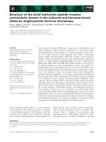
rapidly migrating bands (Fig. 1).
Monosaccharide analysis of the whole LPS indicated the
presence of fucose, 3-O-methyl-6-deoxyhexose, glucose,
galactose, mannose, glucosamine, galactosamine, and man-
nosamine in the proportions 1 : 0.6 : 1.5 : 1.2 : 1.4 :
1.5 : 0.6 : 0.4.
The LPS was O,N-deacylated by strong alkaline treat-
ment. Gel chromatographic separation of the products on
Sephadex G-50 gave two main peaks, which were further
separated by HPAEC to give oligosaccharides 1a, 1b,anda
mixture of 2 and 3 (Scheme 1).
In another experiment, the oligosaccharides obtained
after deacylation and Sephadex G-50 separation were
deaminated with nitrous acid and reduced with NaBH
4
.
This led to removal of all amino sugar residues except B and
O, which were transformed into 2,5-anhydromannitol and
2,5-anhydrotalitol, respectively. The products were separ-
ated by HPAEC and the oligosaccharides 4 and 5 were
isolated.
Mild hydrolysis of the LPS with acetic acid and
subsequent separation of the products by gel chromato-
graphy gave three oligosaccharide fractions. These were
reduced with NaBH
4
and purified by HPAEC to give
oligosaccharides 6, 7a,and8. The longer oligosaccharide 7b
was also analysed by NMR without reduction and HPAEC,
which allowed detection of O-acetylation. Separation by
HPAEC led to O-deacetylation of the reduced oligosaccha-
ride 7b because of the alkaline chromatography conditions.
In 1D and 2D NMR spectra of compound 1a (Fig. 2),
spin systems of 13 monosaccharides were identified. These
were three a-Glc residues (H, J, K), three a-Man residues (E,
F, G), one a-Gal residue (I), two a-GlcN (A, W) residues,
one b-GlcN (B) residue, and three Kdo residues (C, D, X).
The spectra were completely assigned (Table 1), and the
sequence of the hexoses was determined from NOE and
HMBC data, in which all respective strong transglycosidic
correlations were observed. Assignments were made using
methodology outlined in [15]. Oligosaccharides 1a and 1b
are the fragments of the larger structure 2, thus the NMR
data for 1a,b are very close to those for the respective
residues in 2 and are not presented.
The position and anomeric configuration of Kdo
residues was not as easy to assign. The
1
Hand
13
C
chemical shifts of Kdo residues C and X agreed well with
Fig. 1. Deoxycholate-PAGE profiles of the LPS. Lane 1, Salmonella
enteriditis;lane2,P. frisingensis E-82164; lane 3, P. frisingensis type
strain E-79100T; lane 4, P. cerevisiiphilus type strain E-79103T.
3038 E. Vinogradov et al.(Eur. J. Biochem. 270) Ó FEBS 2003
Scheme 1. Structures of the isolated compounds and proposed structure of the carbohydrate backbone of P. frisingensis VTT E-82164 LPS.
Ó FEBS 2003 P. frisingensis lipopolysaccharide (Eur. J. Biochem. 270) 3039
their a-configuration [16], while the H3 signals of Kdo D
appeared at 1.97 (ax) and 2.53 (eq) p.p.m., which may
correspond to a b-configuration [16]. However, a NOE
correlation observed between H3 of Kdo C and H6 of
Kdo D is possible only in the case of an a-configuration
of residue D, linked to O4 of Kdo C, as follows from
molecular modeling. The unusual position of the H3
signals of residue D in product 1a (aswellasin1b, 2,and3)
Fig. 2. Sections of COSY, TOCSY, and
NOESY spectra of the oligosaccharide 1a,
containing correlations from anomeric protons.
Scheme 1. (Continued).
3040 E. Vinogradov et al.(Eur. J. Biochem. 270) Ó FEBS 2003
Table 1. Assigned NMR spectral data for the isolated oligosaccharides obtained in
2
H
2
Oat25°C. Residue nomenclature and oligosaccharide
structures are given in Scheme 1.
Unit, compound Nucleus 12(3eq) 3 (3ax) 456(5b) 7 (6b) 8a (OMe) 8b
a-GlcNP A, 2, 3
1
H 5.61 3.35 3.87 3.45 4.13 4.21 3.79
13
C 91.8 55.6 70.8 70.9 74.1 70.2
b-GlcN B, 2, 3
1
H 4.96 3.04 3.61 3.49 3.61 3.58 3.54
13
C 100.8 56.8 73.5 71.1 75.7 62.5
a-Kdo C, 2, 3
1
H 2.04 1.99 4.17 4.32 3.58 3.69 3.89 3.58
13
C 101.5 35.4 70.5 72.7 73.8 70.5 64.8
Kdo-ol C, 7a,b
1
H 1.98 2.04 3.95 3.90 3.66 3.69 3.87
13
C 38.8 81.6 71.3 72.1 64.2
a-Kdo D, 2, 3
1
H 2.63 1.91 3.95 4.23 3.74 3.97 3.95 3.81
13
C 100.4 34.9 78.6 66.3 73.0 71.2 64.0
a-Kdo-ol D, 8
1
H 4.14 2.14/2.08 4.19 4.14 3.75 3.75 3.69 3.86
13
C 71.4 38.0 80.5 71.4 72.1
a
73.6
a
65.1
a-GlcN W, 2, 3
1
H 5.12 3.38 3.88 3.34 3.73 3.86 3.78
13
C 98.5 55.1 70.8 71.1 74.9 62.3
a-GlcN6P W, 8
1
H 5.43 3.37 3.93 3.62 4.04 4.15 4.21
13
C 97.4 56.6 71.8 71.3 73.8 66.4
a-Kdo X, 2, 3
1
H 2.09 1.85 4.07 4.02 3.76 3.93 3.85 3.69
13
C 103.8 35.5 66.6 67.4 73.5 70.3 63.3
a-Man E, 2, 3
1
H 5.13 4.09 4.01 3.68 4.26 3.74 4.02
13
C 100.2 71.7 72.3 76.4 71.5 63.6
a-Man E, 7a,b
1
H 5.07 4.04 4.06 3.83 3.97
13
C 103.3 72.0 72.0 76.3 73.1
a-Man F, 2, 3
1
H 5.58 4.20 3.86 3.84 3.75 3.86 3.75
13
C 101.0 80.3 76.0 66.6 74.0 67.6
a-Man F, 7a,b
1
H 5.63 4.12 3.96 3.81 3.91 3.79 4.02
13
C 101.1 82.0 71.7 68.0 72.9 67.1
a-Man G, 2, 3
1
H 4.84 4.15 3.89 3.81 3.82 3.85 3.76
13
C 100.6 70.7 81.9 66.9 73.7 62.3
a-Man G, 7a,b
1
H 4.91 4.16 3.89 3.88 3.78
13
C 100.8 70.8 81.7 66.9 73.9
a-Glc H, 2, 3
1
H 5.20 3.78 4.01 3.56 3.87 3.82 3.78
13
C 102.2 72.8 83.0 68.9 73.4 61.1
a-Glc H, 7a,b
1
H 5.22 3.62 3.97 3.57 3.90 3.83
13
C 102.7 72.8 83.2 69.3 72.9 61.4
a-Gal I, 2, 3
1
H 5.25 3.90 3.91 4.24 3.98 3.73 3.69
13
C 102.3 68.1 75.3 66.4 73.4 62.3
a-Gal I, 7a,b
1
H 5.19 4.01 3.95 4.32 4.07
13
C 102.7 68.3 75.2 66.2 72.6 62.3
a-Glc J, 2, 3
1
H 5.30 3.68 3.88 3.48 3.95 3.86 3.77
13
C 93.1 76.7 72.3 70.6 72.1 61.5
a-Glc J, 7a,b
1
H 5.36 3.69 3.93 3.52 3.98 3.80 3.89
13
C 92.8 76.6 72.4 70.6 72.7 61.9
a-Glc K, 2, 3
1
H 5.09 3.55 3.74 3.42 3.93 3.81 3.76
13
C 97.7 72.3 73.9 70.5 73.0 61.5
a-Glc K, 7a,b
1
H 5.14 3.60 3.80 3.50 3.96
13
C 97.6 72.4 74.0 70.5 73.0 61.5
b-GalN L, 2, 3
1
H 4.88 3.38 4.00 4.25 3.71 3.82 3.75
13
C 101.6 53.5 76.2 64.7 76.3 62.1
b-GalN L, 7a,b
1
H 4.78 4.11 3.86 4.18 3.71
13
C 103.0 52.3 77.0 65.3 75.9
a-Gal M, 2, 3
1
H 5.25 3.93 4.09 4.28 3.96 3.73 3.69
13
C 96.4 69.0 70.1 77.3 72.7 62.3
a-Gal M, 7a,b
1
H 5.10 3.82 3.90 4.23 3.90
13
C 97.0 69.1 70.5 77.3 71.8
b-ManN N, 2, 3
1
H 5.11 3.87 4.27 3.79 3.58 3.94 3.83
13
C 103.0 56.1 71.1 74.3 75.8 61.8
b-ManN N, 7a,b
1
H 4.98 4.61 4.16 3.68 3.53 3.92 3.99
13
C 100.9 54.8 73.7 76.7 76.1 61.6
Ó FEBS 2003 P. frisingensis lipopolysaccharide (Eur. J. Biochem. 270) 3041
was probably due to its substitution by an a-GlcN
residue. A similar effect was observed for the products
obtained from Acinetobacter LPS [17,18]. Indeed, the
configuration of Kdo D was unambiguously determined
on the basis of NMR analysis of oligosaccharide 4,in
which Kdo D was not substituted and its H3 signals
appeared at 1.70 and 1.94 p.p.m., corresponding to an
a-configuration.
Table 1. (Continued).
Unit, compound Nucleus 12(3eq) 3 (3ax) 456(5b) 7 (6b) 8a (OMe) 8b
a-GalN O, 2
1
H 5.59 3.66 4.04 4.08 4.07 3.79 3.73
13
C 99.0 51.2 77.4 69.4 72.6 62.2
a-GalN O, 3
1
H 5.61 3.69 4.06 4.13 4.08 3.79 3.73
13
C 98.7 51.1 77.7 69.3 72.6 62.2
a-GalN O, 7a,b
1
H 5.22 4.44 3.98 4.04 4.09 3.79 3.83
13
C 100.9 50.2 75.2 69.9 73.1 62.3
a-Fuc P, 2
1
H 5.11 3.81 4.08 4.04 4.21 1.34
13
C 98.6 69.5 76.0 80.2 69.1 16.9
a-Fuc P, 3
1
H 5.09 3.89 4.07 4.08 4.26 1.30
13
C 103.0 73.2 76.0 80.0 69.4 17.1
a-Fuc P, 7a,b
1
H 5.10 3.71 4.08 4.02 4.17 1.39
13
C 101.9 69.4 76.1 80.6 68.4 16.9
b-GlcA R, 2
1
H 4.62 3.68 3.71 3.71 3.61
13
C 102.9 76.3 78.3 72.0 79.4
b-GlcA R, 7a,b
1
H 4.72 3.68 3.78 3.70 3.86
13
C 103.2 75.4 78.1 72.5 77.0 173.3
a-GlcA R, 3
1
H 5.02 3.75 3.86 3.82 4.39
13
C 100.5 74.2 70.8 71.6 72.2
a-6dTal U, 2
1
H 5.07 4.05 3.57 3.93 4.23 1.18 3.47
13
C 104.2 68.0 75.0 70.3 68.6 16.6 56.1
a-6dTal U, 3
1
H 5.10 4.06 3.51 3.90 4.33 1.221 3.45
13
C 104.0 68.2 75.3 70.3 68.8 16.7 56.2
a-6dTal U, 7b
1
H 5.08 5.24 3.67 3.82 4.21 1.22 3.44
13
C 102.8 68.5 74.8 69.2 68.2 16.7 56.8
a-6dTal U, 7a
1
H 5.13 4.14 3.55 3.82 4.20 1.24 3.51
13
C 104.0 67.6 75.3 70.9 68.4 16.6 56.1
9
1
H 4.83 4.01 3.54 3.95 4.00 1.29 3.46
9, J
n,n+1
, Hz 1 3.5 3.5 1 6.6
9
13
C 102.5 68.1 75.7 70.4 68.2 16.6 56.1
10
1
H 4.48 4.13 3.46 3.88 3.69 1.32 3.56
13
C 102.7 68.9 78.6 70.0 72.7 16.5 57.9
a-11
1
H 5.24 4.00 3.61 3.95 4.18 1.27 3.45
13
C 95.6 68.8 75.2 70.4 68.1 16.7 56.3
b-11
1
H 4.78 4.06 3.48 3.88 3.70 1.30 3.45
13
C 95.0 69.6 78.5 69.5 72.3 16.6 56.3
a-Fuc S, 2
1
H 5.56 4.05 4.08 4.20 4.56 1.27
13
C 99.4 69.2 73.6 78.8 68.0 17.4
a-Fuc S, 3
1
H 5.38 4.03 4.11 4.24 4.39 1.25
13
C 98.9 69.2 74.1 78.8 68.5 17.2
a-Fuc S, 7a,b
1
H 5.50 4.01 4.09 4.08 4.56 1.29
13
C 99.6 69.4 72.7 79.0 67.9 17.4
b-GlcN T, 2,3
1
H 4.77 3.02 3.58 3.43 3.43 3.93 3.76
13
C 100.7 57.8 74.3 70.8 77.8 61.5
b-GlcN T, 7a,b
1
H 4.71 3.85 3.62 3.44 3.47 3.76 4.04
13
C 102.2 57.2 74.9 71.6 77.3 62.1
a-GlcN V, 2
1
H 5.43 3.34 3.88 3.57 3.88 3.82 3.75
13
C 97.8 55.3 73.2 70.3 73.2 61.1
a-GlcN V, 3
1
H 5.44 3.33 3.91 3.51 3.88 3.82 3.75
13
C 97.8 55.3 73.4 70.6 73.2 61.1
a-GlcN V, 7a,b
1
H 5.24 3.96 3.84 3.66 3.90
13
C 99.3 54.7 72.5 70.4 72.8
b-Ara4N Y, 8
1
H 5.56 3.79 4.21 3.76 3.88 4.26
13
C 97.6 70.1 67.5 54.1 61.5
a
Assignments might be interchanged.
3042 E. Vinogradov et al.(Eur. J. Biochem. 270) Ó FEBS 2003
The position of the Kdo residue X was identified on the
basis of the NOE correlation between its H6 and H2 of the
Man residue F (which is analogous to the NOE between
protons C3 and D6). This conclusion was confirmed by the
results of the methylation analysis of compound 4.The
methylated oligosaccharide was hydrolyzed, and the mono-
saccharides converted into alditol acetates with deuterium
label at C1 using NaBD
4
reduction, acetylated, and
analyzed by GC-MS, which allowed identification of all
partially methylated alditol acetates expected for structure 4.
The
31
P-NMR spectrum of 1a contained only one signal
at 2 p.p.m., correlating with H1 of the a-GlcN residue A,
with a coupling constant of 6.5 Hz. Thus oligosaccharide 1a
was phosphorylated at A1.
The negative ion mode ES mass spectrum of 1a gave a
molecular mass of 2378 Da, which corresponded to the
expected composition Hex
7
HexN
3
Kdo
3
P
1
.
The minor product 1b contained one hexose residue less
than 1a according to the mass spectrum (molecular mass of
2216 Da, Hex
6
HexN
3
Kdo
3
P
1
). This is confirmed in the
NMR data by the absence of the glucose residue K,
consistent with the structures shown in Scheme 1.
Oligosaccharides 2 and 3 were isolated in a mixture at a
ratio of about 5 : 1. Analysis of the major series of signals in
the NMR spectra of this mixture led to the identification of
all components of oligosaccharide 1a and also 10 mono-
saccharide spin systems (Fig. 3). The NMR spectra of this
product were complex, but, at 800 MHz with the use of
the standard 2D techniques DQFCOSY, TOCSY, NOESY,
HSQC, HMBC, HSQC-TOCSY, HSQC-NOESY, the
signal spread was sufficient for identification of all mono-
saccharides and linkages between them, as presented in
Scheme 1. The most problematic assignment was related to
the group of signals near 5.1 p.p.m., belonging to ManN N,
Fuc P, 3-O-methyl-6-deoxytalose U (from 3), GlcN W, and
Glc K. Assignment of the signals of residue N and
determination of its position in the structure was possible
using
1
H-
13
C correlation spectra (HSQC, HMBC, HSQC-
TOCSY, HSQC-NOESY). The monosaccharide sequence
was deduced from the observed transglycosidic correlations
from proton-NOE to proton(s)/HMBC to carbon: B1-A6/
A6; E1-C5,C7,D7/C5; W1-D4,D5/D4; I1-F2,X6/F2; F1-
E4/E4; F2-X6; G1-F6/F6; H1-G2,G3/G3; J1-I3,I4/I3; K1-
J2,I4/J2; L1-H3/H3; M1-L3,L4/L3; N1-M4/M4; P1-O3/
O3; R1-P4/P4; U1-P3/–; S1-R2/R2; T1-S4/S4; V1-S3/S3.
Determination of the substitution position of glucuronic
acid R was difficult because of extensive overlapping of its
1
Hand
13
C NMR signals. It was found to be substituted at
O2 from the methylation analysis and from the data for
other oligosaccharides. The problems with residues N, R, U
were resolved in the analysis of the oligosaccharide 7,which
showed no signal overlap for the corresponding residues. In
general, all assignments were confirmed by methylation
analysis.
The residue 3-O-methyl-6-deoxyhexose (U) had all small
intra-ring coupling constants (<3 Hz) in the
1
H-NMR
spectrum, which could correspond to an a-talo-oran
a-gulo- configuration. For the reliable determination of its
configuration, a model compound methyl 3-O-methyl-6-
deoxy-a-
D
-talopyranoside (9), and its b-anomer (10), were
synthesized. This was achieved by configuration inversion at
C2andC4inthemethyl3-O-methyl-2,4-di-O-trifluoro-
methylsulfonyl-6-deoxy-a,b-
D
-glucopyranoside. NMR data
(
1
Hand
13
C chemical shifts and vicinal coupling constants)
for the synthetic compound 9 were close to those of the
residue U in the oligosaccharides (Table 1). Monosaccha-
rides were furthermore identified by GC as alditol acetates.
Thus the residue of 3-O-methyl-6-deoxyhexose had a talo-
configuration. 3-O-Methyl-6-deoxy-
D
-talopyranose, 11,was
isolated from the hydrolysate of the LPS. It contained
a-pyranose and b-pyranose anomeric forms (NMR data in
Table 1), and a smaller amount of furanoside forms (data
for furanoses not presented).
In addition, a minor series of signals in the spectra of
the 2 + 3 mixture could be attributed to structure 3,with
a single difference from 2 to an altered anomeric
Fig. 3. Sections of COSY, TOCSY, and
NOESY spectra of the mixture of the oligo-
saccharides 2 (letter labels) and 3 (letters with
apostrophe labels), containing correlations from
anomeric protons.
Ó FEBS 2003 P. frisingensis lipopolysaccharide (Eur. J. Biochem. 270) 3043
configuration of the residue of GlcA R, being a in 3.The
origin of a-GlcA is not clear; it was not present among the
products of mild acid hydrolysis and thus may be an artefact
of alkaline treatment.
Structures 2 and 3 were in agreement with ESI-MS data,
which determined a molecular mass of 3973.5 Da (Hex
8
-
HexN
8
HexA
1
dHex
3
Kdo
3
P
1
Me
1
).
Methylation analysis of the O-deacylated LPS was
performed using the Ciucanu-Kerek method [12]. Methy-
lated product was converted into a mixture of partially
methylated alditol acetates by acid hydrolysis, reduction
with NaBD
4
, and acetylation. On another sample, the
methylated product was depolymerized by acid methano-
lysis, treated with NaBD
4
to reduce carboxy groups,
hydrolysed, reduced with NaBD
4
, and acetylated. The
second procedure led to the reduction of the GlcA residue
with the introduction of two deuterium labels at C6.
Comparison of the two chromatograms allowed unambi-
gous confirmation that GlcA is substituted at position 2.
The substitution positions of all the other monosaccharides
were confirmed by GC-MS data of the methylated products
to be as presented in Scheme 1.
Deamination of the products of complete deacylation of
the LPS led to the oligosaccharides 4 and 5, representing
undecasaccharide and pentasaccharide fragments of oligo-
saccharides 1a and/or 2. These products were isolated by
HPAEC (after borohydride reduction) and analysed by
NMR spectroscopy, ESI MS, and methylation. The most
important result obtained from NMR analysis of com-
pound 4 was the determination of the anomeric configur-
ation of Kdo D (see above).
Mild acid hydrolysis of the LPS with subsequent borohy-
dride reduction and separation of the products by HPAEC in
alkaline buffer led to the isolation of three main compounds
6, 7a,and8. The 18-residue oligosaccharide 7a contained all
the components of oligosaccharide 2,excepttheKdo
residues D and X, GlcN residues A, B, and W. All amino
sugars were N-acetylated. NMR spectra of this oligosac-
charide were analysed (Table 1) and found to be consistent
with the structure presented in Scheme 1. Especially useful
for the assignment was the well-separated position of the H1
signal of ManN N, which allowed unambigous determin-
ation of its anomeric configuration as b,basedonthe
intraresidual NOE between H1 and H3,5 (all axial) and the
low-field position of its C5 at 76.1 p.p.m. No a-GlcA was
found in the products, thus we conclude that a-GlcA in
product 3 was a result of configuration inversion during
strong alkaline treatment. ESI MS data confirmed the
structure of 7a (observed mass of 3181 Da) and showed that
it contained minor amount of the structure with missing
hexose. As in products 1–3, Glc residue K was missing.
Analysis of the oligosaccharide 7b, obtained after mild
acid hydrolysis by gel chromatography without reduction,
showed that it contains an O-acetyl group on O2 of 3-O-
Me-a-6dTal residue U. Acetylation of O2 led to low-field
shift of the residue U H2 signal to 5.24 p.p.m. (compare
with 4.14 p.p.m. in 7a). Its C1 signal was shifted 2 p.p.m. to
high field in 7b compared with 7a (Table 1) because of the b-
effect of the acetylation. Acetylation of 7b was confirmed by
ESI MS data, which gave the expected mass of 3221.4 Da.
The spectra of oligosaccharide 6 were completely
assigned, and its structure was determined as presented in
Scheme 1 (NMR data not shown). A variant of 6 without
Glc K was also isolated as a minor compound.
The product 8 contained the residue of 4-amino-4-
deoxyarabinose (Y), which was not found in the products
of alkaline deacylation of the LPS. It was linked to position 6
of the GlcN residue by a phosphodiester bond (
31
Psignalat
)0.3 p.p.m., correlating with H6 of GlcN and H1 of Ara4N).
The residue of Ara4N1P was lost after KOH deacylation
and therefore was not present in oligosaccharides 1–3.
The NOE spectra of oligosaccharides 2, 3,and7a,b
contained a number of correlations from H6 of 6-deoxy-
sugars (Fig. 4). This fact was used as additional proof of the
structural assignment. The terminal heptasaccharide frag-
ment including monosaccharide residues from O to V was
Fig. 4. Part of the NOESY spectrum of
compound 7b, containing correlations of H6 of
6-deoxysugars.
3044 E. Vinogradov et al.(Eur. J. Biochem. 270) Ó FEBS 2003
modeled using the InsightII-Discover program, and mini-
mum energy conformation was obtained using cvff force
field. The minimum energy structure indeed explained most
of the observed NOE contacts; calculated distances were
within a range of 2.5–4 A
˚
. Only the NOE between protons
P6 and V1 remained unexplained. The distance between
these protons was 9A
˚
and it was not clear how the
molecule can be modified in order to shorten this distance.
Modeling also confirmed a
D
-configuration for 3-O-methyl-
6-deoxytalose, as setting the
L
-isomer instead of the
D
-isomer resulted in the disappearance of the contact
between protons U6 and P4.
To determine the absolute configurations of the mono-
saccharides, LPS (300 mg) was hydrolysed with 3
M
trifluoroacetic acid. The product was treated with activated
carbon and sequentially passed through cationite in H
+
form and then anionite in AcO
–
form. Neutral sugars were
separated by paper chromatography, and fractions with a
predominance of Gal, Glc, and Man, as well as pure Fuc
and 3-O-methyl-6-deoxy-
D
-talosewereisolated.Theywere
converted into acetates of (S)-2-butyl glycosides and
analysed by GC using the corresponding standard deriv-
atives prepared with (S)-2-butanol and (R)-2-butanol.
Thus Glc, Gal, Man, and 3-O-methyl-6-deoxytalose were
found to have the
D
-configuration, and Fuc had the
L
-configuration. 3-O-Methyl-6-deoxytalose had a positive
optical rotation, which confirms its
D
-configuration.
However, the value of the optical rotation was much
smaller than expected: + 2° v )14° published for the
L
-isomer [14].
Amino sugars were eluted from cationite with 0.5
M
HCl,
N-acetylated, converted into (S)-2-butyl glycoside acetates,
and analysed by GC. Thus the
D
-configuration of GlcN and
GalN was established. ManN was present in this mixture in
small amounts, and its configuration could not be reliably
determined; it was deduced to be
D
from NMR data.
To confirm the absolute configurations of the mono-
saccharides,
13
C-NMR spectra of linear trisaccharide sub-
structures with different combinations of the absolute
configurations of the components were calculated [19] and
the results compared with observed spectra. Chemical shifts
of C1 and carbon atoms at substitution and neighboring
positions were taken into consideration. The results agreed
with the presented structure and showed, in particular, the
configuration of ManNAc to be
D
.
From the combined the data on the structures of the
isolated oligosaccharides, the overall structure of the
P. frisingensis LPS carbohydrate backbone shown in
Scheme 1 is proposed.
Smooth LPS from P. frisingensis type strain E-79100
T
was de-O,N-acylated by strong alkaline treatment, the
products separated by gel chromatography on Sephadex G-
50, and the major oligosaccharides 12a,b isolated by
HPAEC as described above. The structures of the oligo-
saccharides were analysed by NMR and MS. Negative
mode ESI mass spectra of oligosaccharides 12a and 12b
corresponded to molecular masses of 2378 and 2216 Da,
identical with those of oligosaccharides 1a and 1b, respect-
ively. NMR analysis revealed one difference from oligosac-
charides 1a,b: altered glycosylation position of the Man
residue G, being O3 in 1a,b and O2 in 12a,b.
Discussion
The carbohydrate backbone of the P. frisingensis VTT E-
82164 LPS was shown to comprise two major compo-
nents, a 24-saccharide chain and a 14-saccharide chain
(Scheme 1), and corresponding minor components lack-
ing terminal glucose residue K. The presence of two
oligosaccharides of different length is in agreement with
the electrophoretic pattern of the LPS of this strain,
exhibiting two well-resolving bands (Fig. 1). Smooth type
LPS molecules from other Pectinatus strains show a low
molecular mass band of the same mobility as in the
strain E-82164, and a ladder-like pattern, characteristic of
the presence of the O-chain. No bands analogous to the
high-molecular-mass band of strain E-82164 LPS is
present on PAGE of smooth LPS molecules. We thus
conclude that the shorter structure with the backbone of
oligosaccharide 1 corresponds to a core-lipid fragment of
this LPS, and the additional components present in
oligosaccharide 2 replace the O-specific polysaccharide
part. This unusual construction would be better named
lipo-oligosaccharide or LOS, although usually the term
LOS is used to denote LPS from natural rough strains
with core-lipid A parts only [18]. This conclusion is
supported by the discovery of a similar core part in
the polysaccharide O-chain-containing strain E-79100
T
(O-chain structure described in [5]).
The inner-core region of the LPS analysed included the
usual a-Kdo-(2–4)-a-Kdo- fragment, linked to lipid A
disaccharide. Here the sugar chain extending from the
Kdo region contained mannose residues and no heptose.
Similar structures with Kdo replaced by mannose residues
have been reported in several micro-organisms, including
Legionella pneumophila,differentRhizobium species, and
other bacteria [20]. In P. frisingensis VTT E-82164, the
Kdo-proximal region consists of three mannose units in a
branched structure, one carrying an additional a-Kdo
residue. The outer part of the oligosaccharide is rich in
amino sugars (five residues), including three different
aminohexoses GalN, GlcN and ManN. Relatively small
amounts of structural variants were found, mostly missing
one glucose residue. The previously discovered a-
D
-
GlcN6P-(1–4)-Kdo disaccharide [7] obviously corresponds
to the fragment W-D. We found that the phosphate
group at position 6 of GlcN carries the residue of
b-Ara4N.
An interesting feature of the structure determined is
that it contains a trisaccharide fragment, in common with
the following part of the Rhizobium etli LPS structure
[21]:
The absolute configuration of the 3-O-methyl-6-deoxytalose
residue in R. etli LPS has not been determined, however.
This monosaccharide was found in other sources as the
D
(tentatively, in Pseudomonas maltophila) and (usually in
plant sources)
L
isomers and has been given the trivial name
acovenose [14,22].
Ó FEBS 2003 P. frisingensis lipopolysaccharide (Eur. J. Biochem. 270) 3045
Acknowledgements
This work was supported by the Canadian Bacterial Diseases Network.
We thank Donald Krajcarsky (NRC Canada) for the ESI MS analysis.
The spectra at 800 MHz were obtained on the Varian Unity Inova
spectrometer of the Danish Instrument Center for NMR Spectroscopy
of Biological Macromolecules.
References
1. Lee, S.Y., Mabee, M.S. & Jangaard, N.O. (2002) Pectinatus,anew
genus of the family Bacteroidaeceae. Int. J. Syst. Bacteriol. 28, 582–
594.
2. Haikara, A. & Helander, I. (1992) Pectinatus, Megasphaera and
Zymophilus. In The Prokaryotes. A Handbook on the Biology of
Bacteria: Ecophysiology, Isolation, Identification, Applications
(Balows,A.,Truper,H.G.,Dworkin,M.S.,Harder,W.&
Schleifer, K H., eds), Springer Verlag
2
.
3. Suihko, M L. & Haikara, A. (2001) Characterization of Pecti-
natus and Megasphaera strains by automated ribotyping. J. Inst.
Brew. 107, 175–184.
4. Helander, I.M., Hurme, R., Haikara, A. & Moran, A.P. (1992)
Separation and characterization of two chemically distinct lipo-
polysaccharides in two Pectinatus species. J. Bacteriol. 174,
3348–3354.
5. Senchenkova, S.N., Shashkov, A.S., Moran, A.P., Helander, I.M.
& Knirel, Y.A. (1995) Structures of the O-specific polysaccharide
chains of Pectinatus cerevisiiphilus and Pectinatus frisingensis
lipopolysaccharides. Eur. J. Biochem. 232, 552–557.
6. Helander, I.M., Kilpelaˆ inen, I., Vaara, M., Moran, A.P., Lindner,
B. & Seydel, U. (1994) Chemical structure of the lipid A compo-
nent of lipopolysaccharides of the genus Pectinatus. Eur. J.
Biochem. 224, 63–70.
7. Helander, I.M., Moll, H. & Za
¨
hringer, U. (1993) 4-O-(2-amino-2-
deoxy-a-
D
-glucopyranosyl)-3-deoxy-
D
-manno-2-octulosonic acid,
a constituent of lipopolysaccharides of the genus Pectinatus. Eur.
J. Biochem. 213, 377–381.
8. Brade, H. & Galanos, C. (1982) Isolation, purification, and che-
mical analysis of the lipopolysaccharide and lipid A of Acineto-
bacter calcoaceticus NCTC 10305. Eur. J. Biochem. 122, 233.
9. Kjaer, M., Andersen, K.V. & Poulsen, F.M. (1994) Automated
and semiautomated analysis of homo- and heteronuclear multi-
dimensional nuclear magnetic resonance spectra of proteins: the
program PRONTO. Methods Enzymol. 239, 288–308.
10. Tsai, C.M. & Frasch, C.E. (1982) A sensitive silver stain for
detecting lipopolysaccharides in polyacrylamide gels. Anal. Bio-
chem. 119, 115–119.
11. Vinogradov, E.V., Cedzynski, M., Ziolkowski, A. & Swierzko, A.
(2001) The structure of the core region of the lipopolysaccharide
from Klebsiella pneumoniae O3. 3-Deoxy-a-
D
-manno-octulosonic
acid (a-Kdo) residue in the outer part of the core, a common
structural element of Klebsiella pneumoniae O1, O2, O3, O4, O5,
O8, and O12 lipopolysaccharides. Eur. J. Biochem. 268, 1722–
1729.
12. Ciucanu, I. & Kerek, F. (1984) A simple and rapid method for
the permethylation of carbohydrates. Carbohydr. Res. 131,
209–217.
13.
3
Holst, O., Thomas-Oates, J.E. & Brade, H. (1994) Preparation
and structural analysis of oligosaccharide monophosphates
obtained from lipopolysaccharide of recombinant strains of Sal-
monella minnesota and Escherichia coli, expressing the genus-spe-
cific epitope of Chlamydia lipopolysaccharide. Eur. J. Biochem.
222, 183–194.
14. Kapur, B.M. & Allgeier, H. (1968) 3-O-Methyl-6-deoxy-
L
-talose,
Synthese und Identifizierung mit L-Acovenose. Helv. Chim. Acta
51, 89–94.
15. Duus, J.Ø., Gotfredsen, C.H. & Bock, K. (2000) Carbohydrate
structural determination by NMR spectroscopy: modern methods
and limitations. Chem. Rev. 100, 4589–4614.
16. Birnbaum, G.I., Roy, R., Brisson, J.R. & Jennings, H.J. (1987)
Conformations of ammonium 3-deoxy-
D
-manno-2-octulosonate
(KDO) and methyl a-and b-ketopyranosides of KDO: X-ray
structure and
1
HNMRAnalyses.J. Carbohydr. Chem. 6, 17–39.
17. Vinogradov, E.V., Duus, J.Ø., Brade, H. & Holst, O. (2002) The
structure of the carbohydrate backbone of the lipopolysaccharide
from Acinetobacter baumannii strain ATCC 19606. Eur. J. Bio-
chem. 269, 422–430.
18. Vinogradov, E.V., Petersen, B., Thomas-Oates, J., Brade, H. &
Holst, O. (1998) Characterization of a novel branched tetra-
saccharide of 3-deoxy-D-manno-oct-2-ulopyranosonic acid. The
structure of the carbohydrate backbone of the lipopolysaccharide
from Acinetobacter baumannii strain NCTC 10303 (ATCC 17904).
J. Biol. Chem. 273, 28122–28131.
19. Lipkind, G.M., Shashkov, A.S., Knirel, Y.A., Vinogradov, E.V. &
Kochetkov, N.K. (1988) A computer-assisted structural analysis
of regular polysaccharides on the basis of
13
C-NMR data.
Carbohydr. Res. 175, 59–75.
20. Holst, O. (1999) Chemical structure of core region of lipopoly-
saccharides. In EndotoxininHealthandDisease(Brade, H.,
Morrison, D.C., Opal, S.M. & Vogel, S., eds), pp. 115–154. Marcel
Dekker Inc., New York.
21. Forsberg, L.S., Bhat, U.R. & Carlson, R.W. (2000) Structural
characterization of the O-antigenic polysaccharide of the
lipopolysaccharide from Rhizobium etli Strain CE3. A unique
O-acetylated glycan of discrete size, containing 3-O-methyl-6-
deoxy-
L
-talose and 2,3,4-tri-O-methyl-
L
-fucose. J. Biol. Chem.
275, 18851–18863.
22. Weckesser, J., Drews, G. & Mayer, H. (1979) Lipopolysacchari-
des of photosynthetic prokaryotes. Annu. Rev. Microbiol. 33,
215–239.
3046 E. Vinogradov et al.(Eur. J. Biochem. 270) Ó FEBS 2003